Are central executive functions working in
patients with focal frontal lesions?
Pilar Andrés
a,∗, Martial Van der Linden
b aDepartment of Psychology, University of Plymouth, UK bNeuropsychology Unit, University of Liege, Liege, BelgiumReceived 10 July 2000; received in revised form 13 July 2001; accepted 16 August 2001
Abstract
The aim of this study was to examine the hypothesis of a link between frontal cortex and two executive functions in working memory: the capacity to perform a dual task and the ability to inhibit irrelevant information. A dual task designed to assess the capacity to perform storage and processing simultaneously and a directed forgetting task designed to assess the capacity to actively inhibit no-longer relevant information were administered to a group of patients with focal frontal lesions and to a group of control participants. The results revealed that despite showing reduced short-term storage, frontal patients performed the dual task and inhibited the no-longer relevant information as well as control participants. These findings suggest that not all-executive processes are exclusively sustained by the frontal cortex [Quart J Exp Psychol 9 (1996) 5; Curr Opin Neurobiol 10 (2000) 195; Neuropsychology (1994) 544; The Cognitive Neuropsychology of Alzheimer-type dementia. Oxford, New York: Oxford University Press, 1996]. © 2002 Elsevier Science Ltd. All rights reserved.
Keywords: Central executive; Working memory; Inhibition; Dual task; Frontal
1. Introduction
The term working memory [14] refers to a system in-volved in the short-term maintenance and manipulation of information necessary for the performance of complex cog-nitive tasks. The model proposed by Baddeley [9] includes two slave systems ensuring temporary storage of informa-tion, the phonological loop and the visuospatial system, and an attentional system, the central executive (CE). Baddeley suggested that the CE is essentially equivalent to the Super-visory Attentional System (SAS), which is needed in novel or problematic situations, such as planning future actions and decision-making [43]. One important characteristic of the SAS is its non-unitary nature [48–54]. Baddeley [10] also distinguished between different CE functions, among which the ability to select and manipulate information in long-term memory, to select relevant information while rejecting (in-hibiting) irrelevant material, and to coordinate two or more concurrent activities.
In a recent review of the neuroimaging studies on work-ing memory, Smith and Jonides [56] showed that short-term storage (on the order of seconds) for verbal materials de-pends on frontal areas such as Broca (Brodmann area (BA)
∗Corresponding author. Tel.:+44-1752-233804; fax: +44-1752-233176.
E-mail address: pandres@plymouth.ac.uk (P. Andr´es).
44) and the left supplementary and premotor areas (BA 6). D’Esposito et al. [26] showed that other frontal areas (BA 9, 45, 46) might also be involved in short-term storage. How-ever, although storage processes (the phonological loop and the visuospatial sketchpad) have been investigated from a neuropsychological point of view in numerous studies, the CE has received little attention in comparison. This led Smith and Jonides [56] to suggest that the ‘highest priority is to turn now further attention to executive processes and their implementation in the frontal cortex’ (p. 1660).
Empirical evidence from neuroimaging studies (e.g. [23,44]) and from studies with brain-damaged patients [21,25,39,45] supports the involvement of the frontal cortex (i.e. dorsolateral frontal areas, BA 9 and 46 and anterior cingulus, BA 24) in executive processes such as coordi-nating a dual task and inhibition. The picture is not that straightforward however, and the univocal relation between these functions and the frontal cortex is still debated. With regard to the ability to perform concurrent tasks, Frisk and Milner [30] did not observe any impairment in patients who had undergone frontal lobectomies, and neither did Vilkki et al. [61] in patients with focal frontal lobe lesions follow-ing surgery for excision of tumors. Also, in the last study looking at the effect of focal frontal lesions on performance in dual task situations, Baddeley et al. [13] showed that whilst frontal patients with signs of dysexecutive syndrome 0028-3932/02/$ – see front matter © 2002 Elsevier Science Ltd. All rights reserved.
836 P. Andr´es, M. Van der Linden / Neuropsychologia 40 (2002) 835–845
(DS) presented poor dual task performance, frontal patients with no such DS signs did not (see also [3]). Finally, two recent neuroimaging studies show that dual task perfor-mance is not specifically supported by prefrontal activation [1,15]. With regard to the ability to inhibit irrelevant in-formation, recent neuropsychological studies also failed to show impaired performance in patients with focal frontal damage in classical tests of inhibition such as the Wisconsin card sorting test (e.g. [4,5]) and the Stroop test (e.g. [2]), and recent neuroimaging studies have shown widely distri-buted cortical activation in inhibitory tasks [31,32].
It appears that relations between dual task performance, inhibition and the frontal cortex are not yet clearly under-stood and are in need of further study. There can be several possible reasons for the inconsistencies observed between studies on dual tasks involving frontal patients, for example, different methods of administration of the tasks and mea-sures, different aetiologies of the lesions, discrepancies in the criteria used to select patients and variability of the time lag between the onset of the lesions and the testing phase. As for inhibition, despite the increasing interest that this con-cept has attracted in cognitive psychology (e.g. [20]) and its proposed role in the functioning of the CE, there is as yet no study examining working-memory inhibitory capacities in patients with frontal lesions.
Although classical views of executive functions tend to locate these functions in the frontal cortex [9,35,48,49], more recent views suggest that executive functions might be sustained by a broader cortical neural network rather than by solely the frontal cortex [19,40,41] (see also [8,11,12,29,31,32,61]). In order to contribute to the distinc-tion between these two views, we have limited our research to patients with focal lesions of the frontal lobes, and have attempted to characterize the lesion location and extent as well as possible in naturally occurring (with the possible ex-ception of traumatic brain injury) lesions in human patients. Indeed, only patients with frontal lesions not extending to other cerebral areas (in contrast to the patients examined in the studies that mainly support Shallice and coworkers view, i.e. [16,17,48]) were considered in our study (also see [8,57,58]).
The aim of this study was to explore further the hypotheti-cal link between the frontal cortex and the ability to perform two concurrent activities and to inhibit no-longer relevant in-formation in working memory. The first was assessed by us-ing the computation span task [47]. This task allows compar-ing recall in a simple condition in which maintenance only is required with recall in a dual condition in which both main-tenance and information processing are required. The ability to inhibit no-longer relevant information was assessed by using a short-term directed forgetting task [46], which taps the capacity to recall some information and actively inhibit other that was initially processed but subsequently became irrelevant. Two methodological issues received particular attention in the current study: the type of lesions considered, which had to be limited to the frontal lobe, and the stability
of the clinical state (see [61], for the importance of this factor).
If an unequivocal link between executive functions and the frontal cortex does exist (the frontal cortex is the neces-sary and ‘sufficient’ region for all the executive functions), patients with focal frontal damage should be impaired in the capacity to undertake simultaneously storage and pro-cessing and in the ability to inhibit no-longer relevant in-formation. However, if, as recently suggested by Baddeley [11,12], Carpenter et al. [19] and Morris [40,41] (also see [8]), executive functions are sustained by parts of the brain other than the frontal cortex, patients with focal frontal le-sions could present with some executive functions spared.
2. Experiment
2.1. Participants
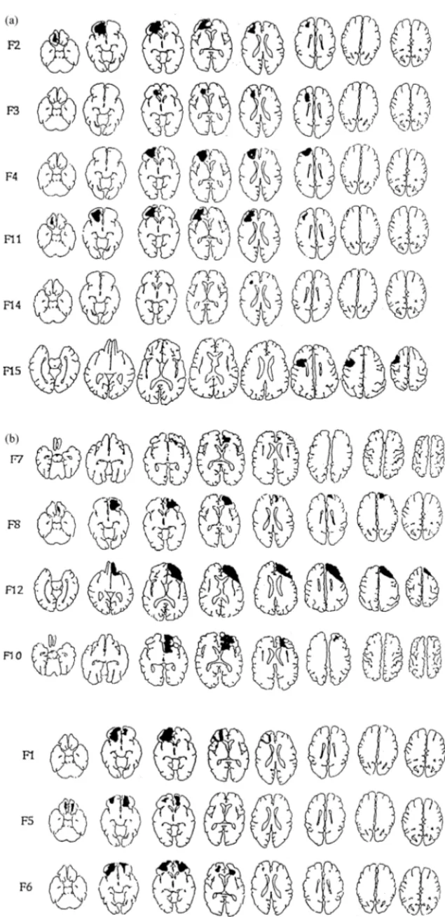
Patients with possible (or putative) frontal lesions were screened by neurologists in five French-speaking Belgian hospitals. The instructions concerning the selection criteria emphasized that a CT scan and/or MRI should confirm the frontal lesion. Only patients with lesions strictly restricted to the frontal lobe could be included in the study. Any hint of lesion in regions other than the frontal lobe led to the exclusion of the patient from the study. Other restrictions were that participants had to be younger than 55 and could not present any antecedent of alcohol or drug abuse, or of any psychiatric disorder. Finally, patients were only included if examined at least 5 weeks after the occurrence of the lesion, or, in the case of neurosurgical patients, the date of surgery. Under these conditions, among the 43 patients referred to us as ‘frontal’ patients, only 13 were included in the study. Four patients had cerebral vascular accidents: two due to anterior communicating artery aneurysm (F3 and F10), one due to anterior cerebral artery aneurysm (F7) and one of unknown origin (F11). Seven patients had traumatic brain injury: five due to motor vehicle accidents (F1, F4, F5, F6 and F14) and two due to falls (F2 and F8). Finally, two patients had been operated for excision of an astrocitomas (F12 and F15). Six patients had a left-sided lesion, four a right-sided lesion and three a bilateral lesion. Location of lesions is shown in Fig. 1a and b. These illustrations repro-duce the last radiographic examination undertaken prior to the testing. The affected regions were identified using the methodology of Damasio and Damasio [22] with the help of an experienced neurologist working blind to the purpose of the study.
Damasio and Damasio’s [22] methodology comprises two main steps. The first consists in transposing the lesion from the CT scan or MRI scan into a set of detailed anatom-ical templates (Damasio and Damasio [22] p. 207–17). The second step consists in identifying the region damaged (e.g. ‘F07’ or prefrontal region) and to translate it into the Brodmann nomenclature with reference to the key codes
Fig. 1. Reconstruction of (a) left frontal; (b) right and bilateral frontal lesions based on CT scan and magnetic resonance scans. For each patient, the horizontal cuts, from left to right, go from most inferior to most superior.
838 P . Andr ´es, M. V a n der Linden /Neur opsyc holo gia 40 (2002) 835–845
Table 2
Age and duration of education (in years) of participantsa
Participant Age Education
Frontal Control Frontal Control
F1, C1 19 19 11 12 F2, C2 54 51 9 9 F3, C3 51 52 16 17 F4, C4 40 40 16 16 F5, C5 18 18 10 10 F6, C6 19 19 11 12 F7, C7 43 41 12 12 F8, C8 21 20 15 14 F10, C10 23 24 11 12 F11, C11 38 39 16 16 F12, C12 51 50 8 8 F14, C14 21 21 9 9 F15, C15 34 33 10 12
aF corresponds to frontal patients and C to control participants.
provided on p. 219 of Damasio and Damasio [22] (e.g. ‘BA 8, 9, 46’, see Table 1).
The standard Damasio and Damasio [22] procedure for the analysis of CT and MRI was applied to our study as follows (see p. 143–7 of Damasio and Damasio [22]).
The set of best-fitting templates was first chosen on the basis of the angle of incidence in which CT or MRI scans were obtained. The lesion was then charted on the templates at every level at which it occurred. Subsequently, an appro-priate “in-register” transparency containing anatomical cells representing neural “areas of interest” in both gray and white matter structures was superimposed over the template in or-der to establish the anatomical areas damaged.
The results of this analysis were then filled in as a hard-copy visual record (see Fig. 1a and b) and keyed in Table 1 to the codes mentioned (e.g. ‘F07’ or prefrontal region corresponds to ‘BA 8, 9, 46’).
Frontal patients were examined after a post-surgery pe-riod long enough to avoid the presence of a “mass effect”1 often observed when patients are examined in the acute pe-riod. The mean delay between the occurrence of the lesion and the neuropsychological evaluation was of 179.8 days (range = 39–467), the mean delay between the occurrence of the lesion and the latest radiographic examination was 99.9 days (range = 1–467) and the mean delay between the radiographic examination and testing was 84.18 days (range= 1–215). Patients were not on anticonvulsant med-ication at the time of testing.
Frontal patients were matched to control participants on the basis of their individual age, sex, type and duration of ed-ucation (see Table 2). Patients, all males, were 33.2 (S.D. = 13.75) years old and had 11.8 (S.D. = 2.9) years of educa-1Canavan et al. [18] found, for example, that important DS symptoms observed in brain-damaged patients examined in the acute period disap-peared after the post-operative period. This indicated that the DS were the consequence of some diffuse brain damage observed in the acute period rather than the consequence of a focal frontal brain lesion.
Table 3
Mattis dementia rating scale: results for each subscalea
Frontal Control Attention 36.4 (0.9) 36.9 (0.3) Initiation 33.2 (4.8) 36.4 (1.7) Construction 6 (0) 6 (0) Concepts 35.5 (5.1) 36.8 (1.9) Memory 24 (1.5) 24.9 (0.6)
aStandard deviations in brackets.
tion. Control participants were 32.8 (S.D. = 13.2) years old and had 12.23 (S.D. = 0.8) years of education. A one-way analysis of variance confirmed that the two groups were cor-rectly matched in terms of age (F(1, 12) = 1.35; P = 0.268) and years of education (F(1, 12) = 3.26; P = 0.1).
All participants volunteered and gave their informed con-sent.
The global cognitive profile was evaluated by means of the Mattis dementia rating scale (DRS) [38]. The mean over-all score was significantly lower for frontal patients (M = 135; S.D. = 9.62) than for control participants (M = 141; S.D. = 2.6) (F(1, 12) = 6.8; P < 0.05). The analysis of the different subscales (Table 3) revealed significant differences in the attention (F(1, 12) = 6.255; P < 0.05) and initia-tion subscales (F(1, 12) = 8.094; P < .05). This profile of performance is characteristic of that generally observed in other studies examining frontal patients (e.g. [55]).
2.2. Materials, procedure and scoring 2.2.1. Computation span task
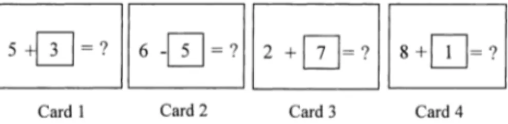
To evaluate the capacity to undertake two tasks simul-taneously, we adapted the dual task method used by Salt-house and Babcock [47]. The materials comprised a series of cards on each of which was presented an arithmetic prob-lem without the solution (see Fig. 2). The second number in each arithmetic problem was framed and the solution was re-placed by a question mark. The arithmetic problems were all sums (X + Y ) or differences (X − Y ) between two one-digit numbers (excluding 0). The values of the digits were further restricted as follows. Y was different from the solution and different from the digit framed in the next problem. Finally, the solution of each problem was always a number between 1 and 18. The number of arithmetic problems presented on each trial increased successively from two to nine, with three trials presented for each series length. Testing discontinued when two trials were failed at a particular series length.
Fig. 2. Example of a series of four cards presented for the span four at the computation span task.
840 P. Andr´es, M. Van der Linden / Neuropsychologia 40 (2002) 835–845
Two conditions were compared: one in which participants were required to remember the digits framed but not to solve the problems (simple span condition), and a second in which they were required to remember the digits while solving the problems. In the simple condition, the cards were pre-sented at the rate of 1 s and participants had to remember the framed digits in order. At the end of the series a blue “recall” card was presented and participants had to recall the se-ries of digits. In the computation span (dual task), the cards were presented at a rate of one every 3 s and participants had to solve the arithmetic problems and give the solution aloud while remembering the framed digit from each prob-lem. When the blue “recall” card appeared at the end of the series, participants had to recall the series of framed digits. Two scores were considered in both the simple and dual conditions. The span was designated as the highest number of target items recalled correctly (all items recalled in orig-inal order) on at least two of the three trials for a particu-lar sequence length. In the dual condition, recall of digits was considered only when the solution to the arithmetical problem was correct. A second score, less restrictive, was obtained by assigning one point for each digit correctly re-called and then adding the number of items correctly rere-called across the experiment.
2.2.2. Directed forgetting
In order to evaluate the ability to actively inhibit no-longer relevant information in working memory, the procedure of Reed [46] was adapted. There were three experimental con-ditions, with the following sequence of events occurring in each trial: (a) presentation of the material to be remembered (one or two trigrams of consonants presented on cards for 2 s each, for example ‘DRG’), (b) an interpolated activity (reading aloud strings of numbers for 10 s), and (c) serial recall. In the ‘single-item’ condition (control condition), a single trigram was presented for retention. Participants were then required to read strings of numbers aloud (interpolated activity) before recalling the trigram in its correct order. In the ‘interference’ condition, an additional (interfering) tri-gram was presented for retention immediately after the first one. Participants had to recall both trigrams at the end of the interpolated task in the order of their presentation. In the ‘directed forgetting’ condition, two trigrams were presented consecutively, as in the interference condition. However, immediately after the presentation of the second trigram, a card was displayed for 500 ms with the inscription ‘to be forgotten’, which prompted participants to forget the second trigram since they would not be required to recall it later. Participants were asked to recall the three letters of the tri-grams in strict serial order after the 10 s of the interpolated activity and were allowed to take as long as they needed to respond. Three practice trials, one per condition, were pre-sented prior to the beginning of the task. Participants were then presented with 30 trials, 10 per experimental condition, presented in the same pre-established random order for all participants.
Table 4
Mean recall (span and number of correctly recalled items) for simple and dual tasks in the computation spana
Frontal Control
Simple span 5.3 (1.4) 6.8 (2.2)
Dual span 2.9 (1.4) 4.8 (2.2)
Simple score 48.8 (23.6) 83.1 (45.6)
Dual score 15.4 (14.9) 41.1 (30.7)
aStandard deviations in brackets.
Participants’ responses were scored following Reed’s cri-terion [46] by assigning one point for each letter recalled (regardless of its serial position within the trigram) and an additional point when this letter was recalled in its correct serial position (maximum score in each condition was there-fore 60). In the interference condition, only the first trigram was scored. Inhibitory capacity was measured by the differ-ence in recall performance between the single-item and di-rected forgetting conditions (didi-rected forgetting cost) [37]. Sensitivity to interference was measured by the difference in performance between the single-item and interference con-ditions.
3. Results
The results are presented for each task separately.
3.1. Computation span
The mean span and mean number of correctly recalled items are presented in Table 4.2 A 2 (group) × 2 (type of span) ANOVA for repeated measures performed on the span measure revealed a significant main effect of group (F(1, 11) = 5.584; P < 0.05), showing that frontal pa-tients presented lower recall performance than control partic-ipants. There was also a significant effect of the type of span (F(1, 11) = 92.172; P < 0.0001), revealing that the perfor-mance in the computation span (dual condition) was weaker than in the simple span. There was no significant interaction between these two factors (F(1, 11) = 0.25; P = 0.627). An ANOVA on the number of correctly recalled items revealed the same pattern of results. There were significant main ef-fects of group (F(1, 11) = 5.962; P < 0.05) and type of span (F(1, 11) = 63.502; P < 0.0001), but again there was no significant interaction between these two factors (F(1, 11) = 0.627; P = 0.445).
In conclusion, patients with focal frontal lobe lesions pre-sented impaired performance compared to control partici-pants, but to a similar extent for the simple and computation spans.
2One patient (F5) and his control participant (C5) could not be admin-istered this task.
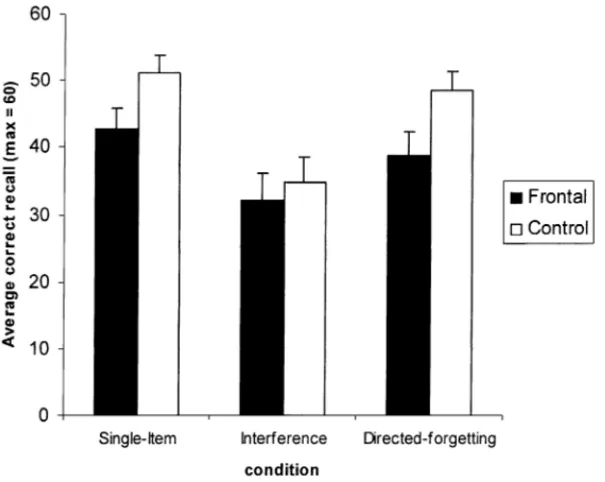
Fig. 3. Mean correct performance in the directed forgetting task by condition. Error bars illustrate standard errors.
3.2. Directed forgetting
The mean recall performance in the single-item, interfer-ence and directed forgetting conditions is shown in Fig. 3. A 2 (group)×3 (conditions) ANOVA for repeated measures revealed a significant main effect of group (F(1, 12) = 4.506; P < 0.05) indicating that frontal patients recalled fewer items than control participants. The effect of condi-tion was also significant (F(2, 24) = 27.355; P < 0.0001). Newman–Keuls post-hoc comparisons revealed that, for both groups, performance in the interference condition was lower than performance in both the single-item condition (P < 0.001), and the directed forgetting condition (P < 0.05). No difference was found between the single-item and directed forgetting conditions (P > 0.05). The interac-tion between group and condiinterac-tion was not significant (F(2, 24) = 2.461; P = 0.107). Directed forgetting cost indices (single-item minus directed forgetting) were calculated for the frontal patients (mean= 3.9; S.D. = 10.1) and the con-trol participants (mean= 2.6; S.D. = 6.7), and revealed no difference between groups (F(1, 12) = 0.238; P = 0.634). Intrusion errors, that is, the recall of consonants of the sec-ond trigram as items of the first, were analyzed in order to obtain an additional measure of the capacity to suppress the no-longer relevant information. The mean number of intrusions was 1.46 (S.D. = 1.6) for the frontal patients and 1.6 (S.D. = 1) for the control participants in the in-terference condition, and 0.62 (S.D. = 1.6) for the frontal patients and 1.54 (S.D. = 2.1) for the control participants in the directed forgetting condition. Given the rarity of such errors, no statistical test could be carried out. One can note, however, that the number of intrusions was equivalent for both groups in the interference condition and that the
differ-ence in the number of intrusions in the directed forgetting condition was in favor of the frontal patients.
In sum, patients with focal frontal lesions showed an over-all lower recover-all performance in the short-term directed for-getting task, but this deficit did not interact with condition. Thus, their efficiency in inhibiting the no-longer relevant information was as good as that of control participants (as confirmed by the equivalent directed forgetting cost indices [37]) and sensitivity to interference was equivalent for both groups.
4. Discussion
The purpose of the present study was to examine to what extent a group of patients with lesions restricted to the frontal cortex present deficits in two working memory tasks expected to implicate the CE [9]. The executive functions investigated were the capacity to perform two tasks simul-taneously, evaluated by the computation span task [47] and the ability to suppress no-longer relevant information, eval-uated by a directed forgetting task [46]. Special care was taken with regard to two methodological aspects: the type of lesion, which had to be restricted to the frontal cortex, and the stability of the clinical state, i.e. patients had to be examined at least 5 weeks after the occurrence of the lesion. Furthermore, the majority of our patients (n = 11) presented a lesion of the dorsolateral frontal lobe, hypothe-sized to be importantly involved in executive functions (see Table 1 for specific location of lesions in our patients).
In the computation task, the results revealed that patients with focal frontal lesions showed lower recall in both the simple and dual conditions. This result is consistent with
842 P. Andr´es, M. Van der Linden / Neuropsychologia 40 (2002) 835–845
the studies showing that short-term storage of verbal infor-mation depends on frontal regions such as Broca’s area (BA 44), the ventral cortex (BA 45) and the left premotor cortex (BA 6) [25,56]. Contrary to what is predicted by the hypoth-esis of a unequivocal link between frontal cortex and dual task management, the results also showed that the decrement in recall induced by dual tasking was equivalent in patients with focal frontal lesions and control participants. Thus, patients and control participants were comparable in their ability to undertake storage and processing simultaneously. This result is in agreement with those of Frisk and Milner [30] and Vilkki et al. [61]. It is also compatible with the observation by Baddeley et al. [13] (also see [3]) that only frontal patients with behavioral signs of DS present impaired performance in dual tasks since our patients did not show any clinical sign of DS (e.g. behavioral disinhibition). What is needed to explain is the paradox observed between the findings from D’Esposito et al.’s [23] neuroimaging study showing specific frontal activations (dorsolateral prefrontal cortex) during dual task management and studies such as the current one (and [30,61]) showing no deficit of dual task performance in patients with lesions of this area. In this vein, a recent neuroimaging study of working memory by Klingberg [34] shows that the prefrontal region is activated during both single working memory and dual working mem-ory tasks. Therefore, contrary to the study by D’Esposito et al. [23], no specific prefrontal activation during the dual task was observed in this study. As Klingberg argued, it is possible that in D’Esposito et al.’s study, the prefrontal ac-tivation during the dual task was simply due to the increase in working memory demand occurring during the simul-taneous performance of two non-working memory single tasks. For example, the stimulus processing or response in one task must be delayed while the other task is given priority, thus inducing a working memory requirement. It may thus be possible that performing working memory in-duces the activation of prefrontal areas as has been shown in several studies (e.g. [25,34,56]), but there is not yet ev-idence to presuppose any prefrontal areas specific to dual task performance ‘per se’ (see [1,15], for similar results).
The directed forgetting task administered in the current study required the maintenance of information no-longer available in the environment during an interval occupied by a secondary task. Additionally, in the directed forgetting condition participants had to actively suppress the no-longer relevant trigram that had been presented. Our data showed that although frontal patients were globally impaired in the capacity to maintain the relevant information, they could inhibit the no-longer relevant information just as well as control participants. Again, the deficit observed in recall is consistent with the numerous studies showing the impli-cation of the frontal cortex in short-term storage of verbal information (see [25,56] for reviews). It is also consistent with the observation by D’Esposito and Postle [25] that the frontal cortex is necessary for some rehearsal processes (also see [56]). The current results do not support however,
the hypothesis of the frontal cortex as the exclusive neural substrate of inhibitory mechanisms.
In conclusion, our results show that whilst patients with lesions restricted to frontal regions evaluated in a stable pe-riod present impaired short-term storage, they also show nor-mal performance in measures of executive processes such as the capacity to undertake two tasks simultaneously and the capacity to inhibit no-longer relevant information. It could be argued that the lower recall performance observed in both conditions of the computation span might have been influ-enced by an executive aspect of this task. Actually, given the characteristics of the type of material, even the sim-ple condition differs from a classical span task: participants have to select the information to be remembered (digit in the frame) and reject the irrelevant information (rest of the string). Therefore, correct recall in this task might depend on short-term storage capacity as well as on the inhibition of the concurrent irrelevant information. However, an exec-utive deficit is not likely to account for the results observed. Indeed, it seems implausible that the low level of interfer-ence induced by the irrelevant information in the computa-tion span (the to-be-remembered numbers were framed) led to such a pronounced difference between groups whereas the much more powerful manipulations of interference (dual compared to simple conditions in the computation span and interference and directed forgetting conditions compared to single-item in the directed forgetting task) revealed no par-ticular group differences.
The literature provides little information about the ques-tion of the necessity of bilateral lesions to impair cognitive inhibition and dual task in working memory and the few neu-roimaging studies of these functions have yielded contradic-tory findings. Whereas D’Esposito et al. [23] and Klingberg [34] found greater activation of the right cortex during dual task, Bunge et al. [15] found a left dominance. From four neuroimaging studies investigating the areas activated by in-hibition tasks, three revealed a right hemispheric dominance [31,32,60] and one left prefrontal activation [33]. It might be possible that unilateral lesions (which were the majority in our study) would not be sufficient to impair inhibition and dual task management. In this context, studies of brain damage and stroke recovery suggest an increasing ability to recruit regions that are contralateral or adjacent to the lesioned area [42,59]. This dynamic organization of cortical functions could also apply to inhibition and dual task man-agement. In a modest attempt to assess the effect of bilateral frontal lesions, we analyzed the individual performance of the two patients of our study with bilateral lesions of BA 9/46. We considered patients’ performance ‘impaired’ when beyond the interval defined by the mean value of the control group± 2 S.D. and below the lowest performance in the control group. The analysis revealed that no patient showed an increased dual task decrement or any impairment of the inhibitory measures (performance on the directed forgetting condition, directed forgetting cost or intrusion errors). This of course may not represent the entirety of patients with
bilateral frontal lesions. To tackle this question more directly, further studies should involve the comparison of patients with bilateral and unilateral frontal lesions. Large groups are needed to address these questions (bilaterality and laterality) and this type of research can only be considered in multicen-ter studies involving multiple research teams and hospitals. Finally, we should note that, although the hypothesis that bifrontal lesions are necessary to impair dual task and inhibi-tion mechanisms in working memory is yet to be tested, the evidence described in the recent literature [8,19,40,41,61] suggests that the hypothesis of more distributed neu-ral network as an alternative for the neuneu-ral substrate of inhibition and dual task management deserves further investigation.
Measures were taken in the present study to minimize the potential influence of mass effects on executive functions ([18,36,61], i.e. as Vilkki et al., we intended to control for the acuteness of the injury by testing patients outside the time period following immediately the onset of the lesion). The fact that we found no deficit of dual task or inhibition in our frontal patients when tested well after a potential “mass effect” converges with the results of other studies [18,36,61] in which DS symptoms were found to disappear after some post-operative period. In this vein our results, together with those of Vilkki et al. and Canavan et al. provide additional support to the suggestion that more cerebral regions than frontal sustain executive functions.
The dissociation observed in patients with frontal le-sions between short-term storage and executive processes in working memory is important for the hypotheses con-cerning the neural substrate of executive functions. As a whole, the current results suggest that not all executive functions are uniquely sustained by the frontal cortex. This finding provides additional support to the hypothesis that some executive functions may be sustained by a distributed cortical neural network rather than by a unique frontal re-gion [11,12,19,32,34,40,41] (see also [6,8,24,31,32,61]). Although the frontal cortex must be involved in executive functions (see for example [8] for some executive deficits in patients with focal frontal lesions examined in a stable period), the current findings suggest that it cannot serve, on its own, as the CE of the brain. It rather must play a role in a distributed set of neural networks concerned with executive functions [24,62]. This idea is also supported in the study by Foster et al. [29] in which performance of nor-mal elderly participants on executive tests correlated more strongly with global cerebral measures than with frontal regional measures.
We should note that it might be possible that some further recovery had happened in three of our patients who received a MRI or CT scan less than 2 months after the lesion. Strictly speaking, if this were the case, this could have contributed to some extent to the good performance on dual task and inhi-bition of our patients as a group. In this vein, further studies excluding this possibility would provide stronger evidence in supporting our results.
We cannot exclude the possibility that some invisible (be-low the threshold detection of CT or MRI scans) microscopic pathology in non-frontal sites existed in our patients with traumatic brain injury. However, it must be clarified that this possibility is irrelevant to our central argument. Indeed, our argument is that the good performance on executive tasks in working memory does not depend necessarily upon having an intact prefrontal cortex. Our results show that patients with frontal lesions are able to perform executive processes (dual task and inhibition) in working memory as well as nor-mal participants, in spite of their reduced short-term storage. Since we are drawing attention to the intact performance of these patients on key executive processes of working mem-ory, hypothetical extra-frontal damage is not relevant to our main argument.
A possible limitation of our study might be the small num-ber of participants, which is a consequence of the rarity3 of patients with lesions restricted to the frontal cortex. It might be argued that this could lead to insufficient statistical power to detect significant differences in executive measures. This is not likely to be the case for three reasons, however. First, significant differences between our patients and control par-ticipants were observed for several measures (MDRS and short-term memory capacity in both working memory tasks). Insufficient statistical power should have masked such dif-ferences. Second, if the frontal cortex is necessary and suf-ficient to perform executive processing, a unique lesion of that area should affect performance in tests of executive functions. Despite the fact that the majority of our patients presented lesions in BA 24, 9 and 46 (see Table 1), which are hypothesized to be particularly important for executive functions [26,56], none of them exhibited significant deficits in such tests when analyzed using a single-case approach. Third, it would be difficult to argue that the frontal cortex is the unique area responsible, for example, for the inhibitory mechanisms involved in directed forgetting when studies with similar sample sizes found deficits in a directed for-getting task with temporal patients (e.g. [28]). Altogether, it seems unlikely that a lack of statistical power is responsible for the good performance of our patients on inhibition and dual task.
As described in the introduction, Baddeley [10–12] sug-gests that the CE is a fractionable system with multiple inde-pendent functions. There is indeed strong neuropsychologi-cal evidence supporting this view [27,48–54]. Additionally, in a recent study [7], we have shown that performance on executive tasks (Hayling and Brixton tests) conceived in the theoretical framework of Norman and Shallice [43] do not correlate in a group of elderly and/or young participants once processing speed has been carried out. In this context, we should note that although the executive processes involved in our tasks (inhibition and coordination of storage and pro-cessing) do not seem to be impaired in patients with focal 3It should be noted that 4 years were needed to accumulate 13 patients for this study.
844 P. Andr´es, M. Van der Linden / Neuropsychologia 40 (2002) 835–845
frontal lesions, other executive processes (e.g. more active manipulation of information in working memory) could be disrupted. Moreover, it remains to be solved whether those patients present a deficit of executive functions when the amount of information to be held in working memory is, contrary to the current study, close to and beyond the span of the participant.
Further research should be conducted in order to break down the CE component of working memory and investigate the neural substrate of its different executive functions. In this vein, it might be likely that some executive functions were sustained by a diffuse cortical (and subcortical) neural network and other by more localized areas.
Acknowledgements
The work reported in this paper was supported by the Spanish Foreign Office (C.G.R.I. in Belgium) and by the Camille Hela Foundation (University of Liege) in the form of a project grant to the first author and is based on part of her doctoral dissertation at the University of Liege. Part of this work was presented at the meeting of the British Psy-chological Society, University of Bristol, September 1997. Thanks are due to Cecilia Brañas, Bruno Kachsten, Bernard Sadzot and Eric Salmon for their neurological expertise and to Marie-Anne Van der Kaa and Eric Vincent for their help with the recruitment of patients. We are also very grateful to Paul Broks, Jonathan K. Foster, Murray Maybery, Tim Perfect, Fabrice Parmentier and two anonymous reviewers for their many useful comments on an early version of this paper.
References
[1] Adcock RA, Constable RT, Gore JC, Goldman-Rakic P. Functional neuroanatomy of executive processes involved in dual-task performance. PNAS 2000;97:3567–72.
[2] Ahola K, Vilkki J, Servo A. Frontal tests do not detect frontal infarctions after ruptured intracranial aneurysm. Brain Cognition 1996;31:1–16.
[3] Alderman N. Central executive deficit and response to operant conditioning methods. Neuropsychol Rehabilitation 1996;6:161–86. [4] Anderson CV, Bigler ED, Blatter DD. Frontal lobe lesions,
diffuse damage, and neuropsychological functioning in traumatic brain-injured patients. J Clin Exp Neuropsychol 1995;17:900–8. [5] Anderson SW, Damasio H, Jones RD, Tranel D. Wisconsin card
sorting test performance as a measure of frontal lobe damage. J Clin Exp Neuropsychol 1991;13:909–22.
[6] Andrés P, Van der Linden M. Inhibition capacity: a ‘frontal’ function? Eur Rev Appl Psychol 1998;48:33–8.
[7] Andrés P, Van der Linden M. Age-related differences in Supervisory Attentional System functions. J Gerontol Psychol Sci 2000;55: 273–80.
[8] Andrés P, Van der Linden M. Supervisory Attentional System in patients with focal frontal lesions. J Clin Exp Neuropsychol 2001;23:225–39.
[9] Baddeley AD. Working memory. Oxford: Psychology Press, 1986. [10] Baddeley AD. Exploring the central executive. Quart J Exp Psychol
1996;9:5–28.
[11] Baddeley AD. The central executive: a concept and some misconceptions. J Int Neuropsychol Soc 1998;4:523–6.
[12] Baddeley AD. Recent developments in working memory. Curr Opin Neurobiol 1998;8:234–8.
[13] Baddeley AD, Della Sala S, Papagno C, Spinnler H. Dual-task performance in dysexecutive and non-dysexecutive patients with a frontal lesion. Neuropsychology 1997;11:187–94.
[14] Baddeley AD, Hitch GJ. Working memory. In: Bower G, editor. The psychology of learning and motivation, vol. 8. New York: Academic Press, 1974. p. 47–90.
[15] Bunge SA, Klingberg T, Jacobsen RB, Gabrieli JD. A resource model of the neural basis of executive working memory. PNAS 2000;97:3573–8.
[16] Burgess PW, Shallice T. Response suppression, initiation and strategy use following frontal lobe lesions. Neuropsychologia 1996;34:263– 73.
[17] Burgess PW, Shallice T. Bizarre responses, rule detection and frontal lobe lesions. Cortex 1996;32:241–59.
[18] Canavan A, Janota I, Schurr P. Luria’s frontal lobe syndrome: psychological and anatomical considerations. J Neurol Neurosurg Psychiatry 1985;48:1049–53.
[19] Carpenter PA, Just MA, Reichle ED. Working memory and executive function: evidence from neuroimaging. Curr Opin Neurobiol 2000;10:195–9.
[20] Clark JM. Contributions of inhibitory mechanisms to unified theory in neuroscience and psychology. Brain Cognition 1996;30:127–52. [21] Cowey CM, Green S. The hippocampus: a “working memory”
structure? The effect of hippocampal sclerosis on working memory. Memory 1996;4:19–30.
[22] Damasio H, Damasio AR. Lesion analysis in neuropsychology. New York: Oxford University Press, 1989.
[23] D’Esposito ME, Detre JA, Alsop DC, Shin RK, Atlas S, Grossman M. The neural basis of the central executive system of working memory. Nature 1995;378:279–81.
[24] D’Esposito ME, Grossman M. The physiological basis of executive function and working memory. Neuroscientist 1996;2:345–52. [25] D’Esposito ME, Postle BR. The dependence of span and
delayed-response performance on prefrontal cortex. Neuropsychologia 1999;37:1303–15.
[26] D’Esposito ME, Postle BR, Ballard D, Lease J. Maintenance versus manipulation of information held in working memory: an event-related fMRI study. Brain Cognition 1999;41:66–86. [27] Eslinger PJ, Damasio A. Several disturbance of higher cognition after
bilateral frontal lobe ablation. Patient EVR Neurol 1985;35:1731–41. [28] Fleck DE, Berch DB, Shear PK, Schefft BK, Privitera MD, Strakowski SM. Directed forgetting deficits in patients with temporal lobe epilepsy: an information processing perspective. J Int Neuropsychol Soc 1999;5:549–55.
[29] Foster JK, Black SE, Buck BH, Bronskill MJ. Ageing and executive functions: a neuroimaging perspective. In: Rabbitt P, editor. Methodology of frontal and executive function. Oxford: Psychology Press, 1997. p. 117–35.
[30] Frisk V, Milner B. The relationship of working memory to the immediate recall of stories following unilateral temporal or frontal lobectomy. Neuropsychologia 1990;28:121–35.
[31] Garavan H, Ross TJ, Stein EA. Right hemispheric dominance of inhibitory control: an event-related functional MRI study. PNAS 1999;96:8301–6.
[32] Garavan H, Ross TJ, Li SJ, Stein EA. A parametric manipulation of central executive functioning. Cerebral Cortex 2000;10:585–92. [33] Jonides J, Smith EE, Marshuetz C, Koeppe RA. Inhibition in verbal
working memory revealed by brain activation. PNAS 1998;95:8410– 3.
[34] Klingberg T. Concurrent performance of two working memory tasks: potential mechanisms of interference. Cerebral Cortex 1998;8:593– 601.
[36] Luria AR, Pribram KH, Homskaya ED. An experimental analysis of the behavioral disturbance produced by a left frontal arachnoidal endotheliona. Neuropsychologia 1964;2:257–80.
[37] MacLeod CM. Directed forgetting: the human memory literature. In: Golding JM, MacLeod CM, editors. Intentional forgetting: interdisciplinary approaches. Mahwah, NJ: Erlbaum, 1998. p. 1–56. [38] Mattis S. Mental status examined for organic mental syndrome in the elderly patients. In: Bellak L, Kaarasu TB, editors. Geriatric psychiatry. New York: Grune Stratton, 1976.
[39] Milner B. Some effects of frontal lobectomy in man: In: Warren JM, Akert K, editors. The frontal granular cortex and behavior. New York: McGraw-Hill, 1964. p. 313–34.
[40] Morris RG. Working memory in Alzheimer-type dementia. Neuropsychology 1994:544–54.
[41] Morris RG. The cognitive neuropsychology of Alzheimer-type dementia. New York: Oxford University Press, 1996.
[42] Musso M, Weiler C, Kiebel S, Muller S, Balau P, Rijntzes J. Training induced brain plasticity in aphasia. Brain 1999;122:1781–90. [43] Norman DA, Shallice T. Attention to action: willed and automatic
control of behavior. Center for human information processing. Technical report no. 99, 1980. In: Davidson RJ, Schartz GE, Shapiro, editors. Consciousness and self-regulation. Advances in research, vol. 4. New York: Plenum Press, 1986. p. 1–18.
[44] Pardo JV, Pardo PJ, Janer KW, Raichle ME. The anterior cingulate cortex mediates processing in the Stroop attentional conflict paradigm. Proc Natl Acad Sci USA 1990;87:256–9.
[45] Perret E. The left frontal lobe of man and the suppression of habitual responses in verbal categorical behavior. Neuropsychologia 1974;12:323–30.
[46] Reed H. Studies of the interference process in short-term memory. J Exp Psychol 1970;84:452–7.
[47] Salthouse TA, Babcock RL. Decomposing adult age-differences in working memory. Dev Psychol 1991;27:763–76.
[48] Shallice T. Specific impairments of planning. Philos Trans Royal Soc London, B 1982;298:199–209.
[49] Shallice T. From neuropsychology to mental structure. Cambridge, UK: Cambridge University Press, 1988.
[50] Shallice T. Multiple levels of control processes. In: Umilta C, Moscovitch M, editors. Attention and performance, vol. XV. Cambridge: MIT Press, 1994. p. 395–420.
[51] Shallice T, Burgess P. Deficits in strategy application following frontal lobe damage in man. Brain 1991;114:727–41.
[52] Shallice T, Burgess P. Higher-order cognitive impairments and frontal lobe lesions in man. In: Levin HS, Eisenberg HM, Benton AL, editors. Frontal lobe function and dysfunction. New York: Oxford University Press, 1991. p. 125–37.
[53] Shallice T, Burgess P. Supervisory control of action and thought selection. In: Baddeley AD, Weiskrantz L, editors. Attention: selection, awareness and control. A tribute to Donald Broadbent. Oxford: Oxford University Press, 1993. p. 171–87.
[54] Shallice T, Burgess P. The domain of supervisory processes and temporal organization of behavior. Philos Trans Royal Soc London, B 1996;351:1405–12.
[55] Shimamura AP, Janowsky JS, Squire LR. What is the role of frontal lobe damage in emory disorders? In: Levin HS, Eisenberg HM, Benton AL, editors. Frontal lobe function and dysfunction. New York: Oxford University Press, 1991. p. 173–95.
[56] Smith EE, Jonides J. Storage and executive processes in the frontal lobes. Science 1999;283:1657–61.
[57] Stuss DT, Toth JP, Franchi D, Alexander MP, Tipper, Craik FIM. Dissociation of attentional processes in patients with focal frontal and posterior lesions. Neuropsychologia 1999;37:1005–27. [58] Stuss DT, Alexander MP. Executive functions and the frontal lobes:
a conceptual view. Psychol Res 2000;63:289–98.
[59] Thulborn KR, Carpenter PA, Just MA. Plasticity of language-related brain function during recovery from stroke. Stroke 1999;30:749–54. [60] Uhl F, Podreka I, Deecke L. Anterior frontal cortex and the effect of proactive interference in word learning. Results of brain-SPECT. Neuropsychologia 1994;32:241–7.
[61] Vilkki J, Virtanen S, Surma-Aho O, Servo A. Dual task performance after focal–cerebral lesions and closed head injuries. Neuropsychologia 1996;34:1051–6.
[62] Weinberger DR. A connectionist approach to the prefrontal cortex. J Neuropsychiatry Clin Neurosci 1993;5:241–53.
![[PDF] Cours complet pour apprendre Ruby | Formation informatique](data:image/gif;base64,R0lGODlhAQABAIAAAP///wAAACH5BAEAAAAALAAAAAABAAEAAAICRAEAOw==)