ORIGINAL ARTICLE
Migraineurs Without Aura Show Microstructural
Abnormalities in the Cerebellum and Frontal Lobe
C. Granziera&D. Romascano&A. Daducci&A. Roche&M. Vincent&G. Krueger&N. Hadjikhani
Published online: 25 May 2013
# Springer Science+Business Media New York 2013
Abstract The involvement of the cerebellum in migraine pathophysiology is not well understood. We used a biparametric approach at high-field MRI (3 T) to assess the structural integrity of the cerebellum in 15 migraineurs with aura (MWA), 23 migraineurs without aura (MWoA), and 20 healthy controls (HC). High-resolution T1 relaxation maps were acquired together with magnetization transfer images in order to probe microstructural and myelin integ-rity. Clusterwise analysis was performed on T1 and magne-tization transfer ratio (MTR) maps of the cerebellum of MWA, MWoA, and HC using an ANOVA and a non-parametric clusterwise permutation F test, with age and
gender as covariates and correction for familywise error rate. In addition, mean MTR and T1 in frontal regions known to be highly connected to the cerebellum were com-puted. Clusterwise comparison among groups showed a cluster of lower MTR in the right Crus I of MWoA patients vs. HC and MWA subjects (p=0.04). Univariate and bivar-iate analysis on T1 and MTR contrasts showed that MWoA patients had longer T1 and lower MTR in the right and left pars orbitalis compared to MWA (p<0.01 and 0.05, respec-tively), but no differences were found with HC. Lower MTR and longer T1 point at a loss of macromolecules and/or micro-edema in Crus I and pars orbitalis in MWoA patients vs. HC and vs. MWA. The pathophysiological implications of these findings are discussed in light of recent literature. Keywords Cerebellum . Migraine . Cortical spreading depression . Structural MRI
Introduction
Migraine is a common and disabling neurological disorder, affecting 15–25 % of women and 6–8 % of men [1–4]. It is characterized by severe, recurrent headaches, often accom-panied by nausea, vomiting, and light sensitivity. In some migraineurs, the headaches are preceded by neurological disturbances known as“aura.”
The cerebellum may play a role in migraine pathophys-iology [5], but its importance still needs to be clarified. Subtle vestibulocerebellar dysfunction have been reported interictally both in patients with [6] and without aura [7,8] (MWA and MWoA, respectively). Stabilometric studies have revealed ictal and interictal abnormalities specific to MWoA patients compared to controls [9], and studies pooling MWA and MWoA [10] showed balance differences between migraineurs as one group and nonmigraineurs.
Cerebellar dysfunction in migraine may have both circula-tory and neurophysiological origins. Although interictal
C. Granziera, D. Romascano, G. Krueger, and N. Hadjikhani equally contributed.
C. Granziera (*)
:
N. HadjikhaniGRHAD, BMI, SV, EPFL, Lausanne, VD, Switzerland e-mail: [email protected]
C. Granziera
Department of Clinical Neurosciences,
Centre Hospitalier Universitaire Vaudois and University of Lausanne, 1011, Lausanne, VD, Switzerland
C. Granziera
:
D. Romascano:
A. Daducci:
A. Roche:
G. Krueger
Advanced Clinical Imaging Technology Group, Siemens-CIBM, EPFL, Lausanne, VD, Switzerland
A. Daducci
STI/IEL/LTS5, EPFL, Lausanne, VD, Switzerland G. Krueger
Healthcare Sector IM&WS S, Siemens Schweiz AG, Renens, VD, Switzerland
M. Vincent
Serviço de Neurologia, HUCFF-UFRJ, Rio de Janeiro, Brazil N. Hadjikhani
Martinos Center for Biomedical Imaging,
Massachusetts General Hospital, Harvard Medical School, Charlestown, USA
perfusion changes have not been demonstrated in the cerebel-lum of MWA and MWoA patients [11], attack-related cere-bellar circulatory changes have been shown in familial hemi-plegic migraine [12,13]. Moreover, subclinical cerebellar in-farcts occur at higher frequency in MWA patients [14].
Abnormal ion channels expressed in the cerebellum [15] may also provoke either direct damage by glutamate and other neurotoxic substances or indirect effect by an increase in cellular excitability leading to cortical spreading depres-sion (CSD). CSD is the likely substratum of migraine aura [16], but it has also been suggested that it may be present in MWoA [17,18].
Cerebellar dysfunction in migraineurs with and without aura could also be the consequence of CSD in the frontal lobe [18]. The so-called crossed-cerebellar diaschisis, a phe-nomenon already described in familial hemiplegic migraine [12], corresponds to a decrease in cerebellar metabolism, caused by the disruption of afferent excitatory inputs sec-ondary to contralateral cerebral hemispheric lesions or CSD [19]. Likewise, CSD in the frontal cortex during migraine could provoke a contralateral cerebellar hypoperfusion.
Finally, impaired function of the cerebellum of migraineurs might be due to dysfunctional trigeminal inputs. Migraine is known to keenly affect the trigeminal system [20], and tri-geminal projections to the cerebellum have been detected in many species [21,22].
Only few studies previously reported structural cerebellar alterations in migraine; cerebellar hyperintensities have been shown to be significantly higher in migraineurs compared to healthy controls [23], and decreased gray matter volume in the cerebellum has been recently described in MWoA patients in a voxel-brain morphometry study [24]. Moreover, a small study applying quantitative MRI spectroscopy in MWA patients showed lower choline levels in the cerebellum of migraineurs vs. controls, pointing at the presence of alterations in cellular membrane composition in MWA.
In this context, we conceived a study aiming at probing the microstructural integrity of the cerebellum and of frontal areas functionally connected to it, by using a multicontrast magnetic resonance imaging approach at 3 T. We used T1 relaxometry to assess the structural properties of the tissue and magnetization transfer imaging to detect eventual myelin-related abnormali-ties. Our hypothesis was that migraineurs would exhibit T1 and magnetization transfer ratio (MTR) alterations in the cerebel-lum and the frontal lobe compared to subjects without migraine.
Methods MRI Acquisition
We enrolled 15 patients with MWA, 23 patients with MWoA, and 20 healthy controls (HC), who underwent a MRI scan in a
3-T Tim Trio scanner (Siemens, Erlangen) equipped with a 32-channel head matrix coil. Clinical characteristics are presented in Table1. Three MWA, two MWoA, and two HC subjects smoked five to ten cigarettes/day at the study enroll-ment. One patient with MWoA, one with MWA, and none of the healthy controls were under hormone replacement therapy. Eight patients with MWoA, five with MWA, and five healthy controls were under oral contraception.
MWA and MWoA patients were diagnosed based on the 2004 IHS criteria [25] and had not experienced attacks at least 3 days prior MRI scanning. None was under prophylactic treatment for at least 6 months. Patients did not suffer from any other neurological or psychiatric disease and had normal neurological examination. HC had no history of migraine or other headache types and did not suffer from other neurolog-ical or psychiatric disease nor followed any pharmacologneurolog-ical treatment in the 3 months prior to the study.
The MRI protocol included four different sequences: MPRAGE as described in the ADNI protocol (http:// www.loni.ucla.edu/ ADNI/Research/Cores/ADNI_Siemens_ 3T_TrioTimVB13.pdf.) (TR/TE=2,400/3 ms, voxel size= 1×1×1.2 mm3, FoV=256×240×160), MP2RAGE (TR/TE= 5,000/3 ms, inversion time=700 ms, FA=4°, voxel size=1× 1×1.2 mm3, FoV=256×240×160) and magnetization transfer imaging (MTI) (TR/TE=48/23 ms, voxel size=2×2×2 mm3, FoV=240×256×96). The MP2RAGE volumes were used to compute the T1 maps. MTR maps were computed from the MTI volumes as follows: MTR=(M0−MT)/M0×100, where MT and M0 are, respectively, the image intensities acquired with and without magnetization transfer saturation pulse. Subsequently, T1 and MTR maps were linearly registered to the MPRAGE volume with 6 degrees of freedom and mutual information cost function using ELASTIX [26].
Cerebellum Structural Integrity
MPRAGE images were nonlinearly registered to the SUIT cerebellum atlas [27–29] using the SUIT toolbox [27–29] for SPM. The same parameters were then applied to register each subject's T1 and MTR map to the SUIT atlas. T1 and MTR maps in SUIT space were then smoothed using an 8-mm FWHM Gaussian kernel.
We assessed whether the groups (MWA, MWoA, and HC) had any difference in MTR and T1 distributions using an ANOVA and a non-parametric clusterwise permutationF test with 10,000 permutations and a cluster-forming thresh-old of p=0.001 (http://www.fmrib.ox.ac.uk/fsl/randomise/ index.html). This test uses random permutations to approx-imate the distribution of the mass of supra-threshold voxel clusters under the null hypothesis (H0: there is no difference
of MTR or T1 distribution between the groups). The distri-bution of the test statistic is then used to infer thep value of
the clusters, which is corrected for the familywise error rate [30]. Age and gender were entered as additional covariates. Significant clusters were then used as a mask to compute the mean value of the corresponding map inside the cluster for each group and to mask theF values map. Post hoc t tests were performed to assess the direction of detected differences between pairs of groups.
Lesions in hemispheric and cerebellar white and gray matter were manually counted on FLAIR and MP2RAGE images as reported in [31].
Structural Integrity of Frontal Cortical Areas Connected to Crus I
Using the methods described above, we identified a region that was structurally different in MWoA compared to MWA and HC, which was located mainly in the Crus I of the cerebellum. We subsequently analyzed the average T1 and MTR contrasts in the latero-orbital cortex, the pars orbitalis, pars opercularis, pars triangularis, and the superior frontal gyrus [32], which are the regions with the strongest func-tional connections to Crus I [32].
Using FreeSurfer (www.freesurfer.org), we computed the parcellation of these five areas, and we registered it to a gray matter probability map calculated using an in-house software based on [33]. We then averaged T1 and MTR in each region considering the voxels with at least 75 % probability to belong to gray matter, in order to reduce partial volume effects. Statistical comparison among MWA, MWoA, and HC groups was then performed using a permutation-based univariate t test (T1 and MTR) and bivariate Hoteling test (T1–MTR), using age and gender as covariates. Correction for familywise error rate was performed for multiple comparisons.
Correlations of MTR and T1 Changes with the Number of White Matter Lesions
Spearman correlations were performed between MTR and T1 values in the cerebellum and frontal cortical regions that
showed significant differences among MWA, MWoA, and HC (left/right pars orbitalis) with the number of hemispheric and cerebellar lesions.
Correlations of MTR and T1 Changes with Attack Frequency and Duration
Spearman correlations were performed between MTR and T1 values in the cerebellum and frontal cortical regions that showed significant differences between MWA and MWoA patients (left/right pars orbitalis) with attack frequency and duration.
Concepts of Quantitative and Semi-quantitative MRI Contrasts (MTR and T1)
The semi-quantitative MTR is a marker of structural integ-rity, which is sensitive to the relative proportion of macro-molecules (myelin and cellular proteins) and water [34]. A reduced MTR indicates therefore a loss of macromolecules and/or microscopic edema [34].
Similarly, the quantitative T1 assessment probes micro-structural properties, and longer T1 values indicate a loss in tissue structure [35]. However, quantitative T1 measure-ments are slightly biased by local iron content, with higher iron levels leading to shorter T1 values [35].
Results
Patients with migraine (MWoA and MWA) did not differ from HC for age (p=0.8) and gender (p=0.2), and MWoA did not differ from MWA patients for disease duration (p=0.4) and migraine frequency (p=0.5). MWoA patients had a significantly higher number of hemispheric WM lesions compared to HC (p=0.01) (Table1).
In the cerebellum, MTR clusterwise comparison among groups showed a cluster in the right Crus I (Fig. 1, total cluster volume=0.9 mm3, p=0.04); a posteriori t tests
Table 1 Clinical characteristic and number of lesions
F female, M male, WM white
matter,GM gray matter
MWoA MWA HC p value
N 23 15 20 n/a
Age 39±14 38±11 37±12 0.9
Gender (F/M) 18/5 10/5 12/8 0.2
Migraine duration (years, mean ± SD) 24.2±15 20.6±12.5 N/A 0.4
Migraine frequency/month (mean ± SD) 5.6±6.9 4.2±5.6 N/A 0.5
Hemispheric WM lesions 151 89 51 0.01 (MWoA vs. HC)
Hemispheric GM lesions 0 0 0 n/a
Cerebellar WM lesions 4 2 0 0.2
showed that the MTR is lower in MWoA patients compared to MWA (MWoA, 0.21±0.01; MWA, 0.23±0.02;p<0.001) and controls (HC, 0.23±0.01,p<0.0001) (Fig.1).
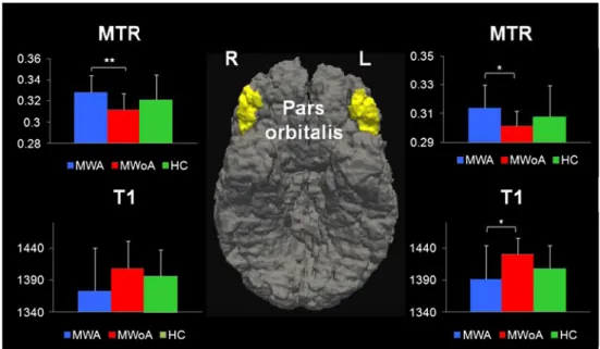
In the frontal lobe, univariate analysis on the T1 contrast showed that MWoA patients had significantly longer T1 in the left pars orbitalis compared to MWA (MWoA, 1,433±25 ms, vs. MWA, 1,391±53 ms, p=0.02) and HC (HC, 1,408± 37 ms), though this latter did not reach significance (p= 0.09) (Fig.2). On the other side, univariate analysis on the MTR contrast showed that MWoA patients had significantly lower MTR in the right and left pars orbitalis compared to MWA (right: MWoA, 0.31±0.01, vs. MWA, 0.33±0.02,p= 0.004, and MWoA, 0.30±0.01, vs. MWA, 0.31±0.01, p= 0.03). No differences were found between MWoA and HC (right: MWoA, 0.31±0.01, vs. HC, 0.32±0.02, p=0.2, and left: MWoA, 0.30±0.01, vs. HC, 0.31±0.02,p=0.6) (Fig.2).
Bivariate analysis of the T1 and MTR contrasts showed that MWoA and MWA differed in both the left and the right pars orbitalis (p=0.03, respectively), whereas no significant difference was found with the HC (p>0.3). No significant correlations were found between MTR and T1 values in the cluster located in Crus I and L/R pars orbitalis of MWoA patients with hemispheric and cerebellar lesions as well as with migraine frequency and duration.
Discussion
Our study shows microstructural alterations in the Crus I of the cerebellum as well as in the pars orbitalis of the prefrontal cortex in patients with MWoA. These findings were restricted to MWoA and absent in MWA confirming partially our hypothesis.
Fig. 1 Voxel-based comparison of the MTR contrast in the cerebellum of
MWA, MWoA, and HC.Top:
coronal and sagittal view of the significant cluster on a background showing the
cerebellum atlas.Bottom: axial
view of the significant cluster on cerebellum atlas background and comparison of the mean MTR in the cluster among MWA, MWoA, and HC
(**p<0.01, ***p<0.0001)
Fig. 2 Region of interest evaluation of MTR and T1 contrasts in the pars orbitalis of the prefrontal cortex in MWA,
MWoA, and HC (*p<0.05,
**p<0.01). Center: 3D bottom
view of right and left pars orbitalis from the FreeSurfer cortical parcellation (www.freesurfer.org)
We applied a biparametric approach at high-field MRI in order to study the microstructural integrity (T1) and the my-elin content (MTR) of the cerebellum and of connected areas of the frontal lobe. The MTR was reduced in the right Crus I of the cerebellum of MWoA vs. MWA patients as well as healthy subjects. Crus I is a posterolateral cerebellar lobe involved in language as well as spatial transformation and working mem-ory; for review, see [36]. It is also implicated in emotion, with responses to unpleasant images [37], fear, and anger [38] as well as in pain processing to noxious heat [37].
Crus I is functionally interconnected with the prefrontal cortex (PFC), especially with pars triangularis, orbitalis, opercularis, and the superior frontal gyrus [32]. Therefore, we subsequently focused our analysis on those three areas and observed bilateral abnormalities in the pars orbitalis of MWoA compared to MWA patients. These abnormalities were characterized by longer T1 and lower MTR. The same characteristics were observed when comparing MWoA vs. HC, though the difference did not reach significance due to the gender unbalance between groups.
The pars orbitalis of the PFC is one of the regions showing the highest peaks of functional connectivity with Crus I [32]. It is located in the inferior frontal gyrus and functionally appears to be involved in value representation and stimulus evaluation [39,40] as well as, to a minor extent, to processing of affective social signals [41–44]. Interestingly, migraine patients (with and without aura) were shown to exhibit stron-ger activations in this area as a response to pain-related vs. nonpain-related negative affective adjectives [45], suggesting an enhanced affective involvement towards pain cues.
Lower MTR and longer T1 point at a lower content of macromolecules (myelin and/or cellular proteins) or at a micro-edema effect in these regions [35]. The presence of these phenomena may be the consequence of repeated micro-inflammatory processes due to recurrent CSD during migraine attacks [46]. Even though CSD has been essential-ly described in migraine aura [16, 47], there is in fact evidence suggesting hyperexcitability of the central nervous system [48–50] and the presence of CSD-like phenomena in MWoA [17,18]. In addition, many studies have underlined the commonalities that exist between both migraine groups and epilepsy (for review, see [51]) and the fact that CSD suppression seems pivotal in migraine prophylaxis [52].
Cortical demyelination has been previously linked to increased CSD velocity and cortical excitability, most prob-ably due to decreased myelin-dependent stabilization and buffering of extracellular ion content [53]. Moreover, a decreased expression of ion channels in the cellular mem-brane, as it has been shown in migraine like basilar-type [54] and familiar hemiplegic migraine type 1–3 [55–57], might also lead to the same phenomena. Therefore, lower macromolecular content in Crus I and pars orbitalis of MWoA might be at the origin of silent cortical spreading
depression waves in migraineurs without aura, as previously suggested by Vincent et al. [5].
On the other hand, we did not find any significant corre-lation between MTR/T1 abnormalities in Crus I/pars orbitalis of MWoA patients and migraine frequency and duration. This may be due to the limited number of subjects studied but could also suggest that the observed microstruc-tural alterations are a condition promoting migraine attacks. No alterations of the frontocerebellar circuitry were ob-served in MWA patients, in contrast with behavioral studies showing that both MWA and MWoA seem to have cerebel-lar dysfunction. Several hypotheses can be put forward to explain these findings: (1) CSD-related circulatory changes may predominate in MWA, leading to microlesions in the posterior fossa [58], though we did not observe any differ-ence in the lesion load in the present study. (2) The possi-bility that the group of MWA patients studied here were not presenting cerebellar signs: one limitation of the present study is the lack of stabilometric or other cerebellar function measures in MWA and MWoA patients, and further studies should address this question. (3) The presence of alterations in indirect circuits comprising pars orbitalis and Crus I (e.g., the nonmotor basal ganglia loop [59, 60] or the anterior cingulate gating loop [39]).
In summary, our work provides evidence of microstruc-tural alterations in the cerebellum–prefrontal circuit in MWoA patients, which could promote increased excitability of the prefrontal cortex and cerebellum, leading to “silent CSD” [5]. Future electrophysiological and functional stud-ies should help to determine the implication of the reported structural abnormalities in migraineurs without aura.
Acknowledgments This work was supported by the Stoicescu
Foundation, the Swiss National Science Foundation Grant
PZ00P3_131914/1 and by the Centre d'Imagerie BioMédicale (CIBM) of the University of Lausanne (UNIL), the Swiss Federal Institute of Technology Lausanne (EPFL), the University of Geneva (UniGe), the Centre Hospitalier Universitaire Vaudois (CHUV), the Hôpitaux Universitaires de Genève (HUG), and the Leenaards and the Jeantet Foundations.
Conflict of interest Dr Roche and Dr Krueger work for Siemens AG.
The other authors have nothing to disclose.
References
1. Lipton RB, Stewart WF. Migraine headaches: epidemiology and
comorbidity. Clin Neurosci. 1998;5(1):2–9.
2. Roncolato M et al. An epidemiological study to assess migraine prevalence in a sample of Italian population presenting to their
3. Warshaw LJ, Burton WN. Cutting the costs of migraine: role of the
employee health unit. J Occup Environ Med. 1998;40(11):943–53.
4. Stewart WF et al. Prevalence of migraine headache in the United States. Relation to age, income, race, and other sociodemographic
factors. JAMA. 1992;267(1):64–9.
5. Vincent M, Hadjikhani N. The cerebellum and migraine.
Headache. 2007;47(6):820–33.
6. Sandor PS et al. Subclinical cerebellar impairment in the common types of migraine: a three-dimensional analysis of reaching
move-ments. Ann Neurol. 2001;49(5):668–72.
7. Wieser T et al. Persistent ocular motor disturbances in migraine
without aura. Neurol Sci. 2004;25(1):8–12.
8. Harno H et al. Subclinical vestibulocerebellar dysfunction in
mi-graine with and without aura. Neurology. 2003;61(12):1748–52.
9. Rossi C et al. Balance disorders in headache patients: evaluation
by computerized static stabilometry. Acta Neurol Scand.
2005;111(6):407–13.
10. Ishizaki K et al. Static stabilometry in patients with migraine and tension-type headache during a headache-free period. Psychiatry Clin Neurosci. 2002;56(1):85–90.
11. Arkink EB et al. Cerebral perfusion changes in migraineurs: a voxelwise comparison of interictal dynamic susceptibility contrast MRI measurements. Cephalalgia. 2012;32(4):279–88.
12. Crawford JS, Konkol RJ. Familial hemiplegic migraine with crossed cerebellar diaschisis and unilateral meningeal enhance-ment. Headache. 1997;37(9):590–3.
13. Lee TG et al. Reversible cerebellar perfusion in familial hemiple-gic migraine. Lancet. 1996;348(9038):1383.
14. Kruit MC et al. Migraine is associated with an increased risk of deep white matter lesions, subclinical posterior circulation infarcts and brain iron accumulation: the population-based MRI CAMERA study. Cephalalgia. 2010;30(2):129–36.
15. van den Maagdenberg AM et al. A Cacna1a knockin migraine mouse model with increased susceptibility to cortical spreading depression. Neuron. 2004;41(5):701–10.
16. Hadjikhani N et al. Mechanisms of migraine aura revealed by functional MRI in human visual cortex. Proc Natl Acad Sci USA. 2001;98(8):4687–92.
17. Woods RP, Iacoboni M, Mazziotta JC. Brief report: bilateral spreading cerebral hypoperfusion during spontaneous migraine
headache. N Engl J Med. 1994;331(25):1689–92.
18. Vincent MB, Hadjikhani N. Migraine aura and related phenomena:
beyond scotomata and scintillations. Cephalalgia.
2007;27(12):1368–77.
19. Gold L, Lauritzen M. Neuronal deactivation explains decreased cerebellar blood flow in response to focal cerebral ischemia or suppressed neocortical function. Proc Natl Acad Sci USA.
2002;99(11):7699–704.
20. Moskowitz MA, Macfarlane R. Neurovascular and molecular mechanisms in migraine headaches. Cerebrovasc Brain Metab
Rev. 1993;5(3):159–77.
21. Jacquin MF et al. Trigeminal primary afferents project bilaterally to dorsal horn and ipsilaterally to cerebellum, reticular formation, and cuneate, solitary, supratrigeminal and vagal nuclei. Brain Res.
1982;246(2):285–91.
22. Huerta MF, Frankfurter A, Harting JK. Studies of the principal sensory and spinal trigeminal nuclei of the rat: projections to the superior colliculus, inferior olive, and cerebellum. J Comp Neurol.
1983;220(2):147–67.
23. Kruit MC et al. Brain stem and cerebellar hyperintense lesions in
migraine. Stroke. 2006;37(4):1109–12.
24. Jin C et al. Structural and functional abnormalities in migraine
patients without aura. NMR Biomed. 2013;26(1):58–64.
25. International Headache Conference (IHC). The international clas-sification of headache disorders: 2nd edition. Cephalalgia. 2004;24
suppl 1:9–160.
26. Klein S et al. elastix: a toolbox for intensity-based medical image
registration. IEEE Trans Med Imaging. 2010;29(1):196–205.
27. Diedrichsen J et al. A probabilistic MR atlas of the human
cere-bellum. NeuroImage. 2009;46(1):39–46.
28. Diedrichsen J. A spatially unbiased atlas template of the human
cerebellum. NeuroImage. 2006;33(1):127–38.
29. Diedrichsen J et al. Imaging the deep cerebellar nuclei: a probabilistic
atlas and normalization procedure. NeuroImage. 2011;54(3):1786–94.
30. Bullmore ET et al. Global, voxel, and cluster tests, by theory and permutation, for a difference between two groups of structural MR
images of the brain. IEEE Trans Med Imaging. 1999;18(1):32–42.
31. Kober T et al. MP2RAGE multiple sclerosis magnetic resonance
imaging at 3 T. Investig Radiol. 2012;47(6):346–52.
32. Krienen FM, Buckner RL. Segregated fronto-cerebellar circuits revealed by intrinsic functional connectivity. Cereb Cortex.
2009;19(10):2485–97.
33. Roche A et al. On the convergence of EM-like algorithms for image segmentation using Markov random fields. Med Image
Anal. 2011;15(6):830–9.
34. Henkelman RM, Stanisz GJ, Graham SJ. Magnetization transfer in MRI: a review. NMR Biomed. 2001;14(2):57–64.
35. Deoni SC. Quantitative relaxometry of the brain. Top Magn Reson Imaging. 2010;21(2):101–13.
36. Stoodley CJ, Schmahmann JD. Evidence for topographic organi-zation in the cerebellum of motor control versus cognitive and affective processing. Cortex. 2010;46(7):831–44.
37. Moulton EA et al. Aversion-related circuitry in the cerebellum: responses to noxious heat and unpleasant images. J Neurosci. 2011;31(10):3795–804.
38. Baumann O, Mattingley JB. Functional topography of primary emotion processing in the human cerebellum. NeuroImage. 2012;61(4):805–11.
39. Kringelbach ML, Rolls ET. The functional neuroanatomy of the human orbitofrontal cortex: evidence from neuroimaging and neu-ropsychology. Prog Neurobiol. 2004;72(5):341–72.
40. Anders S et al. Compensatory premotor activity during affective face processing in subclinical carriers of a single mutant Parkin allele. Brain. 2012;135(Pt 4):1128–40.
41. Sprengelmeyer R et al. Neural structures associated with recogni-tion of facial expressions of basic emorecogni-tions. Proc Biol Sci.
1998;265(1409):1927–31.
42. Wildgruber D et al. Distinct frontal regions subserve evaluation of linguistic and emotional aspects of speech intonation. Cereb
Cortex. 2004;14(12):1384–9.
43. Ethofer T et al. Decoding of emotional information in
voice-sensitive cortices. Curr Biol. 2009;19(12):1028–33.
44. Lotze M et al. Reduced ventrolateral fMRI response during obser-vation of emotional gestures related to the degree of dopaminergic
impairment in Parkinson disease. J Cogn Neurosci.
2009;21(7):1321–31.
45. Eck J et al. Affective brain regions are activated during the pro-cessing of pain-related words in migraine patients. Pain.
2011;152(5):1104–13.
46. Moskowitz MA. Basic mechanisms in vascular headache. Neurol
Clin. 1990;8(4):801–15.
47. Lauritzen M. Pathophysiology of the migraine aura. The spreading
depression theory. Brain. 1994;117(Pt 1):199–210.
48. Boulloche N et al. Photophobia in migraine: an interictal PET study of cortical hyperexcitability and its modulation by pain. J
Neurol Neurosurg Psychiatry. 2010;81:978–84.
49. Denuelle M et al. A PET study of photophobia during spontaneous
migraine attacks. Neurology. 2011;76(3):213–8.
50. Lai KL et al. Subcortical hyperexcitability in migraineurs: a
high-frequency oscillation study. Can J Neurol Sci. 2011;38(2):309–16.
51. Rogawski MA. Common pathophysiologic mechanisms in
52. Ayata C et al. Suppression of cortical spreading depression in
migraine prophylaxis. Ann Neurol. 2006;59:652–61.
53. Merkler D et al. Propagation of spreading depression inversely
cor-relates with cortical myelin content. Ann Neurol. 2009;66(3):355–65.
54. Ambrosini A et al. Familial basilar migraine associated with a new
mutation in the ATP1A2 gene. Neurology. 2005;65(11):1826–8.
55. Ophoff RA et al. Familial hemiplegic migraine and episodic ataxia type-2 are caused by mutations in the Ca2+ channel gene
CACNL1A4. Cell. 1996;87(3):543–52.
56. Ducros A et al. Mapping of a second locus for familial hemiplegic migraine to 1q21-q23 and evidence of further heterogeneity. Ann
Neurol. 1997;42(6):885–90.
57. Vanmolkot KR et al. Novel mutations in the Na+, K+-ATPase pump gene ATP1A2 associated with familial hemiplegic migraine
and benign familial infantile convulsions. Ann Neurol.
2003;54(3):360–6.
58. Kruit MC et al. Infarcts in the posterior circulation territory in migraine. The population-based MRI CAMERA study. Brain.
2005;128(Pt 9):2068–77.
59. Lotze M, Sauseng P, Staudt M. Functional relevance of ipsilateral motor activation in congenital hemiparesis as tested by
fMRI-navigated TMS. Exp Neurol. 2009;217(2):440–3.
60. Lehericy S et al. Diffusion tensor fiber tracking shows distinct