682
Acta Cryst. (1995). D51, 682-694
TEM1/3-Lactamase Structure Solved by Molecular Replacement and Refined Structure
of the $235A Mutant
BY E. FONZI~,* P. CHARL1ER* AND Y. T O ' T H
Centre d'lngOnierie des Prot~ines, Unitd de Cristallographie, Universitd de Liege, Institut de Physique, B5,
B4000 Sart Tilman, Libge, Belgium M. VERMEIRE
Unitd de Cristallographie, Universitd de LiOge, Institut de Physique, B5, B4000 Sart Tilman, LiOge, Belgium
AND X. RAQUET, A . DUBUS AND J . - M . FRI~RE
Centre d'lngOnierie des Protdines and Laboratoire d'Enzymologie, Universitd de Libge, Institut de Chimie, B6, B4000 Sart Tilman, LiOge, Belgium
(Received 20 June 1994; accepted 12 December 1994)
Abstract
/3-Lactamases are bacterial enzymes which catalyse the hydrolysis of the /%lactam ring of penicillins, cephalosporins and related compounds, thus inactivating these antibiotics. The crystal structure of the TEM1 /;/-lactamase has been determined at 1.9A resolution by the molecular-replacement method, using the atomic coordinates of two homologous f3-1actamase refined structures which show about 36% strict identity in their amino-acid sequences and 1.96 A r.m.s, deviation between equivalent C~ atoms. The TEM1 enzyme crystallizes in space group P212~2~ and there is one molecule per asymmetric unit. The structure was refined by simulated annealing to an R factor of 15.6% for 15 086 reflections with I > 2or(/) in the resolution range 5.0-1.9 A,. The final crystallographic structure contains 263 amino-acid residues, one sulfate anion in the catalytic cleft and 135 water molecules per asymmetric unit. The folding is very similar to that of the other known class A/3-1actamases. It consists of two domains, the first is formed by a five-stranded /';/-sheet covered by three c~-helices on one face and one ~-helix on the other, the second domain contains mainly ¢~-helices. The catalytic cleft is located at the interface between the two domains. We also report the crystallographic study of the TEM $235A mutant. This mutation of an active- site residue specifically decreases the acylation rate of cephalosporins. This TEM $235A mutant crystallizes under the same conditions as the wild-type protein and its structure was refined at 2.0A resolution with an R value of 17.6%. The major modification is the appearance of a water molecule near the mutated residue, which is incompatible with the OG 235 present in the wild-type enzyme, and causes very small perturbations in the interaction network in the active site.
* Authors to whom correspondence should be addressed. © 1995 International Union of Crystallography
Printed in Great Britain - all rights reserved
Introduction
/~-Lactamases are penicillin-destroying enzymes which were discovered even before the introduction of fl-lactams as chemotherapeutic agents. New enzymes, which exhibit very diverse substrate specificities and sometimes very different primary structures, are continuously discovered and described.
The chemistry of their catalytic mechanisms divides the/3-1actamases into two groups. The first includes zinc enzymes and are indexed as class B enzymes. All the other fl-lactamases described so far fall into the second group and are active-site serine enzymes. According to their amino-acid sequences they are subdivided into three classes A, C and D, but they all follow a similar kinetic scheme, involving an acyl-enzyme intermediate ES* (Waley, 1992; Ledent, Raquet, Joris, van Beeumen & Fr~re, 1993),
E + S k~+l ES k-~2 ES* k-~ 3 E + P .
k_~ H20
Class A and C /3-1actamases also share the same general secondary-structure pattern as shown by su- perimposing the known three-dimensional structures determined by X-ray crystallography: Staphylococcus
aureus PC1 (Herzberg, 1991), Bacillus licheniformis
749/C (Moews, Knox, Dideberg, Charlier & Fr~re, 1990), Streptomyces albus G (Dideberg et al., 1987)
and Escherichia coli TEM (Strynadka et al., 1992;
Jelsch, Mourey, Masson & Samama, 1993) enzymes for the 2 9 - 3 0 k D a class A /;/-lactamases, Citrobacter freundii (Oefner et al., 1990) and Enterobacter cloacae
P99 (Lobkovsky et al., 1993) enzymes for the 39 kDa class C ~-lactamases. It is also interesting to mention structural similarities with the Streptomyces R61 DD- carboxypeptidase-transpeptidase (Kelly et al., 1986), which corroborate the hypothesis of a common gene ancestor for all active-site serine penicillin-recognizing Acta Crystallographica Section D
enzymes. Nevertheless, the observed divergence between the different /7-1actamase classes and/or the different penicillin-binding protein (PBP) groups, is too high to derive accurate three-dimensional models for a class D /~-lactamase or a PBP from the presently known structures (Joris et al., 1988).
The class A /4-1actamases are synthesized either by Gram-negative or Gram-positive bacteria, and the cor- responding genes are found both on plasmids and on the chromosome. Their catalytic properties are extremely different, reflecting a wide diversity in their primary structures, in contrast to the other fl-lactamase classes.
Following the massive clinical utilization of 14- lactamase-stable compounds, such as the oxyimino-fl- lactam cefotaxime, ceftazidime and aztreonam, against the most widespread pathogenic strains, an increasing number of resistant isolates have emerged within a few years all over the world. The resistance could be attributed to a combination of different phenomena amongst which the occurence of mutants in the SHV and TEM class A enzymes exhibiting hydrolytic properties versus the so-called fl-lactamase-stable antibiotics (see for instance, Jacoby & Medeiros, 1991). These extended- spectrum /7-1actamases show mutations at strategic positions in the active site or in its vicinity, such as E104K near the SDN motif, R164S/H upstream helix h7, A237T/G, G238S and E240K on the b3 strand. Generally the increase of activity against oxyimino compounds is accompanied by a decrease of activity versus good substrates such as benzylpenicillin and cephaloridin (Healey, Labgold & Richards, 1989; Sowek et ai., 1991). Usually these enzymes remain sensitive to the inactivators clavulanate and sulbactam.
We report here the structure of the TEM 1 D4-1actamase as determined by molecular replacement, the first /3- lactamase structure entirely solved by this method, ex- cept those of two/4-1actamase mutants determined from their corresponding native enzyme: the Bacillus licheni- f o r m i s 749/C E166A mutant (Knox, Moews, Escobar & Fink, 1993) and the Staphylococcus aureus PC1 D179N mutant (Herzberg, 1991), whose PDB entry codes are 1MBL and 1BLP, respectively (Bernstein et al., 1977). The use of the molecular-replacement methodology in previous structural studies of TEM1 /'3- lactamase, carried out by Strynadka et al. (1992) and Jelsch et al. (1993) has failed. One explanation could be too low a similarity with the probe structure, namely the Staphylococcus aureus PC I /3-1actamase (1BLM and 3BLM entry codes). To construct a search model of TEM1, we could rely on crystallographic structures of two more similar class A/'4-1actamases, those of Bacillus licheniformis 749/C, named B1 (PDB entry code 4BLM), and of Streptomyces albus G, or SaG, refined at 1.7/~ resolution (personal unpublished results). So, this was a good opportunity to test the limitation of molecular replacement in the case of a class of enzymes that exhibits a wide variety of amino-acid sequences. On
the other hand, the enzymatic properties of the TEM enzyme have been widely studied. Kinetic information about both naturally occuring and site-directed mutants is available. We plan to analyse their structures in order to contribute to an improved insight into the catalytic mechanism and the substrate specificity of the class A fl-lactamases. Here we describe the structure of the $235A TEM mutant, obtained by site-directed mutagenesis (Dubus, Wilkin, Raquet, Normark & Fr6re, 1995) and determined from the atomic coordinates of the native enzyme. Residue 235 is part of the highly conserved KS(T)G motif, which forms a wall of the catalytic cleft. This mutation has little impact on the penicillinase activity, but decreases the cephalosporinase activity in a much more significant manner (Dubus et al., 1995).
The TEM1 structure is also compared with the three other known/7-1actamase structures: BI, SaG and Sa for the Staphylococcus aureus PC I enzyme.
Materials and methods
Purification and crystallization
The TEM enzymes were produced and purified from Escherichia coli harbouring either the ptacl 1 (wild type) or pAD27 ($235A mutant) plasmid (Amman, Brosius & Ptashne, 1983; Dubus et al., 1995). Purification was achieved in two steps by successive chromatographies on a Q-Sepharose Fast Flow column at pH 7.5 and 6.5, respectively.
TEM1 wild-type crystals were grown by the vapour- diffusion method using a wild-type protein concentra- tion of 20 mg ml -I in various buffer and pH conditions (from pH 6.75 to 7.50) with ammonium sulfate as precipitating agent. The best crystals were prisms of 0.9 × 0.25 × 0.25 mm and grew over a period of one to four weeks at 292 K in 0.1 M imidazole pH 7.0, con- taining 10 mM NaN3 and ammonium sulfate at 43-48% saturation.
The crystallization of the TEM $235A mutant was initiated under the same conditions by the microseeding technique, small crystals of the TEM1 wild-type enzyme being used as microseeds. Microcrystals obtained from this cross-seeding were used to repeat experiments to dilute out the effect of heterogenous seeds.
Data collection and processing
The TEM1 crystals are orthorhombic with unit-cell parameters a = 41.8, b = 62.7 and c = 89.8 A, space group P212121. There is one molecule per asymmetric unit which gives a crystal volume per unit molecular mass of 2.03/~3 Da-i and a solvent content of 39.5% by volume. X-ray diffraction data were collected at 293 K with a Siemens X100 area detector. The X-ray source was graphite-monochromated CuKt~ radiation produced by a Rigaku RU-200 rotating-anode generator operating
684 fl-LACTAMASE
Fig. 1. Superposition o f the three fl-lactamase structures represented as ribbons: Bacillus licheniformis (green), Strepto- m y c e s albus G (magenta) and Staphylococcus aureus (blue). Prepared with O (Jones, Zou, C o w a n & Kjelgaard, 1991).
at 40 kV and 120 mA. The detector was positioned at 150 m m from the crystal. Five complementary sets were collected on the same crystal. It was first mounted with the long axis parallel to the incident beam, two sets at high resolution (20 = 30 °) with two different positions of qo-circle (~ = 0 ° and 45 °) and one set at low resolution (20 = 15 °) were collected. The same crystal
0.4 0.3 0.2 0.1 f 0.30 / J / ~ j 0.25 J J j J j 0.20 J J ~ _--- 0.10 0.15 0.25 0.35 0.45 0.55 1/d Fig. 2. Luzzati (1952) plot for the refined TEM1 structure: R factor versus
the inverse o f the resolution, with the theoretical curves corresponding to mean coordinate errors o f 0.10 to 0.30 A.
was then moved inside the capillary so that the long axis was perpendicular to the incident beam: two sets (20 = 15 ° and 30 °, qD = 0 °) were again recorded. For each orientation of the crystal, 500 frames were collected, covering on the whole a 100 ° rotation of the w-circle. In a single frame, reflections resulting from a 0.2 ° oscillation of the crystal were recorded in 60 s.
Crystal orientation and integrated intensities were cal- culated with the X E N G E N Version 2.0 software (Howard et al., 1987). After scaling, 67 369 recorded reflections were obtained with a weighted-squared merging R s y m o n intensity of 4.22%. These merged data yielded 17 252 unique reflections (Table 1).
For the $235A TEM mutant, cell parameters were a = 4 1 . 9 , b = 6 3 . 2 and c = 88.7 ]k and the other crystal properties were the same as those of the wild-type enzyme. The X-ray diffraction data were collected and processed as described above and yielded 48 113 scaled reflections with a n R s y m of 3.5%. After merging 17 496 unique observations were obtained (Table 1).
Structure determination and refinement
The crystal structure of the TEM1 /3-1actamase was determined by the molecular-replacement technique. The M E R L O T (Fitzgerald, 1988) package of programs, which
Fig. 3. Schematic (stereo) drawing o f T E M I : ~-strands are drawn as red arrows, helices as spi- rals, in green and in magenta for the c~-helices and 310-helices, respectively. The active serine $70 is represented in C P K mode. Prepared with O (Jones et al., 1991).
includes the fast rotation function of Crowther (1972), the rotation function of Lattman & Love (1970) and, a translation function of Crowther & Blow (1967), was used throughout. The starting model for the ro- tational and translational parameter investigations was based on the two most similar class A /3-1actamase refined structures, those of Bacillus licheniformis (B1)
and Streptomyces albus G (SAG) which exhibit 35% of
identical residues. The TEM1 model was constructed using the FRODO program package (Jones, 1985) on an Evans & Sutherland PS330. It corresponds to the B1 structure where the side chains have been replaced by the corresponding ones in the TEM1 sequence except
in the 84-90 loop where the SaG structure was used (Fig. 1). Another TEM1 model, called partial, was also tested, and corresponds to the polyalanine chain of well conserved regions between the three class A fl-lactamase structures (B1, SaG and Sa) in addition to the strictly conserved side chains between B1 and TEM1.
The orientation of the TEM1 model in the unit cell P212121 was searched with both complete and partial models at various resolution ranges: 20-4, 10-4, 8-3 and 8-2.5/~. The highest peaks of the rotation functions varied around Eulerian angles o~ = 120,/3 = 63, ~' = 300 °. This orientation was refined using the X-PLOR PC- refinement (Briinger, 1992), and a significant correlation
o< 10 .E 20 30 20 80 110 140 170 Residue number 200 230 260 290
Fig. 4. Temperature-factor distri- bution, along the amino-acid sequence of TEM1 for the backbone (upper) and side-chain (lower) atoms, respectively.
Fig. 5. Stereoview of the active serine and the SDN loop in the
2Fo-Fc electron-density map contoured to 1.1tr above the mean.
686 /4-LACTAMASE
Table 1. Summary of observations by resolution shell for the TEM1 wild-type enzyme and the TEM $235A mutant enzyme
I r e p r e s e n t s the a v e r a g e intensity for all s c a l e d o b s e r v a t i o n s o f a reflection. S h e l l l o w e r A v e r a g e ° A v e r a g e
limit (~,) r e s o l u t i o n (A) l/a(l) (a) T E M I w i l d - t y p e e n z y m e 3.34 4.49 127.2 2.65 2.94 51.3 2.32 2.47 24.5 2.10 2.20 16.6 ! .95 2.03 9.9 1.84 1.91 5.2 Totals 2.58 45.9 No. o f B r a g g reflections C o m p l e t e n e s s P o s s i b l e C o l l e c t e d (%) 3699 3636 98.3 3503 3328 95.0 3477 3053 87.8 3449 2894 83.9 3460 2731 78.9 3397 1610 47.4 2(~85 17252 82.2 (b) T E M $ 2 3 5 A m u t a n t e n z y m e 3.20 4.30 96.3 4243 4294 98.8 2.54 2.81 32.5 4(147 3779 93.4 2.22 2.36 16.5 4000 3227 80.7 2.01 2.11 10.2 3984 2852 71.6 1.87 1.94 5. I 3978 2284 57.4 1.76 1.83 2.5 3922 1160 29.6 Totals 2.55 35.6 24174 17496 72.4
between Fobs and F c a l c w a s only observed with the partial model. This can be explained by the sum of errors intro- duced in the complete model by modelling of less well conserved regions. The final orientation was determined between 8 and 2.5 A resolution and corresponded to the Eulerian angles a = 121.00, ¢'] = 63.19, 3' = 299.80 °.
To position the oriented model in the crystal, several tests were performed with the complete and partial models, at different resolution ranges. Only the complete model at a resolution of 3/~ or more yielded a clear translation solution x = 0.380, y = 0.035, z = 0.135, com- patible with the three Harker sections u = 1/2, v = 1/2, w = 1/2. The corresponding molecular packing was quite acceptable. It is worth noting here that the TEM 1 crystal packing is much denser than those of the other class A/3- lactamases because of the smaller unit-cell dimensions. To improve the molecular-replacement solution, several rigid-body refinements (X-PLOR) were applied to the partial model at 3 ]k resolution and yielded an R factor of about 48% with a correlation of 39.9% between the ob- served and calculated structure factors. This polyalanine positioned model was then used to compute first maps (2Fo - Fc and Fo - F~) between 20.0 and 3.0 A resolution, which showed the well conserved secondary elements and the largest side chains.
It is interesting to note that at this stage, we received the AMoRe molecular-replacement package (Navaza, 1994) and tested its efficiency. At all resolution ranges, with both complete or partial models, the molecular- replacement solution (rotation, translation and rigid-body refinement) was obtained in a very short time.
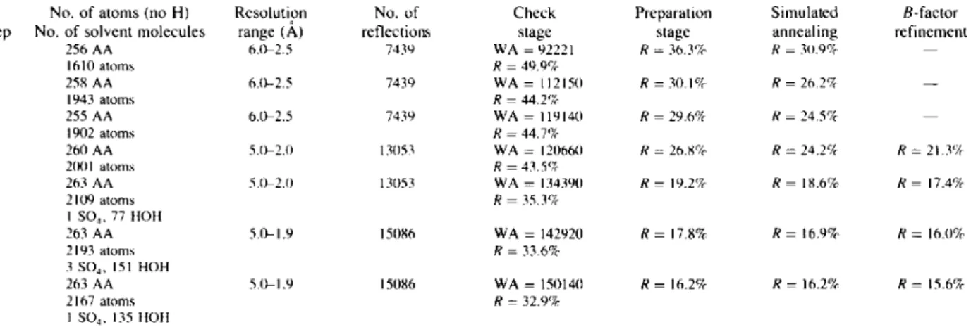
The refinement procedure was performed by simu- lated annealing with the X-PLOR program package. The several alternating cycles of refinement (positions, tem- perature factors) and model refitting (FRODO, PS330) are summarized in Table 2. The final R value is 15.6% for 15 086 reflections with I > 2~r(/) between 5.0 and 1.9 A
resolution. At each step, the structure was constructed on the basis of the 2Fo-Fc and F o - F c maps, always computed with a 20/~ lower resolution limit. Omit maps also had to be generated to interpret the regions that were very different in TEM1 with regard to the initial model. In the final structure, we only conserved the first layer of solvent molecules that showed a very well defined electron density and a temperature factor less than or equal to 60/~ 2. Comparison between the R factor in terms of resolution and theoretical curves with various mean coordinate errors (Luzzati, 1952) shows that the upper limit of the mean positional atomic error is about 0.20 A (Fig. 2).
Results and discussion
Overall structure
The refined structure contains 263 amino-acid residues and the polypeptide chain exhibits the typical folding observed in the known class A fl-lactamase structures (Fig. 3). It consists of two globular domains: one is formed by a five-stranded antiparallel/3-sheet (bl to b5) covered by the two terminal o~-helices (hi, hl 1) and a short 310-helix (hl0) on one face and one o~-helix (h8) on the other face. The second domain contains a big central r~-helix (h2) surrounded by four t~-helices (h4, h5, h6 and h9) and three 310-helices (h3a, h3b and h7). The electron density overall is well defined,with the exception of some solvent-exposed side chains (Q39, Q88, Q90, Q99, E l 0 4 , K111, K146, K215 and E274) and which correspond to the highest temperature-factor values (Fig. 4). A classical 2Fo -F¢ map computed at the • last refinement cycle shows the electron density around the active serine $70 (Fig. 5). The stereochemistry is good with r.m.s, deviations of 0.013/~, for bond lengths, 2.8 ° for bond angles, 24.1 ° for fixed dihedral angles and 1.2 ° for improper dihedral angles. The distribution of
Table 2. Refinement parameters and process of the R factor as a function of refinement steps using the X-PLOR
molecular dynamics program
W A is computed in the check stage and represents the ideal weight between crystallographic and empirical energies. Only reflections such as
/ > 2 a ( / ) are used.
N o . o f residues
N o . o f atoms (no H) Rcsolution N o . o f Check Preparation Simulated B-factor
S t e p N o . o f solvent molecules range (A) reflections stage stage annealing refinement
I 256 A A 6 . 0 - 2 . 5 7 4 3 9 W A = 9 2 2 2 1 R = 3 6 . 3 % R = 3 0 . 9 % - - 1610 a t o m s R = 4 9 . 9 % 2 258 A A 6 . 0 - 2 . 5 7 4 3 0 W A = 11215(I R = 3 0 . 1 % R = 2 6 . 2 % - - 1943 a t o m s R = 4 4 . 2 % 3 255 A A 6 . 0 - 2 . 5 7 4 3 9 W A = 1 1 9 1 4 0 R = 2 9 . 6 % R = 2 4 . 5 % - - 1902 a t o m s R = 4 4 . 7 % 4 2 6 0 A A 5 . 0 - 2 . 0 13(153 W A = 1 2 0 6 6 0 R = 2 6 . 8 % R = 2 4 . 2 % R = 2 1 . 3 % 2(X)I a t o m s R = 4 3 . 5 % 5 263 A A 5.0-2.(I 13053 W A = 1343911 R = 19.2% R = 1 8 . 6 % R = 17.4% 2 1 0 9 a t o m s R = 3 5 . 3 % I SO~, 77 IIO11 6 263 A A 5 . 0 - 1 . 9 15086 W A = 1 4 2 9 2 0 R = 17.8% R = 1 6 . 9 % R = 16.0% 2 1 9 3 a t o m s R = 3 3 . 6 % 3 SO4, 151 H O H 7 263 A A 5 . 0 - 1 . 9 15086 W A = 1 5 0 1 4 0 R = 16.2% R = 16.2% R = 15.6% 2 1 6 7 a t o m s R = 3 2 . 9 % 1 SO4, 135 IIOH
the ~,~/~ torsion angles that define the polypeptide back- bone (Fig. 6) reveals specific conformations for Met69 (9o=54.9 ° and ' ( ) = - 1 4 4 . 8 °) and Leu220 ( ~ = - 1 0 4 . 7 ° and ~/J = - 1 2 1 . 5 ° ) , as observed in all class A structures. Interestingly, these peculiar positions are situated at the junction between the two domains.
The protein is solvated by one sulfate anion in the catalytic cleft and 135 ordered water molecules mainly located in the first coordination shell, directly hydrogen bonded to protein atoms. Their mean temperature-factor value is 36.5 A2. The lowest B-factor values correspond either to outer water molecules involved in intermolec- ular water bridges in the crystal packing, or to inner water molecules located either in the active site such as WAT292 and WAT298 (Table 6), or located between secondary-structure elements (Table 3).
The three-dimensional structure of TEM 1 is stabilized by a series of interactions: one cystine disulfide bridge
1 8 0 - - . • • ° ~. ~. ;. : :'. ",:..,:
v(°)
o " , - c , " " e . . i • R 2 2 1 1 • M 6 9 1 8 0 80" " '0 80,(o)
F i g . 6. Ramachandran plot for all residues except glycines (Ramachan- dran, Ramakrishnan & Sasisekharan, 1 9 6 3 ) .
'Fable 3. Internal water molecules located between
s e c o n d a r y - s t r u c t u r e e l e m e n t s
Water molecule B factor Amino-acid Distance
identification (,~2) identification (,~,) W A T 3 0 1 13.9 M 6 8 N H 3.05 M 6 9 Nit 2.75 L i 6 9 0 2.89 W A T 3 0 2 10.5 L 169 O 2.62 A I 7 2 N H N H 2 . 8 0 W A T 3 0 9 O 2.79 W A T 3 1 0 16.0 1173 O 2.95 D 1 7 6 0 2.72 W A T 3 0 9 O 2 . 6 4 W A T 3 1 I 25.0 R 1 6 4 N H I 3.19 I 173 N H 2.95 D 176 O D 1 2 . 8 0 W A T 3 0 9 O 2 . 8 4 W A T 3 3 7 13.2 D 2 1 4 O 2.74 A 2 1 7 N H 2.87 W A T 2 3 8 O 2.63 W A T 3 5 4 10.1 A 2 1 7 O 2.83 L 2 2 1 N H 2.81 R 2 2 2 N H 2.86 D 2 3 3 O D I 2.87 W A T 3 9 7 19.3 K 2 1 5 O 2.63 G 2 1 8 N H 2.89 W A T 3 6 1 19.6 E 4 8 O E 1 3.17 R 2 5 9 N H 2 3. I I L 2 8 6 O 2.89 W 2 8 9 O 3. I(1 W A T 4 1 8 O 2.91 W A T 3 0 7 20. I $ 2 4 2 O 2.73 $ 2 6 8 N H 2 . 8 6 W A T 3 8 7 28.0 L 193 O 2.77 G 1 9 6 0 2.84 L I 9 9 N i l 2.91 R 2 0 4 N E 3.35
(Cys77-Cys123) which contributes to fix the central h2 c~-helix within the ~-domain, and a dozen of salt bridges listed in Table 4. Among these interactions, only the salt bridge involving R164 and D179 is conserved in all class A/3-1actamase structures, ensuring the stability of helix h7 that defines the bottom of the active site
688 /3-LACTAMASE
(Herzberg, Kapadia, Blanco, Smith & Coulson, 1991;
Sowek et al., 1991). Sequence alignments show that
both residues are very well conserved in all class A
/%lactamases (Ambler et al., 1991; Nordmann & Naas,
1994) with the interesting exception of some TEM vari- ants (Jacoby & Medeiros, 1991). These alignments also
reveal that only a few class A/3-1actamases (Klebsiella
pneumoniae, PIT-2; Pseudomonas aeruginosa, PSE-4; Rhodomonas capsulata and Streptomyces cacaoi, blaU)
have cysteine residues in equivalent positions (Ffaud 123). According to the mutagenesis experiment carried
out on C77 (Schulz, Dalbadie-McFarland, Neitzel &
Richards, 1987), it thus appears that this covalent link
is not essential in the protein folding and in enzymatic activity.
Active-site structure
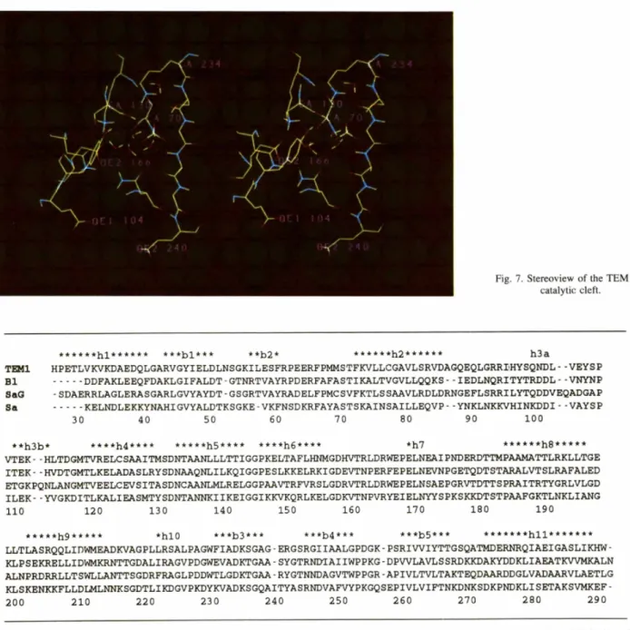
The catalytic cleft (Fig. 7), located at the interface
between the two domains of the protein, is limited by the b3 strand, in particular by the K234-S235-G236 motif, the S130-D131-N132 loop, and the h7 helix
%
i i ~
Fig. 7. Stereoview of the TEM 1
catalytic cleft. T ~ I B 1 S a G S a * * * * * * h l * * * * * * * * * b l * * * * * b 2 * * * * * * * h 2 * * * * * * h 3 a H P E T L V K V K D A E D Q L G A R V G Y I E L D L N S G K I L E S F R P E E R F P M M S T F K V L L C G A V L S R V D A G Q E Q L G R R ~ H Y S Q N D L . . v E Y S P ... D D F A K L E E Q F D A K L G I F A L D T - G T N R T V A Y R P D E R F A F A S T I K A L T V G V L L Q Q K S - - I E D L N Q R I T Y T R D D L - - V N Y N P . S D A E R R L A G L E R A S G A R L G v Y A Y D T . G S G R T V A Y R A D E L F P M C S V F K T L S S A A V L R D L D R N G E F L S R R I L Y T Q D D v E Q A D G A P ... K E L N D L E K K Y N A H I G V Y A L D T K S G K E - V K F N S D K R F A Y A S T S K A I N S A I L L E Q V P - - Y N K L N K K V H I N K D D I - - V A Y S P 30 40 50 60 70 80 90 1 0 0 * * h 3 b * * * * * h 4 * * * * * * * * * h 5 * * * * * * * * h 6 * * * * *h7 * * * * * * h 8 * * * * * v T E K . . H L T D G M T v R E L C S A A I T M S D N T A A N L L L T T I G G P K E L T A F L H N M G D H v T R L D R W E P E L N E A I P N D E R D T T M P A A M A T T L R K L L T G E I T E K • • H v D T G M T L K E L A D A S L R Y S D N A A Q N L I L K Q I G G P E S L K K E L R K I G D E V T N P E R F E P E L N E v N P G E T Q D T S T A R A L v T S L R A F A L E D E T G K P Q N L A N G M T V E E L C E v S I T A S D N C A A N L M L R E L G G P A A V T R F v R S L G D R V T R L D R W E P E L N S A E P G R v T D T T S P R A I T R T Y G R L V L G D I L E K • . Y v G K D I T L K A L I E A S M T Y S D N T A N N K I I K E I G G I K K v K Q R L K E L G D K V T N • V R Y E I E L N Y Y S P K S K K D T S T P A A F G K T L N K L I A N G 1 1 0 120 1 3 0 140 150 160 170 180 190 * * * * * h 9 * * * * * * h l 0 * * * b 3 * * * * * * b 4 * * * * * * b 5 * * * * * * * * * * h l l * * * * * * * L L T L A S R Q Q L I D W M E A D K V A G P L L R S A L P A G W F I A D K S G A G - E R G S R G I I A A L G P D G K - P S R I V V I Y T T G S Q A T M D E R N R Q I A E I G A S L I K H W - K L P S E K R E L L I D W M K R N T T G D A L I R A G V P D G W E V A D K T G A A - S Y G T R N D T A I I W P P K G - D P V V L A V L S S R D K K D A K Y D D K L I A E A T K V V M K A L N A L N P R D R R L L T S W L L A N T T S G D R F R A G L P D D W T L G D K T G A A - R Y G T N N D A G V T W P P G R - A P I V L T V L T A K T E Q D A A R D D G L V A D A A R V L A E T L G K L S K E N K K F L L D L M L N N K S G D T L I K D G V P K D Y K V A D K S G Q A I T Y A S R N D V A F V Y P K G Q S E P I V L V I F T N K D N K S D K P N D K L I S E T A K S V M K E F - 2 0 0 2 1 0 2 2 0 2 3 0 2 4 0 2 5 0 2 6 0 2 7 0 2 8 0 2 9 0
Fig. 8. Alignment of the amino-acid sequences of the four class A ~-lactamases determined from X-ray structure superposition: TEM1 from E.
coli, BI from Bacillus licheniformis, SaG from Streptomyces albus G, and Sa from Staphylococcus aureus. The secondary structures of TEM1,
Table 4. Salt bridges in the TEM1 structure A m i n o - a c i d A m i n o - a c i d D i s t a n c e i d e n t i f i c a t i o n i d e n t i f i c a t i o n (A) K32 NZ D35 OD1 2.96 K34 NZ D38 OD2 3.02 R43 NH 1 E64 OE2 2.98 R61 NE E37 OE2 2.72 NH2 OE1 3.02 NH 1 E64 OE 1 2.80 R83 NH1 E89 OE1 3.16 R93 NE E89 OE2 2.84 NH2 OE 1 2.90 R161 NE D163 OD1 2.94 NH2 OD2 2.75 R164 NH1 D 179 ODI 2.76 NH2 El71 OE2 2.70 R178NE D176OD1 2.76 NE OD2 3.09 NH2 ODI 2.77 R222 NE D233 OD 1 3.12 NH2 OD2 2.99 R259 NH2 E48 OE 1 2.99
that contains two amino-acid residues pointing into the site, E166 and N170. The active site also contains the nucleophilic serine residue $70 on the h2 c~-helix and, three residues downstream, lysine K73. All these amino- acid residues are very well or strictly conserved among all class A fl-lactamases, and many of them are also found in the class C and class D fl-lactamases and in
the penicillin-binding proteins (Joris et al., 1991). Mul-
tiple site-directed mutagenesis experiments have been performed on these residues and have led to a better understanding and caracterization of their role in the catalytic process (Ellerby, Escobar, Fink, Mitchinson & Wells, 1990; Gibson, Christensen & Waley, 1990; Jacob, Joris, Lepage, Dusart & Fr~re, 1990; Branni-
gan, et al., 1991; Juteau, Billings, Knox & Levesque,
1992; Knox et al., 1993; reviewed in Matagne & Fr~re,
1995). Located at strategic positions in the active site, these amino-acid residues are involved in a structurally conserved dense hydrogen-bonding network (Table 5). Some solvent molecules, listed in Table 6, participate in that interaction scheme. The two water molecules, WAT292 and WAT298, are observed in the four class A
Table 5. List of interactions made by the conserved
residue side chains in the catalytic cleft A m i n o - a c i d A m i n o - a c i d D i s t a n c e o b s e r v e d (,A) i d e n t i f i c a t i o n i d e n t i f i c a t i o n T E M 1 $ 2 3 5 A $70 OG K73 NZ 2.79 2.94 S I 3 0 O G 3.11 2.93 K73 NZ S 1 3 0 0 3.01 3.01 N 132 OD 1 2.79 2.80 E166OE1 3.44 3.31 S 130 OG K234 NZ 2.83 2.99 NI32OD1 EI66OE1 3.21 3.13 E166 OE2 3.27 3.34 N 132 ND2 E 104 O 2.97 2.93 E166 OE2 N170 ND2 2.94 2.88 N170 ND2 P167 O 3.28 3.29 K234 NZ $235 O 2.81 2.93
fl-lactamase structures and the first one has an identical position in all enzymes, underlining its crucial functional
interest (Lamotte-Brasseur et al., 1991).
Near the catalytic cleft, residues E104-Y105 lie in front of the SDN loop, and A237-E240 at the end of the b3 strand; as in SaG and B1 enzymes there is no residue in position 239. These amino acids are involved in substrate recognition by side-chain interactions and thus participate in determining the enzymatic profile or substrate specificity (Lenfant, Labia & Masson, 1990;
Sowek et al., 1991; Huletsky, Knox & Levesque, 1993).
Comparison with the other known class A fl-lactamase structures
According to the sequence alignment generated by the superposition of the four class A /3-1actamase refined structures (Fig. 8), the percentage of identical residue between the primary structures and the r.m.s, deviations between corresponding Co~ atoms have been determined (Fig. 9). To compute these r.m.s, values, the 250 struc- turally equivalent CA atoms common to all four enzymes have been used; the following residues: 26-30, 52-57,
Residue identity (%) TEM1 I B| SaG Sa T E M 1 ~,:i~..,.~i~ ~.'.::~:i-'.':~:.::?-.::~:~:!:~:~:.::i:~:~:~:i 3 5 3 7 3 2 r . m . s . B! 1.97 ~:...~ii 43 42 (,A) S a G 1.94 [ 1.05 ~ ! i 28 Sa 2.42 i 1.37 1.95
Fig. 9. L o w e r part, r.m.s, d e v i a t i o n s b e t w e e n the 250 e q u i v a l e n t C a a t o m s o f the four class A ~3-1actamase refined structures. U p p e r part, p e r c e n t a g e s o f identical r e s i d u e b e t w e e n the c o r r e s p o n d i n g a m i n o - a c i d sequences.
Fig. 10. Structural c o m p a r i s o n o f the orientation o f the R244 side chain in T E M 1 and R 2 2 0 in S a G (in grey) relative to the active serine $70 o f T E M 1 . The C a trace is r e p r e s e n t e d as a ribbon.
690 /3-LACTAMASE
Table 6. Solvent molecules in the TEM1 fl-lactamase active site, associated temperature factors B and distances between the 0 atoms of the water molecules, the SO 4 ion and the amino-acid residues, and a comparison with the
solvent molecules in the other fl-lactamase structures
S o l v e n t B factor A m i n o - a c i d D i s t a n c e E q u i v a l e n t m o l e c u l e in
m o l e c u l e ( A : ) i d e n t i f i c a t i o n (~.) BI S a G
SUL291 0 2 43.7 G237 NH 2.98 WAT692 ttOH 1
R244 NH2 2.76
SUI.291 0 4 46.7 S.130 OG 2.83
$235 O 2.85
WAT292 16.2 $70 OG 2.84 WAT712 HOH 43
$70 Nit 3.1)6
E I 6 6 O E I 2.66
N 171) OD I 2.73
WAT293 39.3 S 130 O 3.17 SUL600 HOH 72
N I 3 2 O D I 3.17
SUL291 OI 3.02
WAT294 34. I SUL291 0 3 2.71 WAT931 - -
Y 105 OH 2.97
WAT295 25.7 $70 O(3 2.73 WAT672 HOH 73
S7()N|t 3.51 G237 NH 2.76 SUL291 OI 2.75 WAT297 26.9 V216 O 2.73 WAT673 - - R244 NH2 3.()6 SUL291 03 2.82
WAT298 23.9 D214 OD2 2.75 WAT634 HOH 37
K234 NZ 2.71 $235OG 2.73 $235 NH 2.83 Sa HOH 64 HOH 7 I HOH I11 HOH 81 HOH 42 HOH 103 HOH 22 HOH 54 HOH 34
86-87, 102a-102b--11 la-11 lb of SaG, and 239-256 of Sa have been omitted.
When compared to the three other /3-1actamases, TEM1 appears to be the most different structure with a mean r.m.s, of 2.11/1, compared with 1.47, 1.65 and 1.91/~ for BI, SaG and Sa, respectively. However, the main variations in the general folding do not affect the amino-acid residues directly involved in the active site: (1) the N- and C-terminal residues adopt different conformations in the four enzymes; (2) the 51-55 loop is similarly folded in the TEM1 and Sa structures but is different in SaG and BI where one amino acid has been deleted; (3) the 84-89 structural region is certainly the most variable in the class A/3-1actamases in correlation with the observed amino-acid sequence differences; (4) the 254-257 loop folds differently in TEM1 compared to the three other structures, certainly because of the presence of a proline in position 257 instead of a proline 258; and (5) surprisingly, the loop (b5, h l l ) which is very well conserved in the BI, SaG and Sa fl-lactamases, adopts a very different conformation in TEM 1. This may partly explain the more compact crystal structure. This perturbation is certainly due to the presence of a glycine in position 267 which permits special torsion angles for the TEM1 polypeptide chain (~p = 102, ~ = -19 °) and the presence of a bulky residue (Met) in replacement of Ala or Asp in the other fl-lactamases.
With respect to the two previously reported TEM1 structures, our results can only be compared with those of Jelsch et al. (1993); Strynadka et al. (1992) have not yet published details of their work. The TEM1 structure described by Jelsch has been refined at a higher resolution (1.8/~) and contains more solvent molecules.
Roughly, the geometric parameters such as deviations from ideal values and coordinate errors are close to ours. Their structure analysis reveals the same features: principal interactions, differences with the other class A f/-lactamase structures, active-site topology, (~,(v) and temperature-factor distributions. Meanwhile, their B factors are lower than ours, certainly because they have worked at a higher resolution with more complete data and they have refined the atomic occupancy factors. Their catalytic cleft is also solvated by one sulfate anion and by water molecules WAT297, WAT323, WAT402 and WAT309 equivalent to WAT292, WAT295, WAT297 and WAT298 described here, respectively (Table 6). The only observed difference is that the SO4 anion (SUL291) is positioned further within the active-site pocket and closer to Arg244 and the b3 strand. This proximity to the oxyanion hole, formed by Ser70 NH and Gly237 NH, implies a slight displacement of WAT295 which interacts with GIy237NH, SUL291 O1 and Ser70OG, whereas the equivalent water molecule in the other class A fl-lactamase structures is in contact with GIy237NH, Gly237 O and Ser70 OG.
Accessible surface area
The accessible surface area of the amino acids of the TEM1 structure was computed with the DSSP program (Kabsch & Sander, 1983). The value for the whole mole- cule is 11 344/~2 and its repartition along the sequence is quite similar to that of the BI structure (Knox & Moews, 1991), which has the same fold. The central c~-helix h2 and the /3-sheet are entirely inaccessible to solvent except the b2 strand which is exposed; o~-helices h4 and h5 are less buried, and helices hi, h6, h7, h8, h9 and
Table 7. Accessible surface area for residues defining the active-site walls and for all charged residues of the TEMI
s l r u c l u r e
A c c e s s i b l e s u r f a c e area ( A S A ) is c o m p u t e d with the DSSP p r o g r a m ( K a b s c h & S a n d e r , 1983). T h e v a l u e s are c o m p a r e d with those o f the
c o r r e s p o n d i n g r e s i d u e s in the o t h e r three k n o w n class A / 3 - 1 a c t a m a s e s t r u c t u r e s : BI, S a G a n d Sa. ( A S A ) r e p r e s e n t s the m e a n a c c e s s i b i l i t y o b s e r v e d t h r o u g h o u t the T E M 1 s t r u c t u r e s for an a m i n o - a c i d type.
T E M 1 B 1 S a G Sa
A A A S A (,~2) A A A S A ( , ~ ) A A A S A ( , ~ ' ) A A A S A ( A ~) A A ( A S A ) (,~-') (a) R e s i d u e s o f the a c t i v e site
$70 6 S71) 9 $70 6 $70 13 S 25
K73 0 K73 0 K73 I) K73 O K IO2
S 130 10 S 130 14 S 130 22 S 130 25 S 25
DI31 0 [)131 0 DI31 () DI31 0 D 51
N132 14 NI32 10 NI32 17 N132 12 N 94
EI66 I EI66 1 EI66 0 E l 0 6 5 E 78
N I 7 0 13 NI70 28 NI71) 19 N I 7 0 14 N 94
K234 0 K234 0 K234 0 K234 2 K 102
$235 0 T235 4 T235 4 $235 ll) S 25
G236 I G236 4 G236 2 G236 4 G 25
A237 31 A237 34 A237 40 Q237 95 A 21
(b) C h a r g e d r e s i d u e s
D50 29 D50 30 D5II 15 D50 17 D 5 I
DI31 1} D131 (} DI31 () DI31 1)
D157 8 D157 7 D157 4 D157 9
D179 1 DI79 0 DI79 2 DI79 I
D214 4 N214 10 N214 23 N214 2
D233 t) D233 f) D233 5 D233 ()
E37 25 E37 26 E37 22 E37 40 E 78
E48 2 A48 0 A48 l) A48 l)
E89 9 E89 124 E89 I 17 N89 I 16
E166 I E166 1 E166 0 EI66 5
K32 26 D32 78 R32 99 E32 128 K 102 K73 0 K73 0 K73 0 K73 0 K234 0 K234 0 K234 I) K234 0 Rlful 27 R164 58 R164 50 R164 40 R 86 R244 24 R244 25 N244 0 R244 24 R259 46 V259 8 I259 3 1259 0 R275 31 D275 88 D275 48 N275 90
h l l present an amphiphilic profile. The other structural elements and loops are exposed to the solvent.
The solvent accessibilities of the amino-acid residues defining the active-site walls are compared for the four class A /4-1actamase structures (Table 7a). Relative to the mean values observed for each type of amino acid throughout the TEM1 structure, the accessibility of residues $70, K73, S130, N132, E166, N170, K234, $235 and G236, that are directly involved in the dense hydrogen-bonding network of the catalytic cleft, is very low. The side chains of D131 and A237 are outside the active pocket. The first, D131, is entirely buried and interacts with side chains and backbone atoms of the (~-domain. It would stabilize the position of the SDN loop relatively to the catalytic cleft (Jacob et al., 1990). The second one, A237, is exposed to the solvent and is more variable among class A ~-lactamases than the other active-site residues. It is replaced by Gly, Ser, Thr or Gln. Its backbone atoms, pointing towards the active site, would interact with 13-1actam compounds during their hydrolysis (Lamotte-Brasseur et al., 1991).
The mean-accessibility value associated with glycine residues is relatively high compared to those observed for alanine and serine. This is due to the fact that the glycine residues in TEM1 are more often located on solvent-exposed loops.
The accessibility analysis also reveals very low values for some charged residues (Table 7b). These buried charged residues are generally very well conserved in the class A /3-1actamases. They are either involved in the enzymatic activity and/or in the structure stability On the one hand, K73, E166 and K234 would directly participate in the/3-1actam ring hydrolysis. On the other hand, D131, R164 and D179 would have an indirect function in maintaining the active-site geometry, as described below.
Arg244, located near the b3 strand, is not a conserved residue in class A /4-1actamases, indeed in the SaG structure, it is replaced by an asparagine. However, structure comparisons show that the guanidinium group of R220 in SaG is in a position similar to that of R244 in TEM1 (Fig. 10), and the class A f4-1actamase sequence alignment reveals that if there is no arginine in position 244, there is generally one in position 220 (or less often in 276, structurally close to 220). This suggests that both arginines R220 and R244 could have the same function in class A /3-1actamases. In fact, site-directed mutagenesis experiments and enzymatic analysis have been carried out on TEM 1 (Delaire, Labia, Samama & Masson, 1992; Zafaralla, Manavathu, Lerner & Mobashery, 1992) and on SaG (Jacob-Dubuisson, Lamotte-Brasseur, Dideberg, Joris & Fr~re, 1991). They
692 /7-LACTAMASE confirm that the positive charge of the arginine plays an
important role in positioning the substrate in the active site.
Residues E27, E48, E89, K32 and R259 are directly involved in salt bridges specific to the TEM1 structure, except for the E37-R61 interaction which is relatively well conserved among other class A enzymes, and which links the N-terminal helix to the/7-sheet.
The other charged residues, with weak solvent ex- posure D50, D157 and R275, make internal electro- static interactions, allowing the stabilization of the (bl, b2) loop, ensuring a large polypeptide chain curvature (G156-D157), and bending the C-terminal helix to the /7-sheet.
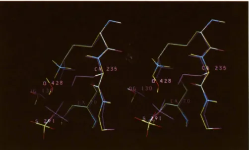
The role of Asp214 and Asp233 is more ambiguous and some site-directed mutagenesis experiments would be required to elucidate their possible structural and/or functional implication. They are located just above the KTG motif and are involved in interactions with K234 and S 130 via the internal water molecule WAT298 rela- tively well conserved among class A structures. In fact D214 and D233 are hydrogen bonded, suggesting that at least one of them is protonated (Fig. 1 la). In the other class A structures, Asp214 is replaced by an asparagine,
but an aspartate at position 246 is also buried and is involved in the same type of interaction with D233 (Fig. l lb). Interestingly, all class A /7-1actamase sequences contain D214-I246 or N214-D246 pairs, suggesting a special function for the conserved amino acid D233.
Refined structure o f the TEM1 $235A m u t a n t
The crystal structure of the $235A mutant was rapidly determined by the molecular-replacement method and Fourier difference synthesis using the atomic coordinates of the native structure. Initial maps were computed with coefficients (2Fo - Fc) and (Fo - Fc), and the model was modified according to the electron density. After several cycles of simulated annealing and individual B- factor refinements, the crystallographic R-factor value was 17.6% in the 6-2/~ resolution range, including
13 171 reflections with I > 2o-(/).
As expected, the final refined mutant structure is very close to the native one, and presents an insignificant r.m.s, value of 0.41/~ between all the atoms of the polypeptide chain. In addition to the absence of the OG235 side chain, the comparison of the mutant and the native TEM1 structures reveals an additional water
(a)
(b)
Fig. 11. Two possible interactions of Asp233. (a) D214-D233-I246 in TEM1 and (b) N214-D233- D246 in SaG_
Fig. 12. Superposition of the active sites of TEM1 (magenta) and of its $235A mutant.
molecule, WAT428, near the CB235, which should be incompatible with a bigger side chain at this position (Fig. 12). Indeed, the distance between the O atom of WAT428 and OG235 would be 2.2/~. This new solvent molecule in the active site introduces some very small perturbations such as the displacement of the sulfate ion and of the other water molecules. Consequently, very slight motions of the side chains 70, 130, 166, pointing into the active site, are also observed (Table 5).
In fact, the hydroxy group of the conserved residue 235, which is either a serine or a threonine in all /3-1actamases, appears to be well positioned to interact with the substrate-free carboxylate (Lamotte-Brasseur et al., 1991). Nevertheless, the kinetic parameters obtained for both wild-type and $235A mutant enzymes reveal that the disappearance of this functional group has little impact on the penicillinase activity. On the con- trary, the cephalosporinase profile is much more affected (Dubus et al., 1995). The hydroxy group of residue 235 would then be more important for positioning the cephalosporins in the active site of the class A /3- lactamases. On the basis of these results, it might be possible to solve the structure of the TEM $235A mutant /3-1actamase complexed with a cephalosporin- type substrate.
The atomic coordinates of the TEM1/3-1actamase and of the $235A mutant have been deposited in the Protein Data Bank.*
This work was supported in part by the Belgian pro- gramme on Inter-University Poles of Attraction initiated by the Belgian State, Prime Minister's Office, Science * Atomic coordinates have been deposited with the Protein Data Bank, Brookhaven National Laboratory. Free copies may be obtained through The Managing Editor, International Union of Crystallography, 5 Abbey Square, Chester CH1 2HU, England (Reference: GR0390).
Policy Programming (PAl No. 19), an Action Con- cert6e with the Belgian Government (89-94/130), and a Convention tripartite between the Region Wallonne, SmithKline Beecham, UK, and the University of Liege.
References
AMBLER, R. P., COULSON, A. F. W., FRI~RE, J.-M., GHUYSEN, J.-M., JORIS, B., FORSMAN, M., LEVESQUE, R. C., TIRABY, G. 8z WALEY, S. G. (1991). Biochem. J. 276, 269-272.
AMMAN, E., BROSIUS, J. & PTASHNE, M. (1983). Gene, 25, 168-178. BERNSTEIN, F. C., KOETZLE, T. F., WILLIAMS, G. J. B., MEYER, E. F.,
BRICE, M. D., RODGERS, J. R., KENNARD, O., SHIMANOUCHI, T. & TASUMI, M. (1977). J. Mol. Biol. 112, 535-542.
BRANNIGAN, J., SPRATT, B. G., MATAGNE, A., JACOB, F., DAMBLON, C., JORIS, B. & FR~RE, J.-M. (1991). Biochem. J. 278, 673-678. BRfJNGER, A. T. (1992). X-PLOR. A System for Crystallography and
NMR, Version 3.0, Manual, Yale Univ., New Haven, CT, USA. CROWTHER, R. A. (1972). The Molecular Replacement Method, edited by
M. G. ROSSMANN, pp. 173--185. New York: Gordon and Breach. CROWTHER, R. A. & BLOW, D. M. (1967). Acta Cryst. 23, 544-548. DELAIRE, M., LABIA, R., SAMAMA, J.-P. & MASSON, J.-M. (1992). J.
Biol. Chem. 267, 20600-20606.
DIDEBERG, O., CHARLIER, P., WERY, J.-P., DEHOTTAY, P., DUSART, J., ERPICUM, T., FRERE, J.-M. & GHUYSEN, J.-M. (1987). Biochem. J. 245, 911-913.
DUBUS, A., WILKIN, J.-M., RAQUET, X., NORMARK, S. & FRI~RE, J.-M. (1995). Biochem. J. In the press.
ELLERBY, L. M., ESCOBAR, W. A., FINK, A. L., MITCHINSON, C. • WELLS, J. A. (1990). Biochemistry, 29, 5797-5806.
FITZGERALD, P. M. D. (1988). J. Appl. Cryst. 21, 273-278.
GIBSON, R. M., CHRISTENSEN, H. t~ WALEY, S. G. (1990). Biochem. J. 271, 399-406.
HEALEY, W. J., LABGOLD, M. R. & RICHARDS, J. H. (1989). Proteins Struct. Funct. Genet. 6, 275-283.
HERZBERG, O. (1991). J. Mol. Biol. 217, 701-719.
HERZBERG, O., KAPADIA, G., BLANCO, B., SMITH, T. S. & COULSON, A. (1991). Biochemistry, 30, 9503-9509.
HOWARD, A. J., GILLILAND, G. L., FINZEL, B. C., POULOS, Z. L., OHLENDORF, D. n. & SALEMME, F. R. (1987). J. Appl. Cryst. 20, 383-387.
HULETSKY, A., KNOX, J. R. & LEVESQUE, R. C. (1993). J. Biol. Chem. 268, 3690-3697.
6 9 4 / % L A C T A M A S E JACOB, F., JORIS, B. LEPAGE, S. DUSART, J. & FRI~RE, J.-M. (1990).
Biochem. J. 271, 399-406.
JACOB-DUBUISSON, F., LAMOTrE-BRASSEUR, J., DIDEBERG, O., Joals, B. & FRI~;RE, J.-M. (1991). Protein Eng. 4, 811-819.
JACOBY, G. A. t~ MEDEIROS, A. A. (1991). Antimicrob. Agents Chemother. 35, 1697-1704.
JELSCH, C., MOUREY, L., MASSON, J.-M. & SAMAMA, J.-P. (1993). Proteins Struct. Funct. Genet. 16, 364-383.
JONES, T. A. (1985). Methods Enzymol. 115, 157-171.
JONES, T. A., Zou, J. Y., COWAN, S. W. ~; KJELDGAARD, M. (1991). Acta Cryst. A47, I l O-119.
JoaIs, B. GHUYSEN, J.-M., DIVE, G., RENARD, A., DIDEBERG, O., CIIARLLiER, P., FRF';RE, J.-M., KELLY, J. A., BOYiNGTON, J. C., MOEWS, P. C. & KNOX, J. R. (1988). Biochem. J. 250, 313-324.
Josls, B., LEDENT, P., DiDEBERG, O., FONZIq., E., LAMOTIE-BRASSEUR,
J., KELLY, J. A., GHUYSEN, J.-M. & FRI~RE, J.-M. (1991). Antimicrob. Agents Chemother. 35, 2294-2301.
JUTFAU, J.-M., BiLLiNGS, E., KNOX, J. R. ~ LEVESQUE, R. C. (1992). Protein Eng 5, 693-701.
KABSCH, W. ~ SANDER, C. (1983). FEBS Lett. 155, 179-182. KEI.LY, J. A., DiDEBERG, O., CHARLiER, P., WERY, J.-P., LIBERT, M.,
MOEWS, P. C., KNOX, J. R., DUEZ, C., FRAIPONT, C., JORIS, B., DUSART, J., FRERE, J.-M. & GHUYSEN, J.-M. (1986). Science, 231, 1429- 1431.
KNOX, J. R. & MOEWS, P. C. (1991). J. Mol. Biol. 220, 435-455. KNOX, J. R., MOEWS, P. C., ESCOBAR, W. A. & FINK, A. L. (1993).
Protein Eng. 6, 11-18.
LAMO'rrE-BRASSEUR, J., DIVE, G., DIDEBERG, O., CHARLIER, P., FRI~RE, J.-M. & GHUYSEN, J.-M. (1991). Biochem. J. 279, 213-221. LATrMAN, E. E. & LOVE, W. E. (1970). Acta Cryst. B26, 1854-
1857.
LEDENT, P., RAQUET, X., JORIS, B., VAN BEEUMEN, J. • FRI~RE, J.-M. (1993). Biochem. J. 292, 555-562.
LENFANT, F., LABIA, R. & MASSON, J.-M. (1990). Biochimie, 72, 495- 503.
LOBKOVSKY, E., MOEWS, P. C., Lit;, H., ZHAO, H., FR~RE, J.-M. & JAMES, R. K. (1993). Proc. NatlAcad. Sci. USA, 90, 11257-11261. LUZZATI, V. (1952). Acta Cryst. 5, 802-810.
MATAGNE, A. & FR~RE, J.-M. (1995). Biochim. Biophys. Acta. In the press.
MOEWS, P. C., KNOX, J. R., DIDEBERG, O., CHARLIER, P. & FRi~RE, J.-M. (1990). Proteins Struct. Funct. Genet. 7, 156-171.
NAVAZA, J. (1994). Acta Cryst. AS0, 157-163.
NORDMANN, P. & NAAS, T. (1994). Antimicrob. Agents Chemother. 38, 104-114.
OEFNER, C., D'ARCY, A., DALY, J. J., GUBERNATOR, K., CHARNAS, R. L., HEINZE, I., HUBSCHWERFEN, C. & WINKLER, F. K. (1990). Nature (London), 343, 284-288.
RAMACHANDRAN, G. N., RAMAKRISHNAN, C. & SASISEKHARAN, V. (1963). J. Mol. Biol. 7, 955-999.
SCHULZ, S. C. , DALBADIE-MCFARLAND, G., NEITZEL, J. J. & RICHARDS, J. H. (1987). Proteins Struct. Funct. Genet. 2, 290-297.
SOWEK, J. A., SINGER, S. B., OHRINGER, S., MALLEY, M. F., DOUGHERTY, T. J., GOUGOUTAS, J. Z. & BUSH, K. (1991). Biochemistry, 30, 3179-3188.
STRYNADKA, N. C. J., ADACHI, H., JENSEN, S. E., JOHNS, K., SIELECKI, A., BETZEL, C., SUTOH, K. ~ JAMES, M. N. G. (1992). Nature (London), 359, 700-705.
WALEY, S. G. (1992). The Chemistry of ,3-Lactams, edited by M. I. PAGE, pp. 198--225. Glasgow: Blackie.
ZAFARALLA, G., MANAVATHU, E. K., LERNER, S. A. & MOBASHERY, S. (1992). Biochemistry, 31, 3847-3852.