Université du Québec
Institut National de la Recherche Scientifique Énergie, Matériaux et Télécommunications
BIO/CHEMICAL SENSING AND GENERATION OF REACTIVE
OXYGEN SPECIES BASED ON UPCONVERTING NANOPARTICLES
Par
Joe Gerald Jesu Raj
Mémoire ou thèse présentée pour l’obtention du grade de Philosophiae doctor (Ph.D.)
en sciences de Énergie, Matériaux et Télécommunications Jury d’évaluation
Président du jury et Marc-André Gauthier
examinateur interne INRS-EMT, Université du Québec
Examinateur externe Fabio Cicoira
Institution École Polytechnique de Montréal
Examinateur externe Louis A. Cuccia
Institution Concordia University, Montreal
Directeur de recherche Fiorenzo Vetrone
ABSTRACT
The intention of this thesis is to explore new applications of hybrid near-infrared (NIR) perturbable lanthanide (Ln3+) upconverting nanoparticles (UCNPs) in the fields of DNA biosensing and copper ion (Cu2+) chemical sensing. Furthermore, the use of these UCNPs as well as superparamagnetic iron oxide nanoparticles (SPIONs) functionalized with an organometallic iridium (Ir) complex for the generation of reactive oxygen species (ROS), which are reactive intermediates in photodynamic therapeutic applications, is investigated. First, we investigated DNA biosensing by synthesizing Ln3+-based, UV/blue emitting, thulium (Tm3+) and ytterbium (Yb3+) co-doped NaYF4 UCNPs as the energy donor upon 980 nm NIR excitation. These UV/blue emitting UCNPs were then integrated into polymer-based nanoparticle and this polymer/upconverting nanohybrid material was appropriately functionalized with the probe DNA sequence via its amino end. In a separate synthesis monodispersed gold nanoparticles (AuNPs) and an Ir complex were prepared. The presence of two carboxyl functional groups on the Ir complex provided suitable conjugation chemistry for the covalent attachment of the complex to the surface of AuNPs through a suitable linker molecule having a thiol functional group. The other end of the probe DNA sequence possessing a thiol end was directly functionalized on the AuNPs thus forming the energy acceptor nanohybrid material. The donor UCNP/polymer nanohybrid and the acceptor AuNP/Ir nanohybrid were brought to close proximity through the helical structure formation of the probe DNA. This facilitates the energy transfer between the Tm3+/Yb3+ co-doped UCNPs and the Ir complex functionalized AuNPs. Subsequent addition of target DNA sequence resulted in the DNA hybridization and elongation thus increasing the distance between
the donor and acceptor species. Since this energy transfer is distance dependent, a highly sensitive and selective DNA nanosensor was developed that is sensitive to picomolar concentration of target DNA and highly selective up to a single base mismatch. Optical nanosensors based on the Tm3+/Yb3+ doping pair in a different upconverting host crystal such as LiYF4 offers many advantages. In contrast to the NaYF4 host matrix, LiYF4 offers more intense NIR to UV/blue upconversion. These optical features of LiYF4 thus enable the designing of multifunctional nanoplatforms based on LiYF4 UCNPs with combined dual-mode (that is, upconversion and conventional luminescence) detection.
In another part of this thesis, we have integrated a naturally occurring, medicinally important organic compound, curcumin, in combination with the Tm3+/Yb3+ co-doped LiYF4 UCNPs. This NIR perturbable organic/inorganic nanohybrid material has been successfully used as a chemical sensor for the detection of Cu2+ ions and showed high sensitivity, selectivity, biocompatibility and excellent Stern-Volmer features. Energy transfer has been achieved between the donor UCNPs and the locally decorated curcumin as an acceptor or reporter molecule. The energy transfer has been a key feature in sensing of Cu2+and was achieved by careful synthesis of UV/visible and NIR emitting LiYF4:Tm3+/Yb3+ co-doped UCNPs followed by coating of a thin layer of silica (SiO2) in order to render them water dispersible. These SiO2 coated UCNPs act as suitable donor material when functionalized with highly biocompatible curcumin leading to the formation of the inorganic/organic nanohybrid, which has been proven to be an excellent material for sensing of Cu2+ ions with high selectivity. The detection limit of this nanohybrid has been found to be 4.75 nM, which is far lower than the allowed Cu2+ limit
for drinking water allowed by the United States Environmental Protection Agency (USEPA). This curcumin tagged nanohybrid material also showed a robust selectivity towards Cu2+ ion even in the presence of other metal ions including heavy metals. Another section of this thesis deals with the synthesis of monodispersed LiYF4:Tm3+, Yb3+@SiO2 UCNPs and their functionalization with varying concentrations of the Ir complex, used in the DNA sensing chapter 3 of this thesis, on their surface. The photophysical properties of these LiYF4:Tm3+,Yb3+@SiO2-Ir nanostructures were investigated and it was demonstrated that upon surface functionalization with the Ir complex, a complete quenching of the upconverted UV emission from the Tm3+ ions was observed. Following absorption of the upconverted UV light, the Ir complex demonstrated a capacity to generate ROS, which was measured in the presence of a probe molecule 1,3-Diphenylisobenzofuran (DPBF). Spectroscopic studies of the upconversion luminescence showed that the photosensitization of the Ir complex was radiative in nature. To confirm that the generation of ROS was in fact due to photosensitization of the Ir complex from the UCNPs, two control experiments were also carried out. First, LiYF4:Tm3+,Yb3+@SiO2 UCNPs alone (without the Ir complex) were studied to observe if the upconverted UV light itself was capable of generating ROS. Second, the Ir complex alone was studied following excitation at 980 nm. In both cases, no ROS was observed indicating that both the UCNPs and the Ir complex are required to generate ROS. This newly developed LiYF4:Tm3+,Yb3+@SiO2-Ir nanoplatform lays the foundation for the NIR triggered generation of ROS, which effectively eliminates the need for low penetrating, high energy external UV excitation, normally required for such photosensitizers. In addition we have synthesized and integrated a multi-modal
nanoprobe that consists of an Ir complex functionalized on the surface of SiO2 coated SPIONs. This particular nanohybrid offers generation of ROS under direct UV illumination, however, it allows for the possibility of exploiting the paramagnetic component of the system for potential applications such as magnetically guided targeting and magnetic resonance imaging. The production of ROS is vital in applications with regard to surface oncology. Since the as synthesized Ir complex exhibits intense visible emission under UV excitation, this Ir complex decorated SPIONs system offers multiple applications such as imaging, and ROS generation hence could be used as a multi-modal nanoprobe.
ACKNOWLEDGEMENTS
It is my honor and privilege to express my deepest and sincere gratitude to Professor Fiorenzo Vetrone, for giving me this wonderful opportunity to do my doctoral
research under his supervision. It has been a pleasure working with him and I am forever and extremely grateful for his dedicated and sincere guidance, constant encouragements, his trust and the valuable freedom to think and work independently and at my own pace.
I express my sincere gratitude to my colleague Dr. Marta Quintanilla, who had been instrumental during the nascent stage of my research as well as setting up the newly procured optical instrument for measurements in the lab. I am also very much thankful to Dr. Eva Hemmer, Dr. Antonio Benayas Hernandez for their valuable help with the TEM measurements. Special thanks are due to Mr. Yue Huang who worked in tandem with me in the lab and offering his valuable time and help for characterization of the materials whenever needed.
I would like to express my sincere thanks to Professor Federico Rosei, Director of the institute, for extending financial support during my research. Many thanks are also due to INRS-EMT administrative staff Ms. Hélène Sabourin, Ms. Nathalie Graveline, Ms. Nathalie Métras, Ms. Michelle Marcotte, Mr. Sylvain Gingras for their help.
I am forever indebted my late Father Mr. Jesu Raj Michael, my Mom Mrs. Fatima Alphonsa Mary Lourdusamy and my dearest and ever enchanting younger sister Lourdu Jenifa Jesu Raj who has always been a part my life wherever I am.
TABLE OF CONTENTS
CHAPTER 1 – INTRODUCTION
1.1 Upconverting Nanoparticle (UCNPs)---2
1.1.1 Upconversion Host Crystal---3
1.1.2 Upconversion Sensitizer---4
1.1.3 Upconversion Activator---5
1.2 Upconversion Mechanisms---5
1.2.1 Excited State Absorption (ESA)---6
1.2.2 Cross Relaxation (CR)---6
1.2.3 Energy Transfer Upconversion (ETU)---6
1.2.4 Photon Avalanche (PA)---7
1.2.5 Energy Migration Mediated Upconversion (EMU)---7
1.3 General Synthetic Methods for Upconverting Nanoparticles---9
1.3.1 Co-Precipitation Method---9
1.3.2 Hydrothermal (Solvothermal) Method---9
1.3.3 Sol-Gel Method---10
1.3.4 Thermal Decomposition Method---10
1.4 Surface Modification of UCNPs---11
1.4.1 Surface Modification Strategies---11
(i) Modification of the Capping Ligand---11
(ii) Coating with Amphiphilic Reagents---12
(iii) Silica Encapsulation (Shell Formation)---13
(iv) Replacement of the Capping Ligand---13
(a) Single Step Replacement of Capping Ligand---14
(b) Two Step Replacement of Capping Ligand---14
CHAPTER 2 – APPLICATIONS OF UPCONVERTING NANOPARTICLES 2.1 Biosensing---15
2.1.1 DNA Biosensing---16
2.3 Reactive Oxygen Species---19
2.3.1 Generation of ROS at NIR Excitation---20
2.3.2 Generation of ROS with UV Excitation---21
2.4 References---22
AIM OF THE THESIS---44
CHAPTER 3 – SENSITIVE DETECTION OF ssDNA USING AN LRET-BASED UPCONVERTING NANOHYBRID MATERIAL 3.1 Abstract---45
3.2 Introduction---46
3.3 Results and Discussion---48
3.3.1 Morphological characterization---48
3.3.2 Surface characterization---50
3.3.3 Photophysical characterization---55
3.3.4 Luminescence Resonance Energy Transfer and ssDNA Sensing---56
3.4 Experimental Section---61
3.4.1 Chemicals---61
3.4.2 Instrumentation---62
3.4.3 Synthesis of precursor complex [(ppy)2Ir(µ-Cl)]2---63
3.4.4 Synthesis of [(ppy)2Ir(dcbpy)]+ PF6− ---64
3.4.5 Synthesis of activated [(ppy)2Ir(dcbpy)]–Sulfo-NHS ester---64
3.4.6 Synthesis of AuNPs---65
3.4.7 Attachment of activated [(ppy)2Ir(dcbpy)]-NHS ester to AuNPs---65
3.4.8 Synthesis of poly(styrene-co-acrylic acid) (PSA) nanoparticles---65
3.4.9 Synthesis of PSA/SiO2 nanostructures---66
3.4.10 Synthesis of oleate-capped NaYF4:Tm3+, Yb3+ co-doped upconverting nanoparticles---66
3.4.11 Synthesis of hydrophilic NaYF4:Tm3+, Yb3+ UCNPs---66
3.4.12 Synthesis of NaYF4:Tm3+, Yb3+ decorated PSA/SiO2 nanohybrids---67
3.4.13 Surface modification NaYF4:Tm3+, Yb3+ decorated PSA/SiO2 nanohybrids with epoxy groups---67 3.4.14 Conjugation of epoxy modified NaYF4:Tm3+, Yb3+ decorated PSA/SiO2
3.4.15 Conjugation of Ir(III)-AuNPs conjugates with UCNP-decorated
PSA/SiO2NPs//ssDNA-Probe---68
3.4.16 DNA hybridization assay for LRET measurements---68
3.5 Conclusion---68
3.6 References---69
CHAPTER 4 – DIRECT NEAR-INFRARED SENSING of Cu
2+IONS
USING CURCUMIN TAGGED LiYF
4:Tm
3+, Yb
3+UPCONVERTING
NANOHYBRID MATERIAL
4.1 Abstract---734.2 Introduction---74
4.3 Results and Discussion---76
4.3.1 Morphological characterization---76
4.3.2 Surface characterization---78
4.3.3 Photophysical characterization---80
4.3.4 Energy Transfer from the UCNPs to the surface tagged curcumin at 980 nm NIR excitation---81
4.3.5 Sensing of Cu2+ at the visible wavelength---83
4.3.6 Sensing of Cu2+ at the NIR wavelength---85
4.3.7 Sensing of Cu2+ at the UV absorption wavelength---86
4.3.8 Selectivity of the LiYF4:Tm3+, Yb3+@SiO2-curcumin nanosensor in the visible region ---87
4.3.9 Selectivity of the LiYF4:Tm3+, Yb3+@SiO2-curcumin nanosensor in the NIR region---88
4.3.10 Reusability of the LiYF4:Tm3+, Yb3+@SiO2-curcumin nanosensor---89
4.4 Experimental Section---91
4.4.1 Materials and Chemicals---91
4.4.2 Instrumentation---91
4.4.3 Synthesis of oleic acid capped upconverting nanoparticles---92
4.4.4 Synthesis of LiYF4:Tm3+, Yb3+@SiO2 upconverting nanoparticles---92
4.4.6 Sensing of Cu2+ using LiYF4:Tm3+, Yb3+@SiO2-curcumin nanohybrid---93
4.4.7 Regeneration of LiYF4:Tm3+, Yb3+@SiO2 -curcumin nanohybrid material---93
4.5 Conclusion---93
4.6 References---94
CHAPTER 5 – NEAR-INFRARED TRIGGERED GENERATION OF
REACTIVE OXYGEN SPECIES FROM UPCONVERTING
NANOPARTICLES DECORATED WITH AN ORGANOIRIDIUM
COMPLEX
5.1 Abstract---1015.2 Introduction---101
5.3 Results and Discussion---105
5.3.1 Morphological characterization---105
5.3.2 Surface characterization---108
5.3.3 Photophysical characterization---111
5.3.4 Luminescence Resonance Energy Transfer (LRET) between the UCNP and the Ir complex---113
5.3.5 Generation of ROS at NIR excitation---115
5.4 Experimental Section---118
5.4.1.Materials and chemicals---118
5.4.2 Instrumentation---118
5.4.3 Synthesis of precursor complex [(ppy)2Ir(µ-Cl)]2 and Synthesis of [(ppy)2Ir(dcbpy)]+ PF6−---119
5.4.4 Synthesis of oleate-capped LiYF4:Tm3+,Yb3+ UCNPs---120
5.4.5 Synthesis of LiYF4:Tm3+,Yb3+@SiO2 UCNPs---120
5.4.6 Synthesis of LiYF4:Tm3+,Yb3+@SiO2-[(ppy)2Ir(dcbpy)]+PF6---121
5.5 Conclusions---121
5.6 References---122
CHAPTER 6 - SUPERPARAMAGNETIC IRON OXIDE NANOPARTICLES
TAGGED WITH HETEROLEPTIC IRIDIUM PENDANTS FOR THE
GENERATION OF REACTIVE OXYGEN SPECIES
6.1 Abstract---1286.2 Introduction---128
6.3 Results and Discussion---131
6.3.1 Morphological characterization ---131
6.3.2 Surface characterization---132
6.3.3 Photophysical characterization---134
6.3.4 Generation of ROS with UV Excitation---135
6.4 Experimental Section---138
6.4.1 Materials and chemicals---138
6.4.2 Instrumentation---138
6.4.3 Synthesis of oleate-capped SPIONs (Fe3O4)---138
6.4.4 Synthesis of silica coated SPIONs (Fe3O4@SiO2)---139
6.4.5 Synthesis of Ir complex---139
6.4.6 Synthesis of Ir complex functionalized SPION@SiO2-Ir ---141
6.4.7 Detection of ROS---141
6.5 Conclusion---141
6.6 References---142
CHAPTER 7 – CONCLUSION AND FUTURE PERSPECTIVES
7.1 Conclusion---147LIST OF FIGURES
Figure 1.1 Fundamental upconversion mechanism and anti-Stokes shift following NIR
excitation.---2
Figure 1.2. UCNP with host crystal and emissive dopants.---3 Figure 1.3. Schematic of upconversion luminescence mechanisms. a) Excited-State
Absorption (ESA), b) Cross Relaxation (CR), c) Energy Transfer Upconversion (ETU), d) Photon Avalanche (PA), and e) Energy Migration Mediated Upconversion (EMU). GS, ES1, and ES2 represent ground state, intermediate state, and excited state, respectively. GS, ES, ET denote ground state, excited state and energy transfer, respectively..---9
Figure 2.1 Basic principle and types of a sensor showing different components.---15 Figure 2.2 Mechanism for the detection of ssDNA target based on Fluorescence Resonance
Energy Transfer (FRET).---17
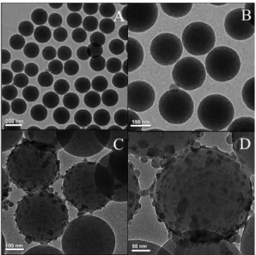
Figure 3.1 TEM images of A) The as synthesized polystyrene-co-acrylic acid (PSA)
nanoparticles, B) Silica coated PSA NPs (PSA/SiO2), C) NaYF4:Tm3+/Yb3+ tagged PSA/SiO2
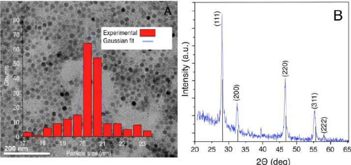
nanoparticle D) HRTEM of NaYF4:Tm3+/Yb3+ tagged PSA/SiO2 nanoparticle.---48 Figure 3.2 TEM (A) and XRD (B) analysis of as synthesized NaYF4:Tm3+, Yb3+ UCNPs.---49
Figure 3.3 Left: FTIR spectra of PSA nanoparticles, PSA/SiO2 and PSA/SiO2
(epoxy)/NaYF4:Tm3+, Yb3+; Right: Optical upconversion emission images of (A) as-synthesized
NaYF4:Tm3+,Yb3+ in hexane, (B) citrate stabilized NaYF4:Tm3+,Yb3+ in water, (C)
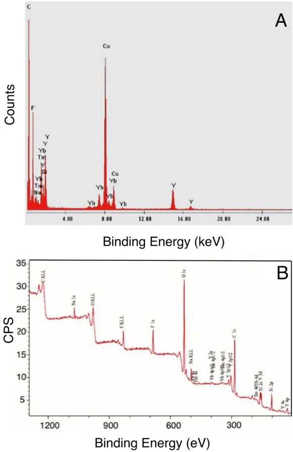
NaYF4:Tm3+,Yb3+ decorated PSA/SiO2 nanohybrid.---50 Figure 3.4 Representative (A) EDX and (B) XPS of NaYF4:Tm3+, Yb3+ decorated PSA/SiO2
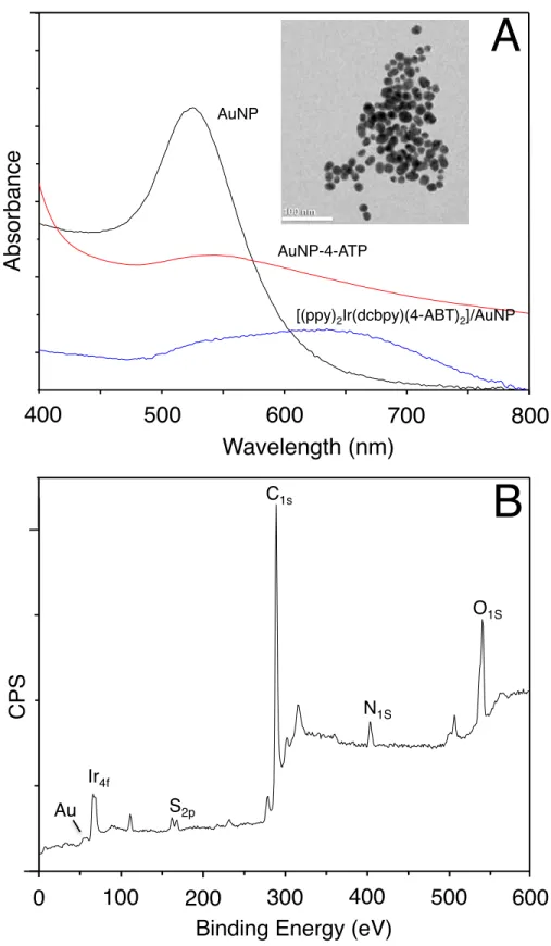
nanoparticles. Both EDX and XPS confirmed the presence of dopant ions (Tm3+, Yb3+) of the NaYF4 host crystal lattice on the PSA/SiO2 nanoparticles.---52 Figure 3.5 (A) Comparison of UV absorptions of AuNPs, 4-ABT modified AuNP and Ir(III)
attached AuNPs through thiol linker 4-ABT (inset: TEM of AuNPs) and (B) XPS of Ir (III) decorated AuNPs through 4-ABT linker.---54
Figure 3.6 Photophysical properties of the synthesized Ir complex, [(ppy)2Ir(dcbpy)]+PF6- (the
structure and optical images of Ir complex under natural and UV light are shown in the inset).--- 55
Figure 3.7 (A) UV-Vis absorption spectrum of the AuNPs (red line) and Ir complex
[(ppy)2Ir(dcbpy)(4-ABT)2] immobilized on AuNPs (black line). (B) Upconversion emission
spectrum of NaYF4:Tm3+, Yb3+ in hexane with corresponding energy level transitions following
excitation with 975 nm.---56
Scheme 3.1 (A) and (B) Proposed LRET-based detection of ssDNA using UCNP decorated
PSA/SiO2 nanohybrids and Ir(III)-AuNPs and (C) Plausible luminescent resonance energy
transfer from UCNP to Ir decorated AuNPs.---57
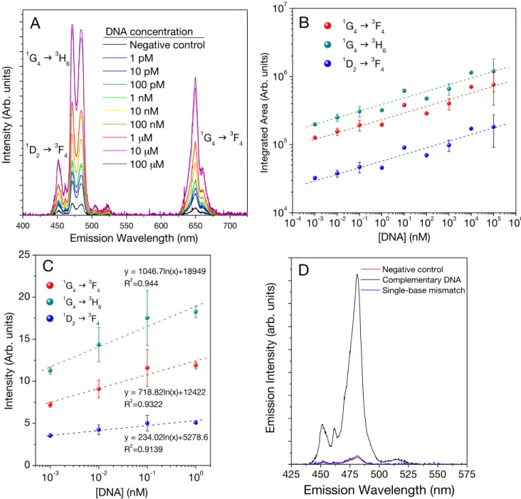
Figure 3.8 (A) Integrated area under the blue emission vs. concentration of complementary
DNA sequence (B) Upconversion spectra in the blue region of the sensor upon adding different concentrations of target ssDNA sequence. (C) Linear dependence of emission intensity at the lower target DNA concentration levels. (D) Comparison of luminescent emission intensities of complementary, negative control and single base mismatch DNA sequences.---60
Figure 3.9 Optical images of (A) as-synthesised AuNPs, (B) [(ppy)2Ir(dcbpy)]+ PF6− in
Ethanol, (C) PSA/SiO2 /NaYF4:Tm3+,Yb3+//ssDNA(Probe)//Ir(III)-AuNP, (D) PSA/SiO2
/NaYF4:Tm3+,Yb3+// dsDNA(Probe+Complementary)//Ir(III)-AuNP, before precipitation, (E)
PSA/SiO2/NaYF4:Tm3+,Yb3+// dsDNA(Probe+Complementary)//Ir(III)-AuNP after precipitation.---61 Figure 4.1 XRD spectrum of oleic acid capped UCNPs (black), with reference to the standard
(red) LiYF4 crystals (JCPDS No. 01-077-0816).---76 Figure 4.2 Transmission electron microscopic image of the synthesized (A) oleate-capped
LiYF4:Tm3+, Yb3+ (B) silica coated UCNPs (C) Particle size distribution of the oleate capped
UCNPs for length and width..---77
Figure 4.3 FTIR spectra of oleate-capped LiYF4:Tm3+, Yb3+ UCNPs (blue), SiO2 coated
UCNPs, LiYF4:Tm3+, Yb3+@SiO2 (black) and curcumin functionalized SiO2 coated UCNPs,
LiYF4:Tm3+, Yb3+@SiO2-curcumin (green).---78 Figure 4.4 Loading of curcumin on the LiYF4:Tm3+, Yb3+@SiO2 UCNPs.--- 79
Figure 4.5 Photophysical properties of curcumin, UCNPs and Cu2+ ion. Upconversion
emission spectrum of LiYF4:Tm3+, Yb3+@SiO2 (black) with their respective energy level
transitions; UV-vis absorption spectrum curcumin (blue); emission spectrum of curcumin (green, λ =360 nm); UV-vis absorption spectrum Cu2+ (red).---81
Scheme 4.1 Energy level transitions in the LiYF4:Tm3+, Yb3+ UCNPs and the plausible LRET
between the UCNPs and surface functionalized organic natural product, curcumin.---82
Figure 4.6 (A) UV to NIR UC Luminescence spectra of the curcumin loaded LiYF4:Tm3+,
Yb3+@SiO
2 UCNPs. (B) Gradual decrease in the UV emission intensity of UCNP upon
successful loading of curcumin. (C) Decrease in the blue emission of Tm3+ in the UCNP due
to strong absorption by the surface tagged curcumin. (D) Increase in the green emission at 515 nm due to the LRET from the curcumin to the UCNP.---83
Figure 4.7 (A) Gradual decrease in the upconverted green luminescence intensity of the
LiYF4:Tm3+, Yb3+@SiO2-curcumin UCNPs upon successful titration of various concentrations
of Cu2+ ions. (B) Decrease in the upconverted green emission intensity with reference to the upconverted blue emission confirming the absence of energy transfer due to the presence of increased Cu2+ concentrations. (C) Increase in the upconverted blue emission intensity at 450
nm with reference to the upconverted green emission at 510 nm also confirms the absence of energy transfer between curcumin and the UCNPs.---84
Figure 4.8 (A) Gradual decrease in the upconverted NIR intensity of LiYF4:Tm3+, Yb3+@SiO2
-curcumin UCNPs upon successful titration of different concentrations of Cu2+ ions. (B)
Stern-Volmer plot of Cu2+ triggered luminescence quenching of the upconverted NIR emission of the LiYF4:Tm3+, Yb3+@SiO2-curcumin UCNPs. (C) Plot of log[(I0−I)/I] against log[Cu2+] for the
LiYF4:Tm3+, Yb3+@SiO2-curcumin UCNPs and the quencher (Cu2+).---86 Figure 4.9 UV-vis absorption spectrum of LiYF4:Tm3+, Yb3+@SiO2-curcumin UCNPs in the
presence of different concentrations of Cu2+ ions. (B). The decrease in absorbance intensity
showing excellent linear relationship.---87
Figure 4.10 UV-vis absorbance of LiYF4:Tm3+, Yb3+@SiO2-curcuminUCNPs against different
metal ions in the showing the high selectivity of the proposed nanohybrid material (Top); Optical images of LiYF4:Tm3+, Yb3+@SiO2-curcuminUCNPs in the presence of different metal
ions (Bottom) (Grey bar: LiYF4:Tm3+, Yb3+@SiO2-curcuminUCNPs in the presence of various
metal ions before adding Cu2+; Burgundy bar: LiYF
4:Tm3+, Yb3+@SiO2-curcuminUCNPs in the
presence of various metal ions after adding Cu2+).---88 Figure 4.11 The selectivity of Cu2+ ion towards curcumin at the NIR region. Among all the
ions tested, only Cu2+ absorbs in the NIR showing significant reduction in the NIR emission of UCNPs due to [Cu2+
Figure 4.12 The absorbance spectrum of LiYF4:Tm3+, Yb3+@SiO2-curcuminUCNPs in the
absence of Cu2+ (Black), in the presence of Cu2+ (Red) and after reaction with EDTA (Blue). The splitting of the peak upon addition of Cu2+ is due to the π→π* and ligand to metal charge
transfer transition.---90
Scheme 5.1 980 nm excitation of the LiYF4:Tm3+,Yb3+@SiO2-Ir nanostructure for the NIR
triggered generation of ROS.---104
Figure 5.1 (A) Characteristic TEM image of the oleate-capped LiYF4:Tm3+,Yb3+ and (B)
LiYF4:Tm3+, Yb3+@SiO2 UCNPs. (C) Particle size distribution of the oleate-capped UCNPs
showing both the lengths and the widths. The aspect ratio was calculated to be approximately 1.73.--- ---106
Figure 5.2 DLS results of the LiYF4:Tm3+, Yb3+@SiO2-Ir nanostructures. Average particle
diameter was determined to be 135 nm. Note: All DLS measurements were performed in PBS buffer solution and the LiYF4:Tm3+, Yb3+@SiO2-Ir nanostructures were highly stable.---106 Figure 5.3 XRD pattern of LiYF4:Tm3+, Yb3+@SiO2 UCNPs along with the corresponding
reference pattern for LiYF4 crystals (JCPDS-01-077-0816).---107 Figure 5.4 (A) FTIR spectra of the parent oleate-capped LiYF4:Tm3+, Yb3+ UCNPs (black
line), UCNPs@SiO2 (blue line) and UCNPs@SiO2-Ir nanostructures (green line). (B) XPS
spectrum of the LiYF4:Tm3+, Yb3+@SiO2-Ir nanostructures.---109 Figure 5.5 Loading efficieny of Ir complex on the UCNPs@SiO2 nanohybrid.---110 Figure 5.6 ESI-MS Spectrum of the final Ir complex.---110 Figure 5.7 (A) Absorption spectrum of the synthesized Ir complex (blue line) and
upconversion luminescence spectrum of the LiYF4:Tm3+,Yb3+@SiO2 UCNPs (black line) and
(B) upconversion luminescence spectrum of the UCNPs following functionalization with the Ir complex. λexc = 980 nm.---112
Figure 5.8 Energy level diagrams of Yb3+ and Tm3+ ions showing the different mechanisms
leading to the observed upconverted emissions following 980 nm excitation. Curved arrows indicate nonradiative energy transfer from excited Yb3+ ions to the Tm
3+ ions sequentially
populating the various excited states; The dotted arrows pointing downwards indicate multiphonon relaxation to the lower-lying levels; the connected arrows in various shades of
Figure 5.9 (A) Total upconversion emission spectra of the LiYF4:Tm3+,Yb3+@SiO2 UCNPs
obtained following functionalization with different concentrations of the Ir complex. The spectra were normalized to the NIR emission band. The inset (B) shows the expanded region between 300 and 400 nm where quenching is observed. (C) Plot demonstrating the decrease in the UV emission intensity upon increased concentration of Ir complex for the LiYF4:Tm3+,Yb3+@SiO2-Ir nanostructures. (λexc = 980 nm).--- 114
Figure. 5.10 (A) Decrease in the absorption of the DPBF probe molecule in the presence of
LiYF4:Tm3+,Yb3+@SiO2 UCNPs decorated with the Ir complex at various time intervals. (B)
Comparison of the absorbance peak of DPBF at 420 nm after NIR irradiation at different times and for various samples, including LiYF4:Tm3+,Yb3+@SiO2 without the Ir complex,
LiYF4:Tm3+,Yb3+@SiO2 functionalized with two different concentrations of Ir complex (1 mM
and 6 mM), and the Ir complex alone (i.e. without UCNPs). (C) Comparative plot of ln(At/A0)
as a function of time. A0 is the initial absorbance, and At is the absorbance at different 980 nm
NIR irradiation times.---116
Scheme 6.1 Principle of ROS generation of SPION-Ir nanohybrid at direct UV excitation.-130
Figure 6.1 (A) and (C) are the TEM images of bare and SiO2 coated SPIONs, respectively,
with their respective particle size distribution histograms (B) and (D). (E) XRD pattern of the both bare (a) and SiO2 coated (b) SPIONs (JCPDS file No. 65-3107)..---132 Figure 6.2 (A) FTIR Spectra of (a) oleate-capped Fe3O4 SPIONs; b) SiO2 coated Fe3O4
SPIONs (the broken square box indicates the reduced intensity of the symmetrical and asymmetrical stretching vibrations of C-H of oleate and c) Ir complex decorated Fe3O4@SiO2
(the Fe3O4@SiO2-Ir) SPIONs. (B) XPS of the Fe3O4@SiO2-Ir SPIONs.---133
Figure 6.3 The UV-vis absorption and emission spectra of the Ir complex loaded SiO2 coated
SPIONs ; inset is the molecular structure of the Ir complex (left); The optical images of (a) Fe3O4@SiO2-Ir in the presence of (b) in the absence of magnet, (c) the cationic Ir complex
alone in 1:1 ethanol:water solution.---134
Figure 6.4 (A) Loading of the Ir complex on the SiO2 coated magnetic nanoparticles. The
loading percentage was calculated to be 68% Increased loading showed increased absorbance confirming the successful encapsulation of the Ir complex on the surface of SiO2.
(B) Decrease in the absorption of the DPBF probe in the presence of Fe3O4@SiO2 SPIONs
peak of DPBF at 420 nm after 360 nm irradiation at different time intervals including Fe3O4@SiO2 SPIONs without the Ir complex, Fe3O4@SiO2 SPIONs functionalized with two
different concentrations of Ir complex (5 mM and 10 mM). (D) Comparative plot of ln(At/A0) as
a function of time. A0 is the initial absorbance, and At is the absorbance at different time
LIST OF ABBREVIATIONS AND NOTATIONS
AuNPs Gold Nanoparticles CR Cross Relaxation
DLS Dynamic Light Scattering DNA De-oxy Ribonucleic Acid
EDC 1-ethyl-3-(3-dimethylaminopropyl)carbodiimide EDTA Ethylenediaminetetraacetic acid
EDX Energy Dispersive X-ray Spectroscopy EMU Energy Migration Mediated Upconversion ESA Excited State Absorption
ETU Energy Transfer Upconversion
FRET Fluorescence Resonance Energy Transfer FTIR Fourier Transform Infrared Spectroscopy GSA Ground State Absorption
I Intensity of Emitted Light I0 Intensity of Incident Light
Ir (III) Iridium Complex LOD Limit of Detection
LRET Luminescence Resonance Energy Transfer MB Molecular Beacon mM Millimolar NHS N-hydroxysuccinimide NIR Near-infrared nm nanometer nM Nanomolar
NMR Nuclear Magnetic Resonance Spectroscopy NPs Nanoparticles OA Oleic acid ODE 1-Octadecene OM Oleylamine PA Photon Avalanche PAA Polyacrylic acid
PBS Phosphate Buffer Saline PDT Photodynamic Therapy PEG Polyethyleneglycol PEI Polyethyleneimine PL Photoluminescence pM Picomolar
PSA Polystyrene-co-acrylic Acid PSD Particle Size Distribution PVP Polyvinylpyrrolidone ROS Reactive Oxygen Species
SPIONs Superparamagnetic Iron Oxide Nanoparticles TEM Transmission Electron Microscopy
TFA Trifluoroacetic Acid UC Upconversion
UCNPs Upconverting Nanoparticles UV-Vis Ultraviolet-Visible
XPS X-Ray Photoelectron Spectroscopy XRD X-Ray Diffraction Spectroscopy
λem Emission Wavelength
λex Excitation Wavelength
CHAPTER 1 - INTRODUCTION
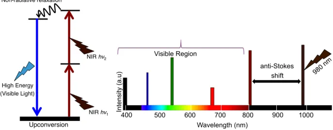
Upconversion is a non-linear optical phenomenon in which low energy NIR light is converted to higher energy UV, visible, and NIR emissions. The upconversion phenomenon has been observed mainly in the lanthanide (Ln3+) materials. Tri-positive Ln3+ ions have 4f n 5d 0-1 inner electronic configurations that are strongly shielded by overlying 5s and 5p orbitals. In addition, they have abundant, ladder-like 4f energy levels, which facilitate energy transfer between adjacent levels [1]. Ln3+ ions possess sharp emission peaks due to intra 4f-4f transitions, and although this transition is symmetry forbidden, it becomes partially allowed due to intermixing of 4f orbitals with the 5d orbitals in Ln3+ [2,3]. Due to the versatility of the energy levels involved, Ln3+ ions offer extensive photophysical and photochemical properties [4-6]. In particular, the anti-Stokes upconversion phenomenon is different from the conventional two-photon (or multiphoton) absorption process in which, two or more low energy photons are absorbed simultaneously through its virtual metastable state. On the other hand, upconversion involves sequential absorption of NIR photons and makes use of the real, long-lived (µs to ms) intermediate states [7]. The presence of these real intermediate states in the upconversion process acts as photon reservoirs, and hence, the efficiency of the upconversion process is generally several orders of magnitude higher than that of two-photon absorption [8]. This unique advantage of upconversion enables the process to be realized by an inexpensive continuous wave (CW) diode laser instead of the much more expensive ultrafast pulsed (femtosecond) lasers [8]. Since this unique frequency upconversion does not exist in biomolecules, UCNPs are well suited for bio-applications in sensing, imaging, therapy and drug delivery [9,10]. In particular, the large anti-Stokes shift paves the way for considerable difference between excitation and emission wavelengths in the electromagnetic spectrum, which permits multiplexed sensing, and imaging [11].
Non-radiative relaxation! Upconversion! NIR hv1! High Energy ! (Visible Light)! NIR hv2! 400! 500! 600! 700! 800! 900! 1000! 980nm ! Anti-Stokes! Shift! Visible Region! Wavelength (nm)! In te n si ty (a.u )! 400 500 600 700 800 900 1000 980 n m anti-Stokes shift Visible Region Wavelength (nm) In te nsi ty (a.u )
Figure 1.1 Fundamental upconversion mechanism and anti-Stokes shift following NIR excitation
Since the transition occurs within the 4f energy levels in the Ln3+, the emission intensities are sharp and cannot be influenced by external factors. Hence, they are resistant to photo-bleaching [12] yielding stable luminescence and offer the advantage of being used for long-term repetitive and dynamic imaging [13]. Since the process of upconversion takes places following excitation with NIR wavelengths in the biologically transparent window, there is little to no background autofluorescence since the NIR excitation wavelength is specific only to the UCNPs. This allows for deep tissue penetration in biological tissues, less scattering and enhanced imaging capabilities [14].
1.1 Upconverting Nanoparticles (UCNPs)
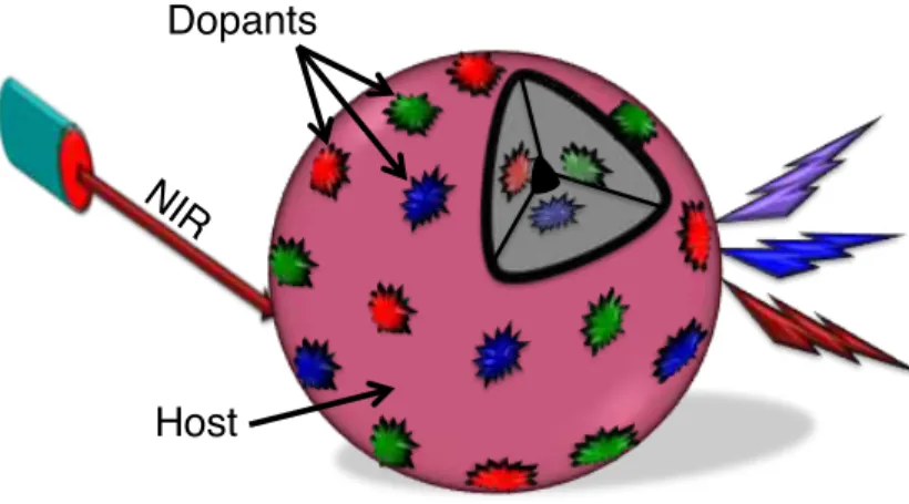
UCNPs are considered as binary systems that contain a host crystal and optically active Ln3+ dopants with dimensions preferably less than 100 nm. The Ln3+ dopants are well dispersed within the host crystals and also at the surface [29]. Upon excitation at NIR wavelengths, the emission wavelength could range from the deep UV to visible to NIR. One of the intriguing facts about UCNPs is that they have similar optical properties as that of bulk materials due to their intra 4f-4f forbidden transitions, which are well protected from the outer 5s and 5p orbitals [15,16]. However, their luminescence efficiency is very low compared to that of bulk materials due to nanoscale induced surface defects. Due to the high surface area-to-volume ratio of UCNPs, most of the optically active emissive Ln3+ dopants are exposed to deleterious surface deactivation
these impurities increases the phonon decay process and thus reduces upconversion luminescence efficiency [17]. The surface deactivation effect occurs in the following ways: (i) The presence of surface quenching centers located near the optically emissive dopants enhances the phonon energy loss; and (ii) random migration of energy from the optically active dopants which are located at the center of the UCNPs to their surface and subsequent energy loss [18]. In addition to the effect of host lattice, the concentration of Ln3+ dopants can also influence the upconversion efficiency. At higher concentration of emissive dopants, back energy transfer dominates which can lead to energy loss due to cross-relaxation process (to be discussed later in the thesis) and thus reduces upconversion efficiency [19]. Hence it has been a challenge to reduce the size of the nanocrystal and increase the luminescence efficiency. In order to achieve both, careful and judicious nanochemistry and surface engineering and functionalization of UCNPs are needed.
Figure 1.2 UCNP with host crystal and emissive dopants 1.1.1 Upconversion Host Crystal
Selection of a suitable upconversion host material is very crucial since they do influence the upconversion quantum yield and tuning of wavelength [20]. An ideal upconversion host material must be transparent in the wavelength range of interest, show high chemical stability, and should have high optical damage threshold. The host crystal would also have low lattice phonon energies in order to minimize non-radiative relaxation and thereby to enhance the upconversion luminescence [21]. Also, the host crystal should have minimum lattice mismatch with dopant ions. Among many reported
NIR !
Host! Dopants!
been employed extensively because of their low phonon energy, minimum lattice mismatch with dopants and stability towards moisture and optical transparency [22]. An extensive literature review reveals that hexagonal phase β-NaYF4 is the most preferred host material over the cubic phase α-NaYF4 due to stronger upconverted luminescence emission [23-25]. A significant advancement in reducing the size (less than 20 nm) of the UCNPs while simultaneously increasing upconversion intensity was reported by Yang. et al. for the Er3+ and Tm3+ doped NaLuF4 host crystal [26]. Although multi-colored emissions were observed with various dopants in the β-NaYF4 upconversion host, emission in the UV spectral range has been a challenge since it requires greater than three incident photons, which are less efficient than the two-photon visible emitting upconversion process. This was overcome by using LiYF4 as the host crystal, which was reported by Mahalingam et al. [27]. In their work, the authors synthesized, Tm3+/Yb3+ co-doped LiYF4 by a thermal decomposition process. Their work resulted in the highly monodispersed hydrophobic UCNPs which was eventually made water dispersible through silica coating. In the present work, we have synthesized and used fluoride based host crystals such as NaYF4 and LiYF4 as the UV, visible and NIR emitting donor for biosensing, chemical sensing and the generation of ROS.
1.1.2 Upconversion Sensitizer
Ytterbium ion (Yb3+) is the most widely used sensitizer for the upconversion process since the Yb3+ ion has a higher absorption cross section at the 980 nm NIR excitation wavelength [28]. Moreover, the Yb3+ ion has only one excited energy level and hence minimizes the non-radiative energy loss. The electronic energy level structure of Yb3+ is simple consisting of the ground state, 2F7/2 and only one excited state, 2F5/2. Since the sensitizer has only one excited state, changing its concentration will not cause any concentration related quenching (up to a certain point) [29]. High concentration of Yb3+ (greater than 18%) could help enhance the upconversion luminescence and has been reported along with Tm3+, Er3+, Ho3+ activator ions to achieve high upconversion luminescence [1].
1.1.3 Upconversion Activator
Tuning of upconversion luminescence requires judicious selection of activator/sensitizer combination. The Ln3+ dopant that emits the higher energy photon is known as the activator, and the most commonly used activator ions are erbium (Er3+), holmium (Ho3+) and thulium (Tm3+) as dopants in host crystals due to their abundant, long-lived and ladder-like electronic energy states [29]. Heer et al. first reported efficient upconversion emission in colloidal Ln3+-doped UCNPs [30]. They synthesized green emitting Yb3+/Er3+ and blue emitting Yb3+/Tm3+ co-doped NaYF4 host UCNPs. Later, Boyer et al. reported on the synthesis of blue and green emitting, α-phase NaYF4 UCNPs with similar dopant combination in a single, one-pot thermal decomposition method [31]. Other ions from the Ln3+ series have also been reported as activators for various applications [32]. In our work, we have focused only on the blue emitting Tm3+ activator doped in different host materials (NaYF4 or LiYF4). The UV/blue emission is of particular interest in our work because of the expected luminescence energy transfer to the appropriate acceptor for bio and chemical sensing as well as ROS generation.
1.2 Upconversion Mechanisms
The upconversion process has been extensively investigated in recent years and has been proved to be a valuable method for generating UV, visible, NIR emission upon lower energy NIR excitation. Upconversion is a non-linear optical process where two or more low energy photons are converted to higher energy emission, most commonly in the visible range. Since the emitted photons have energy higher than the excitation energy, this process has been defined as an anti-Stokes effect. Different upconversion luminescence mechanisms have been proposed. However, there are four fundamental upconversion mechanisms such as excited state absorption (ESA), energy transfer upconversion (ETU), photon avalanche (PA) and cross relaxation (CR). Also, energy migration mediated upconversion (EMU), a recent mechanism proposed for core/shell type nanoparticles has also been discussed.
1.2.1 Excited State Absorption (ESA)
Excited state absorption (also called sequential absorption) involves sequential absorption of two photons (Figure 1.3a). The mechanism was first proposed by Bloembergen [33]. ESA is a single ion process and takes place in materials with low Ln3+ dopant concentration. In the first step, a photon is absorbed to promote the ion from the ground state (GS) to its first excited state also known as the metastable state (ES1). This step is known as ground state absorption (GSA). In the next step, another photon is absorbed to excite the ion from the intermediate state to its ES2 level. Subsequently, the excited ion radiatively relaxes back to the ground state by emitting a higher energy photon.
1.2.2 Cross Relaxation (CR)
This energy transfer process typically results from the ion-ion interaction between two identical Ln3+ dopants. In this process, two neighboring Ln3+ ions (Ln3+(1) and Ln3+(2)) are simultaneously excited to their first intermediate state by the incoming NIR photons. Subsequently, one of the Ln3+ ions, Ln3+(1), transfers its energy to the second Ln3+ ion, Ln3+(2) promoting Ln3+(1) from the first intermediate state (ES1) to the second intermediate state (ES2). Ln3+(2) then returns to the ground state (GS) (Figure 1.3b). The efficiency of the cross relaxation process depends on the dopant concentration and usually occurs in more heavily doped single ion systems. However, very high concentrations can result in back energy transfer and lead to vibrational decay, which results in the quenching of the upconversion luminescence.
1.2.3 Energy Transfer Upconversion (ETU) Among all the upconversion processes, energy transfer upconversion (ETU) is the most
efficient in Ln3+-doped UCNPs and was first reported by Auzel [34]. Typically, two different Ln3+ ions (sensitizer and activator) are involved in the ETU process. In a typical ETU process, the sensitizer ion accepts the incident low energy photons in its ground state (GSA) thereby promoting it to the upper excited level. Subsequently, the sensitizer ion in the excited state transfers its energy to a neighboring Ln3+ ion in its proximity (the activator) promoting it from the ground state to its first intermediate excited state (Figure
to the upper excited state. This is the most common mechanism in co-doped systems such as Er3+/Yb3+, Tm3+/Yb3+, and Ho3+/Yb3+ [29].
1.2.4 Photon Avalanche (PA)
Photon Avalanche (PA), sometimes called absorption avalanche, was first discovered by Chivian et al. and it has been considered as a more advanced and efficient upconversion process, although it is more rarely observed [35]. A two-ion system is demonstrated in Figure 1.3d. Ln3+(2) is involved in a weak ground state (non-resonant) absorption process exciting the ion to the first excited state (ES1). Low energy NIR light is also absorbed by Ln3+(1) and promoting it to the first excited level ES1. Subsequent energy transfer from the ES1 level of Ln3+(1) to the ES1 state of Ln3+(2) populates this level. This population promotes Ln3+(2) from energy level ES1 to ES2. This is followed by cross relaxation between ES2 of Ln3+(2) and ES1 of Ln3+(1). In the next steps, energy transfer from ES1 of Ln3+(1) to the ES1 level of Ln3+(2) populates this level and again promoting Ln3+(2) to its ES2 level. This creates a looping process and exponentially increases the populations of the ES2 level of Ln3+(2) and hence enhances the upconversion emission to a great extent thus leading to an avalanche process [36]. 1.2.5 Energy Migration Mediated Upconversion (EMU)
Energy migration mediated upconversion is usually observed in core-shell type UCNPs. There are four types of Ln3+ dopants employed such as the usual sensitizer, accumulator, migrator and activator (Figure 1.3e). The Ln3+ sensitizer dopant is used to collect the low energy NIR photons and transfers its energy to the adjacent accumulator ion at its ES1 level. Subsequent energy transfers promote the ion to its ES2 level. The role of the migrator ion is to receive the energy from the accumulator ion at its ES2 and transfer it through its sub-lattice and finally to the activator ion. All these energy transfers occur at their respective excited states only. The location of sensitizer/accumulator and accumulator/activator are controlled in such a way in the core/shell structure to reduce the energy loss by adverse cross relaxation process. The efficiency of EMU could be increased by a range of migrator ions through its core/shell structure to facilitate the energy transfer from the accumulator to the activator. An
acquired by the sensitizer can be transferred and stored in the accumulator which in turn transfers its energy in a step-wise manner to the adjacent migrator ion. The migrator ions carry this throughout the core/shell structure and finally to the activator ion. The process amasses the energy, and when the excited ion relaxes back from its ES2 level to the ground state, it emits higher energy photons. The core/shell composite structure offers the advantage of changing the concentration of the activator dopant at the same time minimizes the upconversion quenching. Hence, tunable upconversion luminescence can be achieved even for activators with short-lived intermediate states [37]. GS! ES1! !em ! h"! ET! ET!
Sensitizer! Accumulator! Migrator! Migrator! Activator! nX! Core! Shell! NIR Shell! Core! ES2! GS! ES1! ES2!
Excited State Absorption (ESA)! Ln3+ (1)! !em ! h"1 ! h"2 ! GS! ES1! ES2! GS! ES1! Cross Relaxation (CR)! Ln3+ (1)! ES2!Ln 3+ (2)! h"2 ! h"1 ! (a)! (b)! GS ES1 ES2
Energy Transfer Upconversion (ETU) GS ES1 !!"## ET h" ET Ln3+ (1) Ln3+ (2) (c)! (e)! GS! Photon Avalanche (PA)!
GS! ES1! ES1! ES2! !ex ! !em ! Ln3+ (1)! Ln3+ (2)! h" ! (d)! h" ! ET
Figure 1.3. Schematic of upconversion luminescence mechanisms. a) Excited-State Absorption (ESA), b) Cross Relaxation (CR), c) Energy Transfer Upconversion (ETU), d) Photon Avalanche (PA), and e) Energy Migration Mediated Upconversion (EMU). GS, ES1, and ES2 represent ground state, intermediate state, and excited state, respectively. GS, ES, ET denote ground state, excited state and energy transfer, respectively.
1.3 General Synthetic Methods for Upconverting Nanoparticles
There have been different chemical methods used for the synthesis of UCNPs including co-precipitation, hydrothermal (solvothermal), sol-gel, and thermal decomposition. 1.3.1 Co-Precipitation Method
The co-precipitation synthetic method offers several advantages such as, simple operating procedure, fast synthesis, and mild reaction conditions. In addition, this method does not require any expensive laboratory setup [38]. Moreover, the size of the UCNPs can be readily controlled by using suitable surface ligands such as polyethyleneimine (PEI) [39], polyvinylpyrrolidone (PVP) [40], and by a strong hexadentate ligand, such as ethylenediaminetetraacetic acid (EDTA) [41] in the reaction mixture. Despite all these advantages, in some situations, the synthesis requires post heat treatment [42,43]. Moreover, the synthesis usually results in the formation of less efficient α-phase UCNPs. Since β-phase UCNPs have higher upconversion efficiency [44], it is imperative that calcination becomes necessary in order to obtain the β-phase. However, calcination can only be carried out at higher temperature and this poses a major problem for laboratory conditions [45].
1.3.2 Hydrothermal (Solvothermal) Method
The hydrothermal (solvothermal) method has been used to synthesize both α-phase and β-phase UCNPs. In this method the solubility of precursor materials can be increased under the hydrothermal or solvothermal conditions, which effectively increases the rate of the reaction [46-48]. This method produces UCNPs with excellent crystallinity, tunable size, and crystal morphology by controlling the reaction temperature, time, precursor concentration, as well as pH value [49-52]. In addition, this
no post heat treatment is required. Like any other method, the hydrothermal (solvothermal) method has a few drawbacks; requirement of specialized reaction container (autoclave) and manipulation of the reactions under a set of reaction temperature as well as pressure conditions over a long period of time. In most of these reactions, water is a major solvent however in some situations other hydrophilic solvents could be used in order to improve the solubility of the precursor materials. Such reactions are known as solvothermal synthesis [55].
1.3.3 Sol-Gel Method
The sol-gel method is a typical wet-chemical process generally used for the synthesis of UCNPs. This method is distinct in the sense that the synthesis has been carried out by hydrolysis and polycondensation of metal alkoxide precursors and the nanoparticle growth is feasible in reverse micelle emulsions. Over the years, this method was extended to the synthesis of various UCNPs with mixed metal oxides such as Er3+ -doped TiO2, ZrO2, BaTiO3, and YVO4 [56-58]. Sivakumar et al. produced white light upconversion as well as red and green following NIR excitation using sol-gel based Ln3+-doped UCNPs [59,60]. However, in spite of all these advantages, the nanoparticles synthesized by the sol-gel method find little or no applications in the development of biological assays due to particle aggregation and production of polydispersed nanoparticles [29].
1.3.4 Thermal Decomposition Method
The thermal decomposition method is by far the best method for the controlled synthesis of highly monodispersed and precisely defined UCNPs [27,31,61-65]. In this method, metal trifluoroacetate (TFA) precursors are dispersed in a high boiling, non-coordinating solvent such as octadecene and with a suitable capping ligand such as oleic acid, oleylamine, or trioctylphosphine (at temperatures between 250 °C and 330 °C. The presence of capping ligand/surfactant helps control the growth of the UCNPs in the solution mixture. Due to the high temperature and the sensitivity of UCNPs towards oxygen impurities, control over the reaction must be precise in order to produce good-quality monodispersed nanoparticles. Other factors such as pressure, capping ligand,
solvent and reagent concentrations also play an important role in the crystalline size and morphology [66]. Since this method uses the air sensitive TFA precursors, toxic fluorinated and oxyfluorinated compounds are released as byproducts during the reaction hence this synthesis must be carried out in a well-ventilated fume hood. In addition, this method produces UCNPs, which are hydrophobic (oleate-capped) and hence further surface modification is required in order to make them water dispersible for biological applications [27].
1.4. Surface Modification of UCNPs
For targeted biological applications it is imperative that the synthesized UCNPs can be dispersed in aqueous media and bear specific functional groups amenable to biomolecules at their surface [67]. Hence, surface functionalization of UCNPs is needed for immunoassay [68-70], imaging [71-77], DNA encoding [78], cancer therapeutic [79-82] and biosensing [83-86] applications. In order to make the UCNPs water-dispersible and apply them successfully in biomedical research, suitable surface functionalization and bioconjugation chemistries are prerequisites. [87-89]
1.4.1 Surface Modification Strategies
Over the years, many different strategies have been developed to convert hydrophobic UCNPs into more water dispersible hydrophilic particles [90]. These strategies could be divided into four categories: (1) chemical modification of the hydrophobic surface ligand such as oleic acid and oleylamine (2) coating with dual layer molecules such as amphiphilic reagent and polymers, (3) formation of an additional layer on the surface of UCNPs (4) complete displacement of the original native hydrophobic ligand followed by the addition of hydrophilic ligand. These strategies will be discussed briefly in the following sections.
(i) Modification of the Capping Ligand
In this method, the hydrophobic molecule present on the surface of the UCNPs could be altered/modified by using a suitable reagent, however, it has been infrequently used. The carbon-carbon double bond present in the oleic acid or oleylamine capping ligands
oxidizing agents such as the Lemieux-Von Rudloff reagent [91]. Other reagents such as ozone [92] and 3-chloroperoxybenzoic acid [93] have also been used to oxidize the surface ligand to incorporate appropriate functional groups that facilitate water dispersibility. Upon modification, these UCNPs could be attached to cancer drugs such as doxorubicin (DOX) to enable controlled drug delivery applications [94].
(ii) Coating with Amphiphilic Reagents
This method involves coating of the hydrophobic UCNPs with long alkyl chain molecules to form a dual layer at its surface. This layer is stabilized by Van-der-Waals attractive forces between the hydrophobic oleate moiety and the hydrophobic end of the amphiphilic reagent [95,96]. There are a few advantages of using amphiphilic molecules such as they (a) show strong Van-der-Waals interaction, (b) offer facile modification of surface charge, and (c) offer multiple layer formation thus protecting the UCNP surface from possible quenching effects. The attraction at the hydrophobic end results in the availability of hydrophilic functional groups directed outward and leads to the formation of a bilayer at the surface of the UCNPs making them well dispersible in water or any buffer solution for further bioconjugation. Another important class of amphiphilic reagent that is used to modify the surface of nanoparticles is the phospholipids. Phospholipid modified UCNPs could be promptly absorbed or internalized by cells [97] henceforth used for imaging purposes. For drug delivery applications, phospholipids have been widely used. Recently, Huang et al. developed green emitting UCNPs encapsulated in liposomes and loaded with DOX as a hybrid nanocomposite for drug delivery following 980 nm NIR excitation [98]. Different phospholipids such as maleimide or biotin are readily available commercially and their size could be engineered with different chain lengths or modified by adding poly(ethyleneglycol) (PEG) molecules, since PEG offers high biocompatibility [99,100]. In addition to UCNPs, other nanoparticles such as metal plasmonic AuNPs, quantum dots, or superparamagnetic iron oxide nanoparticles (SPIONs) have also been modified with amphiphilic polymers [101]. Other polymers such as poly(acrylic acid) with long alkyl chains could also be attached to the surface of hydrophobic oleate-capped UCNPs [102,103]. This endows negative charges on the surface of UCNPs due to the availability of carboxylic acid functional groups at neutral
could also help conjugate the nanoparticles to diverse biomolecules [104]. Reports also show that UCNPs may be coated with amphiphilic chitosan in light triggered drug delivery application such as photodynamic therapy [105].
(iii) Silica Encapsulation (Shell Formation)
Hydrophobic UCNPs can be made water dispersible (hydrophilic) by forming a thin layer of shell on their surface. Silica (SiO2) is the most commonly used shell material for this purpose. The formation of a SiO2 shell on the surface of UCNPs is a useful method to bring in different functionalities as well as making them water or buffer dispersible. Strategies such as the Stöber method or the reverse microemulsion method have been used for uniform shell formation. In the Stöber method, the nanoparticles were directly coated with SiO2 through a seeded polymerization technique using the tetraethyl orthosilicate (TEOS) precursor without the use of any stabilizer [106]. However, in the reverse microemulsion method, a surfactant known as Igepal CO-520 is widely used since it forms fairly stable reverse microemulsions for polymerization of the TEOS [107]. During the synthesis, ammonia is used as a catalyst since it causes the formation of silicic acid at a concentration above the nucleation concentration ensuring a steady growth of the SiO2 shell on the surface of the UCNPs [108,109]. In spite of the many advantages of SiO2 coating strategy, it does have a few disadvantages; SiO2 coated UCNPs might lead to polydispersity and necking, which is undesirable for bioapplications. Incomplete formation of silica shell will be hard to predict and additionally they tend to aggregate quickly.
(iv) Replacement of the Capping Ligand
Ligand replacement or substitution is another important strategy for surface modification of UCNPs. This can be achieved by two methods. The first is the direct (or single step) replacement of the capping ligand and second is a two-step process in which, first the capping ligand is completely removed from the surface of the UCNPs followed by attachment of the new hydrophilic ligand on its surface.
(a) Single Step Replacement of Capping Ligand
In this single-step method, the capping hydrophobic organic ligand on the surface of the UCNPs is completely replaced by the incoming more polar ligand, which gives the UCNPs water dispersibility. The advantage of this method is that the process is fast and multiple functional groups could be made available at the surface. In a typical procedure, the UCNPs with its capping ligand (in most cases oleic acid) and the new ligand are stirred at a suitable temperature followed by multiple washings. The resultant hydrophilic nanoparticles could be dispersed either in water or in a buffer solution [110]. However, the main disadvantage is particle aggregation, and frequent sonication is required before suitable bioapplications [111].
(b) Two-Step Replacement of Capping Ligand
Bogdon et al. [112] have developed a method that involves the reaction of oleate-capped hydrophobic UCNPs with hydrochloric acid. This reaction with a strong acid, HCl, strips the surface capping ligand, leaving naked UCNPs making them water or buffer solution-dispersible. In the next step, the bare UCNPs were coated with suitable ligands for targeted applications.
CHAPTER 2 – APPLICATIONS OF UPCONVERTING NANOPARTICLES
2.1 Biosensing
Sensing is an analytical process, in which a target compound/analyte is being detected by means of a detectable change in the signal produced. Based on the signal obtained the sensing mechanism can further be classified as optical, electrical, mass, thermal, magnetic and pressure (Figure 2.1). In all of these sensing principles, the pool containing the target analyte is subjected to a molecular recognition event in which the analyte will aptly bind to a suitable receptor. This binding event produces a measurable change in the signal. The change in the signal is further processed and displayed on a display unit [113,114]. Based on the type of target analyte/compound the sensors can broadly be classified as biosensor and chemical sensor. If the target analyte is biological in nature, then the sensor is known as a biosensor. On the other hand, if the analyte is chemical in nature then it is called a chemical sensor.
Figure 2.1 Basic principle and types of a sensor showing different components
The International Union of Pure and Applied Chemistry (IUPAC) defines the biosensor as a self-contained integrated receptor-transducer device that is capable of providing selective, quantitative or semi-quantitative analytical information using a biological
Detection principle and types of sensors!
!
Bio/Chemical Receptor! !
Molecular Recognition Event! Sample! Protein! Toxin! Peptide! Vitamin ! Sugar! Metal ion! Optical ! ! Electrical (Voltammetry, Potentiometry, Conductivity)! ! Mass! (QCM)! ! Thermal! ! Magnetic! ! Pressure! !
recent times, the significant challenge is the sensitive and selective signal production due to the biological recognition event (transduction) of the target analyte at the lowest concentrations [114].
2.1.1 DNA Biosensing
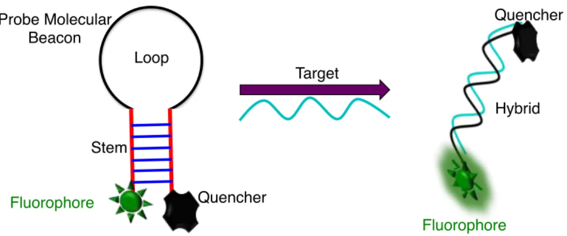
Recently, DNA biosensors that are based on the hybridization of nucleic acids have gained much interest [115,116]. To realize high sensitivity and selectivity, it is imperative that the DNA biosensors should be more robust. Recently efforts have been made to develop new DNA biosensors that use nanoparticles [117-119]. Using nanoparticles for DNA biosensor brings new advantages to this field. Firstly, since the biosensor depends on the size of the nanoparticles, more atoms are exposed as the size is reduced because of increased surface area. This enhances the available binding sites on the nanoparticle surface thus increasing the sensitivity many folds. Different types of nanoparticles have been employed in DNA biosensors including metal nanoparticles, such as gold or silver [118-120], semiconductor inorganic quantum dots [121-123] or carbon dots [124]. Although most of these nanoparticles are good candidates for DNA biosensing applications, inorganic semiconductor quantum dots possess inherent toxicity since most of the commonly used quantum dots contain toxic heavy metals thus limiting their applications in biological sciences. Also, they have blinking effects, which affects their application in long-term repetitive imaging. Most importantly, in many cases, inorganic quantum dots and carbon dots are excited under high energy UV light, which has limited penetration depths and increased scattering thereby reducing their sensitivity [125]. One of the most common mechanisms involved in nanoparticle-based sensing relies on the fluorescence resonance energy transfer (FRET) between the donor and acceptor. Use of FRET-based nanoparticles for sensing can have some drawbacks that must be taken into careful consideration such as inefficient energy transfer, uncertainty in the relative orientation of the donor-acceptor pair either before or after hybridization (Figure 2.2), reduced lifetime of the fluorophore, large background signals arising from the biological samples, and the requirement of labeling [126].
Figure 2.2 Mechanism for the detection of ssDNA target based on Fluorescence Resonance Energy Transfer (FRET).
In order to overcome some these undesirable properties, Ln3+ based UCNPs are beginning to gain attention in the field of biosensing. In our work, we intended to develop a new DNA nanobiosensor that uses, Ln3+ based UCNPs (see Chapter 3). Since these UCNPs could be excited at the biologically transparent near-infrared wavelength (980 nm), drawbacks such as background autofluorescence and scattering could be avoided. In addition, these UCNPs do not show any blinking, they have high chemical stability, reduced toxicity and the sharp emission wavelengths resulting from the intra 4f-4f transition could be effectively used in multiplexed detection of many target analytes [127].
2.2 Chemical Sensing
A chemical sensor, or a chemosensor, is a responsive material or a compound that shows an observable change in its physical property such as electrical, electronic, magnetic, or optical signal when it comes in contact with a target chemical analyte. Among many other sensors, fluorescent chemosensors offer significant advantages owing to their ease of development, sensitivity, and specificity and also, their response time can be easily monitored [128]. Heavy metal ions are considered as environmental pollutants, and due to their toxic nature, receptors that are capable of detecting a particular heavy metal ion in a complex mixture is of significant interest [129].
Mechanism of FRET Based ssDNA Detection!
Stem! Loop! Probe Molecular ! Beacon! Fluorophore! Quencher! Target! Hybrid! Fluorophore! Quencher!
2.2.1 Cu2+ Chemical Sensing
Among all the heavy metal ions, Cu2+ ion has been a very special element since it plays a pivotal role in the human body and also in other living organisms [133,134]. Although Cu2+ plays an important role in human health, its presence in higher concentrations may cause serious health issues such as Alzheimer and Parkinson diseases [135-137] Additionally, Cu2+ is one of the most versatile heavy elements that is found in drinking water in many developing and developed countries. Hence, the detection of Cu2+ ions in the presence of other metal ions and impurities is of significant importance. Developing a suitable sensitive as well as selective material for Cu2+ detection has been a challenge. Research has been carried out to develop sensors for Cu2+ based on fluorescence methods [138-140]. Thus far, most of the reported sensors for Cu2+ use water-insoluble organic dye molecules, which are not suitable for detection in real biological samples and also in limited resource environment. Recently there has been an increase in interest to develop nanoparticle-based Cu2+ detection with high sensitivity and selectivity [141-143]. Most of the nanosensors are based on ligand functionalization of metal plasmonic nanoparticles such as silver, gold, quantum dots, carbon dots as well as polymer-based nanoparticles [144-152]. Although these nanoparticles have advantages of their own, they suffer from broad absorption and emission wavelengths, size, and shape dependent optical properties, which could alter the limit of detection (LOD) in the nanoparticle solution if polydispersed [153,154]. Use of toxic quantum dots with heavy metal ions either in its core or shell could compromise the sensing of the target metal ion analyte due to leaching effects. All these limitations have significantly hampered the development of practical nanosensors for Cu2+ detection. Henceforth, NIR based chemosensors have gained much attention to overcome all these deleterious effects. In one of the methods for the detection of Cu2+, Zhang et al. [155] used green emitting Yb3+/Er3+ co-doped NaYF4 UCNPs as the energy donor following NIR excitation and an organic dye, rhodamine B hydrazide, as the acceptor through fluorescence resonance energy transfer. As mentioned previously, the reported organic dye used in this work has limited or no solubility in aqueous media hence affecting its LOD and makes the method not easily reproducible. In order to overcome all these
that the nanohybrid system composed of the highly biocompatible, natural product curcumin functionalized on the surface of UV to NIR emitting LiYF4:Tm3+, Yb3+ UCNPs can be used for sensitive detection of Cu2+ at NIR wavelengths following NIR excitation. The use of NIR as the detection wavelength increases its sensitivity and selectivity since most of the other metal ions, including toxic metal ions, do not absorb in the NIR region except for Cu2+ (see Chapter 4).
2.3 Reactive Oxygen Species
The term reactive oxygen species (ROS) is used to denote any molecular species or an ion derived from molecular oxygen with a free radical intermediate. Free radicals are species with a single or unpaired electron. The ground state of molecular oxygen has two single unpaired electrons (also called bi-radical) in its outermost orbital. Since these two electrons have the same spin in its ground state (triplet state), they remain stable and not reactive. In a situation where if one of the two single (odd) electrons gets excited to the upper energy level, which results in the electron having opposite spin then the resulting molecular oxygen species becomes highly reactive, called a singlet oxygen species [156]. Gerschman et al. suggested that molecular oxygen species with free radicals could have toxic effects for aerobic organisms [157]. Over the decades, the terms reactive oxygen species (ROS), reactive oxygen intermediates (ROI) and reactive nitrogen species (RNS) have been used to define any molecular species with different reactive intermediates. In general, these reactive species are endogenous and highly reactive molecules having either oxygen or nitrogen with an unpaired electron. When molecular oxygen species undergoes incomplete reduction, it could produce the superoxide radical anion (O2˙−) and hydroxyl radicals (OH˙). The term ROS includes both reactive oxygen intermediate and also (O3) and singlet oxygen (*1O2) [158]. Sometimes the ROS defines species with more than one oxygen atom and includes the peroxy (ROO˙), alkoxy (RO˙), semiquinone anion (SQ˙−) and carbonate anion (CO3˙−) radicals [159]. Research has concluded that ROS such as hydroxyl and peroxy radicals are responsible for oxidative damage to biomolecules such as DNA, fatty acids and many other cellular components [160] and overproduction of ROS is the major reason
![Figure 3.6 Photophysical properties of the synthesized Ir complex, [(ppy) 2 Ir(dcbpy)] + PF 6 - (the structure and optical images of Ir complex under natural and UV light are shown in the inset)](https://thumb-eu.123doks.com/thumbv2/123doknet/5006278.125035/75.918.230.646.304.623/figure-photophysical-properties-synthesized-structure-optical-complex-natural.webp)
![Figure 3.7 (A) UV-Vis absorption spectrum of the AuNPs (red line) and Ir complex [(ppy) 2 Ir(dcbpy)(4- Ir(dcbpy)(4-ABT) 2 ] immobilized on AuNPs (black line)](https://thumb-eu.123doks.com/thumbv2/123doknet/5006278.125035/76.918.151.702.150.582/figure-absorption-spectrum-aunps-complex-dcbpy-immobilized-aunps.webp)