HAL Id: tel-00806206
https://tel.archives-ouvertes.fr/tel-00806206
Submitted on 29 Mar 2013HAL is a multi-disciplinary open access archive for the deposit and dissemination of sci-entific research documents, whether they are pub-lished or not. The documents may come from teaching and research institutions in France or abroad, or from public or private research centers.
L’archive ouverte pluridisciplinaire HAL, est destinée au dépôt et à la diffusion de documents scientifiques de niveau recherche, publiés ou non, émanant des établissements d’enseignement et de recherche français ou étrangers, des laboratoires publics ou privés.
of paraplegics using FES: From simulations to
experiments
Jovana Jovic
To cite this version:
Jovana Jovic. Towards a functional assistance in transfer and posture of paraplegics using FES: From simulations to experiments. Other. Université Montpellier II - Sciences et Techniques du Languedoc, 2012. English. �tel-00806206�
UNIVERSITÉ MONTPELLIER 2
SCIENCES ET TECHNIQUES DU LANGUEDOC-THÈSE
pour obtenir le grade de
DOCTEUR DE L’UNIVERSITÉ MONTPELLIER 2
Discipline: Génie Informatique, Automatique et Traitement du SignalFormation Doctorale: Systèmes Automatiques et Microélectronique École Doctorale: Information, Structures et Systèmes
présentée et soutenue publiquement par
Jovana J
OVI ´
C
le 26 Octobe 2012
Titre :
Vers une assistance fonctionnelle du transfert et
de la posture chez le sujet paraplégique sous
électrostimulation : de la simulation a
l’ expérimentation.
JURY:
M. Dejan Popovi´c Professeur, ETF Belgrade Rapporteur Mme. Agnès Roby-Brami Directrice de recherche, ISIR Paris Rapporteur M. Philippe Souères Directeur de recherche, CNRS, LAAS Examinateur M. Philippe Fraisse Professeur, LIRMM, UM2 Directeur de thèse Mme. Christine Azevedo-Coste Chargée de recherche, INRIA Co-directrice de thèse M. Charles Fattal Médecin, CMN PROPARA Co-directeur de thèse
A N D P O S T U R E O F PA R A P L E G I C S U S I N G F E S :
F R O M S I M U L AT I O N S T O E X P E R I M E N T S
j ova na j ov i ´
c
LIRMM
Laboratoire d’Informatique, de Robotique et de Microélectronique de Montpellier INRIA
Institut national de recherche en informatique et en automatique
October 26, 2012
posture chez le sujet paraplégique sous électrostimulation : de la simulation a l’ expérimentation) c October 26, 2012
This research project would not have been possible without the support of many people. In the first place I wish to express my gratitude and appreciation to my supervisors, Christine Azevedo Coste and Philippe Fraisse for giving me the opportunity to work in the research area of rehabilitation engineering and for offering me invaluable assistance and guidance during this research. Among the many kindnesses they have shown me, they generously gave their encouragement and support whenever I needed it and for that I am most thankful. It has been a great pleasure working with them. I also extend my deepest gratitude to a member of the supervisory committee, Charles Fattal whose knowledge and assistance were vital to the success of this study.
I would like to acknowledge the members of the defense committee, Dejan Popovi´c, Agnès Roby-Brami and Philippe Souères for their enlightening suggestions, which helped to improve the quality of this dissertation, and for inspiring discussion during the oral defense, which I will always remember.
I would also like to thank all the subjects who participated in my studies, for being patient and having confidence in our work.
Special thanks to David Giraud and all the DEMAR team members for many scientific discussions, help and friendship over the past three years. I have enjoyed the work I have done in collaboration with Vincent Bonnet and Sébastien Lengagne and for that I wish to thank them as well. I would like to thank Master student Camilla Pierella who aided me in completing this research. I am most grateful to Anne Aliaga for dealing with administration and bureaucratic matters during my stay in France, and to Catherine Carmeni for improving the text and my English writing.
I have made many friends along the way. Many thanks to Pedro, Renata, Mariana, Syria, Elia, Alvaro, Vincent, Marcus, Olivier, Jérémie, Georgios, Miguel, Johann, Alonso, Rogerio, Camilla and Antoine. Special thanks to closest friends Divine, Pawel, Julia, Alejandro, and Maxim who have always been there for me. It has been a wonderful experience and an honor knowing all of you and spending a part of the life with you. I am sure that our friendship will continue no matter how far we are one from each other.
I especially wish to thank Flore Lévy and all the members of ArtPole Studio in Mont-pellier for sharing the same passion and for all great times we spent dancing together.
Let me not forget my Serbian friends Milica, Drazen, komsa Igor, Tamara, Ivan baraba, Joja, Bole, Dare, Jovan, Dragana, Mr and Mrs Martinovic, Mr and Mrs Svilar and Mr and Mrs Tomic for their support. Special thanks to Bojana, Alex and Buda, who, under supervision of Goran Svilar, had a small part in writing this document. Finally, I am most grateful to my dearest medical doctors, Ivanovic, Stanulovic and Jelenkovic, for their support and help.
have done for me. I also wish to express my love and gratitude to my beloved family for their support and love through the duration of these studies. Jelena, Gorica, Mosa, Jovan, Ivanka, Sladja i Sasa, hvala vam na svoj pomoci i podrsci tokom poslednjih godina. Najvece hvala Slavici, Veri i Vuletu koji nisu vise sa nama.
Today there are around 90 million people suffering from Spinal Cord Injury (SCI) world-wide. Thanks to the progresses in emergency medical care, problems faced by SCI indi-viduals after an accident are no longer life threatening. This has generated a shift of the priorities in medical research and practice towards improvements in their quality of life. The use of Functional Electrical Stimulation (FES) for motion restoration in paralyzed limbs proved to have a potential to provide both functional and therapeutic benefits. The aim of this thesis is to investigate solutions which would improve quality of life of para-plegics by restoring the sit-to-stand, transfer from one surface to another and standing tasks. We aim at finding a good trade-off between achievable functionality of the FES system and its simplicity in terms of number of required sensors and computational cost and, accordingly, its applicability in clinical practice and daily life of paraplegic individuals.
R É S U M É
Aujourd’hui, 90 millions de personnes dans le monde souffrent de lésions de la moelle épinière. Grâce aux progrès de la prise en charge des patients, leur espérance de vie est comparable à celle du reste de la population et la priorité de la recherche médicale est désormais consacrée à l’amélioration de leur qualité de vie. La stimulation électrique fonctionnelle (SEF) permet de restaurer les mouvements des membres paralysés et a des bénéfices thérapeutiques et fonctionnels. L’objectif de cette thèse est de proposer des so-lutions pour l’assistance au lever de chaise, au transfert d’une surface à une autre et à la station debout. Nous souhaitons trouver un compromis entre fonctionnalité et simplicité d’utilisation en termes de nombres de capteurs requis et de complexité calculatoire et donc favoriser l’application en environnement clinique et privé.
International Journals
1. J. Jovic, C. Azevedo Coste, P. Fraisse, C. Fattal
Upper and lower body coordination in FES-assisted sit-to-stand transfers in paraplegic subjects - A case study.
Paladyn, Journal of Behavioral Robotics 2(4): 211-217, 2011.
2. J. Jovic, S. Langagne, P. Fraisse, C. Azevedo Coste
Impact of Functional Electrical Stimulation of knee joints during Sitting Pivot Transfer motion for paraplegic people.
International Journal of Advanced Robotic Systems, N/A, 2012.
Reviewed Conference proceedings
1. J. Jovic, V. Bonnet, C. Azevedo Coste, P. Fraisse A Paradigm for the Control of Upright Standing in Paraplegic Patients.
EMBC’2012 : 34th Annual International Conference of the IEEE Engineering in Medicine and Biology Society, USA (2012).
2. S. Lengagne, J. Jovic, C. Pierella, P. Fraisse, C. Azevedo Coste Generation of Multi-Contact Motions with Passive Joints: Improvement of Sitting Pivot Transfer Strategy for Paraplegics.
BioRob’2012 : IEEE International Conference on Biomedical Robotics and Biomecha-tronics, Italy (2012).
3. J. Jovic, C. Azevedo Coste, P. Fraisse, V. Bonnet, C. Fattal Optimization of FES-assisted rising motion in individuals with paraplegia.
Skills Conference 2011, France (2011).
4. J. Jovic, C. Azevedo Coste, P. Fraisse, C. Fattal Decreasing the arm participation in complete paraplegic FES-assisted sit to stand.
IFESS’2011 : 16th Annual International FES Society Conference, Brazil (2011).
5. J. Jovic, V. Bonnet, P. Fraisse, C. Fattal, C. Azevedo Coste Improving Valid and De-ficient Body Segment Coordination to Improve FES-Assisted Sit-To-Stand in Paraplegic Subjects.
ICORR’2011 : 12th International Conference on Rehabilitation Robotics, Switzer-land (2011).
1. J. Jovic, C. Azevedo Coste,M. Benoussaad, P. Fraisse, C. Fattal Optimizing FES-assisted sit to stand transfer initiation in paraplegic individuals using trunk movement information.
ISEK’2010: International Society of Electrophysiology and Kinesiology, Aablorg, Danemark (2010).
Today, approximately 90 million people worldwide live with Spinal Cord Injury (SCI). The type of paralysis- tetraplegia (quadriplegia) or paraplegia- depends on the lesion level. In tetraplegia, the person has sustained a loss of feeling and/or movement in both upper and lower limbs. Paraplegia is the general term for the loss of feeling and/or movement in the lower part of the body. This thesis focuses on individuals who are complete paraplegic, i.e., with the loss of sensory and motor functions in their lower extremities. Thanks to improved emergency medical care, many of the problems faced by individuals with paraplegia following an accident are no longer life threatening, and their life expectancy is now comparable to that of able-bodied individuals. This has generated a shift in the priorities of medical research and practice away from survival and toward improvements in the quality of life for those living in a wheelchair. Inde-pendence is a major goal in rehabilitation. The ability to move and transfer is central to autonomous living, and therefore the manual wheelchair has become an important assistive device. However, wheelchair users are subjected to intense loads on the upper trunk and Upper Limb (U/L) muscles and joints during wheelchair propulsion, and al-most every other daily activity such as transfer, driving and household activities. Conse-quently, musculoskeletal pain is a common complication. While the primary injury itself greatly limits personal independence, any further functional limitation due to secondary complications, could cause a decrease or even a total loss in the remaining functional independence. Due to limited mobility, the paraplegic population also faces many other medical problems related to bone loading, cardio-circulatory stimulation, and metabolic changes which increase the risk of diabetes, joint extension, pressure sores, and muscle spasticity.
Functional Electrical Stimulation (FES) to restore motion in paralyzed limbs has shown the potential to provide both functional and therapeutic benefits. The advantages of FES over conventional rehabilitation treatments are numerous. Movement restoration by FES in patients with SCI has been a subject of research for many years. Nevertheless, muscle fatique occurs rapidly in the context of FES induced muscle contractions and thus the duration of functional movements is quite limited. Consequently, the number of effective FES systems is still limited.
In light of these observations, this work aimed to develop solutions to improve the quality of life of people with SCI by restoring the movements of sit-to-stand, transfer from one surface to another and standing that are lost due to SCI. We aimed to find a good trade-off between the achievable functionality of the FES system and its simplicity in terms of the number of required sensors and the computational cost and,
Chapter 1 gives a brief description of the natural control of movement in physically intact individuals and introduces SCI pathology. A short review of some of the solutions proposed to restore movement in paraplegic patients is presented, and the principles of FES are described.
Chapter 2deals with FES-assisted sit-to-stand movement in the paraplegic population. Ways to move that minimize arm efforts and reduce the muscle fatigue induced by FES have been investigated. The chapter also validates a new closed-loop system for sit-to-stand transfer to meet the needs of paraplegic individuals through experiments with six paraplegic patients. The criterion for patient selection, the experimental setup and the protocols are explained.
In Chapter 3 the benefits of FES during sitting-pivot-transfer motion in paraplegic patients are investigated using an optimization process and biomechanical modeling of the human body. The simulation results are presented.
Chapter 4proposes a new solution for 3D control of standing posture in SCI patients by means of FES. The proposed controller should enable prolonged standing and should be able to cope with the patient’s voluntary movements. The simulation results are pre-sented.
Chapter 5summarizes the results and conclusions of this thesis and proposes perspec-tives for future investigations.
Additional aspects related to this work are described in the Appendix. This include the statement of the local ethics committee’s approval of the protocol for performing experiments with paraplegic patients, additional results fromChapter 2and descriptions of the software developed for the movement analysis presented in Chapter 2, and a description of the marker placements in the experiments described inChapter 3.
1 nat u r a l a n d a r t i f i c i a l c o n t r o l o f m ov e m e n t 1 1.1 Natural control of movement 1
1.1.1 Nervous system 1 1.1.2 Skeletal muscles 8 1.2 Anatomy of lower limbs 11 1.3 Spinal cord injury 16 1.4 Orthoses 18
1.5 Functional electrical stimulation 19 1.6 Conclusion 23
2 f e s-assisted sit-to-stand motion 25
2.1 Sit-to-stand movement in able-bodied individuals 25 2.2 Sit-to-stand movement in paraplegic individuals 27 2.3 FES control strategies: State of the art 27
2.4 Motivation and goal of the chapter 30 2.5 Optimization of sit to stand movement 30
2.5.1 Human data collection 32 2.5.2 Biomechanical model 33 2.5.3 Optimization process 34 2.5.4 Data processing 37 2.5.5 Results 37
2.5.6 Conclusion 38
2.6 Coordination of lower and upper parts of the body during STS
move-ment 39
2.6.1 Method 40
2.6.2 Subject selection 47 2.6.3 Data processing 47 2.6.4 Results 48
2.6.5 Discussion and conclusion 55 2.7 Conclusion 56
3 f e s-assisted sitting-pivot-transfer motion 59
3.1 Sitting pivot transfer movement in paraplegic individuals 59 3.2 Motivation and goal of the chapter 64
3.3 Kinematic model 65
3.3.1 Dynamic modeling and balance 66 3.3.2 Computation of the contact forces 66 3.4 Optimization process 68
3.5 The scenarios analyzed in the study 68
3.6 Experimental validation 69 3.7 Data processing 70
3.8 Results and discussion 71
3.8.1 The ability of the optimization process to predict SPT trajectories 71 3.8.2 Influence of FES assistance on hand forces during SPT motion 77 3.9 Conclusion 78
4 f e s-assisted unsupported standing 81
4.1 Prolonged standing in able-bodied individuals 81 4.2 FES control strategies: State of the art 83
4.3 Motivation and goal of the chapter 87 4.4 Biomechanical model 88
4.5 CoM modeling 88 4.6 Postural controller 92 4.7 Human data collection 95 4.8 Data processing 96
4.9 Results and discussion 97 4.10 Conclusion 98 5 d i s c u s s i o n a n d c o n c l u s i o n 105 i a p p e n d i x 109 a a p p e n d i x a 111 b a p p e n d i x b 199 c a p p e n d i x c 203 d a p p e n d i x d 207 b i b l i o g r a p h y 211
SCI Spinal Cord Injury
U/L Upper Limb
FES Functional Electrical Stimulation
CNS Central Nervous System
PNS Peripheral Nervous System
ASIA American Spinal Injury Association
MRC Medical Research Council
AFO Ankle Foot Orthosis
KAFO Knee Ankle Foot Orthosis
HKAFO Hip Knee Ankle Foot Orthosis STS Sit-To-Stand
PID Proportional Integral Derivative
GA Genetic Algorithm
DoF Degree of Freedom
HAT Head-Arm-Trunk
VSF Vertical Shoulder Force
HSF Horizontal Shoulder Force
BW Body Weight
WL Window Length
WE Window End
Imax Maximum stimulation amplitude
Tramp Duration of the ramp
M Mean value
Max Maximal value
I Initial value
DE Detection Error
SPT Sitting Pivot Transfer
Sc Scenarios
CoM Center of Mass
RMS Root Mean Square error
CoM Center of Mass
AP Anterior-Posterior
ML Medio-Lateral
CoP Center of Pressure
EMG Electromyography
LQG Linear Quadratic Gaussian
PD Proportional Derivative
PI Proportional Integral
1
N AT U R A L A N D A R T I F I C I A L C O N T R O L O F M O V E M E N T
The main goal of this thesis is to propose solutions for posture and transfer movement restoration by means of FES for persons with paraplegia due to SCI. In order to design the artificial controller, it was necessary to understand the natural control of movements, and the complexity of the organs involved. This chapter therefore describes the basics of the physiological control of human movement, with a short description of the hu-man neural and musculoskeletal system at the the beginning of the chapter1
. Special attention is given to the anatomy of the lower limbs1
. Next, we explore the reasons for SCI occurrence, the problems that the SCI population must cope with and the proposed systems for the restoration of functional movements of paralyzed limbs2
. The chapter ends with the description of FES principles2
.
1.1 nat u r a l c o n t r o l o f m ov e m e n t
1.1.1 Nervous system
Any human activity, from a slight gesture to physical exercise requires muscle contrac-tions to achieve the desired movement, or to maintain a desired posture. These muscle contractions are the final result of a complex series of tasks: motion planning, generation of muscle control signals and monitoring of sensory information to allow appropriate corrections of the original plan. These tasks are performed by the nervous system which is unique in the vast complexity of thought processes and control actions it can perform. Every minute, it receives millions of bits of information from the sensory nerves and or-gans and then integrates all of it to determine the responses to be made by the body. The nervous system is made up of two types of cells, the neurons, the main functional units, and glial cells, which are non-neuronal and primarily support and protect the neurons.
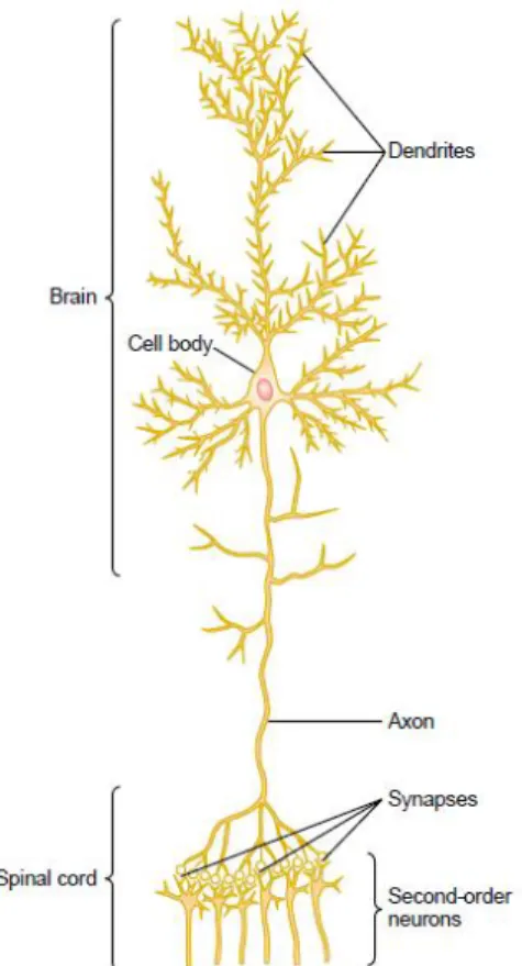
n e u r o n: the basic functional unit The nervous system contains more than 100 billion neurons. Neurons are specialized in receiving, conducting and transmitting electrochemical impulses, known as action potentials. Figure 1 shows a typical neuron of a type found in the brain motor cortex. A typical neuron possesses a cell body (often called the soma), dendrites, and an axon. Incoming signals enter this neuron through synapses, which are specialized connections with other cells, located mostly on the neu-ronal dendrites, but also on the cell body. Depending on the type of neuron, there may
1 The content of this section is mainly based on the following references [34], [45] and [55].
2 The content of this section is mainly based on the following reference [115].
be only a few hundred or as many as 200,000 such synaptic connections from input fibers. The output signal from the neuron travels by way of a single axon leaving the neuron. This axon has many separate branches to other parts of the nervous system or peripheral body. A special feature of most synapses is that the signal normally passes only in the forward direction (from the axon of a preceding neuron to dendrites on cell membranes of subsequent neurons). This forces the signal to travel in the directions required to perform specific nervous functions.
Figure 1: Structure of a large neuron in the brain, showing its important functional parts. Adapted from [55].
s e n s o r y pa r t o f t h e n e r v o u s s y s t e m Most activities of the nervous system are initiated by sensory experience that excite sensory receptors, such as visual receptors in the eyes, auditory receptors in the ears, tactile receptors on the body surface, and other types of receptors. This sensory experience can either cause an immediate reaction from the brain, or a memory of the experience can be stored in the brain for minutes, weeks, or even years to determine bodily reactions at some future date.
m o t o r pa r t o f t h e n e r v o u s s y s t e m The most important role of the nervous system is to control the various bodily activities. This is achieved by controlling (1)
con-traction of appropriate skeletal muscles throughout the body, (2) concon-traction of smooth muscle in the internal organs, and (3) secretion of active chemical substances by both exocrine and endocrine glands in many parts of the body. These activities are collec-tively called the motor functions of the nervous system, and the muscles and glands are called effectors because they are the actual anatomical structures that perform the functions dictated by the nerve signals. Figure 2shows the motor nerve axis of the ner-vous system for controlling skeletal muscle contraction. Note inFigure 2that the skeletal muscles can be controlled from many levels of the central nervous system, including the spinal cord or different parts of the brain. Each of these areas plays its own specific role, the lower regions concerned primarily with automatic, instantaneous muscle responses to sensory stimuli, and the higher regions with deliberate complex muscle movements controlled by the thought processes of the brain.
Figure 2: Skeletal motor nerve axis of the nervous system. Adapted from [55].
Anatomically, the nervous system consists of the Central Nervous System (CNS) (sys-tema nervosum centrale), the processing area and the Peripheral Nervous System (PNS) (systema nervosum periphericum) which connects the CNS with various receptors and effectors (Figure 3).
Figure 3: Nervous system of a human. Adapted from [55].
1.1.1.1 Central nervous system
The human nervous system has inherited special functional capabilities from each stage of human evolutionary development. From this heritage, three major levels of the central nervous system have specific functional characteristics: (1) the lower brain or subcortex, (2) the higher brain or cortex, and (3) the spinal cord.
l o w e r b r a i n o r s u b c o r t e x Many, if not most, of what we call the subconscious activities of the body are controlled in the lower areas of the brain: in the medulla, pons, mesencephalon, hypothalamus, thalamus, cerebellum, and basal ganglia. For in-stance, subconscious control of arterial pressure and respiration is achieved mainly in the medulla and pons. Control of equilibrium is a combined function of cerebellum, medulla, pons, and mesencephalon. And many emotional patterns, such as anger, ex-citement, sexual response, reaction to pain, and reaction to pleasure, can still occur after destruction of much of the cerebral cortex.
h i g h e r b r a i n o r c o r t e x The cerebral cortex is essential for most of our thought processes, and it is also an extremely large memory storehouse. One of the most
impor-tant functions of the cortical level is to process incoming information in such a way that appropriate mental and motor responses will occur. The cortex never functions alone but always in association with lower centers of the nervous system. Without the cerebral cortex, the functions of the lower brain centers are often imprecise. The cerebral cortex usually converts these functions to determinative and precise operations.
s p i na l c o r d The spinal cord is the main pathway for information connecting the brain and the peripheral nervous system. However, the spinal cord is not only a conduit for signals from the periphery of the body to the brain, or in the opposite direction from the brain back to the body. Even after the spinal cord has been cut in the high neck region, many highly organized spinal cord functions still occur. For instance, neuronal circuits in the cord can cause walking movements, reflexes that withdraw parts of the body from painful objects, reflexes that stiffen the legs to support the body against gravity, and reflexes that control local blood vessels, gastrointestinal movements, or urinary excretion. In fact, the upper levels of the nervous system often operate not by sending signals directly to the periphery of the body but by sending signals to the control centers of the cord, simply "commanding" the cord centers to perform their functions.
The terms used to describe spinal cord functions are the following:
1. Spinal nerve. The term spinal nerve generally refers to a mixed spinal nerve, which carries motor, sensory, and autonomic signals between the spinal cord and the body. Humans have 31 left-right pairs of spinal nerves, each roughly corresponding to a segment of the vertebral column: eight cervical spinal nerve pairs (C1-C8), 12 thoracic pairs (T1-T12), five lumbar pairs (L1-L5), five sacral pairs (S1-S5), and one coccygeal pair. Each spinal nerve is formed by the combination of nerve fibers from the dorsal and ventral roots of the spinal cord. The dorsal roots carry sensory axons, while the ventral roots carry motor axons. The spinal nerve emerges from the spinal column through an opening (intervertebral foramen) between adjacent vertebrae. Outside the vertebral column, the nerve divides into branches.Figure 4 shows the scheme of a spinal nerve on the right, and the body functions which spinal nerves located on different levels of the spinal cord control control, on the left.
2. Dermatome. A dermatome is defined as the cutaneous area whose sensory inner-vation is derived from a single spinal nerve (i.e., dorsal root) (seeFigure 5). There are eight cervical nerves, 12 thoracic nerves, five lumbar nerves and five sacral nerves. Each of these nerves relays sensation from a particular region of skin to the brain. For example, the T1 dermatome comes to the mid-line of the forearm, the T4 dermatome is at the level of the nipples, the T10 dermatome includes the navel, the L1 dermatome is in the groin, and the S1 dermatome is at the outer edge of the foot and heel. The division of the skin into dermatomes reflects the segmental organization of the spinal cord and its associated nerves.
Figure 4: Spinal nerve: scheme and functions. Adapted from [8]
3. Myotome. A myotome is defined as the muscular distribution of a single spinal nerve (i.e., ventral root), and is thus the muscular analogue of a cutaneous der-matome. Each muscle in the body is supplied by a particular level or segment of the spinal cord and by its corresponding spinal nerve. Knowledge of the myotomes of each spinal nerve enables the clinical localization of lesions causing motor dys-function (seeFigure 5). Examples of myotome distributions in the upper and lower extremities are as follows: C1/C2-neck flexion/extension, C3-neck lateral flexion, C4-shoulder elevation, C5-shoulder abduction, C6-elbow flexion/wrist extension, C7-elbow extension/wrist flexion, C8-thumb extension, T1-finger abduction, L2-hip flexion, L3-knee extension, L4-ankle dorsi-flexion, L5-great toe extension, S1-ankle plantar-flexion, S2-knee flexion.
1.1.1.2 Peripheral nervous system
The peripheral nervous system consists of the nerves and ganglia outside of the brain and spinal cord. The main function of the PNS is to connect the central nervous system to the limbs and organs.
Figure 5: Dermatomes and myotomes. Adapted from [55].
Functionally, the PNS can be divided into the somatic nervous system and the au-tonomic nervous system. The somatic nervous system (or voluntary nervous system) is associated with the voluntary control of body movements via skeletal muscles. The somatic nervous system consists of the nerves responsible for stimulating muscle con-traction, including all the non sensory neurons connected to skeletal muscles and skin. The autonomic nervous system (or visceral nervous system or involuntary nervous sys-tem) acts as a control system functioning mostly below the level of consciousness, and controlling visceral functions. It affects heart rate, digestion, respiratory rate, salivation, perspiration, pupillary dilation, urination, and sexual arousal.
1.1.2 Skeletal muscles
About 40 per cent of the body is skeletal muscle, and perhaps another ten per cent is smooth and cardiac muscle. In this chapter, we will focus mainly on the function of skeletal muscles because they are responsible for voluntary motions of body segments.
p h y s i o l o g i c a l a nat o m y o f s k e l e ta l m u s c l e Each skeletal muscle is com-posed of several muscle fibers (Figure 6). The cell membrane of the muscle fiber is called the sarcolemma. The sarcolemma consists of the plasma membrane, and an outer coat made up of a thin layer of polysaccharide material that contains numerous thin collagen fibrils. At each end of the muscle fiber, this surface layer of the sarcolemma fuses with a tendon fiber, and the tendon fibers in turn collect into bundles to form the muscle tendons that then insert into the bones.
Each muscle fiber contains several hundred to several thousand myofibrils, which are represented by the many small open dots in the cross-sectional view of Figure 6, part C. Each myofibril (Figure 6, parts D and E) is composed of about 1500 adjacent myosin filaments and 3000 actin filaments, which are large polymerized protein molecules that cause the actual muscle contraction. These are represented diagrammatically inFigure 6, parts E through L. The thick filaments in the diagrams are myosin, and the thin filaments are actin. Note inFigure 6, part E that the myosin and actin filaments partially interdig-itate and thus cause the myofibrils to have alternate light and dark bands, as illustrated inFigure 6. The light bands contain only actin filaments and are called I bands because they are isotropic to polarized light. The dark bands contain myosin filaments, as well as the ends of the actin filaments where they overlap the myosin, and are called A bands because they are anisotropic to polarized light. Note also the small projections from the sides of the myosin filaments in Figure 6, parts E and L. These are cross-bridges. The interactions between these cross-bridges and the actin filaments cause contractions. Fig-ure 6, part E also shows that the ends of the actin filaments are attached to a so-called Z disc. From this disc, these filaments extend in both directions to interdigitate with the myosin filaments. The Z disc, which itself is composed of filamentous proteins differ-ent from the actin and myosin filamdiffer-ents, passes crosswise across the myofibril and also crosswise from myofibril to myofibril, attaching the myofibrils to one another all the way across the muscle fiber. Therefore, the entire muscle fiber has light and dark bands, as do the individual myofibrils. These bands give skeletal and cardiac muscle their striated appearance. The part of the myofibril (or of the whole muscle fiber) that lies between two successive Z discs is called a sarcomere. When the muscle fiber is contracted the length of the sarcomere is about 2 micrometers. At this length, the actin filaments completely overlap the myosin filaments, and the tips of the actin filaments are just beginning to overlap one another. This is the length at which the muscle is capable of generating its greatest force of contraction.
Figure 6: Organization of skeletal muscle, from the gross to the molecular level. F, G, H, and I are cross-sections at the levels indicated. Adapted from [55].
Based on observations of their contractile properties (muscle strength, contraction ve-locity and fatigability), muscle fibers can be divided into three types. Type I fibers, oxida-tive fibers, contract slowly and have high fatigue resistance. The force produced by Type I fibers rises and falls slowly, but can be kept consistent for long periods. They are mainly used for maintaining posture and sports like long-distance running. Type IIb fibers,
con-trary to Type I, are glycolytic fibers and respond to the action potential quite fast. They can produce high force, but they tend to fatigue easily and need a long time to recover. This type of muscle fiber is needed to generate instantaneous or vigorous motion, such as jumping or sprinting. Type IIa fibers use both oxidative and glycolytic processes for metabolism. Compared with the other two fiber types, Type IIa fibers are intermediate in terms of contraction speed, force productivity and fatigability. Single muscles may be composed of the three fiber types in different proportions, which results in compound muscle contractile properties in the different muscles [148].
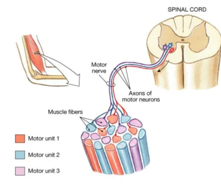
g e n e r a l m e c h a n i s m o f m u s c l e c o n t r a c t i o n Whatever the muscle fiber type, each muscle fiber is exclusively innervated by a single motor neuron; in contrast, a motor neuron can activate a number of muscle fibers with the same muscle fiber type [148]. The
group composed of a motor neuron and all the muscle fibers it innervates is known as a motor unit, as shown inFigure 7.
The initiation and execution of muscle contraction occur in the following sequential steps:
1. Once the decision to move a body segment is made the brain will generate an electrical signal and transmit it to the spinal nerve. Further, an action potential travels along a spinal nerve to its endings on muscle fibers.
2. At each ending, the nerve secretes a small amount of the neurotransmitter sub-stance acetylcholine.
3. The acetylcholine acts on a local area of the muscle fiber membrane to open multi-ple "acetylcholinegated" channels through protein molecules floating in the mem-brane.
4. Opening of the acetylcholine-gated channels allows large quantities of sodium ions to diffuse to the interior of the muscle fiber membrane. This initiates an action potential at the membrane.
5. The action potential travels along the muscle fiber membrane in the same way that action potentials travel along nerve fiber membranes.
6. The action potential depolarizes the muscle membrane, and much of the action potential electricity flows through the center of the muscle fiber. Here it causes the sarcoplasmic reticulum to release large quantities of calcium ions (Ca++) that have been stored within this reticulum.
7. The calcium ions initiate attractive forces between the actin and myosin filaments, causing them to slide alongside each other, which is the contractile process.
8. After a fraction of a second, the calcium ions are pumped back into the sarcoplas-mic reticulum by a Ca++ membrane pump, and they remain stored in the reticulum
until a new muscle action potential comes along; this removal of calcium ions from the myofibrils causes the muscle contraction to cease.
Figure 7: A motor unit composed by a motor neuron and the muscle fiber it innervates. Adapted from [6].
1.2 a nat o m y o f l o w e r l i m b s
The human leg is the entire lower limb of the human body and the main parts are: the foot, the shank, and the thigh. These three segments are connected at the ankle joint, the knee joint and the hip joint.
s k e l e t o n As shown onFigure 8, the upper part of the lower limb, or the thigh, has a single bone called the femur. This is the largest and longest bone in the human body. It has a rounded head which articulates with the acetabulum pelvis, forming the hip joint. At the knee, the femur is enlarged and it articulates with the tibia, forming the knee joint. The human lower limb also has a sesamoid-type bone that protects the front of the knee, called the patella. This kneecap is actually formed inside the tendon that connects the tibia and femur. The lower part of the lower limb has two bones, the tibia and fibula. These bones are joined by an interosseous membrane and they are relatively fixed and do not rotate around each other. The tibia has a large flat area at the knee called the
tibial plateau, where it articulates with the femur. At its end, the tibia articulates with the talus of the foot and forms the ankle joint.
Figure 8: Bones of human lower limb. Adapted from [5].
h i p m u s c l e s Movements of the hip joint include flexion and extension, abduction and adduction, and rotation. Flexion of the hip occurs when the angle between the torso and thigh is decreased. Conversely, extension occurs when this angle is increased. Abduction is the term representing the motion of bringing away mid-line of body, ad-duction is the movement towards mid-line. Hip abad-duction occurs when the femur moves outward to the side, and hip adduction occurs when the femur moves back to the mid-line. Hip rotation occurs when the femur moves along its longitudinal axis. Lateral hip rotation is when the anterior surface of the femur turns outward. The movement of the anterior surface of the femur inward is medial hip rotation.
There are several muscles1
that contribute to the movement of the hip joint (Figure 9 andFigure 10). These muscles can be clustered in the following groups:
1. Hip flexion: iliopsoas, sartorius, tensor of fascia lata, rectus femoris, pectineus, adductor longus, adductor brevis, and gracilis (when the knee is extended).
2. Hip extension: hamstrings [semitendinosus, semimembranosus, and biceps femoris (the long head)], adductor magnus (hamstring part), and gluteus maximus.
3. Hip adduction: adductor longus, adductor brevis, adductor magnus, gracilis, and pectineus.
4. Hip abduction: gluteus medius, gluteus minimus, tensor of fascia lata, sartorius, piriformis (when the hip is flexed), obturator internus (when the hip is flexed), and gemelli (when the hip is flexed).
5. Hip medial rotation: gluteus medius, gluteus minimus, tensor of fascia lata, pectineus, semimembranosus, and semitendinosus.
6. Hip lateral rotation: obturator externus, obturator internus, gemelli, piriformis, quadratus femoris, gluteus maximus, sartorius, and biceps femoris.
Figure 9: Anterior view of the hip and knee muscles. Adapted from [34]
k n e e m u s c l e s The knee permits flexion and extension around a virtual transverse axis, as well as a slight medial and lateral rotation around the axis of the lower part of the lower limb in the flexed position. The muscles1
responsible for:
1. Knee flexion are: hamstrings (biceps femoris, semitendinosus, and semimembra-nosus), sartorius, gracilis, gastrocnemius, and popliteus.
2. Knee extension are: quadriceps (rectus femoris, vastus lateralis, vastus medialis, and vastus intermedius).
Figure 10: Posterior view of the hip and knee muscles. Adapted from [34]
4. Knee lateral rotation are: biceps femoris and popliteus (unlocks extended knee). SeeFigure 9andFigure 10.
Figure 12: Posterior view of ankle muscles. Adapted from [34]
a n k l e m u s c l e s Movements of the ankle joint include dorsiflexion/plantarflexion, adbuction/adduction, and inversion/eversion. Dorsiflexion is the movement that de-creases the angle between the foot and the shank. Contrarily, the movement that in-creases the angle between the foot and the shank is called plantarflexion. Inversion is a movement in which the inner border of the foot is raised so that the plantar surface faces towards the body mid-line, and eversion is a movement where the outer border of the foot is raised so that the plantar surface feces away from the mid-line.
The muscles1
contributing to the movement of the ankle joint are following (Figure 11 andFigure 12):
1. Ankle dorsiflexion: tibialis anterior, extensor digitorum longus, extensor hallucis longus, and fibularis (peroneus) tertius.
2. Ankle plantarflexion: gastrocnemius, soleus, plantaris, flexor hallucis longus, flexor digitorum longus, tibialis posterior, fibularis (peroneus) longus, and fibularis (per-oneus) brevis.
3. Ankle inversion: tibialis anterior, and tibialis posterior.
4. Ankle eversion: fibularis (peroneus) longus, fibularis (peroneus) brevis, and fibu-laris (peroneus) tertius.
1.3 s p i na l c o r d i n j u r y
Spinal cord injuries or diseases are frequent cause of disability and may result in the total or partial obstruction in the flow of both sensory and motor information [115].
Spinal cord injuries are most often caused by trauma, especially following motor vehicle or sports accidents [115]. The extent of the loss depends on the site of the lesion, since
functions associated with spinal roots below the lesion level will lose their connection with the higher cerebral structures. This means that the brain will not receive sensory feedback from the areas of the body innervated from these roots, nor will it be able to control muscles in these areas, although the reflex responses will remain intact.
The strength of the paralyzed muscle contractions is usually graded on a Medical Research Council (MRC) scale of 0-5:
1. grade 5: muscle contracts normally against full resistance,
2. grade 4: muscle strength is reduced but muscle contraction can still move the joint against resistance,
3. grade 3: muscle strength is further reduced such that the joint can be moved only against gravity with the examiner’s resistance completely removed,
4. grade 2: muscle can move only if the resistance against gravity is removed,
5. grade 1: only a trace or flicker of movement is seen or felt in the muscle or fascicu-lations are observed in the muscle,
6. grade 0: no movement is observed.
The most widely accepted grading system to express the consequences of spinal cord injury is the classification developed with the American Spinal Injury Association (ASIA) scale. Traumatic spinal cord injury is classified into five categories on the ASIA impair-ment scale [1]:
1. A indicates a "complete" spinal cord injury where no motor or sensory function is preserved.
2. B indicates an "incomplete" spinal cord injury where sensory but not motor func-tion is preserved below the neurological level.
3. C indicates an "incomplete" spinal cord injury where motor function is preserved below the neurological level and more than half of the key muscles below the neurological level have a muscle grade of less than 3 on the MRC scale, which indicates active movement with a full range of motion against gravity.
4. D indicates an "incomplete" spinal cord injury where motor function is preserved below the neurological level and at least half of the key muscles below the neuro-logical level have a muscle grade of 3 on the MRC scale or more.
5. E indicates spinal cord injury where motor and sensory scores are normal.
Two main classes of the SCI exist, depending on the level of the spinal cord injury. (Figure 13):
1. Tetraplegia (or quadriplegia), an injury to the spinal cord in the cervical region, with the loss of motor and/or sensory functions of head, neck, shoulder, arms, chest, stomach, hips, lower limbs, and feet.
2. Paraplegia, an injury to the spinal cord in the thoracic, lumbar, or sacral segments, with the loss of sensory and motor functions involving only the lower limbs. The body parts that may be affected are the chest, stomach, hips, lower limbs, and feet.
Figure 13: Levels of spinal cord injury. Adapted from [3].
As mentioned, in this thesis we will focus only on complete SCI individuals unable to voluntarily control their lower extremities. The problems faced by individuals with paraplegia are no longer related to survival after the accident, as was the case in the past. Thanks to improvements in emergency medical care, their life expectancy is now compa-rable to that of the able-bodied population. However, many medical problems arise from living a longer life in a wheelchair, especially in relation to the decrease in muscle mass and bone density in the lower limbs (with consequent predisposition to osteoporosis) and heart and circulatory diseases. An SCI can also result in metabolic changes, increas-ing the risk of diabetes. Durincreas-ing daily activities, such as wheelchair propulsion or transfer from one surface to another, paraplegic patients put an intense load upon the muscles and joints of the upper extremities and, consequently, often experience shoulder compli-cations. In addition to these long-term issues, paraplegic individuals experience other practical problems with a more direct effect on everyday life. These include poor (or
absent) bladder control, impaired trunk balance capabilities, increased risk of pressure sores, and muscle spasticity [138].
1.4 o r t h o s e s
In order to restore standing and walking motion passive mechanical orthoses have been proposed. The common ones are the Ankle Foot Orthosis (AFO), Knee Ankle Foot Or-thosis (KAFO), Hip Knee Ankle Foot OrOr-thosis (HKAFO), Reciprocal Gait orOr-thosis, and the powered orthosis.
AFO stabilizes of the ankle joints. One special designs of AFO used for standing and walking in paraplegics is the Vannini-Rizzoli stabilizing orthosis.
An example of KAFO is shown inFigure 14a. A typical KAFO has a fixed ankle joint. The knee joint is capable of flexion, but during standing and walking it is locked in a position of extension. Paraplegic subjects are taught to stand with hips in full extension [85].
(a) Example of KAFO orthosis.
(b) Reciprocal Gain orthosis. (c) Ekso Bionics exoskele-ton.
(d) Parastep system.
Figure 14: Orthoses for standing and walking after SCI. Adapted from [4], [7], and [127].
Another type of orthosis is HKAFO. Compared with KAFO, in HKAFO the knee joints are also locked with parallel pads. A special type of HKAFO is the Hip Guidance orthosis designed to reduce the energy expenditure of walking.
The Reciprocal Gait orthosis (Figure 14b) provides support for the trunk, pelvis and lower extremities, while allowing ambulation with the use of assistive devices such as a walker, canes or crutches. The cable system enables a coupling mechanism of the hips. While one leg is in stance, the cable provides stability of that hip through the tension created by the opposite advancing leg. As the advancing leg begins stance, the tension
created at the opposite limb assists in overweighting, and a forward movement occurs [127].
An example of powered orthosis is the Ekso Bionics exoskeleton, developed in 2012 (Figure 14c). The Ekso Bionics exoskeleton supports its own 20-kilogram weight via skeletal legs and footrests and takes care of the calculations needed for each step. Pa-tients need to balance their upper body, shifting their weight as they plant a walking stick on the right; a physical therapist then uses a remote control device to signal the left leg to step forward. In a future model, the walking sticks will have motion sensors that communicate with the legs, allowing the user to take complete control [4].
Another approach for movement restoration in the paraplegic population is functional electrical stimulation. The advantage of the FES approach over mechanical orthosis can be listed:
1. The patient’s own muscles are used;
2. FES-provoked movements use the patient’s own metabolic energy;
3. Preserved neuromuscular reflex can be functionally used;
4. FES can prevents muscle atrophy;
5. FES can reduce muscle spasms;
6. FES may improve muscle and skin blood flow and prevent bone demineralization;
7. FES orthosis has a favorable appearance, has no attachments to cause pressure spots, and does not depend on extremity size for fit, thereby eliminating problems due to a change in girth [85].
The principles of FES are described in the following section. A mechanical orthosis can be used in combination with FES, and is then called a hybrid orthosis.
The only commercial product based on FES for standing and walking after SCI is Parastep-1R (Sigmedics, Chicago, IL), which was approved for home usage in 1994 by the Food and Drugs Administration (Figure 14d).
1.5 f u n c t i o na l e l e c t r i c a l s t i m u l at i o n
Functional electrical stimulation can be defined as the use of an electrical stimulus to achieve muscle contraction. FES delivers trains of the electrical charge pulses, mimicking to an extent the natural flow of excitation signals generated by the CNS in non-impaired structures [115]. The muscle contraction is achieved via the depolarization of the neuron
and the provoking of an action potential, determined by an electrical field generated between two electrodes [138]. Further the mechanism of muscle contraction is similar to
muscle can be modulated by varying one of three stimulation parameters: pulse width, pulse amplitude, and pulse frequency. The muscle strength also depends on the position of the electrodes, the muscle condition at the time of stimulation (length of the muscle fibers and contraction speed) and the type of stimulated muscle. A typical FES system consists of a stimulator, electrodes and a control unit. The control unit determines the pattern of electrical stimulus for the desired movement. The stimulator generates and delivers the stimulus to the muscle of interest through the electrodes.
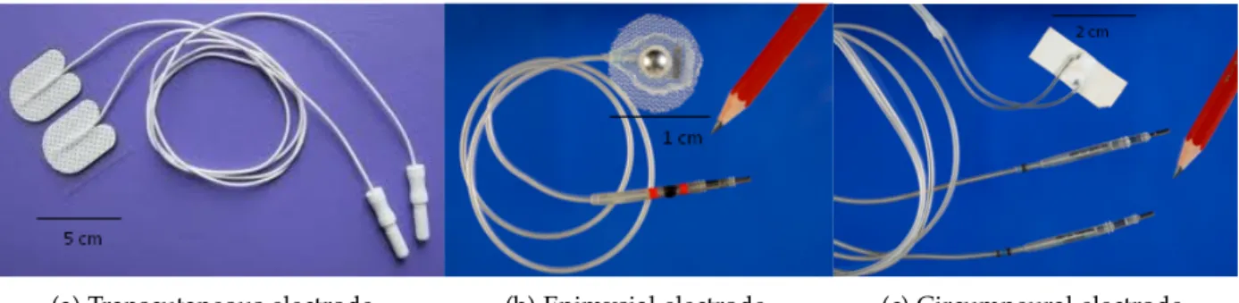
(a) Transcutaneous electrode. (b) Epimysial electrode. (c) Circumneural electrode.
Figure 15: Examples of the electrodes used for motor functions restoration. Electrodes (b) and (c) were developed by SUAW project.
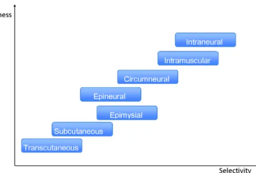
The electrodes used to restore motor function after an injury to CNS can be transcu-taneous (placed on the skin surface, shown inFigure 15a), subcutaneous (placed within a muscle), epimysial (placed on the surface of the muscle, shown in Figure 15b), in-tramuscular (placed inside the muscle), epineural (placed on the surface of the nerve), circumneural (wrapped around the nerve that innervates the muscle of interest, shown in Figure 15c), or intraneural (placed inside the nerve of interest). Therefore, based on the position of the electrodes, FES systems can be on the surface or implantable. The advantages of implanted vs. surface FES systems are better selectivity, repeatable exci-tation, and permanent positioning of the electrodes. The electrodes in implanted FES systems are placed away from pain receptors; therefore, the sensation to the user with a preserved sensory system is more pleasant. However, the disadvantage of implanted FES systems is the risk of damage due to the improper design and implantation of the system. Another major issue is the complicated surgical procedure to position the elec-trodes [19].Figure 16 characterizes electrode types according to their invasiveness and
selectivity.
FES stimulators can be divided into current-regulated and voltage-regulated. The elec-trical charge delivered to the stimulated muscle depends on the amplitude and duration of the stimulation pulses, the output impedance of the stimulator, and the impedance of the electrode-skin contact. Given that the electrode-skin contact has electro-capacitive properties, the use of voltage-regulated stimulators may result in uncontrolled electrical
Figure 16: Characterization of the types of FES electrodes according to invasiveness and selectiv-ity. Adapted from [62].
charges delivered to the stimulated muscles, causing pain or weak muscle contraction. Conversely, current-regulated stimulators precisely control the charge delivered to the system, but they may cause tissue damage if the surface of the used electrode is too small.
The stimulation waveform can be monophasic or biphasic. The biphasic shape is gener-ally used for the following reasons: surface stimulation is more comfortable with bipha-sic stimulation pulses and, for implanted electrodes the risk of tissue damage is less with biphasic stimulation.
One of the biggest limitations of FES systems for restoring functional movements in the SCI population is the rapid onset of the muscle fatigue after FES-induced muscle con-tractions compared with natural muscle concon-tractions. In a muscle contraction controlled by the CNS, the recruitment order is described by Henneman’s Size Principle [60], with
small slow fatigue-resistant motor units activated before the large, fast fatigable units. In FES-controlled muscle contractions, the larger muscle fibers are easily excited com-pared with small fibers. The frequency of natural motor neuron activation needed to achieve continuous contraction is 8-10 Hz [95]. If we take an example of the quadriceps
muscle group in surface FES-controlled muscle contraction (Figure 17), the continuous contraction occurs at around 20 Hz, and in order to achieve higher muscle forces, higher frequencies are usually needed (35-50 Hz) [95]. Also, in FES-induced muscle contraction,
all muscle fibers between the electrodes are activated at the same time, which is different from the asynchronous activation during natural contraction. All these factors result in
a much higher rate of muscle fatigue during FES-induced muscle contraction than that seen during natural contractions.
Figure 17: Knee joint torque obtained stimulating the quadriceps with surface electrodes at dif-ferent frequencies. Adapted from [115].
The history of successful applications of electrical stimulation started with the artifi-cial pacemaker, which was clinically implanted for the first time in 1958. Other appli-cations are cochlear implants, bladder management, deep brain stimulation, drop-foot correctors for hemiplegic patients, tremor compensation and other applications for reha-bilitation and therapy [19].
f e s-assisted movement restoration in paraplegics As mentioned, the pro-longed immobilization after SCI causes many physiological problems. FES-assisted stand-ing can ameliorate many of them. Durstand-ing prolonged bed rest immobilization bone loss occurs very quickly. Disuse of large masses of bone and muscle produces losses in bone calcium, reduces bone density, and induces hypercalciuria. Passive standing therapy (around three hours per day) proved to be sufficient to induce a slow decline in the elevated calcium excretion. Urinary tract infection occurs in more than half people with SCI. Bladder pressure is about three times greater in the standing posture compared with supine posture. Urine is drained more completely during micturition in the stand-ing position, thereby reducstand-ing the risk of bladder infections. Passive standstand-ing has been shown to produce decreased muscle tone in patients with spasticity. Due to the loss of sympathetic vascular tone and the skeletal muscle pump, patients with SCI have prob-lems maintaining blood pressure. Prolonged standing can lead to the cardiovascular system adaptation producing functional circulation. Pressure sores are also an impor-tant medical complications after SCI. Regular standing allows sustained periods of relief
to the sacral and ischial high-pressure areas of the buttocks [16]. Severe muscle atrophy
occurs rapidly following traumatic SCI, and FES therapy proved to be able to prevent this atrophy [17]. In addition to the therapeutic benefits, prolonged standing has a great
potential to provide functional benefits to SCI patients. It would allow paraplegics to reach further than when sitting in a wheelchair and to communicate with other people on an equal level. Also, it is a prerequisite for walking. In complete paraplegic patients, the functional benefits of standing may be greater than those of gait, since, in the foresee-able future, traveling over more than a short distance will still be easier in a wheelchair [16].
FES has proved to have great potential in functional movement restoration in the para-plegic population. The first functional electrical stimulation was applied to parapara-plegic patient by Kantrowitz in 1963. The quadriceps and glutei muscles of a T3 paraplegic subject were stimulated using surface electrodes. In the seventies, eighties and nineties of last century, FES as a technique for movement restoration in SCI received consid-erable scientific attention. A detailed biography review of the proposed systems and methods for FES-assisted standing up and standing are given in Chapter 2 and Chap-ter 4, respectively. However, FES-assisted applications require that the stimulation pro-vide strong, consistent muscle force. Yet, as already mentioned muscle fatigues far more rapidly when artificially stimulated than when excited by the central nervous system. As a result, the successful implementation of FES paradigms for movement restoration has been greatly limited by premature fatigue. Consequently, the number of effective systems used in clinical and every day practice is still limited.
1.6 c o n c l u s i o n
In this chapter, the nature of human motion and, the functioning of the neural and mus-culoskeletal systems are presented. However, the mechanisms of physiological muscle contractions, described in this chapter, do not apply to people with SCI. Moreover, af-ter SCI at thoracic or lumbar spinal cord level, lower extremity functions are limited. Therefore, additional knowledge about lower limb muscle functions is presented. The list of medical complications in people with SCI is long, and solutions to make their lives easier are greatly needed. In the literature mechanical orthoses and FES have been proposed for motion restoration in paraplegic people. This chapter thus contains a brief description of the most often used orthoses. However, the solutions for the artificial con-trol of functional motions proposed in this work are based on the use of FES. Hence, the principles of FES are presented at the end of the chapter.
2
F E S - A S S I S T E D S I T - T O - S TA N D M O T I O N
2.1 s i t-to-stand movement in able-bodied individuals
Standing up is a common daily activity and a prerequisite to standing or walking. This frequently executed task is one of the most biomechanically demanding activities [119].
There is no unic definition of Sit-To-Stand (STS) motion. The manner in which the STS movement is defined in the literature depends on the aim of the study. For example,
Roebroeck et al. defined the STS motion as moving the body’s center of mass upward from a sitting to a standing position without losing balance [120]. Vander Linden et al.
defined STS movement as a translational movement to the upright posture requiring movement of the center of mass from a stable position to a less stable position over extended lower extremities [137]. The STS movement was also described using kinematic
or kinetic variables, defining different phases and events during this postural task [84],
[88], [125]. Frequently used definition in the literature, provided by Schenkman et al.,
comprises four phases (seeFigure 18). Phase I (flexion-momentum) starts with initiation of the movement and ends just before the buttocks are lifted from the seat of the chair. Phase II (momentum-transfer) begins as the buttocks are lifted and ends when maximal ankle dorsiflexion is achieved. Phase III (extension) is initiated just after maximum ankle dorsiflexion and ends when the hips first cease to extend; including legs and trunk extension. Phase IV (stabilization) begins after extension of the hips is reached and ends when all motion associated with stabilization is completed [125].
The STS movement in able-bodied subjects has been studied with the standardized clinical tests that are used in epidemiological studies and clinical testing [52], [114],
[121]. Measurements of the kinetic and kinematic variables of the STS movement have
been obtained using different instruments, such as force plates [88], optoelectronic
sys-tems [63], [112], [126], goniometry [66], [107], and accelerometry [49]. Numerous studies
investigated the factors that influences the rising motion. These factors can be divided into few categories, chair related (e.g., chair height, arm-free vs. armrest-use condition), subject related (per example, age or muscle strength) and strategy related (per example, lower limbs and trunk position before the STS task, speed of task performance, light conditions) [70]. The analysis of these factors has suggested the following general
con-clusions.
A higher chair seat results in lower torque values at hip and knee joints. However, comparing the study results is difficult because study designs differ, and the chair seat height is not always based on lower-extremity length [20], [106], [107], [119], [126], [133].
Using armrests will lower the joint torques needed at the knee, probably without
Figure 18: Four phases of sit-to-stand motion marked by four key events. Adapted from [125]
fluencing the range of joint motion [9], [10]. But to our knowledge, there have been no
reports on the interaction between the height of the armrests, chair seat height, hand positioning, or their cumulative effect on STS movement performance.
The influence of trunk position has also been documented. According toShepherd and Gentile, changing the initial trunk position to have more flexion did not change the peak of a summation of hip, knee and ankle joint torques. The duration of the extension phase was longer and a high value of a summation of hip, knee and ankle joint torques was sustained for a longer proportion of the phase, indicating that more muscle force was required [129]. Schenkman et al., described a "momentum transfer strategy" in which
the momentum generated by the upper-body is used during the extension phase. In fact, healthy adults perform the STS-movement by a small flexion of the trunk, subsequently they start rising from the chair. The movement ends in an erect standing position and the achievement of stability (Figure 18) [125]. In contrast, in people with muscle weakness
rising from a chair is characterized by increased flexion of the trunk prior to rising from the seat [53], [61], [146]. This strategy has been referred to as the "stabilization-strategy".
Effect of knees and feet (posterior, preferred, and anterior positions) positioning prior to the start of the STS movement appears to influence the movement strategy. A shorter movement time has been shown when the feet are placed posterior with the respect to the chair [128]. Positioning the feet more posteriorly enables lower maximum extension
joint torque at the hip during STS movement, as well as lower hip flexion speed [76].
No differences were found in electromyographic activity with respect to the different feet positioning [107]. Positioning the knee more extended than preferred prior to the
STS movement appeared to lead to an increase of the hip joint angular displacement, with an increase of hip extension joint torque [39]. It was also reporter that the preferred
lower-extremity position gives less head movement and lower ground reaction forces [132].
Yoshioka et al. showed the influence of the speed of STS performance on the sum of the peak hip and knee joint torque values, i.e. as the STS movement time increase, the joint torque decreases [147].
No effect on movement time was found when visual control was varied [102], [103].
2.2 s i t-to-stand movement in paraplegic individuals
In spite of the fact that STS plays an important role in everyday life, the biomechanics of this postural task in paraplegic population have not been well documented. To the best of our knowledge, only few studies have been performed involving only couple of paraplegic subjects each.
Bahrami et al. showed that paraplegic patients standing up, with and without as-sistance of FES of quadriceps muscles and using arm support, perform this task in a different way than an able-bodied person, i.e. they do not use the "momentum transfer strategy". These observations suggest that the able-bodied used the constant information from the actual state of the entire body to control the whole body movement. For the paraplegic patients only a part of this information is available and thus, some signifi-cant differences are observed between the maneuver they use and the strategy used by able-bodied subjects [15].
Similar, Kamnik et al.also showed three different ways of performing this motion in paraplegic population. The first group comprised paraplegic patients whose electrically stimulated knee extensor muscles could not provide enough knee joint torque; therefore, they stood up primarily with the help of arm support. The second group was com-posed of regularly FES-trained patients who made better use of lower-limb support and unloaded the upper extremities. The third group was composed of paraplegic patients who simulated the behavior of the able-bodied, i.e. they pushed and pulled their bodies forward prior to standing up in order to gain some linear momentum, which is helpful in the initial standing up phase (Figure 18) [75].
Azevedo-Coste et al. compared the sit-to-stand trajectories of lower-limb joint angles and showed that the main difference between able-bodied and paraplegic subjects is the onset of leg movement with regard to trunk bending; this author hypothesized that in order to be efficient, bending the trunk forward should start before and last through knee and ankle movement [14].
2.3 f e s c o n t r o l s t r at e g i e s: state of the art
The ability to rise from a sitting to a standing position is very important for individuals with paraplegia in order to achieve minimal mobility. This movement also has functional and therapeutic benefits related to bone loading, joint extension, cardio-circulatory stim-ulation, and pressure sore prevention [116]. Persons with spinal cord injuries can recover
the capability of standing up by using implanted or surface FES systems [50], [85], [115].
The principles of FES and its advantages over conventional rehabilitation treatment are given inChapter 1.
The FES-assisted sit-to-stand method, which is used in clinical practice, involves open-loop stimulation of knee extensors activated by hand switches, as proposed byKralj and Bajd.[85] andGuiraud et al. [50]. This technique works adequately in many cases [26];
however, when this strategy is applied, stimulation starts without reference to upper-body movement. Hence, the whole-upper-body motion is not optimal and requires high joint velocity and hight upper-limb forces during the rising motion [74], which, if often
re-peated, may cause both damage to joint tissues and shoulder complications. Also, open-loop FES systems often use higher than needed stimulation parameters, hence the mus-cle contractions induced by FES tend to result in rapid musmus-cle fatigue, which limits the following activities [14].
A number of closed-loop strategies have been proposed to solve some of these prob-lems. Ewins et al. proposed a Proportional Integral Derivative (PID) closed-loop con-troller of knee joints [37]. The system was designed to move the knee angles to a hyper
extended position by modifying the stimulation amplitude of the electrically stimulated knee extensors. Servo potentiometers, attached to the thighs and calves of the patient, were used to monitor the knee angles. The results showed smoother trajectories, but neither reduced upper-limb efforts nor lower terminal velocities in the knees were re-ported. Because of the nonlinear dynamics of muscles and the sit-to-stand motion, a PID controller does not work well for control of the STS task [93].
Other strategies, such as closed-loop ON/OFF [104] controllers have been successful in
reducing terminal knee velocity during sit-to-stand maneuvers, which would preserve knee joints. The closed-loop ON/OFF control had a switching function, based on a predefined phase-plane switching curve of the desired knee angle versus knee velocity, which divided the state space of a system into regions of on and off FES commands. Greater arm force was required compared with open-loop stimulation.
Dolan et al. proposed a switching curve controller for FES-assisted sit-to-stand mo-tion. The controller simulated the behavior of an able-bodied person by observing a phase plane defined by the knee angle-knee angular velocity relationship (Figure 19). The main goal of this controller was to control the end of both sit-to-stand and stand-to-sit motion. The controller was tested in a pilot study on a female paraplegic subject. The controller successfully decreased terminal knee velocities, but greater arm forces during the motion were reported [31]. The reported arm forces could be explained by the
obser-vation that the subject had no experience in using the system and therefore might have been applying more arm support than needed.
Davoodi and Andrewsdeveloped and compared gain scheduling PID and fuzzy logic controllers [25]. These authors used a Genetic Algorithm (GA) as an optimization
ap-proach for tuning the controllers’ parameters. Both controllers, when compared with a PID or ON/OFF controller, resulted in smoother rising maneuvers and the average
elec-Figure 19: Typical graph of knee angular velocity in relation to angle trajectory for able-bodied individuals during standing up and sitting down. Adapted from [31]
trical stimulation required for successful motion was reduced. They were also able to reduce the arm forces but the level of the required arm support was still greater than the forces needed in the open-loop FES method. Further, the high number of GA trials required during calibration makes it inconvenient for practical use. The same group of authors proposed a fuzzy logic controller based on reinforcement machine learning for controlling FES of the lower limbs during rising maneuvers [26]. Three simulation
sce-narios were successfully tested: learning to compensate for weak arm forces, learning to minimize arm forces, and learning to minimize the terminal velocity of the knee and arm forces. Although this method appears to be promising, only its theoretical feasibility has been tested.
Mahboodi and Towhidkhah[93] investigated FES-assisted standing up using the
non-linear model predictive control approach. This theoretical study showed good tracking behavior for lower-limb joint angles, but it has not been experimentally tested. The arm efforts during the motion have not been analyzed.
Contrary to these "control-driven" methods, other "patient-driven" approaches based on an inverse dynamic model have been proposed [32], [33] [117], [118]. Here the action
of the FES controller was adjusted to the voluntary contribution of the patient, i.e., to the hand forces or body posture. Feedback to the system consisted of joint positions or hand reaction forces, which were fed into the inverse dynamic model in order to predict the stimulation pulse duration needed for the movement. Paraplegic patients were able to control the standing up movement by their voluntary body efforts. The upper-body efforts were lower, compared with sit-to-stand motion performed without FES support. However, these strategies require very accurate and realistic models (Figure 20) that are often difficult to obtain [116] and their practical real-time application remains
![Figure 2 : Skeletal motor nerve axis of the nervous system. Adapted from [ 55 ].](https://thumb-eu.123doks.com/thumbv2/123doknet/7727307.248944/20.892.271.577.451.885/figure-skeletal-motor-nerve-axis-nervous-adapted.webp)
![Figure 5 : Dermatomes and myotomes. Adapted from [ 55 ].](https://thumb-eu.123doks.com/thumbv2/123doknet/7727307.248944/24.892.207.626.93.722/figure-dermatomes-myotomes-adapted.webp)
![Figure 9 : Anterior view of the hip and knee muscles. Adapted from [ 34 ]](https://thumb-eu.123doks.com/thumbv2/123doknet/7727307.248944/30.892.97.757.372.717/figure-anterior-view-hip-knee-muscles-adapted.webp)
![Figure 10 : Posterior view of the hip and knee muscles. Adapted from [ 34 ]](https://thumb-eu.123doks.com/thumbv2/123doknet/7727307.248944/31.892.140.778.105.394/figure-posterior-view-hip-knee-muscles-adapted.webp)
![Figure 12 : Posterior view of ankle muscles. Adapted from [ 34 ]](https://thumb-eu.123doks.com/thumbv2/123doknet/7727307.248944/32.892.221.618.112.503/figure-posterior-view-ankle-muscles-adapted.webp)
![Figure 14 : Orthoses for standing and walking after SCI. Adapted from [ 4 ], [ 7 ], and [ 127 ].](https://thumb-eu.123doks.com/thumbv2/123doknet/7727307.248944/35.892.154.826.535.838/figure-orthoses-standing-walking-sci-adapted.webp)