INVESTIGATION DES FONCTIONS DE L’HISTONE H2A.Z DANS
LE GÉNOME HUMAIN ET DE SES MÉCANISMES
D’INCORPORATION DANS LA CHROMATINE
(INVESTIGATING THE FUNCTION OF HISTONE H2A.Z IN THE HUMAN GENOME AND MECHANISMS OF CHROMATIN INCORPORATION)
par
Anahita Lashgari
thèse présentée au Département de biologie en vue de l’obtention du grade de docteur ès sciences (Ph.D.)
FACULTE DES SCIENCES UNIVERSITE DE SHERBROOKE
Le 18 Janvier 2017
le jury a accepté la thèse de Madame Anahita Lashgari dans sa version finale.
Membres du jury
Professeur Luc R. Gaudreau Directeur de recherche Département de Biologie
Professeur Jean-Yves Masson Évaluateur externe
Université Laval
Professeur Nicolas Gévry Évaluateur interne Département de Biologie
Professeur Viktor Steimle Président-rapporteur Département de biologie
iv
SUMMARY
Regulation of transcription is crucial for the appropriate development and function of eukaryotic cells. In eukaryotes, DNA is organized into a dynamic, complex, nucleoprotein structure called chromatin. Chromatin structure provides markedly restricted access of transcription factors to regulatory sites. Several mechanisms have evolved to modulate chromatin dynamics in order to regulate proper gene expression. One of the most intriguing mechanisms that modulate chromatin structure is the exchange of canonical histones with histone variants by chromatin remodeling complexes.
Among the histone variants, H2A.Z is an essential regulator of gene transcription. H2A.Z is enriched at regulatory regions but significant levels of the histone variant can also be found within gene bodies. However, the role of H2A.Z within the gene bodies is still not well understood. Recent evidence suggests that active recruitment of H2A.Z within gene bodies is required to induce gene repression. In contrast to this view, we show that global inhibition of transcription results in H2A.Z accumulation at gene transcription start sites, as well as within gene bodies. Our results indicate that accumulation of H2A.Z within repressed genes can also be a consequence of the absence of gene transcription rather than an active mechanism required to establish repression.
The second part of my Ph.D. project was to investigate the potential role of BRD8 - a subunit of the p400/Tip60 complex - in p53-mediated signaling. We find that knockdown of BRD8 leads to p21 induction and concomitant cell cycle arrest in G1/S. We further demonstrate that the p53 transcriptional pathway is activated in BRD8-depleted cells, and this accounts for upregulation of not only p21 but also proapoptotic genes, an event that leads to consequent apoptosis. Importantly, the DNA damage response is induced upon depletion of BRD8 and DNA damage foci are detectable in BRD8-depleted cells under normal growth conditions, as indicated by immunostaining for γ-H2AX. Notably, H4K16 acetylation is reduced in
BRD8-v
depleted cells suggesting that BRD8 may have a role in recruiting and/or stabilizing the p400/Tip60 complex within chromatin, thus facilitating DNA repair. Consistent with the activated DNA damage response, we find that in BRD8-depleted cells, CHK2 is activated but, surprisingly, CHK1 protein levels are severely reduced. Taken together, our results suggest that BRD8 is involved not only in mediating p53-dependant gene suppression, but also in mediating the DNA damage response.
In the last part of my Ph.D. project, I investigated the possible mechanisms involved in recruitment of the p400 chromatin remodeler complex to chromatin. I showed that histone variant H2A.Z is essential for efficient recruitment of p53 and p400 to the distal p53 binding element of the p21 promoter. Furthermore, using double knockout (DKO) MEFs for p300/CBP I showed that the depletion of p300/CBP lead to a severe decrease in the recruitment of p400 at p21 promoter. Further studies are necessary to fully understand the role of p300/CBP in targeting p400 to chromatin.
In conclusion, my studies provide insights into the molecular mechanisms involved in chromatin regulation by histone variant H2A.Z and chromatin remodeler complex p400.
vi
SOMMAIRE
La régulation de la transcription est un mécanisme crucial pour le bon développement et fonctionnement des cellules eucaryotes. Chez les eucaryotes, l'ADN est organisé dans une structure dynamique de nucléoprotéines appelée chromatine. La structure de la chromatine forme une barrière qui contrôle l'accès des facteurs de transcription à leurs sites de fixation sur l’ADN. Plusieurs mécanismes ont été acquis au cours de l'évolution pour moduler la dynamique de la chromatine afin de réguler de manière adéquate l'expression des gènes. Un des mécanismes les plus intriguant qui module la structure de la chromatine est le remplacement des histones canoniques par des variants d'histones. Il est effectué par des complex de remodelage de la chromatine.
Parmi les variants d'histones, H2A.Z est un régulateur essentiel de la transcription des gènes. H2A.Z est enrichi aux régions régulatrices des gènes, mais des niveaux significatifs de ce variant d'histone peuvent aussi être observés au cœur des gènes. Le rôle de H2A.Z localisé a lèintérieur gènes n'est, pour l'instant, pas bien compris. Des résultats récents suggèrent que le recrutement actif de H2A.Z dans les gènes est requis pour induire leur répression. En opposition à ces résultats, nous montrons que l'inhibition globale de la transcription conduit à l'accumulation de H2A.Z aux sites d'initiation de la transcription, mais aussi au cœur des gènes. Nos résultats indiquent que l'accumulation de H2A.Z dans les gènes réprimés serait une conséquence de l'absence de transcription plutôt qu'un mécanisme actif requit pour établir la répression.
La seconde partie de mon doctorat a été dédiée à l'étude du rôle de BRD8 (une sous-unité du complexe p400/Tip60) dans la signalisation contrôlée par p53. Nous avons trouvé que la déplétion de BRD8 conduit à l'induction de p21 et à l'arrêt concomitant du cycle cellulaire en phase G1/S. Nous montrons aussi que le circuit transcriptionnel de p53 est activé dans les cellules déplétées en BRD8. Cela résulte en l'induction de p21, mais aussi de gènes
vii
proapoptotiques, ce qui conduit la cellule en apoptose. De manière marquante, la voie de réponse aux dommages de l'ADN est induite suite à la déplétion de BRD8, ce qui est observée par l'apparition de foci de dommages à l'ADN révélés par immunocoloration de γ-H2AX. De plus, l'acétylation de H4K16 est réduite dans les cellules déplétées en BRD8, suggérant que BRD8 pourrait avoir un rôle dans le recrutement et/ou la stabilisation du complexe p400/Tip60 dans la chromatine, et pourrait donc faciliter la réparation de l'ADN. En accord avec le fait que la réponse aux dommages de l'ADN soit activée, nous trouvons que dans les cellules déplétées en BRD8, CHK2 est activé mais étonnamment le niveau de la protéine CHK1 était fortement diminué. Ensemble, nos résultats suggèrent que BRD8 est impliqué non seulement dans la répression des gènes régulés par p53, mais aussi dans la réponse aux dommages de l'ADN.
Finalement, dans la dernière partie de mon doctorat j'ai étudié le mécanisme qui pouvait être responsable du recrutement du complexe p400 au niveau de la chromatine. Nous avons montré que le variant d'histone H2A.Z est essentiel pour le recrutement de p53 et de p400 au site distal de fixation de p53 sur le promoteur de p21. De plus, en utilisant des cellules MEF DKO pour p300/CBP, nous avons montré que la déplétion de p300/CBP conduit à une diminution sévère du recrutement de p400 au promoteur de p21.
En conclusion, mes études permettent de mieux comprendre les mécanismes moléculaires impliqués dans la régulation de la chromatine par l'histone H2A.Z et le complexe de remodelage de la chromatine p400.
viii
ACKNOWLEDGMENTS
I am indebted to many people whom I have had the privilege of learning from during my graduate studies. First, I would like to express my sincere gratitude to my supervisor Professor Luc Gaudreau for giving me the opportunity to work in his lab and for his trust and support. He also helped me to refine my thinking and research; I am extremely grateful.
I would also like to thank my thesis committee for their support and insightful comments: Professor Viktor Steimle, Professor Nicolas Gévry and Professor Jean-Yves Masson. I would like to express my appreciation to Professor Pierre-Étienne Jacques for his contribution to our H2A.Z study.
During my time as a PhD student, I have also had great colleagues in the lab that have helped shape me into a better scientist. I sincerely thank Jean-François Millau for his contributions, comments and stimulating discussions. I am especially grateful to Liette Laflamme for her support and guidance at the beginning of my Ph.D. study. Special thanks to Maud Marques who helped me significantly upon my arrival in Quebec. I thank Benoit Guillemette for always being open to questions and discussion and for his guidance in the lab. I thank my fellow lab mates Joannie, Myriam, Hanna, Kathy, Patrice and Ines for all the fun we have had over the last few years.
Last but not the least, I would like to express my deep gratitude to my family, especially my mother who has always been extremely supportive of my studies. I would like to thank my husband, Reza, who has always been very supportive of my aspirations and was there to keep me motivated during the more difficult times.
ix
CONTENTS
SUMMARY ...iv
SOMMAIRE ...vi
ACKNOWLEDGMENTS ...viii
LIST OF ABBREVIATIONS ...xiii
LIST OF TABLES ...xv
LIST OF FIGURES ...xvi
CHAPTER 1 ...1
General Introduction ...1
Chromatin Structure and Regulation of Transcription ...1
1.1 Chromatin structure ...2
1.1.1 Nucleosomes ...2
1.1.2 Structure of the 30 nm fiber ...4
1.1.3 Chromatin structure beyond the 30 nm ...6
1.1.4 Chromatin Dynamics ...6
1.2 Histone Modifications and Modifying Enzymes ...7
1.2.1 Histone Acetylation ...9
1.2.1.1. Histone Acetyltransferase (HATs) ...9
1.2.1.2. Histone Deacetylases (HDACs) ...10
1.2.2 Histone Methylation...10
1.2.2.1. Histone Methyltransferases ...10
x
1.3 Histone Variants...11
1.3.1 Histone Variant H2A.Z ...13
1.3.1.1 H2A.Z Nucleosome properties ...13
1.3.1.2 H2A.Z in Transcriptional Regulation ...17
1.3.1.3 H2A.Z Deposition and Removal ...21
1.3.1.4 H2A.Z in DNA Damage Repair ...23
1.3.1.5 Post Transcriptional modifications of H2A.Z ...24
1.4 Chromatin Remodelers ...26
1.4.1 Chromatin Remodeler Complexes (CRCs) ...26
1.4.2 Nucleosome Recognition by Remodelers ...27
1.4.2.1 Chromatin Remodelers Domains ...27
The Bromodomain ...27
The Chromodomain ...28
The Plant Homeodomain Domain (PHD Finger) ...29
HAND-SANT-SLIDE ...29
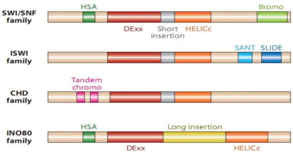
1.4.3 Chromatin Remodeler Complex Families ...30
1.4.3.1. The SWI/SNF Family of Chromatin Remodelers ...31
1.4.3.2. The ISWI Family of Chromatin Remodelers ...32
1.4.3.3. The CHD Family of Chromatin Remodelers ...33
1.4.3.4. The INO80 Family of Chromatin Remodelers ...33
1.5 The p400/Tip60 Chromatin Remodeling Complex ...38
1.5.1 P400 Containing Complex ...39
1.5.2 p400 as an Adenovirus E1A Oncoprotein Interaction Partner...40
1.5.3 p400, Transcription and Chromatin Remodeling: regulating the dynamics of H2A.Z nucleosomes ...40
1.5.4 p400 in Stress and DNA Damage Response (DDR) ...42
xi
1.5.6 p400 and The Cell Cycle...47
General Hypothesis and Objectives ...49
CHAPTER 2 ...52
Investigation of histone variant H2A.Z localization in the human genome ...52
Article Description ...53
Global Inhibition of Transcription Causes Histone H2A.Z to be Incorporated within Gene Bodies ...53
2.1 Abstract ...54
2.2 Introduction ...55
2.3 Materials and Methods ...57
2.3.1 Cells, cell culture, and cell treatment ...57
2.3.2 Chromatin immunoprecipitation ...57
2.3.3 Chromatin immunoprecipitation coupled to sequencing ...57
2.3.4 RT-qPCR ...58
2.3.5 RNA sequencing ...58
2.3.6 Immunoblotting ...59
2.3.7 Knock-down of p400 ...60
2.4 Results and Discussion ...61
2.4.1 H2A.Z level is low within gene bodies and anti-correlates with transcription at nucleosome +1 ...61
2.4.2 Inhibition of transcription specifically increases the incorporation of H2A.Z within genes ...62
xii
2.5 Acknowledgements ...70
2.6 Data availability ...70
2.7 References ...71
2.8 Supplementary Figures ...75
2.9 Supplementary Material and Methods ...79
2.9.1 RNA-seq data analysis ...79
2.9.2 ChIP-seq data analysis ...79
2.9.3 ChIP-seq aggregate profiles ...80
2.10 Supplementary Material and Methods References ...84
CHAPTER 3 ...85
Investigating the role of bromodomain protein 8 (BRD8) in cell cycle regulation and DNA damage response ...85
Article description ...85
Cellular Depletion of BRD8 causes p53-dependent apoptosis and induces a DNA damage response in non-stressed cells ...86
3.1 Abstract ...87
3.2 Introduction ...88
3.3 Results ...91
3.3.1 BRD8 knockdown induces cell cycle arrest and cell death through induction of p53-dependent apoptosis in HCT116 cells ...91
3.3.2 Cellular depletion of BRD8 induces p53-dependent p21 transcription in HCT116 cells ...98
3.3.3 BRD8 is required to prevent DNA damage in non-stressed cells ...103
xiii
3.3.5 Acetylation of histone H4K16 is partially suppressed in BRD8 depleted cells ...110
3.4 Discussion...111
3.5 Acknowledgments ...113
3.6 Materials and Methods ...114
3.6.1 Cell Culture ...114
3.6.2 BRD8 Knockdown ...114
3.6.3 Immunoblotting ...115
3.6.4 Isolation of RNA and quantitative PCR ...116
3.6.5 Cell viability assay ...116
3.6.6 Cell cycle analysis ...117
3.6.7 Apoptosis detection ...117 3.6.8 Laser microirradiation ...117 3.6.9 Immunofluorescence ...118 3.7 Supplementary Tables ...119 3.8 References ...121 3.9 Supplementary Figures ...133 CHAPTER 4 ...136
Investigating the mechanisms by which the p400 complex is targeted to chromatin ....136
4.1 Introduction ...136
4.2 Materials and Methods ...139
4.2.1 Cell culture and lentiviral infections ...139
4.2.2 Immunoblot assay ...140
4.2.3 RT-qPCR ...141
xiv
4.3 Results ...143
4.3.1 H2A.Z depletion decreases the recruitment of p400 at distal p53 response element at the p21 promoter ...143
4.3.2 Deletion of p300/CBP abrogates the presence of p400 at the distal p53 response element on p21 promoter ...146
4.3.3 p400/Tip60 recruitment at the p21 promoter is modestly affected by BRD8 knockdown ...149 4.4 Discussion ...152 4.5 Supplementary Figures ...157 CHAPTER 5 ...159 5.1 GENERAL DISCUSSION ...159 5.2 PERSPECTIVES ...166 BIBLIOGRAPHY ...170
xv
List of Abbreviations
36B4 Acidic ribosomal phosphoprotein P0 gene (RPLP0)
AOM Azoxymethane
ATM Ataxia telangiectasia mutated ATP Adenosine triphosphate
ATR Ataxia Telangiectasia and Rad3-Related Protein
BRD Bromodomain
CDK Cyclin dependent kinase
CDKI Cyclin dependent kinase inhibitor
CDKN1A Cyclin-dependent kinase inhibitor 1A ChIP Chromatin Immunoprecipitation
ChIP-seq Chromatin Immunoprecipitation followed by deep-sequencing CHK1 Checkpoint kinase 1
CHK2 Checkpoint kinase 2 CPT Camptothecin Dauno Daunorobicin
DDIT4 DNA-damage-inducible transcript 4 DKO Double knockout
DMEM Dulbecco’s modified Eagle's medium
DREAM Dimerization partner, RB-like, E2F and Multi-vulval class B DSB Double-strand break
DSS Dextran sodium sulfate
E1A Adenovirus early region 1A protein FACS Fluorescence activated cell sorting FACT Facilitates Chromatin Transcription FBS Fetal Bovine Serum
xvi
FPKM Fragments Per Kilobase of transcript per Million mapped reads
G1 Gap 1 phase
G2 Gap 2 phase
GDF15 Growth Differentiation Factor 15 IF Immunofluorescence
KD Knockdown
M Mitosis
MEF Mouse embryonic fibroblast NFR Nucleosome Free Region NHEJ Non-Homologous End Joining P400 (EP400) E1A-binding protein p400
PCNA Proliferating Cell Nuclear Antigen
PI Propidium Iodide
qPCR Quantitative PCR
Rad21 RAD21 cohesin complex component RNA Pol II RNA Polymerase II
RNA-seq RNA sequencing
RT-qPCR Reverse Transcription followed by quantitative PCR
S Syntesis phase
shRNA Short hairpin RNA siRNA Small interfering RNA
Spt6 Transcription elongation factor SPT6
SRCAP Snf-2-related CREB-binding protein activator protein TSS Transcription Start Sites
xvii
LIST of TABLES
Table 1.1 Different classes of modifications identified on histones and their
functions ...8
Table 1.2 The most studied histone variants and their functions ...12
Table 1.3 Remodeler composition and orthologous subunits ...35
Table 2.S1 ChIP antibodies ...82
Table 2.S2 ChIP primers ...82
Table 2.S3 RT-qPCR primers ...83
Table 2.S4 Western blot antibodies...83
Table 3.S1 Antibodies ...119
Table 3.S2 RT-qPCR Primers ...120
Table 4.1 Antibodies used for Western blot experiments ...141
Table 4.2 Primers used for RT-qPCR analysis ...141
Table 4.3 Antibodies used for ChIP experiments ...142
xviii
LIST of FIGURES
Figure 1.1 Nucleosome structure...3
Figure 1.2 Levels of chromatin compaction ...5
Figure 1.3 Comparison between H2A.Z and H2A ...15
Figure 1.4 H2A.Z enrichment patterns and promoter architecture differences in different organisms ...19
Figure 1.5 Chromatin remodeler families, defined by their ATPase domains ...31
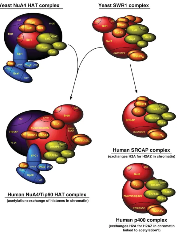
Figure 1.6 SWR1-related complexes in yeast and mammalian cells ...37
Figure 1.7 Schematic representation of the motif structure of p400 ...38
Figure 2.1 The presence of H2A.Z at genes anti-correlates with transcription ...62
Figure 2.2 Inhibition of transcription specifically increases the incorporation of H2A.Z within genes. ...65
Figure 2.3 The p400 chromatin-remodeling ATPase is specifically recruited at gene TSSs following transcription inhibition ...67
Figure 2.4 A schematic model illustrating H2A.Z accumulation at gene following inhibition of transcription. ...69
Figure 2.S1 Average H2A.Z ChIP-seq signal at TSS ...75
Figure 2.S2 Inhibition of transcription using α- amanitin ...77
Figure 2.S3 Inhibition of transcription specifically increases the incorporation of H2A.Z within genes ...77
Figure 2.S4 Knockdown of p400 decreases the incorporation of H2A.Z within chromatin ...78
Figure 3.1 Knockdown of BRD8 using two siRNA in HCT116 p53+/+ cells...92
Figure 3.2 BRD8 knockdown induce cell cycle arrest and cell death ...93
xix
Figure 3.4 BRD8 knockdown induces pro-apoptotic p53 target genes in HCT116
cells ...97
Figure 3.5 Knockdown of BRD8 activate p53 in HCT116 p53+/+ cells ...99
Figure 3.6 Lack of p21 did not rescues proliferation arrest and apoptosis in BRD8 depleted cells ...102
Figure 3.7 BRD8 knock down activates DNA damage pathway ...104
Figure 3.8 Co-staining of BRD8 with DNA damage foci ...106
Figure 3.9 BRD8 knockdown activates CHK2 but abrogates CHK1 ...108
Figure 3.10 Effect of BRD8 knock down on cell cycle distribution and proliferation of HCT116 p53 null cells ...109
Figure 3.11 BRD knockdown decrease H4K16 acetylation...110
Figure 3.S1 The protein levels of H2A.Z and transcription levels of p400, Tip60 and MRG15 does not significantly affected by knock down of BRD8 ...133
Figure 3.S2 Effect of BRD8 knock down on cell cycle distribution and proliferation of HCT116 p21 null cells ...134
Figure 3.S3 BRD8 depletion using shRNA ...135
Figure 4.1 H2A.Z depletion decreases the recruitment of p400 and p53 to the distal p53 response element within the p21 promoter ...145
Figure 4.2 Deletion of p300/CBP abrogates association of p400 at the distal p53 response element within the p21 promoter ...148
Figure 4.3 BRD8 depletion affects the recruitment of p400 at distal p53 RE on p21 promoter. ...151
Figure 4.S1 Recruitment of p300 and CBP at p53 distal element on p21 promoter is increased upon induction of p21 ...157
Figure 4.S2 Recruitment of p400 at p53 distal element on p21 promoter is increased upon induction of p21 ...158
1
CHAPTER 1
GENERAL INTRODUCTION
The present thesis introduction provides a review on the basics of chromatin structure and important regulatory mechanisms involved in chromatin dynamics affecting gene regulation. In addition, different aspects of the histone variant H2A.Z and the chromatin remodeler p400/Tip60 are also reviewed, as they relate to the project and the results presented in chapters 2, 3 and 4.
Chromatin Structure and Regulation of Transcription
It is estimated that the human genome encodes approximately 25,000 genes. All the somatic cells in one organism have the same genome, the same genotype, but at the same time, cells from different tissues drastically differ in appearances, properties and biological functions. Proper regulation of gene expression guarantees the proper phenotype and is crucial during differentiation and development of an organism. In eukaryotes, transcriptional regulation necessitates a balance between repressive packaging of the genome into nucleosomes and enabling access to RNA polymerase II (RNA pol II) and regulatory proteins. In this process, epigenetic mechanisms play a crucial role to regulate the states of chromatin compaction, conformation, modifications and content (Venkatesh and Workman, 2015).
2
1.1 Chromatin Structure
In eukaryotes, the size of DNA is far larger than the size of the compartment in which it is contained. Indeed, the total length of a human being's DNA is approximately two meters, while the average diameter of the nucleus, where the DNA is stored, is approximately six micrometers (Alberts et al., 2014). In order to be able to fit into the nucleus, DNA has to be condensed in some manner. To do so, DNA molecules form millions of ordered nucleoprotein particles, referred to as nucleosomes, which are just the first level of DNA packing. Next, higher ordered structures help to further compact the DNA within the nucleus, which results in an organized and dynamic structure known as the chromatin (Woodcock and Ghosh, 2010). Although these levels of compaction are necessary to help contain DNA in the nucleus, they represent an important accessibility barrier for many cellular processes. Hence, several mechanisms have evolved to overcome this barrier and to regulate proper gene expression, an aspect that is crucial for the appropriate development and function of eukaryotic cells (Clapier and Cairns, 2009).
1.1.1 Nucleosomes
In eukaryotic cells, DNA is associated with about an equal mass of histone proteins in a highly condensed nucleoprotein complex called chromatin. The building block of chromatin is the nucleosome. The nucleosome consists of an octamer of core histone proteins, 2 copies each of canonical histone H2A, H2B, H3, and H4, that is wrapped 1.65 times per 146 base pairs (bp) of DNA (Fig. 1.1). Histones’ N- and C-terminal tails protrude from the nucleosome and can be the harbor sites for various post-translational modifications and make contact with adjacent nucleosomes. Of note, all eukaryotes also contain lower abundance histone variant proteins
3
that can be incorporated into nucleosomes for regulatory purposes (Talbert and Henikoff, 2010) (see also section 4.1).
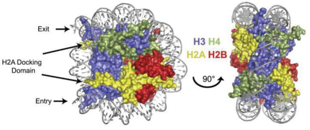
Figure 1.1 Nucleosome structure in front (left) and side view (right), showing that (H3/H4)2 is at the center of the DNA wrap, with two dimers of H2A/H2B docked at the edges, near the DNA entry and exit locations. The different histone proteins (depicted in cartoon representation) are drawn in yellow: H2A, red: H2B and blue: H3, green: H4. The backbone of DNA is in gray (Weber and Henikoff, 2014).
Two adjacent nucleosomes in an array connect to each other by linker DNA. The length of linker DNA is variable between species and tissues and even within a single cellular genome. It ranges between 20 and 90 base pairs (Van Holde et al., 1974). In higher eukaryotes, a linker histone (commonly H1 or H5) associates with the linker DNA at the site of DNA's entry to/exit from the nucleosome and influences the orientation of linker DNA with respect to the nucleosome (Hamiche et al., 1996; Syed et al., 2010; Szerlong and Hansen, 2011).
In low salt concentration and in the absence of divalent cations (e.g. Mg2+), nucleosomes that
are connected by linker DNA have the appearance of “beads on a string” by electron microscopy. This structure is the 10 nm fiber (Olins and Olins, 1974) and has been seen in
4
endogenous and reconstituted chromatin. However under physiological conditions this conformation is not the most favored conformation of the chromatin (Szerlong and Hansen, 2011; Thoma et al., 1979; Woodcock and Ghosh, 2010).
1.1.2 Structure of the 30 nm Fiber
Under physiological conditions, linear chromatin condenses to form chromatin superhelical secondary structure of 30 nm fibers in both interphase and metaphase chromosomes (Szerlong and Hansen, 2011; Woodcock and Ghosh, 2010) (Fig. 1.2). X-ray diffraction revealed 30 nm chromatin fibers in different cell types such as transcriptionally active chicken erythrocytes, HeLa metaphase chromatin and Balbiani ring genes in Chironomus tetans (Midge) (Andersson et al., 1982; Horowitz-Scherer and Woodcock, 2006; Langmore and Paulson, 1983; Langmore and Schutt, 1980; Paulson and Langmore, 1983). Several models have been proposed for the 30 nm fiber:
- Two start helical ribbon model (zigzag) (Dorigo et al., 2004; Schalch et al., 2005; Woodcock and Ghosh, 2010)
- Two start cross-linker model (zigzag) (Szerlong and Hansen, 2011; Woodcock and Ghosh, 2010)
- One start solenoid model (Robinson and Rhodes, 2006; Widom and Klug, 1985)
So far, our knowledge about the detailed structure of the 30 nm chromatin fiber remains controversial. The structure is mostly the result of intrinsic characteristics of native chromatin such as the variation of linker length, histone components, histone variants and their modifications, DNA sequence and architectural chromatin proteins (scaffold protein) (Li and Reinberg, 2011; Szerlong and Hansen, 2011; van Holde and Zlatanova, 2007).
5
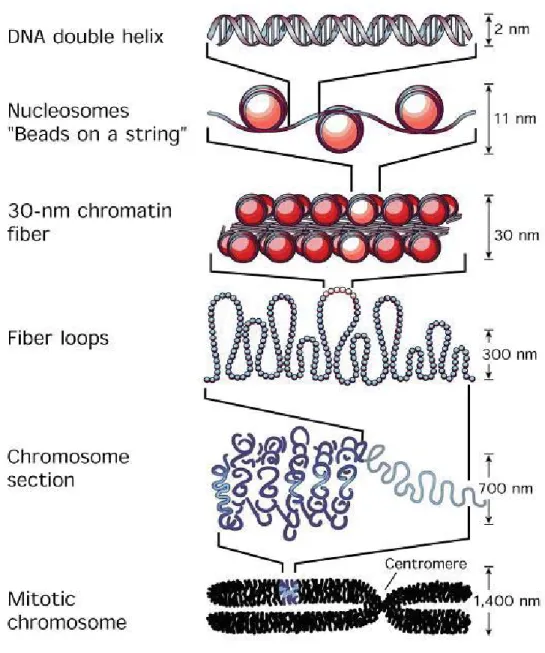
Figure 1.2 Levels of chromatin compaction from double helix DNA toward metaphasic chromosome (Felsenfeld and Groudine, 2003).
6
1.1.3 Chromatin Structure beyond the 30 nm
Chromatin tertiary structures, also referred to as fiber–fiber interactions, are formed from interactions between 30 nm chromatin fibers. Fibrous chromatin loops and other superstructures found in both metaphase chromosomes and specialized regions of interphase chromosomes, such as gene enhancers and insulators, are examples of chromatin tertiary structures. (Li and Reinberg, 2011; Szerlong and Hansen, 2011; Woodcock and Ghosh, 2010). The structural properties of chromatin structure beyond the 30 nm are largely unknown. In addition, chromatin fiber oligomerization is both cooperative and reversible and, like chromatin secondary structure, requires core histone amino-terminal “tail” domains.
1.1.4 Chromatin Dynamics
The mechanisms of gene expression rely on the structure and the composition of chromatin. The packaging of chromosomal DNA by nucleosomes condenses and organizes the genome but occludes many regulatory DNA elements and represents an accessibility barrier for many cellular metabolic processes such as gene transcription, DNA replication, DNA repair and DNA recombination (Clapier and Cairns, 2009). There are different mechanisms in the cell to overcome this barrier that provide a highly regulated system of DNA packaging and unpackaging, thus regulating the accessibility of DNA to DNA-binding factors. Indeed, chromatin structure exhibits a highly dynamic equilibrium between an open and permissive conformation of 10 nm beads on string, and the condensed, non-permissive 30 nm fiber.
The accessibility of DNA in chromatin is modulated by the incorporation of histone variants and two classes of enzymes: histone modifying enzymes and ATP-dependent nucleosome
7
remodelers (Clapier and Cairns, 2009; Kouzarides, 2007). The introduction of each histone variant to the nucleosome conveys a unique structure in chromatin that can influence the accessibility of DNA (Talbert and Henikoff, 2010). Histone modifying enzymes post-transcriptionally modify histones which alters the structure of chromatin and also provides binding sites for regulatory proteins; whereas, chromatin remodeling complexes use the energy of ATP to disrupt histone octamer-DNA contacts by translocating nucleosomes along DNA, removing histones by ejection or exchanging histones. Below, I describe these regulatory elements in more detail.
1.2 Histone Modifications and Modifying Enzymes
Histone modifying enzymes post-transcriptionally modify histones. There are ever-growing numbers of different histone post-translational modifications (PTMs), which introduce meaningful variation into chromatin and are referred to collectively as the “Histone Code”. The “histone code hypothesis,” initially proposed by Allis in 2000, states that “multiple histone modifications, acting in a combinatorial or sequential fashion on one or multiple histone tails, specify unique downstream functions” (Strahl and Allis, 2000). Further studies revealed a wide range of histone modifications and the enzymes responsible for their placement (referred to as writers) and removal (referred to as erasers), as well as the protein domains that can recognize these modifications (referred to as readers). Writers, erasers and readers are responsible for the establishment of homeostasis of combinatorial patterns of histone modifications in a given cellular context (Allis and Jenuwein, 2016).
Most of the histone modifications occur in the N-terminal tails, but histone cores and C-termini can be modified as well. Modifications affect the properties of the histones and the chromatin containing them and, hence, the chromatin structure of the region. In addition to regulating the chromatin structure, they also recruit chromatin remodeling complexes. The recruitment of
8
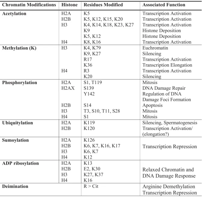
proteins and complexes with specific enzymatic activity is how modifications mediate their function (Bannister and Kouzarides, 2011). In this way, modifications can influence all the DNA-involved processes such as transcription, replication, recombination and repair. Different classes of modifications and their functions are summarized in Table 1.1 and, in the following sections, the most studied modifications, namely acetylation and methylation, will be reviewed.
Table 1.1 Different classes of modifications identified on histones and their functions (Adapted from (Sadri-Vakili and Cha, 2006).
2.1 Histone Acetylation
Chromatin Modifications Histone Residues Modified Associated Function
Acetylation H2A H2B H3 H4 K5 K5, K12, K15, K20 K4, K14, K18, K23, K27 K9 K5, K12 K8, K16 Transcription Activation Transcription Activation Transcription Activation Histone Deposition Histone Deposition Transcription Activation Methylation (K) H3 H4 K4, K79 K9, K27 R17 K36 R3 K20 Euchromatin Silencing Transcription Activation Transcription Elongation Transcription Activation Silencing Phosphorylation H2A H2AX H2B H3 H4 S1, T119 S139 Y142 S14 T3, S10, T11, S28 S1 Mitosis
DNA Damage Repair Regulation of DNA Damage Foci Formation Apoptosis
Mitosis Mitosis
Ubiquitylation H2A
H2B K119 K120 Silencing, Spermatogenesis Transcription Activation/
(elongation?) Sumoylation H2A H2B H3 H4 K126 K6, K7, K16, K17 K6, K7 K12 Transcription Repression
ADP ribosylation H2A
H2B H3 H4 K13 E2, K30 K27, K37 K16
Relaxed Chromatin and DNA Damage Response
Deimination R > Cit Arginine Demethylation
9
1.2.1 Histone Acetylation
1.2.1.1 Histone Acetyl Transferases (HATs)
The HATs utilize acetyl Co-A as a cofactor and catalyze the transfer of an acetyl group to the ε-amino group of lysine side chains and by doing this they neutralize the lysine’s positive charge and cause weaker interaction between histones and DNA. In addition, acetylated lysines of histones provide binding sites for protein domains such as bromodomains and PHD fingers, which are often found in HATs, and chromatin remodeling complexes. Their function may be to open up chromatin by neutralizing the positive charges of histones, and thereby weakening their interaction with DNA (Bannister 2011).
There are two major classes of HATs: type–A and type-B. The type-A HATs are a diverse family. They are classified into three separate families depending on amino acid sequence homology and conformational structure: GNAT, MYST and CBP/p300 (Hodawadekar and Marmorstein, 2007). Each of these enzymes modifies multiple sites within the histone N-terminal tails. This class of enzyme exhibits functions of numerous transcriptional coactivators. (Bannister and Kouzarides, 2011; Das et al., 2009). Like many other histone modifying enzymes, type-A HATs are often found associated in large multiprotein complexes. The components of these complexes have important roles in controlling enzyme recruitment, activity and substrate specificity (Bannister and Kouzarides, 2011). Type-B HATs are predominantly cytoplasmic and acetylate-free histones; they are not able to acetylate histones already incorporated into the chromatin. This class of HATs is highly conserved and has high sequence homology with scHat1. Type-B HATs acetylate newly synthesized histones H4 at K5, K12 and H3. This pattern of acetylation is important for histone deposition and is removed after their incorporation (Bannister and Kouzarides, 2011).
10
1.2.1.2 Histone Deacetylases (HDACs)
HDAC enzymes oppose the effects of HATs, they reverse lysine acetylation and restore the positive charge of lysine residues. By doing this they stabilize the chromatin structure and act as transcriptional repressors. HDACs have relatively low substrate specificity; a single enzyme is capable of deacetylating multiple sites within histones (Bannister and Kouzarides, 2011). There are four classes of HDACs: Classes I and II contain enzymes that are most closely related to yeast scRpd3 and scHda respectively, class III (sirtuins) are homologous to yeast scSir2 and class IV has only one member, HDAC11. In contrast to other HDAC classes, class III requires NAD+ for its activity (Bannister and Kouzarides, 2011).
1.2.2 Histone Methylation
1.2.2.1 Histone Methyltransferases
Histone methylation mainly occurs on side chains of lysine and arginine. This modification does not alter the charge of the histone. Furthermore, lysines may be mono-, di-, or tri-methylated, whereas arginines may be mono- or di- (symmetrically or asymmetrically) methylated. Histone lysine methyltransferases (HKMT) mainly methylate lysines within N-terminal tails of histones. All HKMTs catalyze the transfer of a methyl group from S-adenosylmethionine (SAM) to the ε-amino group of a lysine residue. HKMTs are generally specific for residues and also for modification degrees (i.e., mono-, di- and/or tri-methyl state)
11
(Bannister and Kouzarides, 2011). There are two classes of arginine methyltransferases: type-I and type-type-Itype-I. Type-type-I generates mono-methyl arginine (Rme1) and asymmetric di-methyl arginine (Rme2as), whereas type-II generates Rme1 and symmetric di-methyl arginine (Rme2s). Together, these two types of arginine methyltransferases, referred to as PRMT, have 11 members.
1.2.2.2 Histone Demethylases
Histone demethylase enzymes were discovered more recently. In 2004, the first lysine demethylase was identified: lysine-specific demethylase 1 (LSD1) (Shi et al., 2004). Another class of lysine demethylase JHDM1 (JmjC domain-containing histone demethylase 1) was discovered in 2006 (Tsukada et al., 2006). Today, many additional histone lysine demethylases have been found and, with the exception of LSD1, they all contain a catalytic jumonji domain. Like methyltransferases, demethylases possess specificity for their targets and for the level of demethylation of their targets (Bannister and Kouzarides, 2011). The jumonji protein is also shown to be capable of demethylating arginine (i.e. JMJD6) (Chang et al., 2007). Moreover, there is another mechanism for the reversal of the methylation of arginine residues - the conversion of arginine to citrulline via deimination reaction (Bannister and Kouzarides, 2011; Goto et al., 2002; Sugiyama et al., 2002).
1.3 Histone Variants
A fraction of histones are non-allelic variants that are less abundant compared to canonical histones. The variants are usually present as single-copy genes. Histone variants have distinct biophysical characteristics and localize to specific regions of the genome where they are
12
thought to alter the properties of nucleosomes and nucleosome dynamics within those regions (Kamakaka and Biggins, 2005). These properties of histone variants are important to shape the chromatin landscape of cis-regulatory and coding regions in support of specific transcription programs (Weber and Henikoff, 2014). Unlike canonical histones, the expression of most histone variant genes is not restricted to S-phase but continues throughout the cell cycle (Kamakaka and Biggins, 2005). Their genes contain introns and their transcripts are often polyadenylated. The major histone variants and their general functions are summarized in Table 1.2.
Since my PhD project was focused on the histone variant H2A.Z, I will present this variant in more detail in the following section.
Table 1.2 The most studied histone variants and their functions.
Family Variant Species Localization Function
H3 H3.3 Ubiquitous Transcription region Active transcription triggers deposition/removal
CenH3 Ubiquitous Centromere Kinetochore formation/function
H2A H2A.Z Ubiquitous Promoter, heterochromatin boundary, gene body of repressed genes, DNA DSBs
Transcription activation/
repression, chromatin segregation, DNA repair
H2A.X Ubiquitous DNA damage site DNA repair, recombination, transcription repression H2A.BbD Vertebrates Active X chromosome /
autosome
Transcription activation
Macro H2A Vertebrates Inactive X chromosome X chromosome inactivation H2A.Lap1 Mouse Inactive X chromosome Transcription activation during
13
1.3.1 Histone Variant H2A.Z
H2A.Z is highly conserved with Ң90% sequence identity among different organisms ranging from yeast to humans (Malik and Henikoff, 2003). It replaces H2A in approximately 5% of yeast and 10% of chicken and mammalian nucleosomes. H2A.Z is essential for viability in mice (Faast et al., 2001), Xenopus (Ridgway et al., 2004), Drosophila (van Daal and Elgin, 1992) and Tetrahymena (Liu et al., 1996), while its deletion causes growth retardation in yeast (Redon et al., 2002). H2A.Z is involved in diverse biological functions in different species such as regulation of gene expression both positively and negatively (Adam et al. 2001; Larochelle and Gaudreau 2003, Barski 2007, Gevry 2007), chromosome segregation (Rangasamy 2004, Krogan et al. 2004), cell cycle progression (Dhillon et al. 2006), maintenance of heterochromatin-euchromatin boundaries (Meneghini et al. 2003), the establishment of constitutive heterochromatin (Fan et al. 2004, Sarcinella 2007) and DNA damage repair (Alatwi and Downs, 2015; Gonzalez-Perez et al., 2013; Xu et al., 2012). Despite the fact that H2A.Z has been studied in detail, the role of this variant in the regulation of transcription is as of yet not fully understood.
1.3.1.1 H2A.Z Nucleosome Properties
H2A.Z shares ~60% sequence identity with H2A (Fig. 1.3 A).The three-dimensional structure of an H2A.Z-containing nucleosome is overall similar to that of the H2A nucleosome (Suto et al., 2000) (Fig. 1.3 A). However, there are subtle differences in specific regions between the structures of the two nucleosomes (Suto et al., 2000) that might explain their functional differences. The main structural difference between H2A and H2A.Z resides within the C-terminal “docking domain,” which is the surface interacting with the H3/H4 dimer (Fig. 1.3 A and B). The H2A.Z docking domain has less than 40% amino acid identity with H2A.
14
Structural studies have shown that altering H2A.Z's docking domain affects the interface between the H2A.Z/H2B dimer and the H3/H4 dimer (Malik and Henikoff, 2003; Suto et al., 2000). Indeed, substitution of glutamine 104 (Q) in H2A with glycine (G) in H2A.Z (Fig. 1.3 red arrow) results in the loss of three hydrogen bonds with H3, causing subtle destabilization of the interaction between the H2A.Z docking domain and H3 (Suto et al., 2000). This results in a ‘looser’ packaging of H2A.Z-containing nucleosomes and an open chromatin structure that improves the accessibility of the nucleosomal DNA to the transcriptional machinery. Furthermore, the docking domain of H2A.Z creates (presents) an interaction site for metal ion-binding on the surface of the nucleosome that may create a very specific interaction interface for other factors, whereas the corresponding region in H2A is not able to perform this type of interaction (Suto et al., 2000). Another difference between H2A and H2A.Z is the extension of a patch of acidic residues in the αC helix of the docking domain. In H2A.Z, the acidic patch is composed of four acidic residues compared to three in H2A. Altogether, these differences result in an altered H2A.Z/H2B dimer surface and an uninterrupted acidic surface around the H2A.Z nucleosomes that are important to explain the distinct physiological role of the variant.
Several studies have attempted to understand the importance of the altered C-terminus of H2A.Z. Research on Drosophila revealed that a region of H2A.Z, referred to as M6 and containing the acidic patch, is essential for embryonic development and viability (Clarkson et al., 1999) demonstrating the functional importance of the acidic patch. Furthermore, specific mutations in the acidic patch result in sensitivity to genotoxic stress (Jensen et al., 2011). In addition, the acidic patch and C-terminus contain some residues, such as a metal ion-binding site, that are important for H2A.Z deposition into chromatin and can also serve as a potential interaction platform on the surface of the nucleosome to recruit specific factors or modulators.
15
Figure 1.3 Comparison between H2A.Z and H2A (A) Sequence alignment of Xenopus laevis H2A and mouse H2A.Z. Intervals of 10 amino acids for H2A (filled circle) and H2A.Z (open circle) are indicated. Differences between the two amino acid sequences are colored red. Regions that are essential for the function of H2A.Z are boxed. The docking domain is indicated with a dashed line, secondary structure elements of the histone fold (α1, α2 and α3) and extensions (αN and αC) are indicated. Red arrow shows glutamine 104 [Q] in H2A and glycine [G] in H2A.Z. (B) Superposition of canonical nucleosome and H2A.Z nucleosome, viewed down the superhelical axis. Only 73 bp of the DNA and associated proteins are shown. Regions of protein-DNA interaction are numbered starting from the nucleosomal dyad. H3 is colored blue, H4 green, H2B red, H2A yellow, H2A.Z gray, and DNA brown. (C) Side view of the superimposed nucleosomes in (A) (rotated by 90° around the y-axis) with parts of the DNA removed for clarity. (D) Superposition of H2A and H2A.Z, in a view similar to that in (A). The docking domain is boxed (Suto et al., 2000).
B C D
16
It has been shown that the C-terminal region of H2A.Z can interact with some components of the transcriptional apparatus such as pol II and TBP and the substitution of the H2A C-terminus with H2A.Z is sufficient to provide the H2A.Z’s unique function in positive regulation of gene transcription in yeast cells with a deletion of H2A.Z gene (Adam et al., 2001). The acidic patch of H2A.Z in yeast was shown to be critical for mediating its deposition in chromatin and represents a potential candidate for the interaction of H2A.Z with its deposition and/or targeting machinery (Jensen et al., 2011). Recently, it was shown that a short region of the docking domain of H2A.Z interacts with the histone chaperone ANP32E (Obri et al., 2014). This interaction is important in order to evict H2A.Z/H2B dimers from nucleosomes and to stabilize the dimers once released (Obri et al., 2014). Finally, the acidic patch is also required for H2A.Z to promote higher-order chromatin folding in higher eukaryotes (Fan et al., 2004).
In addition to the docking domain, the loop 1 (L1) region is quite different between H2A and H2A.Z. Loop 1 is where the two H2A (or two H2A.Z) molecules come into contact with each other in the same nucleosome (Luger et al., 1997; Suto et al., 2000). Structural studies indicate that steric clashes preclude heterodimerization of H2A and H2A.Z in the same nucleosome (Suto et al., 2000). However, more recent studies confirm that both homotypic (with 2 H2A.Z/H2B dimers), and heterotypic (with one H2A.Z/H2B dimer), nucleosomes exist (Luk et al., 2010; Weber et al., 2010).
Based on the structural study, incorporation of H2A.Z confers unique structural features on the nucleosome compared to H2A (Suto et al., 2000). Surprisingly, there are contradictory reports indicating that H2A.Z both positively and negatively affects nucleosome stability in vitro (Abbott et al., 2001; Li et al., 2005; Park et al., 2005; Suto et al., 2000). In vitro studies showed that nucleosomes containing recombinant H2A.Z or native chicken erythrocyte H2A.Z are more stable than H2A-containing nucleosomes (Park et al., 2004; Thambirajah et al., 2006). Controversially, there are studies indicating that nucleosomes containing H3.3/H2A.Z have
17
decreased nucleosome stability (Jin and Felsenfeld, 2007; Jin et al., 2009), while another in vitro study reported no difference in stability between H3.3/H2A.Z and H3.3/H2A (Thakar et al., 2009). Overall, it seems that H2A.Z slightly stabilizes in vitro and destabilizes in vivo. This may be partly explained by the different methods used in these studies. Besides, acetylation of H2A.Z in active genes in vivo (Bruce et al., 2005; Valdes-Mora et al., 2012) or the fact that H2A.Z nucleosomes can be either heterotypic or homotypic might explain these discrepancies (Luk et al., 2010; Weber et al., 2010). However, the mechanism of transcription regulation by the incorporation of H2A.Z is as of yet not fully understood.
1.3.1.2 H2A.Z in Transcriptional Regulation
A role for H2A.Z in transcription was first proposed more than 30 years ago by Allis et al. In 1980, they showed that H2A.Z is exclusively present in the transcriptionally active macronucleus in Tetrahymena but not in the inactive micronucleus (Allis et al., 1980). Further studies in yeast showed that H2A.Z regulates gene transcription (Adam et al., 2001; Santisteban et al., 2000). Later, studies on the role of H2A.Z in transcription were also conducted in yeast ,where genome-wide localization analysis demonstrated that H2A.Z-containing nucleosomes occupy specific regions across the genome. It has been observed that H2A.Z marks the 5' ends of both active and inactive genes in euchromatin (Raisner et al., 2005). Furthermore, one or two H2A.Z-containing nucleosomes observed at TSS flanking regions of nearly all euchromatic genes, both active and inactive, and at TATA-less promoters, are perhaps indicative of a role in the formation of nucleosome free regions (NFR) (Guillemette et al., 2005; Li et al., 2005; Raisner et al., 2005; Zhang et al., 2005a). Loss of H2A.Z in yeast leads to defects in transcriptional activation, while its presence at gene promoters seems to be inversely correlated with transcription levels (Guillemette et al., 2005; Li et al., 2005; Raisner et al., 2005; Zhang et al., 2005a). Following gene activation, H2A.Z is rapidly evicted (Adam et al., 2001; Auger et al., 2008; Larochelle and Gaudreau, 2003; Li et al., 2005; Santisteban et
18
al., 2000), suggesting a role for H2A.Z in the chromatin structure of inducible genes (Guillemette et al., 2005; Zhang et al., 2005a).
Intriguingly, the repressive effect of H2A.Z on stress-inducible genes was also observed in yeast (Lindstrom et al., 2006). H2A.Z mutant yeast have been reported to de-repress silencing at the HMR locus (Dhillon and Kamakaka, 2000). H2A.Z found at genes near sub-telomeric regions antagonizes the spreading of heterochromatin onto euchromatin regions (Meneghini et al., 2003). Non-acetylated and ubiquitinated versions of H2A.Z co-localizes with facultative heterochromatin marks, such as H3K9me2, in heterochromatic, peri-centric and sub-telomeric regions (Fan et al., 2004; Hanai et al., 2008; Hardy et al., 2009; Sarcinella et al., 2007; Swaminathan et al., 2005). Furthermore, immunofluorescence experiments in Drosophila and mammalian cells confirmed that H2A.Z is mainly associated with heterochromatin such as peri-centric regions (Rangasamy et al., 2003; Sarcinella et al., 2007; Swaminathan et al., 2005). In 2008, the Zilberman and Henikoff research group’s genome-wide studies on Arabidopsis thaliana showed that DNA methylation could be antagonistic to the incorporation of H2A.Z and thereby regulate gene silencing by excluding H2A.Z (Zilberman et al., 2008).
Beside the activation role of H2A.Z in transcription, it has been shown that H2A.Z has a role in promoting elongation via facilitating RNA pol II passage by affecting the correct assembly and modification status of pol II elongation complexes and by favoring efficient nucleosome remodeling over the gene (Santisteban et al., 2011). More recently, an anti-correlation between H2A.Z occupancy and stalled pol II has been observed (Weber et al., 2014). These observations suggest that H2A.Z/H2B dimers can be more easily removed from nucleosomes than H2A/H2B dimers hence aiding RNA pol II transcriptional elongation. In addition, it proposes that H2A.Z has a role in recruitment or stimulating the activity of the transcription elongation complex FACT (Weber and Henikoff, 2014).
19
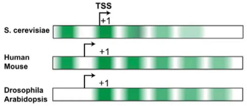
In higher eukaryotes, H2A.Z is highly enriched at promoter regions both at the upstream -1 nucleosome and downstream +1 nucleosome of TSS, similar to its distribution in yeast (Raisner et al., 2005) (Fig. 1.4), and also at regulatory elements such as enhancers and insulators (Barski et al., 2007; Bruce et al., 2005). One difference between yeast and humans, as well as Drosophila is that the presence of H2A.Z at human genes promoters is positively correlated with the presence of RNA pol II and transcription (Barski et al., 2007; Hardy et al., 2009; Mavrich et al., 2008).
Figure 1.4 H2A.Z enrichment patterns and promoter architecture differences in different organisms. Green intensity represents H2A.Z enrichment (Weber and Henikoff, 2014).
A negative role for H2A.Z in transcription has also been reported in mammals. It was shown by our laboratory that H2A.Z negatively regulates p53 target gene p21 (Gevry et al., 2007). Indeed, it was shown that p400 inhibits p21 expression by depositing H2A.Z into nucleosomes at p53 binding sites in the p21 promoter in unstressed condition (Gevry et al., 2007). In stressed conditions, p53 binds to its specific response elements (REs) at the p21 promoter and activates its transcription. Furthermore, the Espinosa laboratory identified a mechanism of repression involving the recruitment of the H2A.Z remodeling complex SRCAP and H2A.Z deposition at the promoters of ∆Np63α target genes (Gallant-Behm and Espinosa, 2013; Gallant-Behm et
20
al., 2012). As mentioned earlier, H2A.Z occupies the +1 nucleosome of the promoter of active genes; it was shown that the +1 nucleosome of active genes silenced during mitosis shifted upstream to occupy TSSs during mitosis thereby significantly reducing the length of the NFR. The mitotic shifting was specific to active genes that were silenced during mitosis and was not seen at promoters which are silenced by methylation or mitotically expressed genes (Kelly et al., 2010).
How can H2A.Z produce both positive and negative effects in transcription? One explanation is that H2A.Z facilitates the recruitment of both activator and repressor complexes by acting as a ‘general facilitator’ that generates access for a variety of complexes both activating and repressive (Creyghton et al., 2008; Hu et al., 2013; Li et al., 2012). Studies in embryonic stem cell (ESC) self-renewal and differentiation provide evidence for this model. H2A.Z facilitates the binding of the PRC2 complex to repressed genes and of MLL complexes to active genes in self-renewing ESCs (Hu et al., 2013). During differentiation of ESCs, H2A.Z facilitates retinoic acid-induced RARα binding, activation of differentiation markers and the repression of pluripotency genes (Hu et al., 2013). H2A.Z and transcription factor Foxa2 both act to regulate nucleosome depletion and gene activation during ESCs differentiation (Li et al., 2012). Interestingly, nucleosome depletion during ESC differentiation is dependent on the SWI/SNF and Ino80 chromatin remodeling complexes (Li et al., 2012), which suggests that the cycle of deposition and removal of H2A.Z modulates the accessibility of regulatory factors or complexes (Weber and Henikoff, 2014).
Another explanation for the role of H2A.Z in both positive and negative regulation of transcription comes from our own laboratory studies in both human cells and yeast, where we showed that H2A.Z is required for the proper positioning of nucleosomes at promoters (Gevry et al., 2009; Guillemette et al., 2005; Marques et al., 2010; Millau and Gaudreau, 2011). As such, one can imagine that stable phasing of nucleosomes by H2A.Z may influence
21
transcription by either favoring the recruitment of transcription factors or the opposite; preventing the recruitment of such factors by masking their DNA binding sites (Marques et al., 2010).
Although genome-wide localization of H2A.Z is principally at gene regulatory regions, it can also be found within gene bodies in both yeast and metazoans (Hardy et al., 2009; Latorre et al., 2015). However, little is known about the role of H2A.Z within gene bodies. In a recent study, the recruitment of H2A.Z within gene bodies by the DREAM complex has been proposed to cause the repression of the targeted gene. They suggested that active recruitment of H2A.Z within gene bodies is required to establish gene transcriptional repression (Latorre et al., 2015). In contrast, Hardy and colleagues previously showed that active gene transcription would prevent H2A.Z from associating to gene bodies (Hardy et al., 2009). They showed that upon shut down of heat-shock genes in yeast, H2A.Z re-associates with gene bodies. Moreover, they found that yeast RNA Pol II-associated histone chaperones FACT and Spt6 prevent accumulation of H2A.Z within gene bodies during transcription by evicting H2A.Z from nucleosomes and exchanging it with H2A (Jeronimo et al., 2015). These experiments suggest that H2A.Z accumulation within gene bodies would be the consequence of gene repression. In chapter 2 we attempt to verify whether the recruitment of H2A.Z within the body of repressed genes is the cause, or the consequence of the gene repression.
1.3.1.3 H2A.Z Deposition and Removal
Deposition of yeast H2A.Z is accomplished by the Swr1 complex (Swr1.com) (Kobor et al., 2004; Krogan et al., 2003; Mizuguchi et al., 2004). Histone chaperones Nap1 and Chz1 preferentially bind to H2A.Z–H2B dimers and deliver them to Swr1.com for deposition into chromatin (Luk et al., 2007). Efficient Swr1.com deposition of H2A.Z at promoter regions is
22
further promoted by a specific pattern of histone H3 and H4 tail acetylation (Altaf et al., 2007; Li et al., 2005; Raisner et al., 2005; Zhang et al., 2005a). H4 Lys 16 is shown to be required for H2A.Z incorporation at telomeres by Swr1(Altaf et al., 2007; Shia et al., 2006). Higher eukaryotes contain two distinct ATP-dependent complexes depositing H2A.Z in vivo and in vitro: SRCAP and p400, (Gevry et al., 2007; Ruhl et al., 2006; Wong et al., 2007) both of which are homologous to yeast Swr1 (see also section 5).
Mutagenesis of a typical promoter revealed that a 22 bp segment of DNA containing a binding site of the Myb-related protein Reb1 and an adjacent dT:dA tract is sufficient to program formation of a NFR flanked by two H2A.Z nucleosomes (Raisner et al., 2005; Ranjan et al., 2013; Yen et al., 2013). NFRs are characterized by two well-positioned flanking nucleosomes principally located at the gene promoter. Whether NFR formation is required for H2A.Z deposition or vice versa is debated. Our laboratory suggested that H2A.Z deposition plays a role in nucleosome positioning, while others proposed that H2A.Z deposition has no role (Guillemette et al., 2005; Li et al., 2005). Later, it has been shown that H2A.Z pre-deposition is dispensable for NFR formation but NFR formation promotes H2A.Z deposition (Hartley and Madhani, 2009). Recruitment of H2A.Z to NFR may explain H2A.Z enrichment at nucleosomes flanking promoters but it cannot explain the lack of upstream H2A.Z nucleosomes in organisms such as Arabidopsis and Drosophila (Mavrich et al., 2008; Zilberman et al., 2008) (Fig. 1.4), and it also cannot explain the enrichment of H2A.Z in the gene bodies of all eukaryotes (see section 3.1.2).
One of the mechanisms for removal of H2A.Z from the nucleosome is through the INO80 complex. It was shown that the INO80 complex exchanges nucleosomal H2A.Z/H2B dimers with free H2A-H2B in vivo and in vitro (Papamichos-Chronakis 2011). Recently, another mechanism for the removal of H2A.Z was found. A new H2A.Z-specific chaperone ANP32E has been identified and characterized as a component of p400/Tip60 complex in 2014 (Mao et
23
al., 2014; Obri et al., 2014). ANP32E interacts with a short region of the C-terminal docking domain of H2A.Z in the H2A.Z/H2B dimer to evict the dimer from the nucleosome (Mao et al., 2014; Obri et al., 2014). Genome-wide chromatin immunoprecipitation followed by high-throughput sequencing showed that ANP32E regulates H2A.Z deposition at TSS’s as well as enhancers and insulators (Mao et al., 2014; Obri et al., 2014). These results suggest that p400/Tip60 targeting by ANP32E regulates the H2A.Z genome-wide localization pattern.
1.3.1.4 H2A.Z in DNA Damage Repair
Cells regularly encounter with endogenous and exogenous stresses that can ultimately lead to DNA damage. To preserve genomic integrity, cells have numerous DNA damage sensors that detect different types of damage and initiate the appropriate repair pathway. DNA damage response pathways include cell cycle arrest, the transcriptional and post-transcriptional activation of a subset of genes including DNA repair factors (Jackson and Bartek, 2009). Amongst different forms of DNA lessions, DNA double strand breaks (DSBs) are particularly important, because they are difficult to repair and extremely toxic. Two principal mechanisms existe for DSBs repair: non-homologous end-joining (NHEJ) and homologous recombination (HR) (Khanna and Jackson, 2001). One of the major challenges in biology today is to understand how the chromatin state can affect the DNA repair and to investigate the role of histone variants and chromatin remodelers in these events.
Previous studies showed that cells lacking either H2A.Z or components of p400/Tip60 complex are hypersensitive to DNA damage and show defects in both NHEJ and HR DNA damage repair (Downs et al., 2004; Ikura et al., 2000; Murr et al., 2006; Xu et al., 2012; Xu et al., 2010). In yeast, H2A.Z is transiently incorporated at DNA damage sites where it regulates DNA end resection and hence homologous recombination repair (Billon and Cote, 2013). It
24
has been proposed that the p400/Tip60 remodeling complex decreases nucleosome stability at DNA damage sites which is required for the RNF8-dependent ubiquitination of chromatin and subsequent recruitment of the repair factors BRCA1 and 53BP1 to DSBs (Xu et al., 2010). Later, the same research group demonstrated that H2A.Z is transiently exchanged into nucleosomes at DSBs by the p400 remodeling complex and shifts the chromatin to an open conformation which is required for acetylation and ubiquitination of histones and for loading of the BRCA1 complex (Xu et al., 2012). They also showed the exchange of H2A.Z restricts single-stranded DNA production which is required for the loading of Ku70/80 proteins at damage sites (Xu et al., 2012). In contrast to the data generated by Price and colleagues, Canitrot, Trouche, and colleagues presented divergent results in 2014 (Taty-Taty et al., 2014). No H2A.Z incorporation was detected at DSBs by immunofluorescence or chromatin immunoprecipitation, thereby, they proposed that p400 function at the break is independent of H2A.Z incorporation and rather mostly through TIP60 and acetylation of chromatin .However, recent studies shed light on this discrepancy. Using micro-irradiation (“laser stripping”), it was shown that H2A.Z rapidly accumulates at DNA damage sites and that H2A.Z is retained for a few minutes before being rapidly removed (Alatwi and Downs, 2015; Gursoy-Yuzugullu et al., 2015a). The data from this study also suggest that H2A.Z is removed from damaged sites by the histone chaperone ANP32E and the INO80 chromatin remodeler. Removal of H2A.Z by ANP32E promotes acetylation of histone H4, (Taty-Taty et al., 2014) which remodels the chromatin and facilitates DNA repair through NHEJ by recruiting Ku70 (Gursoy-Yuzugullu et al., 2015a), while INO80 facilitates HR steps downstream of resection (Alatwi and Downs, 2015).
1.3.1.5 Post-transcriptional Modifications of H2A.Z
Like other histones, H2A.Z undergoes post-translational modifications such as acetylation, ubiquitylation, and sumoylation (Babiarz et al., 2006; Keogh et al., 2006; Millar et al., 2006).
25
H2A.Z can be acetylated at up to four lysine residues on its N-terminal tail in yeast by NuA4 and SAGA histone acetyltransferase complexes. It is acetylated only after its assembly into chromatin (Babiarz et al., 2006; Keogh et al., 2006) which may play a role in its rapid nucleosome exchange during gene activation or repression. It was shown that non-acetylated H2A.Z spreads across the entire promoter of inactive genes but only acetylated H2A.Z localizes at the TSS of active genes; furthermore, acetylated H2A.Z anti-correlates with promoter H3K27me3 and DNA methylation (Valdes-Mora et al., 2011). Deacetylation of H2A.Z led to its degradation via the ubiquitin–proteasome pathway (Chen et al., 2006) while mono-ubiquitilated H2A.Z is found on the X chromosome and is thought to be involved in the maintenance of heterochromatin (Sarcinella et al., 2007). In S. cerevisiae, sumoylation of H2A.Z is important for chromosome fixation to the nuclear periphery in response to persistent DNA double-stranded break (Kalocsay et al., 2009).
26
1.4 Chromatin Remodelers
As discussed above, remodelers are needed for nucleosome dynamics to (Clapier and Cairns, 2009):
i. Deposit and correctly space nucleosomes following replication and also, in a replication-independent manner, fill the gaps where nucleosomes were ejected;
ii. Prevent the impeding of advancing DNA/RNA polymerase by the nucleosomes during replication and transcription by ejecting or chaperoning the histone octamer around the advancing polymerase;
iii. Position the cis-element in nucleosome free regions or in the DNA linker between nucleosomes or to expose the element transiently on the nucleosome surface;
iv. Provide rapid access for DNA damage response factors to the sites of DNA damage by removing or sliding the nucleosomes and also reconstruct the nucleosomes afterward. Likewise in the case of DNA recombination (Clapier and Cairns, 2009).
1.4.1 Chromatin Remodeling Complexes (CRCs)
CRCs are DNA translocases which utilize the energy of ATP hydrolysis to alter histone–DNA contacts and, consequently, they all share a similar ATPase domain (Clapier and Cairns, 2009). The ATPase domain of the remodeling enzymes is composed of two tandem Rec-A like folds consisting of seven conserved helicase-related sequence motifs. The ATPase subunit of all CRCs classifies as part of the Superfamily 2 (SF2) group of helicase-like proteins (Eisen et al., 1995; Flaus et al., 2006). CRCs have similarities in their DEAD/H-containing ATPase subunits and also have other divergent characteristics. Based on the structural similarities of their ATPase domains they are divided into four different families: SWI/SNF, ISWI, CHD and
27
INO80 (Fig. 1.6) (Clapier and Cairns, 2009; Langst and Manelyte, 2015). They all share five basic properties:
i. Affinity for nucleosomes, beyond DNA itself;
ii. Domains that recognize covalent histone modifications except for the ISWI family; iii. A similar DNA-dependent ATPase domain, required for remodeling and serving as a
DNA-translocating motor to break histone-DNA contacts; iv. Domains and/or proteins that regulate the ATPase domain;
v. Domains and/or proteins for interaction with other chromatin or transcription factors.
1.4.2 Nucleosome Recognition by Remodelers
The presence of chromatin-interaction domains like bromo, chromo and SANT domains in different ATPase remodelers suggests that they can be selectively targeted to chromatin regions with distinct modification patterns to carry out specialized roles (Wang et al., 2007). These domains are also called epigenetic reader domains (Taverna et al., 2007). Additionally, they recognize different DNA structures/sequences and RNA signals that target them to specific genome loci (Langst and Manelyte, 2015).
1.4.2.1 Chromatin Remodeler Domains
The Bromodomain
28
histone acetyltranferases and are the only protein domain known to recognize acetyl-lysine residues on proteins (Sanchez and Zhou, 2009). BRDs are found in diverse nuclear proteins such as HATs (GCN5, PCAF, p300/CBP), ATP-dependent chromatin-remodeling complexes (p400/Tip60, BAZ1B), helicases (SMARCA), methyltransferases (MLL, ASH1L), transcriptional coactivators (TRIM/TIF1, TAFs) transcriptional mediators (TAF1), nuclear-scaffolding proteins (PB1), and the BET family (Filippakopoulos et al., 2012). SWI/SNF family remodelers have a bromodomain in the C-terminal region of the ATPase. Some of the SWI/SNF-related complexes like yRSC, dPBAP and hPBAF contain multiple bromodomains. The polybromo in a single subunit or several bromodomains among different subunits (e.g., yRsc1/2/4/10) of one remodeling complex can allow cooperative recognition of separate modifications (Clapier and Cairns, 2009). The bromo-adjacent homology (BAH) domain often resides near bromodomains. It was shown that the BAH domain is also a histone recognition domain (Onishi et al., 2007).
The human genome encodes 61 BRDs that are present in 46 diverse BRD-containing proteins which cluster into eight families based on structure/sequence similarity (Filippakopoulos et al., 2012). Dysfunction of BRD-containing proteins has been linked to disease processes, including cancer, inflammation and viral replication (Sanchez and Zhou, 2009). Despite that recent studies of BRD-containing proteins have highlighted the role of these domains in various biological processes and their association with disease, the function of many of the human BRD-containing proteins is not well characterized.
The Chromodomain
Chromodomain (chromatin organization modifier) is a protein structural domain of about 40-50 amino acid residues that is found in the chromodomain family of remodeling complexes.