HAL Id: hal-02311298
https://hal.sorbonne-universite.fr/hal-02311298
Submitted on 10 Oct 2019
HAL is a multi-disciplinary open access
archive for the deposit and dissemination of
sci-entific research documents, whether they are
pub-lished or not. The documents may come from
teaching and research institutions in France or
abroad, or from public or private research centers.
L’archive ouverte pluridisciplinaire HAL, est
destinée au dépôt et à la diffusion de documents
scientifiques de niveau recherche, publiés ou non,
émanant des établissements d’enseignement et de
recherche français ou étrangers, des laboratoires
publics ou privés.
Histone supply: Multitiered regulation ensures
chromatin dynamics throughout the cell cycle
Shweta Mendiratta, Alberto Gatto, Geneviève Almouzni
To cite this version:
Shweta Mendiratta, Alberto Gatto, Geneviève Almouzni. Histone supply: Multitiered regulation
ensures chromatin dynamics throughout the cell cycle. Journal of Cell Biology, Rockefeller University
Press, 2019, 218 (1), pp.39-54. �10.1083/jcb.201807179�. �hal-02311298�
REVIEW
As the building blocks of chromatin, histones are central to establish and maintain particular chromatin states associated
with given cell fates. Importantly, histones exist as distinct variants whose expression and incorporation into chromatin
are tightly regulated during the cell cycle. During S phase, specialized replicative histone variants ensure the bulk of the
chromatinization of the duplicating genome. Other non-replicative histone variants deposited throughout the cell cycle
at specific loci use pathways uncoupled from DNA synthesis. Here, we review the particular dynamics of expression,
cellular transit, assembly, and disassembly of replicative and non-replicative forms of the histone H3. Beyond the
role of histone variants in chromatin dynamics, we review our current knowledge concerning their distinct regulation
to control their expression at different levels including transcription, posttranscriptional processing, and protein
stability. In light of this unique regulation, we highlight situations where perturbations in histone balance may lead to
cellular dysfunction and pathologies.
Histone supply: Multitiered regulation ensures
chromatin dynamics throughout the cell cycle
Shweta Mendiratta
1,2*
, Alberto Gatto
1,2*
, and Genevieve Almouzni
1,2
Introduction
The eukaryotic genome is packaged and organized in
chroma-tin. The nucleosome, the basic unit of chromatin, consists of an
octamer with two copies each of the core histone H2A, H2B, H3,
and H4 around which is wrapped ∼146 bp of DNA and a variable
linker DNA associated with the linker histone H1. The core
his-tones share a conserved histone-fold domain that mediates their
head-to-tail heterodimerization. In this way, H2A and H2B form
two dimers flanking a (H3-H4)
2tetramer. The less conserved
linker histones, the H1 family, possess a central globular domain
flanked by a short N-terminal tail and a long basic C terminus. All
histone families exist as variants that are differentially expressed
and can undergo several posttranslational modifications. A
vari-ety of nucleosomes thus use distinct histone variants, as well as
posttranslational modifications with different properties often
associated with specific chromatin states (
Talbert and Henikoff,
2017
;
Reinberg and Vales, 2018
). As the major protein component
of chromatin, histones are critical for its dynamic organization,
assembly, and disassembly during most DNA transactions.
How-ever, because of their basic nature, uncontrolled histone
accu-mulation can lead to promiscuous interactions with any acidic
component, forming aggregates that are ultimately cytotoxic.
Nonnucleosomal histones are therefore constantly under check.
From their synthesis to incorporation into chromatin, as well as
during disassembly and disposal, histones are escorted by distinct
histone chaperones (
Gurard-Levin et al., 2014
;
Hammond et al.,
2017
). The first protein designated as a histone chaperone is
nu-cleoplasmin, discovered as the major protein present in Xenopus
laevis oocytes (
Laskey et al., 1978
). Mainly in charge of H2A-H2B,
it functions along with the H3-H4 N1/N2 chaperones to provide
a stockpile of maternal core histones. During early development,
this storage of maternal core histones is required to assemble
newly replicating DNA into chromatin, thereby sustaining the
first rounds of rapid cell division (
Woodland and Adamson, 1977
;
Earnshaw et al., 1980
). Under this unusual situation, particular
chaperones are thus needed to cope with massive amounts of
soluble histones. In contrast, in cycling cells, the soluble
reser-voir is more limited and has long been ignored. Not only are
his-tone chaperones in charge of the cytosolic reservoir of hishis-tones
or histones in transit but, most importantly, they also promote
the deposition, eviction, and recycling of specific histone
vari-ants during DNA replication, transcription, and repair.
Physio-logical changes in the level of histone chaperones and histone
variants occur at various times during development, such as the
up-regulation of nucleoplasmin in Xenopus female germ cells
to accommodate the pool of maternal histones (
Laskey et al.,
1978
), the accumulation of H3.3 in rat postmitotic neurons when
cells have exited from the cell cycle (
Piña and Suau, 1987
), and
the up-regulation of the chaperone ASF1b in highly
proliferat-ing cells (
Corpet et al., 2011
). A major interest has thus arisen
© 2018 Mendiratta et al. This article is distributed under the terms of an Attribution–Noncommercial–Share Alike–No Mirror Sites license for the first six months after the publication date (see http:// www .rupress .org/ terms/ ). After six months it is available under a Creative Commons License (Attribution–Noncommercial–Share Alike 4.0 International license, as described at https:// creativecommons .org/ licenses/ by -nc -sa/ 4 .0/ ).
1Institut Curie, Paris Sciences et Lettres Research University, Centre National de la Recherche Scientifique, UMR3664, Equipe Labellisée Ligue contre le Cancer, Paris,
France; 2Sorbonne Universités, Université Pierre et Marie Curie Paris 06, Centre National de la Recherche Scientifique, UMR3664, Paris, France.
*S. Mendiratta and A. Gatto contributed equally to this paper; Correspondence to Genevieve Almouzni: genevieve.almouzni@ curie .fr. on October 10, 2019
jcb.rupress.org
Downloaded from
http://doi.org/10.1083/jcb.201807179
concerning the interplay between histone variants and their
dedicated chaperones to maintain chromatin integrity during
development, differentiation, and the entire lifespan of an
indi-vidual (
De Koning et al., 2007
;
Filipescu et al., 2013
;
Gurard-Levin
et al., 2014
;
Hammond et al., 2017
).
In this review, considering histone H3 variants and their
ex-pression during the cell cycle, we will describe how different
mechanisms control the amounts of histones. H3 variants recently
emerged as important actors in cancer biology, as they are altered
or misregulated across different types of tumors (
Vardabasso et
al., 2014
;
Weinberg et al., 2017
). Distinct variants are under tight
regulation to meet various demands along the cell cycle and mark
specific functional domains in the genome (
Gurard-Levin et al.,
2014
;
Sitbon et al., 2017
). A notable example is CenH3
CENP-A, a
rapidly evolving variant that specifically marks the centromere.
Other H3 variants show a high degree of sequence similarity and
are associated with different domains. Based on their deposition
pathway, H3 variants can be distinguished as replicative and
non-replicative. Replicative forms are specialized variants whose
expression peaks in S phase and whose incorporation is coupled
to DNA synthesis. During S phase, a provision of replicative H3
variants (H3.1 and H3.2) supports the bulk assembly of
chroma-tin onto newly synthesized DNA. In contrast, the constitutive
expression of the non-replicative variant H3.3 sustains histone
turnover throughout the cell cycle, representing the majority of
histones in quiescent and terminally differentiated cells. The
other non-replicative variant, CenH3
CENP-A, is specifically
de-posited at centromeres and marks the site of kinetochore
assem-bly. Its expression peaks in G2/M phase in mammalian cells, and
its deposition occurs only late in mitosis/early G1. Interestingly,
replicative histone genes show a particular organization in
clus-ters not observed for replacement histones, providing a unique
means for controlling their expression.
We will first briefly review our current knowledge on the
dynamics and deposition of these histone variants onto DNA,
considering both dedicated and general chaperones. Next, we
will describe recent advances concerning the regulation of their
expression and the impact on genome organization and function
throughout the cell cycle. Finally, we will put forward a few
ex-amples of aberrant expression of histone genes and discuss the
consequences of their imbalance.
Dedicated chaperones for dynamics and deposition of
replicative and non-replicative histone variants
The deposition of H3 variants involves different histone
chap-erones and leads to a partitioning of the genome in distinct
chromosomal domains (
Fig. 1, A and B
). In S phase, the doubling
of genomic content requires a massive provision of histones
to ensure the duplication of chromatin (
Corpet and Almouzni,
2009
;
Alabert and Groth, 2012
). Nucleosomes ahead of the
rep-lication fork are displaced, and histones are recycled onto newly
replicated DNA along with de novo deposition of new histones
to restore nucleosome density (
Probst et al., 2009
;
Almouzni
and Cedar, 2016
). This leads to the mixing of new and parental
histones along with their particular marks, thereby enabling
the propagation of active and repressive states to subsequent
cell generations (
Ray-Gallet and Almouzni, 2010
;
Reinberg and
Vales, 2018
). Orchestration of histone incorporation and
re-cycling during S phase involves mechanisms coupled to DNA
synthesis and dedicated histone chaperone complexes. The
chro-matin assembly factor-1 (CAF-1) complex is the H3-H4 histone
chaperone that promotes nucleosome assembly coupled to DNA
synthesis during replication (
Smith and Stillman, 1989
) and
re-pair (
Gaillard et al., 1996
). CAF-1 is recruited at replication forks
through the interaction of its p150 subunit with the proliferating
cell nuclear antigen (PCNA) (
Moggs et al., 2000
), a processivity
factor for the DNA polymerase. The CAF-1 complex associates
specifically with the replicative variant H3.1 and H3.2, coupling
their deposition to DNA synthesis (
Tagami et al., 2004
;
Latreille
et al., 2014
).
Antisilencing function 1 (ASF1) is a H3-H4 chaperone viewed
as an intermediary handing over distinct histone variants to their
specific chaperones (
Mello et al., 2002
;
Daganzo et al., 2003
;
Tang et al., 2006
). ASF1 is rather promiscuous and can interact
with all H3 variants, including H3.1, H3.3, and CenH3
CENP-A.
Ini-tially implicated in the deposition of new histones (
Tyler et al.,
1999
), ASF1 also regulates their supply during replication (
Groth
et al., 2005
). Moreover, it plays a critical role in coupling histone
dynamics with the progression of the replicative helicase during
S phase (
Groth et al., 2007
). At replication forks, ASF1 forms a
complex with the MCM2 subunit of the MCM helicase and binds
H3-H4 dimers associated with the MCM2 histone-binding
do-main (
Groth et al., 2007
;
Huang et al., 2015
;
Richet et al., 2015
).
MCM2 might act as a chaperone to unload parental H3-H4
te-tramers ahead of the fork, while the association with two ASF1
could help dissociating the tetramers in two dimers of parental
histones (
Clément and Almouzni, 2015
). ASF1 is thus ideally
po-sitioned to handle both parental and new histones. The
distri-bution of global and parental H3.1 and H3.3 throughout S phase
has been determined by superresolution microscopy (
Fig. 1 C
),
which demonstrated that, in addition to causing a global loss of
parental H3, ASF1 depletion leads to a relocalization of
paren-tal histones away from replication foci. This affects the
distri-bution of both replicative and non-replicative H3 variants. Both
exhibit a distinct nuclear distribution, and the effect of ASF1
loss differs between variants depending on replication timing
(
Clément et al., 2018
).
Chromatin assembly is also required to sustain the turnover
of histones that occurs independently of the cell cycle. This is
mostly mediated by a replication- and cell cycle–independent
pathway that involves the deposition of the histone variant H3.3
(
Ahmad and Henikoff, 2002
). The HIRA complex associates
spe-cifically with H3.3 (
Tagami et al., 2004
). The complex consists of
three protein subunits: histone cell cycle regulator (HIRA),
ubi-nuclein-1 (UBN1), and calcineurin binding protein 1 (CAB IN1).
H3.3 is incorporated into chromatin throughout the cell cycle
and accumulates in postmitotic cells. It is mainly associated with
transcribed regions and regulatory sites with high nucleosome
turnover (
Goldberg et al., 2010
) but also, more broadly, at any
given location where a gap occurs (
Ray-Gallet et al., 2011
). At
active promoters, HIRA colocalizes with H3.3, UBN1, and ASF1a
(
Pchelintsev et al., 2013
). HIRA coordinates with ASF1a to mediate
the deposition of histone H3.3 in a replication-independent
man-ner. If CAF-1 fails to assemble nucleosomes, the HIRA complex
can provide a fallback strategy. The current model proposes that
HIRA, owing to its ability to bind naked DNA in vitro, promotes
H3.3 deposition in a gap-filling fashion at nucleosome-free
re-gions (
Ray-Gallet et al., 2011
). The recently identified
homotri-merization property of HIRA subunit is required for CAB IN1
binding and is critical for its functional activity (
Ray-Gallet et al.,
2018
). HIRA also promotes nucleosome reassembly after
nonho-mologous end joining in a replication-independent manner, and
CAF-1 in a DNA synthesis-coupled manner (
Li and Tyler, 2016
).
Thus, the two chaperones act in concert, balancing each other
to ensure chromatin maintenance. This recently discovered
ho-motrimerization property of the HIRA subunit resembles the
trimerization of the yeast Ctf4, a component of the replication
machinery present at replication forks (
Simon et al., 2014
), and is
likely required for CAB IN1 binding to ensure histone deposition
at DNA damage sites (
Ray-Gallet et al., 2018
).
Besides transcribed regions, H3.3 is also deposited at
spe-cific chromosomal landmarks independent of HIRA. The death
domain–associated protein DAXX and the chromatin
remodel-ing factor α-thalassemia/mental retardation syndrome protein
(ATRX) promote the enrichment of H3.3 at pericentric
heteroch-romatin and telomeres (
Drané et al., 2010
;
Goldberg et al., 2010
).
The DAXX/ATRX complex associates with H3.3 at telomeres in
a replication-uncoupled manner. H3.3-H4 dimers directly
in-teract with DAXX via its histone-binding domain. ATRX is
dis-pensable for the interaction but contributes to target DAXX at
specific chromatin domains, thereby promoting
DAXX-depen-dent H3.3 accumulation (
Lewis et al., 2010
). In addition, the
on-coprotein DEK has been involved in the differential distribution
of H3.3 by HIRA and DAXX/ATRX in somatic and embryonic
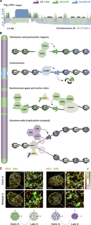
Figure 1. Enrichment of histone H3 variants and their deposition by dedicated chaperones. (A) Genomic distribution of H3.1, H3.3, and CenH3 from published ChIP-Seq data in HeLa cells (Lacoste et al., 2014; Clément et al., 2018). The plot shows the enrichment relative to input for all variants at a representative region spanning the centromere and the proximal short and long arms of chromosome 18 (p11.21-q21.1). The enrichment is com-puted as the log2 ratio between the mean per-base number of reads from
H3.1 (purple), H3.3 (green), and CenH3 (blue) and their respective input, at
consecutive 10-kb bins (smoothed over five nonzero bins). Enriched regions are highlighted in darker colors, illustrating the partitioning of the genome into chromatin domains associated with specific variants. (B) Schematic representation of the histone chaperones involved in the deposition of H3 variants at distinct chromosomal locations. The DAXX/ATRX complex pro-motes the accumulation of the non-replicative variant H3.3 at telomeres and pericentric heterochromatin. H3.3 is also deposited at actively transcribed regions and regulatory sites by HIRA, a complex consisting of three subunits: CAB IN1, HIRA, and UBN1. The non-replicative variant CenH3 is deposited spe-cifically at centromeres by its dedicated chaperone HJU RP, marking the site of kinetochore assembly. Both H3.1 and H3.3 are incorporated at centromeres during S phase, and H3.3 acts as a placeholder for CenH3 loading in late mito-sis and early G1. H3.1 is deposited genome-wide by the CAF-1 complex during S phase, or at DNA repair sites throughout the cell cycle. The CAF-1 complex consists of three subunits: p48, p50, and p160. The p150 subunit interacts with PCNA and promotes CAF-1 recruitment at replication forks. This couples H3.1 deposition to DNA synthesis and ensures proper chromatin assembly during replication. ASF1 is a general chaperone that can handle both H3.1 and H3.3 and hands them over to their dedicated chaperones. (C) Illustration of high-resolution visualization by STO RM of H3.1 and H3.3 along S phase (adapted from Clément et al., 2018). The STO RM images show the nuclear dis-tribution of H3.1 and H3.3 (HA staining, in red) at sites of DNA synthesis (EdU staining, in green) in early and mid/late S. Scale bars represent 5 µm. Insets represent enlarged images of selected areas where scale bars correspond to 600 nm. H3.3 clusters show stable volume, but there is a decrease in H3.3 density as S phase progresses; the late domains likely show a dilution of H3.3 during DNA replication. In contrast, H3.1 clusters change in both volume and density during S phase, with larger H3.1 clusters and low densities in early S and clusters with smaller volumes and higher density in mid/late S phase. See Clément et al. (2018) for further details.
stem cells. DEK can help in loading ATRX, and hence H3.3, on
telomeric regions, thereby maintaining telomere integrity
(
Ivanauskiene et al., 2014
).
The dedicated chaperone Holliday junction recognition
protein (HJU RP) ensures CenH3
CENP-Aloading at centromeres
(
Dunleavy et al., 2009
;
Foltz et al., 2009
). CenH3
CENP-Aspecifi-cally marks the centromere and is required for kinetochore
as-sembly and proper chromosome segregation during cell division.
During replication, CenH3
CENP-Ais diluted while H3.1 and H3.3
are deposited, and H3.3 is likely used as a placeholder for newly
assembled CenH3
CENP-A(
Dunleavy et al., 2011
), which is deposited
later during late telophase/early G1 (
Jansen et al., 2007
;
Bodor
et al., 2013
). Phosphorylation of HJU RP is required to ensure
proper timing of CenH3
CENP-Aincorporation, as
nonphosphory-latable mutants localize to centromeres throughout the cell cycle
(
Müller et al., 2014b
).
As outlined above, histone chaperones are generally in charge
of dedicated histone cargos and promote the deposition of
spe-cific variants, whereas others, like ASF1, are more promiscuous.
For further details on H3 variants and their chaperones, we direct
the reader to recent reviews (
Müller and Almouzni, 2017
;
Sitbon
et al., 2017
). Remarkably, under particular circumstances, some
chaperones can substitute for each other when one is either
miss-ing or limitmiss-ing. Thus, chaperone function can show some degree
of overlap. This has been observed upon DAXX depletion when a
fraction of the replacement variant H3.3 associates with the
rep-licative assembly machinery (
Drané et al., 2010
), or when HIRA
backs up CAF-1 to fill nucleosome gaps behind the fork (
Ray-Gallet et al., 2011
), or when DAXX handles excess CenH3
CENP-Ain place of HJU RP upon CenH3
CENP-Aoverexpression (
Lacoste
et al., 2014
). Such cross-talk between histone chaperones and
their choice of histone variant can thus provide robustness in
the process of histone management and suggests a potential for
chromatin plasticity.
Mechanisms for deposition and dynamics in and out of
chro-matin are thus critical. They involve DNA synthesis–coupled
pathways for replicative variants and DNA synthesis–uncoupled
pathways for non-replicative ones. Beyond handling histones,
their regulation and production play an equally important role to
meet different cellular demands. We will next discuss how the
ex-pression of replicative and non-replicative variants is regulated.
Replicative histone variants: A unique genomic organization
and regulation at multiple levels
Chromatin assembly in S phase requires vast amounts of newly
synthesized histones to ensure its restoration on the duplicated
genome. Replicative histone variants share a unique
transcrip-tional program that ensures higher expression levels for DNA
synthesis–coupled deposition in S phase. In contrast to
replace-ment variants, H3.1 and H3.2 genes show a peculiar organization
in clusters that comprise multiple copies of all core histones and
the H1 linker. This offers a potential means to optimize
coregu-lation. Indeed, in the human and mouse genome, replicative
his-tone genes cluster at three syntenic loci that remained physically
linked through evolution (
Marzluff et al., 2002
). The human
histone cluster 1 (HIST1) is located on chromosome 6 (6p22) and
comprises more than 50 coding genes, while HIST2 and HIST3
are located on chromosome 1 (1q21 and q42) and contain,
respec-tively, 10 and 3 coding genes. Besides their physical proximity,
genes are compartmentalized and processed in nuclear bodies
that concentrate the factors required for their transcription and
processing. These bodies were initially thought to coincide with
Cajal bodies, subnuclear compartments discovered at the
begin-ning of the century (
Cajal, 1903
) and implicated in the biogenesis
of ribonucleoproteins associated with histone pre-mRNAs (
Frey
and Matera, 1995
;
Calvi et al., 1998
;
Liu et al., 2000
). The presence
of separate histone locus bodies (HLBs) dedicated to the
tran-scription of replicative histones was only recognized later (
Liu et
al., 2006
). Similar to Cajal bodies, HLB foci are marked by coilin,
but they form at distinct subnuclear locations and coilin is
dis-pensable for their assembly (
Liu et al., 2009
). The nuclear
com-partmentalization of replicative histone genes is also reflected
by their higher-order organization (
Rao et al., 2014
;
Fritz et al.,
2018
). Higher-order structures can be revealed from
self-inter-acting regions where contacts among genome sequences in
phys-ical proximity occur more frequently (topologphys-ically associating
domains [TADs]). The three gene subsets in the human cluster 1
interact within separate TADs. Notably, these TADs further
in-teract with each other over an ∼1.5-Mb distance, indicating that
separate genes also establish long-range contacts and come in
physical proximity within the nucleus (
Fig. 2 A
). The
distinc-tive interplay among replicadistinc-tive histone genes is thus apparent
at multiple dimensions, but it is still unclear how these layers
of organization relate to each other. HLBs might represent a
case of self-organization (
Matera et al., 2009
) or an example of
phase-separated nuclear bodies that assemble via liquid
demix-ing by macromolecular crowddemix-ing (
Zhu and Brangwynne, 2015
;
Duronio and Marzluff, 2017
). This might resemble the phase
sep-aration recently implicated in the formation of heterochromatin
protein 1 (HP1) foci (
Larson et al., 2017
;
Strom et al., 2017
) and
repetitive RNA foci (
Jain and Vale, 2017
). It would be interesting
to experimentally address the link between phase transitions and
the assembly of HLBs. Transgenic assays in Drosophila
melano-gaster revealed that a sequence located between histone H3 and
H4 genes is important to mediate HLB assembly. Neither the H3
and H4 coding region nor the 3′ signals are required for HLB
formation (
Salzler et al., 2013
). Chromatin-linked adaptor for
male-specific lethal (CLA MP) is a zinc finger protein that binds
the GA repeat motif within bidirectional H3-H4 promoter and
controls chromatin accessibility, thereby enhancing
transcrip-tion and promoting HLB formatranscrip-tion (
Rieder et al., 2017
). Much
remains to be understood concerning how their transcriptional
regulation exploits this particular 3D organization.
Besides their distinctive genomic and subnuclear
organiza-tion, replicative histones differ from replacement variants in
terms of gene architecture and mRNA processing. Virtually all
genes in the histone clusters lack introns, have relatively short
UTRs, and produce transcripts that harbor a conserved 3′
stem-loop structure and do not undergo polyadenylation in many
or-ganisms (
Marzluff et al., 2008
;
Duronio and Marzluff, 2017
;
Mei
et al., 2017
). The only processing needed to form a mature histone
mRNA is the endonucleolytic cleavage of its precursor. This is
mediated in cis by the 3′ stem-loop and a purine-rich sequence
downstream of the cleavage site, the histone downstream element
(HDE). These features are widely conserved in metazoans as well
as unicellular eukaryotes (
Dávila López and Samuelsson, 2008
;
Marzluff et al., 2008
) but are not found in species, such as
Sac-charomyces cerevisiae, that lack DNA synthesis–coupled histone
variants (
Eriksson et al., 2012
). In budding yeast, there is a single
H3.3-like variant whose expression is induced in S phase together
with other histone gene pairs (
Osley et al., 1986
). Histone
repres-sion outside of S phase is mediated by several factors, including
the yeast orthologues of several known chaperones such as HIR1,
HIR2, and HIR3 (
Sherwood et al., 1993
;
Spector et al., 1997
) as well
as Asf1 (
Sutton et al., 2001
). In addition, histone transcription
is also regulated by Spt10, a putative acetyltransferase, together
with its partner Spt21 (
Kurat et al., 2014
). At the protein level,
Rad53 participates as part of a surveillance mechanism that
mon-itors the accumulation of excess histone proteins and triggers
their degradation (
Gunjan et al., 2005
;
Singh et al., 2010
). This
mode of regulation at the level of protein stability is not unique to
budding yeast but is further exploited in other organisms. Indeed,
the human histone chaperone nuclear autoantigenic sperm
pro-tein (NASP), similar to the Xenopus N1/N2, maintains a cytosolic
soluble pool of H3-H4 dimers and protects them from degradation
via chaperone-mediated autophagy (
Cook et al., 2011
).
In organisms with replicative histone variants, their
ex-pression throughout the cell cycle is controlled at multiple
lev-els (
Rattray and Müller, 2012
). Replicative histone genes are
transcribed and processed by several factors within the HLBs
(
Fig. 3
). Transcription initiation is controlled by cyclin E/
CDK2-dependent phosphorylation of the nuclear protein, ataxia-
telangiectasia locus (NPAT) at the G1/S transition. Phosphorylated
NPAT is required to initiate the assembly of HLBs and persists
throughout S phase to activate the expression of replicative
his-tone genes (
Ma et al., 2000
;
Zhao et al., 2000
;
White et al., 2011
).
NPAT also interacts with FLI CE-associated huge protein (FLA SH),
an essential cofactor involved in HLB assembly and 3′ processing of
nascent transcripts (
Barcaroli et al., 2006
;
Yang et al., 2009
;
White
et al., 2011
). Pre-mRNA maturation relies on the recognition of the
cis-regulatory elements at the 3′ end by two factors that are
spe-cific to replicative histones: the stem-loop binding protein (SLBP)
and the U7 snRNP (
Mowry and Steitz, 1987
;
Dominski et al., 1999
;
Sullivan et al., 2001
;
Zhao et al., 2004
). The U7 snRNP is a
ribo-nucleoprotein complex composed of U7 snRNA, Sm proteins, and
U7-specific Lsm10 and Lsm11 proteins (
Pillai et al., 2001
,
2003
).
The 5′ end of the complex binds histone pre-mRNAs via U7 snRNA
hybridization to the 3′ HDE sequence (
Cotten et al., 1988
;
Soldati
and Schümperli, 1988
). SLBP binds the stem-loop upstream and
interacts with the U7 snRNP to stabilize its association. This
stabi-lization is required for proper maturation and cleavage of histone
pre-mRNAs (
Pandey et al., 1994
;
Sullivan et al., 2001
) but might be
dispensable in vitro if the RNA duplex is sufficiently stable (
Streit
et al., 1993
;
Dominski et al., 1999
).
Pre-mRNA cleavage is catalyzed by the CPSF73
endonucle-ase (
Dominski et al., 2005
;
Mandel et al., 2006
). The interaction
Figure 2. Higher-order organization of replicative histone genes and compartmentalization of nuclear factors in HLBs. Replicative histones are redun-dantly encoded by multiple intronless genes that exhibit a conserved cluster organization across several lineages. (A) Location and spatial interactions among histone genes within the human histone cluster 1 (HIST1). The contact matrix was generated using iteratively corrected Hi-C data in GM12878 cells at 10-kb resolution (Rao et al., 2014) and plot with gcMapExplorer (Kumar et al., 2017). Genome bins where histone genes are located are marked in purple, illustrating the presence of three separate subsets within the HIST1 cluster. Neighboring genes in each subset interact within separate TADs, but all three subsets further engage in long-range interactions that bring distant genes together over an ∼1.5-Mb distance. This spatial organization might reflect the compartmentaliza-tion of replicative histone genes in the nucleus. Their pre-mRNAs are indeed transcribed and processed in dedicated nuclear bodies, called HLBs. (B) Nuclear distribution of NPAT, an essential factor driving HLB assembly and transcription initiation. The confocal image was retrieved from the Human Protein Atlas (v.18) and shows NPAT staining in U2-OS cells (in green). The nucleus and microtubules are stained in blue and red, respectively. NPAT concentrates at distinct subnuclear locations and marks the HLBs.
between FLA SH and the U7-specific protein Lsm11 is critical for
CPSF73 recruitment at the cleavage site (
Burch et al., 2011
;
Yang
et al., 2013
). SLBP, U7 snRNP, and FLA SH contribute to recruit
scaffolding factors and additional components of the histone
cleavage complex. The core components include Symplekin
(
Kolev and Steitz, 2005
;
Tatomer et al., 2014
) and CPSF100,
which heterodimerizes with CPSF73 to catalyze the
endonucle-olytic cleavage of the 3′ end (
Dominski et al., 2005
;
Kolev et al.,
2008
). Other factors (
Duronio and Marzluff, 2017
) might
con-tribute to stabilize interactions in the cleavage complex that are
specific to the processing of histone pre-mRNAs. mRNA levels
of replicative histones decrease at the end of S phase, and SLBP
is degraded upon phosphorylation by cyclin A/CDK1 (
Koseoglu
et al., 2008
). The 3′ stem-loop structure is necessary and
suffi-cient for the degradation of histone mRNAs (
Graves et al., 1987
;
Pandey and Marzluff, 1987
). Degradation is also regulated via 3′
end uridylation by the uridylyltransferase TUT7 (
Mullen and
Marzluff, 2008
;
Lackey et al., 2016
), and the 3′hExo enzyme is
needed in the initial steps (
Yang et al., 2006
). The mechanism
underlying 3′ end recognition to allow 3′hExo to initiate
degra-dation of the stem-loop is not well understood but might involve
elements downstream of the stop codon (
Graves et al., 1987
).
Besides factors that are specific to the processing of replicative
histones, several common targets and transcriptional regulators
contribute to expression in the HLB (also summarized in
Fig. 3
).
Human histone genes harbor TATA, CCA AT, and GC boxes in their
promoter region, as well as putative binding sites for several
transcription factors (
Mariño-Ramírez et al., 2006
). In human
Figure 3. Cell cycle timing and regulation of replicative histone genes. The replicative variant H3.1 is deposited by CAF-1 in a DNA synthesis–coupled manner, mostly during replication in S phase. Expression peaks at the G1/S transition, and its transcription is regulated in concert with other replicative histone genes located within the histone clusters. Their pre-mRNAs are processed through a distinct pathway that involves the recognition of two unique cis-regulatory elements: a 3′ stem-loop structure and an HDE. Transcription and pre-mRNA processing are compartmentalized in the nucleus and coordinated in HLBs. HLB assembly and transcriptional activation is initiated by NPAT. NPAT is phosphorylated by the Cyclin E/CDK2 complex at the G1/S transition. The maturation of histone pre-mRNAs requires the endonucleolytic cleavage of its 3′ tail and is mediated by several factors recruited to HLBs. The U7 snRNP binds the HDE via hybridization of the U7 snRNA. It interacts with SLBP, a protein that specifically recognizes the 3′ stem-loop structure and stabilizes the U7 snRNP association. FLA SH is another essential coactivator that promotes the recruitment of transcription factors and interacts with the Lsm11 subunit of the U7 snRNP to recruit the components of the histone cleavage complex. These transcription factors include Symplekin and the CPSF73/CPSF100 heterodimer that catalyzes the 3′ end endonucleolytic cleavage. Mature mRNAs are cleaved downstream of the 3′ stem-loop, which is required for mRNA degradation. SLBP is also degraded at the end of S phase after phosphorylation by the Cyclin A/CDK1 complex. H3 availability is further modulated at the protein level by NASP, a H3-H4 histone chaperone that protects soluble histones from degradation via chaperone-mediated autophagy counteracting Hsc70 and Hsp90.
cells, FLA SH participates in the recruitment of coactivators, such
as p73, that contribute to the transcription of replicative histones
(
De Cola et al., 2012
). In the tandemly arrayed linker and core
histone clusters of Drosophila, the TATA-binding protein TBP
regulates the transcription of core histones, while H1 genes have
TATA-less promoters modulated by the TBP related factor, TRF2
(
Isogai et al., 2007
). The core and linker histones are indeed
dif-ferentially expressed in flies, with H1 transcribed throughout the
S phase and the core histones induced during a short pulse in
early S phase (
Guglielmi et al., 2013
). Myc also colocalizes to the
Drosophila HLB and contributes to the expression of replicative
histone genes (
Daneshvar et al., 2011
). In mouse embryonic stem
cells, several chromatin factors such as E2f1, Ctcf, Smad1, and
Yy1 are likely to be involved in the regulation of both core and
linker histones (
Gokhman et al., 2013
). The contribution of
dif-ferent factors to the transcription of specific genes is still unclear
and might vary among lineages (
Mariño-Ramírez et al., 2006
)
as well as in a tissue- and developmental-specific manner. The
transcription elongation rate also affects the pre-mRNA folding
at the 3′ end. Stem-loop formation is impaired in slow elongation
conditions following UV irradiation or RNA polymerase II
muta-tion, which leads to an accumulation of polyadenylated histone
mRNAs (
Saldi et al., 2018
). The negative elongation factor (NELF)
interacts with the nuclear cap binding complex (CBC) and plays
a role in the 3′ processing. Their combined knockdown leads to
increased expression of replicative histone genes, and CBC was
shown to interact directly with SLBP. Both NELF and CBC
physi-cally associate with the histone gene body, and NELF accumulates
in nuclear foci where histone cleavage factors localize (
Narita et
al., 2007
). Genome-wide RNAi screens in Drosophila showed that
depletion of H3.3 and H2Av disrupts the expression of replicative
histones (
Wagner et al., 2007
). The U7 snRNP fails to accumulate
at the HLB when H2Av is mutated. Although the effect may be
in-direct, this suggests that histone variants themselves might play
a role in transcriptional processing.
The regulation of specific genes within the histone clusters
has yet to be characterized systematically. Nonetheless, their
unique cis-regulatory features and distinctive organization
en-sures a coordinated processing that enables a precise temporal
control of their expression. This regulation is critical for cell
cycle progression and has important implications for both
ge-nome and epigege-nome assembly. The cross-talk between 3D
orga-nization in the nucleus and transcription is surely a remarkable
paradigm, and the link between HLBs and topological domains
is a promising avenue for investigation. Such studies might also
give important insights into the coregulation of genes induced
synchronously by other cues.
Distinct transcriptional regulation and cell cycle expression of
the centromeric variant CenH3
In contrast to replicative variants, CenH3
CENP-Ais encoded by
a single multi-exon gene located outside of histone clusters
(
Sullivan et al., 1994
;
Régnier et al., 2003
). CenH3
CENP-Aexpres-sion is regulated through a distinct, DNA synthesis–independent
pathway (
Fig. 4
). Transcripts lack a 3′ stem-loop and undergo
conventional processing through splicing and polyadenylation.
The expression of CenH3
CENP-Apeaks in G2, and this temporal
control is key for its centromeric targeting (
Shelby et al., 1997
).
Induction of CenH3
CENP-Aduring S phase leads to a loss of specific
targeting and misassembly at chromosome arms. CenH3
CENP-Adeposition occurs in telophase/early G1 (
Jansen et al., 2007
).
HJU RP, its chaperone, is recruited at the centromere during this
critical time window and is otherwise distributed throughout
the nucleoplasm and at nucleoli (
Dunleavy et al., 2009
). A
con-trolled expression of CenH3
CENP-Ais necessary to prevent
promis-cuous interactions with low-affinity chaperones and aberrant
CenH3
CENP-Aloading (
Lacoste et al., 2014
;
Shrestha et al., 2017
).
Thus, ensuring an exclusive handling by HJU RP is likely critical
for proper deposition of CenH3
CENP-A. Its periodic regulation is
indeed a widely conserved feature, and CenH3
CENP-Amistargeting
occurs in a variety of distant species whenever the gene is
ecto-pically expressed at constitutively high levels (
Van Hooser et al.,
2001
;
Heun et al., 2006
;
Gascoigne et al., 2011
;
Mendiburo et al.,
2011
;
Choi et al., 2012
;
Lacoste et al., 2014
).
The regulation of both CenH3
CENP-Aand HJU RP is coordinated
in concert with other late cell cycle genes involved in mitotic
pro-gression, such as CDC25B, AUR KB, PLK1, and CENP-B (
Wang et
al., 2005
). Expression of these genes peak in G2/M, and they
harbor a conserved cell cycle–dependent element (CDE) and cell
cycle genes homology region (CHR) in their promoter (
Müller et
al., 2014a
). During G1 phase, CHR promotes their transcriptional
repression via recruitment of the dimerization partner, RB-like,
E2F, and MuvB (DRE AM) complex (
Sadasivam and DeCaprio,
2013
). Binding of DRE AM components to the CHR can be also
facilitated by the upstream CDE sequence (
Müller et al., 2012
).
When cells progress into S phase, DRE AM components
disso-ciate from the MuvB core complex and B-MYB binds to MuvB.
The B-MYB-MuvB (MMB) complex recruits FOXM1 to the CHR
site in late S phase. B-MYB is hence phosphorylated and
under-goes proteasome degradation, whereas MuvB remains bound to
FOXM1 (
Down et al., 2012
;
Sadasivam et al., 2012
). The
progres-sive phosphorylation of FOXM1 by cell cycle–dependent kinases
finally promotes its activation in G2/M (
Fu et al., 2008
;
Laoukili
et al., 2008
;
Chen et al., 2009
), which leads to maximal induction
of CHR-harboring genes bound by the MuvB-FOXM1 complex.
The CDE/CHR motif in CenH3
CENP-Apromoter was early
recog-nized as a potential cis-regulatory element underlying its periodic
expression (
Shelby et al., 1997
). CenH3
CENP-Ais a direct target of
FOXM1 (
Wang et al., 2005
;
Chen et al., 2013
), together with HJU RP
and other mitotic genes. In human and mouse cells, FOXM1
deple-tion leads to reduced CenH3
CENP-Aexpression and impaired mitotic
progression (
Wang et al., 2005
). Both CenH3
CENP-Aand HJU RP also
proved to be potential targets of the DRE AM repressor complex
(
Fischer et al., 2016
). Notably, their CDE/CHR motif facilitates
transcriptional repression upon p53-dependent recruitment of
DRE AM in mouse cells (
Filipescu et al., 2017
). Furthermore, p53
activation also leads to down-regulation of HJU RP and CENP-A in
human cells. Whether CenH3
CENP-Arepression during G1/S is
like-wise dependent on DRE AM has yet to be confirmed. Nevertheless,
the fine-tuning of CenH3
CENP-Akinetics is likely constrained by the
need to preserve its precise centromeric targeting.
Besides their transcriptional coregulation, CenH3
CENP-Aand
HJU RP are dedicated binding partners, and their interaction
plays an important role in maintaining a homeostatic balance.
CenH3
CENP-Aand HJU RP coexist as a soluble complex in which
each of them favors the reciprocal stabilization of the other
(
Bassett et al., 2012
;
Filipescu et al., 2017
). The N-terminal
por-tion of HJU RP binds both CenH3 and H4, thereby protecting the
dimers locally at the region of contact (
Bassett et al., 2012
). The
specific interaction with HJU RP is driven by the centromere
tar-geting domain (CATD) of CenH3
CENP-Aand favors the stabilization
of nonnucleosomal dimers. Exogenous overexpression of either
CenH3
CENP-Aor HJU RP leads to an increase in the endogenous
levels of their binding partner (
Filipescu et al., 2017
). HJU RP loss
results in CenH3
CENP-Adepletion, and CenH3
CENP-Aknockdown
leads to the proteasome-mediated degradation of HJU RP. The
in-teraction between the two could be further modulated by
post-translational modifications in CenH3
CENP-AN-terminal tail. For
instance, CDK1/cyclin B–dependent phosphorylation at serine
68 during G2/M hinders the interaction with HJU RP, preventing
premature loading of CenH3
CENP-Aat centromeres (
Hu et al., 2011
;
Yu et al., 2015
;
Wang et al., 2017
). Ubiquitylation at lysine 124
in-stead facilitates their interaction and might control the stability
of the complex (
Niikura et al., 2015
). The CUL4A-RBX1-COPS8
complex mediates CenH3
CENP-Alysine 124 ubiquitylation (
Niikura
et al., 2015
,
2017
), but the deubiquitylating enzyme has not yet
been identified. Posttranslational modifications and their effect
on CenH3 stability vary significantly across species, consistent
with the low conservation of its N-terminal tail (
Au et al., 2013
;
Bade et al., 2014
). The underlying pathways might represent
potential adaptations to consider in light of its rapid evolution
(
Malik and Henikoff, 2003
;
Kursel and Malik, 2017
) or
coevolu-tion with their dedicated chaperone (
Sanchez-Pulido et al., 2009
;
Rosin and Mellone, 2016
).
Constitutive expression of the non-replicative H3.3 variant by
independent paralogs
H3.3 is constitutively expressed in a cell cycle–independent
man-ner, supporting histone turnover outside of S phase and in
qui-escent or postmitotic cells (
Wu and Bonner, 1981
;
Wu et al., 1982
,
1983
) and histone replacement after fertilization in Drosophila
(
Loppin et al., 2005
) and mice (
Jang et al., 2015
;
Tang et al., 2015
).
H3.3 is redundantly encoded by two paralogous genes: H3.3A and
H3.3B (
Fig. 5
). These are conserved in many organisms and,
de-spite producing the same protein, they show different coding
sequences, cis-regulatory targets, and intron-exon organization
(
Brush et al., 1985
;
Wells and Kedes, 1985
;
Chalmers and Wells,
1990
;
Akhmanova et al., 1995
;
Albig et al., 1995
;
Bramlage et al.,
1997
). Similar to CenH3
CENP-A, H3.3A and H3.3B are solitary genes
lacking 3′ stem-loop structure. Both give rise to polyadenylated
transcripts, and H3.3B can generate up to three isoforms from
alternative polyadenylation sites (
Albig et al., 1995
;
Bramlage et
al., 1997
;
Feng et al., 2005
). Although both H3.3A and H3.3B can
contribute to new H3.3 synthesis, the two genes show distinct
expression profiles in the male and female germline, among
different tissues, and during development (
Krimer et al., 1993
;
Akhmanova et al., 1995
;
Bramlage et al., 1997
;
Couldrey et al.,
1999
;
Jang et al., 2015
;
Maehara et al., 2015
).
The cis-regulatory elements in the human H3.3B promoter
include an octamer (Oct) motif and a combined cAMP- and
TPA-responsive element (CRE/TRE) flanked by a TATA box and
six CCA AT boxes (
Witt et al., 1997
). Promoter deletion
con-structs show that the proximal promoter region, comprising
Oct, CRE/TRE, and TATA elements, is sufficient to drive
tran-scription in vitro (
Witt et al., 1997
). The CRE/TRE sequence in
H3.3B promoter can recruit TPA-inducible AP-1 factors (
Karin
et al., 1997
) and transcription factors of the CREB/ATF family
that are typically activated via PKA-dependent
phosphoryla-tion in response to cAMP (
Mayr and Montminy, 2001
).
How-ever, cAMP treatment does not significantly affect H3.3B levels
in vitro, whereas TPA induces a strong transcriptional response
Figure 4. Cell cycle timing and regulation of the centromeric variant CenH3. The non-replicative variant CenH3CENP-A is deposited at centromeres
by its dedicated chaperone HJU RP in telophase/early G1. Its expression is reg-ulated in a cell cycle–dependent manner and peaks in G2/M. CenH3CENP-A is
encoded by a single multi-exon gene located outside of the histone clusters that undergoes conventional pre-mRNA processing via splicing and polyad-enylation. Transcription is coordinated in concert with other late cell cycle genes, including HJU RP. This is regulated in cis by the CHR/CDE motif in their promoter region. The recruitment of the DRE AM complex at the CHR is thought to promote transcriptional repression during G1. At the beginning of S phase, the MuvB core of the DRE AM complex remains bound to the CHR/CDE while other components dissociate and are replaced by B-MYB. The B-MYB-MuvB (MMB) complex recruits FOXM1 in late S phase. B-MYB is degraded upon phosphorylation, whereas the progressive phosphorylation of FOXM1 leads to its activation in G2/M. Both CenH3 and HJU RP are induced at the same time and mutually stabilize each other at the protein level. The reciprocal stabilization is affected by posttranslational modifications on the N-terminal tail of CenH3, which further regulate the timing of deposition. S69 phosphorylation prevents interaction with HJU RP and premature load-ing, while K124 ubiquitylation favors HJU RP binding and might contribute to their stabilization.
that is dependent on the presence of an intact CRE/TRE motif
(
Witt et al., 1998
). This indicates a possible role for the
recruit-ment of AP-1 factors through the TPA-inducible PKC pathway to
induce H3.3B in vivo. However, additional players are likely
in-volved in specific tissues or developmental stages and have not
yet been characterized. Concerning H3.3A, it has a GC-rich
pro-moter devoid of TATA and CCA AT boxes and instead harbors an
ATA motif along with four SP1 binding sites. Stepwise deletion
of the proximal promoter region, containing three SP1 motifs,
reduces transcriptional activity in vitro in an additive manner
(
Frank et al., 2003
); however, the binding partners and
mech-anistic basis are largely unexplored. Hence, factors promoting
H3.3A induction or differential expression in various tissues
remain largely unknown.
Consequences of H3 variant alterations and imbalances
As discussed, an incredible orchestration works to control the
provision of histone proteins by multitasking at each possible
level of regulation. Histones are always accompanied by
chap-erones, their guardians, throughout their cellular life (
Gurard-Levin et al., 2014
). The proper dosage of histone variants and
chaperones plays an important role in defining the chromatin
landscape during embryonic development, lineage commitment,
and cell fate decisions (
Filipescu et al., 2014
). Xenopus oocytes
are protected from the surplus of histones by nucleoplasmin,
sustaining chromatin assembly through the rapid cell divisions
that occur during early development. In mammalian cells,
rep-licative aging is associated with impaired histone synthesis
(
O’Sullivan et al., 2010
). This may be linked to chronic damage
signals at telomeres that could affect histone levels (
O’Sullivan
et al., 2010
). Histone supply also affects the length of S phase
and cell cycle progression (
Groth et al., 2005
,
2007
;
Günesdogan
et al., 2014
). Interfering with SLBP, which reduces histone
ex-pression in human cells, also results in reduced cell growth and
impaired S phase progression (
Zhao et al., 2004
). Intriguingly,
the excess H3 during mitosis localizes to the centrosomes for
proteasome-mediated degradation in worms, flies, and human
cells (
Wike et al., 2016
). The cross-talk with the centrosome could
hence play a role in preserving chromosome integrity by
coor-dinating different signaling events. Genes in the HIST1 cluster
are significantly up-regulated across different breast cancer cell
lines and breast tumor specimens (
Fritz et al., 2018
). The
chro-matin organizer CTCF, mutated in several cancers, is found at
the boundaries between TADs associated with distinct gene
sub-clusters. Considering that the nuclear organization of the HLB
is compromised in cancer cells (
Ghule et al., 2009
), this could
contribute to the misregulation of histone genes in tumors.
CenH3
CENP-Aoverexpression is common in many aggressive
tumors (
Tomonaga et al., 2003
;
Amato et al., 2009
;
Hu et al.,
2010
;
Li et al., 2011
;
Wu et al., 2012b
;
Qiu et al., 2013
;
Filipescu et
al., 2017
). These CenH3
CENP-Aimbalances correlate with genomic
instability and malignant progression as well as poor prognosis
and response to treatment (
Sun et al., 2016
;
Zhang et al., 2016
).
CenH3 overexpression also leads to ectopic recruitment of
ki-netochore components and mitotic defects in both Drosophila
(
Heun et al., 2006
) and fission yeast (
Choi et al., 2012
;
Gonzalez
et al., 2014
). In human cells, overexpression of CenH3
CENP-Are-sults in misassembly at chromosome arms (
Lacoste et al., 2014
)
in a cell cycle–independent manner (
Nechemia-Arbely et al.,
2017
). Ectopic deposition is mediated by ATRX/DAXX and
con-fers higher tolerance to DNA damage, a potential mechanism for
resistance. Excess CenH3
CENP-Alocalizes at CTCF binding sites
(
Lacoste et al., 2014
), DNase I hypersensitive sites, and
transcrip-tion factor binding sites across the genome (
Athwal et al., 2015
).
Subtelomeric regions prone to instability are also hotspots for
CenH3
CENP-Aaccumulation in overexpressing conditions (
Athwal
et al., 2015
). Understanding how higher doses of CenH3
CENP-Acan
confer a selective advantage in genomically unstable cells will be
important. Overexpression is associated with better tolerance to
damage, but higher CenH3
CENP-Alevels are also linked to
chromo-some instability and micronuclei formation in both cancer and
stable diploid cells (
Shrestha et al., 2017
). Future studies will
cer-tainly be critical to understand how histone imbalances and
chro-matin misassembly may lead to severe pathological implications.
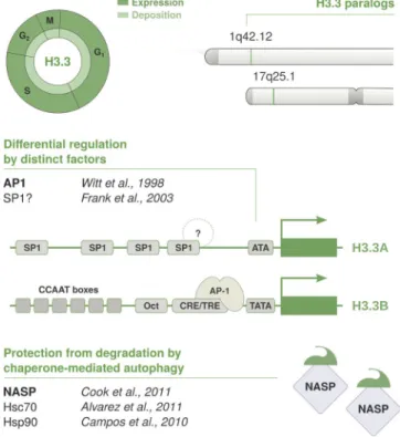
Figure 5. Cell cycle timing and regulation of the replacement variant H3.3. The non-replicative variant H3.3 is deposited throughout the cell cycle by HIRA at regions of high turnover, such as regulatory sites and transcribed regions. H3.3 is expressed constitutively and redundantly encoded by two conserved paralogs: H3.3A and H3.3B. Both genes are located outside of his-tone clusters, contain introns, and give rise to polyadenylated mRNAs. H3.3A and H3.3B encode for the same protein, but their gene architecture is not conserved. They show distinct coding sequences, intron-exon organization, and cis-regulatory elements and are differentially expressed during develop-ment and among tissues. Putative binding sites have been identified in their respective promoter regions, and the CRE/TRE motif in H3.3B promoter was shown to mediate its activation via recruitment of AP-1 transcription fac-tors. However, the basis of differences in expression is poorly understood. Similar to H3.1, the NASP histone chaperone can bind H3.3-H4 dimers and contributes to fine-tune protein levels via protection from chaperone- mediated autophagy.
Concerning H3.3, clear differences in expression between
the two paralogs were observed in the fly and mouse male
ger-mlines (
Akhmanova et al., 1995
;
Bramlage et al., 1997
;
Feng et al.,
2005
) and throughout preimplantation development in
mam-mals (
Couldrey et al., 1999
;
Kafer et al., 2010
;
Xue et al., 2013
).
In flies, H3.3A and H3.3B single knockouts are viable and fertile,
whereas double-null mutants have low viability and are sterile
(
Hödl and Basler, 2009
;
Sakai et al., 2009
). Hypomorphic
H3.3B-null mice die postnatally, but single H3.3A knockouts are viable,
with reduced male fertility (
Couldrey et al., 1999
;
Tang et al.,
2015
). However, the effect of hypomorphic mutations depends
on the genetic background (
Jang et al., 2015
), and it is unclear
what drives this variability among the paralogs. Notably,
differ-ences in expression could have important implications in
tum-origenesis, because mutations in H3.3A and H3.3B are linked
to distinct types of cancers (
Weinberg et al., 2017
). Indeed, the
impact and occurrence of somatic mutations differs between
H3.3A and H3.3B (
Behjati et al., 2013
). K36 mutations, found in
95% of chondroblastomas and 7% of clear-cell chondrosarcomas,
occur predominantly in the H3.3B gene. G34 and K27 mutations
are instead nearly exclusive to H3.3A and linked to other types of
tumors such as high-grade astrocytomas (
Schwartzentruber et
al., 2012
;
Sturm et al., 2012
;
Wu et al., 2012a
;
Aihara et al., 2014
)
and giant cell tumors of bone (
Behjati et al., 2013
;
Sarungbam et
al., 2016
). The lysine 27 is encoded by an AAG codon in H3.3A and
AAA in H3.3B; hence the chance of a specific substitution
occur-ring in a given paralog can be explained in part by codon
differ-ences between H3.3A and H3.3B. K27 mutations are indeed also
found in H3.1-coding genes, where, similar to H3.3A, the lysine
is often encoded by a AAG codon (
Kallappagoudar et al., 2015
).
However, differences in expression may affect the tissue
specific-ity and outcome of specific amino acid substitutions, a possibilspecific-ity
that has not been fully explored. Interestingly, duplication rates
also vary between the two paralogs. Numerous H3.3 pseudogenes
exist in both the human and mouse genome, and the majority
are more closely related to H3.3A (
Wells et al., 1987
;
Ederveen et
al., 2011
;
Maehara et al., 2015
). It is currently unknown whether
genomic context or differential transcription has any impact on
the outcome or propensity to genetic variation. Recent findings
have provided insights into how alterations in multiple histone
variants beyond H3 can cause changes in epigenome plasticity
and genome stability, thereby driving cancer initiation and/or
progression (
Vardabasso et al., 2014
;
Park et al., 2016
;
Buschbeck
and Hake, 2017
). Understanding the regulation of distinct
his-tone variants in normal conditions will be critical to gain insights
into their role in cancer progression and open concrete avenues
for future therapeutic strategies.
Concluding remarks
We have summarized the current knowledge concerning the
transcriptional regulation of replicative and non-replicative
his-tone variants. Their genomic organization and distinct features
are a unique example of how an exquisite orchestration of events
can contribute to handle the demands for histone variants under
distinct physiological conditions. The coregulation of replicative
variants, and the evolution of independent mechanisms that
co-ordinate the production of non-replicative forms, may represent
an interesting paradigm that could apply to other gene families.
We have also learned a lot about the interplay among histone
variants and their chaperones and how they shape the epigenome
and sustain chromatin plasticity. In the future, it will be crucial to
further investigate histone cross-talk in the variety of cell types
that constitute an organism, under normal or pathological
condi-tions. Multidisciplinary approaches and single-cell technologies
will certainly play a pivotal role in the future to resolve the
dy-namics of chromatin architecture across different cells and over
the lifetime of an individual.
Acknowledgments
We thank Dominique Ray-Gallet and Iva Simeonova for critical
reading of the manuscript.
This work was supported by La Ligue Contre le Cancer
(Equipe Labelisée 2016), Parisian Alliance of Cancer Research
Institutes, Agence Nationale de la Recherche (11-LABX-0044
“DEEP,” 10-IDEX-0001-02 “PSL,” ANR-12-BSV5-0022-02
“CHA PIN HIB,” ANR-14-CE16-0009 “Epicure,” ANR-14-CE10-0013
“CEL LEC TCH IP,” 16-CE15-0018 “CHR ODYT,”
ANR-16-CE12-0024 “CHI FT,” and ANR-16-CE11-0028 “REP LIC AF”),
and European Research Council (ERC-2015-ADG project 694694
“ChromADI CT” and ERC-2015-POC project 678563 “EPO CH28”).
A. Gatto is supported by the Horizon 2020 Framework
Pro-gramme for Research and Innovation (H2020 Marie Skłodowska-
Curie Actions grant agreement 798106 “REP LIC HROM4D”).
The authors declare no competing financial interests.
Author contributions: S. Mendiratta, A. Gatto, and G.
Almou-zni wrote the manuscript.
Submitted: 24 July 2018
Revised: 5 September 2018
Accepted: 12 September 2018
References
Ahmad, K., and S. Henikoff. 2002. The histone variant H3.3 marks active chromatin by replication-independent nucleosome assembly. Mol. Cell. 9:1191–1200. https:// doi .org/ 10 .1016/ S1097 -2765(02)00542 -7
Aihara, K., A. Mukasa, K. Gotoh, K. Saito, G. Nagae, S. Tsuji, K. Tatsuno, S. Yamamoto, S. Takayanagi, Y. Narita, et al. 2014. H3F3A K27M mutations in thalamic gliomas from young adult patients. Neuro-oncol. 16:140–146.
https:// doi .org/ 10 .1093/ neuonc/ not144
Akhmanova, A.S., P.C. Bindels, J. Xu, K. Miedema, H. Kremer, and W. Hen-nig. 1995. Structure and expression of histone H3.3 genes in Drosophila melanogaster and Drosophila hydei. Genome. 38:586–600. https:// doi .org/ 10 .1139/ g95 -075
Alabert, C., and A. Groth. 2012. Chromatin replication and epigenome main-tenance. Nat. Rev. Mol. Cell Biol. 13:153–167. https:// doi .org/ 10 .1038/ nrm3288
Albig, W., B. Bramlage, K. Gruber, H.-G. Klobeck, J. Kunz, and D. Doenecke. 1995. The human replacement histone H3.3B gene (H3F3B). Genomics. 30:264–272. https:// doi .org/ 10 .1006/ geno .1995 .9878
Almouzni, G., and H. Cedar. 2016. Maintenance of Epigenetic Information.
Cold Spring Harb. Perspect. Biol. 8:a019372. https:// doi .org/ 10 .1101/ cshperspect .a019372
Alvarez, F., F. Muñoz, P. Schilcher, A. Imhof, G. Almouzni, and A. Loyola. 2011. Sequential establishment of marks on soluble histones H3 and H4. J. Biol.
Chem. 286:17714–17721. https:// doi .org/ 10 .1074/ jbc .M111 .223453
Amato, A., T. Schillaci, L. Lentini, and A. Di Leonardo. 2009. CEN PA overex-pression promotes genome instability in pRb-depleted human cells. Mol.