HAL Id: tel-01911135
https://tel.archives-ouvertes.fr/tel-01911135
Submitted on 2 Nov 2018HAL is a multi-disciplinary open access archive for the deposit and dissemination of sci-entific research documents, whether they are pub-lished or not. The documents may come from teaching and research institutions in France or abroad, or from public or private research centers.
L’archive ouverte pluridisciplinaire HAL, est destinée au dépôt et à la diffusion de documents scientifiques de niveau recherche, publiés ou non, émanant des établissements d’enseignement et de recherche français ou étrangers, des laboratoires publics ou privés.
establishment of hepatitis B virus minichromosome
Maëlle Locatelli
To cite this version:
Maëlle Locatelli. The histone chaperone HIRA is crucial for the early establishment of hepatitis B virus minichromosome. Virology. Université de Lyon, 2018. English. �NNT : 2018LYSE1169�. �tel-01911135�
N°d’ordre NNT : 2018LYSE1169
THESE DE DOCTORAT DE L‘UNIVERSITÉ DE LYON
Opérée au sein deL’Université Claude Bernard Lyon 1
Ecole Doctorale
N° 340Biologie Moléculaire Intégrative et Cellulaire (BMIC)
Spécialité de doctorat : Virology
Discipline : Infectiology
Soutenue publiquement le Mardi 18 Septembre 2018 par
Maëlle LOCATELLI
THE HISTONE CHAPERONE HIRA IS
CRUCIAL FOR THE EARLY ESTABLISHMENT
OF HEPATITIS B VIRUS MINICHROMOSOME
Directeurs de thèse : Professeur Fabien ZOULIM et Docteur Barbara TESTONI
Devant le jury composé de :
Mr. Professeur Michael NASSAL, PU, Université de Fribourg en Brisgau Rapporteur Mme. Docteur Christine NEVEUT, DR2 INSERM, Institut Pasteur, Paris Rapporteur Mr. Professeur Massimo LEVRERO, PU-PH, CRCL/HCL Examinateur Mr. Docteur Jean-Pierre QUIVY, DR2 CNRS, Institut Curie, Paris Examinateur Mr. Professeur Fabien ZOULIM, PU-PH, CRCL/HCL Directeur de thèse Mme. Docteur Barbara TESTONI, CR1 INSERM, CRCL Co-directrice de thèse
Président de l’Université
Vice-président du Conseil Académique Vice-président du Conseil d’Administration
Vice-président du Conseil Formation et Vie Universitaire Vice-président de la Commission Recherche
Directeur Général des Services
M. le Professeur Frédéric FLEURY
M. le Professeur Hamda BEN HADID M. le Professeur Didier REVEL M. le Professeur Philippe CHEVALIER M. Fabrice VALLÉE
M. Alain HELLEU
COMPOSANTES SANTÉ
Faculté de Médecine Lyon Est – Claude Bernard
Faculté de Médecine et de Maïeutique Lyon Sud – Charles Mérieux Faculté d’Odontologie
Institut des Sciences Pharmaceutiques et Biologiques Institut des Sciences et Techniques de la Réadaptation
Département de formation et Centre de Recherche en Biologie Humaine
Directeur : M. le Professeur J. ETIENNE Directeur : Mme. la Professeure C. BURILLON Directeur : M. le Professeur D. BOURGEOIS Directeur : Mme. la Professeure C. VINCIGUERRA Directeur : M. le Professeur Y. MATILLON Directeur : Mme. la Professeure A-M. SCHOTT
COMPOSANTES ET DEPARTEMENTS DE SCIENCES ET TECHNOLOGIE
Faculté des Sciences et TechnologiesDépartement Biologie
Département Chimie Biochimie
Département Génie Electrique et des Procédés Département Informatique
Département Mathématiques Département Mécanique Département Physique
UFR Sciences et Techniques des Activités Physiques et Sportives Observatoire des Sciences de l’Univers de Lyon
Polytech Lyon
Ecole Supérieure de Chimie Physique Electronique Institut Universitaire de Technologie de Lyon 1 Ecole Supérieure du Professorat et de l’Education Institut de Science Financière et d'Assurances
Directeur : M. F. DE MARCHI
Directeur : M. le Professeur F. FLEURY Directeur : Mme. C. FELIX
Directeur : M. H. HAMMOURI
Directeur : M. le Professeur S. AKKOUCHE Directeur : M. le Professeur G. TOMANOV Directeur : M. le Professeur H. BEN HADID Directeur : M. J-C. PLENET
Directeur : M. Y. VANPOULLE Directeur : M. B. GUIDERDONI Directeur : M. le Professeur E.PERRIN Directeur : M. G. PIGNAULT
Directeur : M. le Professeur C. VITON
Directeur : M. le Professeur A. MOUGNIOTTE Directeur : M. N. LEBOISNE
Abstract
The histone chaperone HIRA is crucial for the early establishment of hepatitis B virus minichromosome.
Hepatitis B virus (HBV) chronically infects 240 million people worldwide and is the major cause of hepatocellular carcinoma (HCC). Currently standard-of-care treatments can achieve long-term viral suppression, but are not able to completely eliminate the virus, due to the persistence of the covalently closed circular DNA (cccDNA). cccDNA, the viral minichromosome, resides in the nucleus of infected hepatocytes by virtue of its chromatin structure. Indeed, upon entry into hepatocytes, the partially double stranded viral DNA (relaxed circular (rc)DNA) is released into the nucleus, where it is repaired and wrapped by histones to form an episomal chromatinized structure. The mechanisms leading to cccDNA formation and chromatinization are still largely unknown and their elucidation would be a first step toward the identification of new therapeutic targets to impair cccDNA persistence. To this aim, we investigated the role of host factors belonging to DNA repair and nucleosome assembly pathways in cccDNA formation at early time points (i.e. between 30 minutes and 72 hours) post-infection in both HepG2-NTCP cell line and Primary Human Hepatocytes (PHH). We particularly focused on the histone chaperone Hira, which is known to deposit histone variant 3.3 (H3.3) onto cellular DNA in a replication-independent manner and in association to nucleosome reshuffling during transcription and DNA repair. We were able to detect cccDNA in the nuclear fraction of hepatocytes as early as 30 minutes and 24h post-infection, by qPCR and Southern Blotting (SB), respectively. Knock-down of Hira by RNA interference before virus inoculation led to a strong decrease in cccDNA accumulation (both in qPCR and SB) which was independent from HBx protein expression (using an HBx-defective virus). rcDNA levels remained stable, indicating either a possible incomplete or delayed rcDNA to cccDNA transition. Chromatin Immunoprecipitation analysis showed that Hira was bound to cccDNA already at 2 hours post-infection and that its recruitment was concomitant with the deposition of histone H3.3 and the binding of HBV capsid protein (HBc). After 24 hours of infection, an increase of H3.3 and Pol2 binding on cccDNA was observed, correlating with the initiation of the transcription of the 3.5 kb RNA. By Co-Immunoprecipitation and Proximity Ligation Assay experiments, we showed that Hira was able to interact with HBc in infected hepatocytes and
in a HepaRG cell line expressing HBc in an inducible manner. Altogether, our results suggest that chromatinization of incoming viral DNA is a very early event, requiring the histone chaperone Hira. While HBx is not required for this process, HBc could play a major role, suggesting that the interaction between Hira and HBc could represent a new therapeutic target to be investigated.
Résumé
La chaperone d’histones, HIRA, est essentielle dans l’établissment précoce du minichromosome du virus de l’hépatite B.
Le virus de l’hépatite B (HBV) infecte de manière chronique 240 millions de personnes dans le monde, et est la principale cause de carcinome hépatocellulaire. Actuellement, les traitements standards permettent une suppression virale à long terme, mais ne sont pas capables d’éliminer complètement le virus, en raison de la persistance de l’ADN circulaire et clos de façon covalente (ADNccc).
Ce minichromosome viral réside dans le noyau des hépatocytes infectés, grâce à sa structure chromatinienne. En effet, lors de l’infection d’un hépatocyte, l’ADN viral partiellement double brin (ADN relaché circulaire (rc)) est libéré dans le noyau, où il est réparé et enveloppé par des protéines histones, afin de former une structure d’épisome chromatinisé. Les méchanismes conduisant à la formation ainsi qu’à la chromatinisation de l’ADNccc sont encore largement inconnus, et leur élucidation constituerait une première étape vers l’identification de nouvelles cibles thérapeutiques, susceptibles d’altérer la persistance de l’ADNccc. Dans ce but, nous avons étudié le rôle des facteurs hôtes de réparation de l’ADN, et des voies d’assemblage des nucléosomes, dans la formation de l’ADNccc, à des stades précoces (entre 30 minutes et 72 heures) de l’infection, dans des lignées cellulaires d’HepG2-NTCP, ainsi que dans des hépatocytes primaires humains. Nous nous sommes particulièrement concentrés sur la protéine chaperone d’histones, HIRA, qui est connue pour déposer le variant d’histone 3.3 (H3.3) sur l’ADN cellulaire d’une manière indépendante de la réplication et en association avec le remaniement des nucléosomes pendant la transcription et la réparation de l’ADN.
Nous avons été capables de détecter l’ADNccc dans la fraction nucléaire des hépatocytes dès 30 minutes et 24 heures post-infection, par qPCR et Southern Blotting (SB), respectivement. L’extinction de HIRA par ARN interférent (siARN) avant l’inoculation du virus, a conduit à une forte diminution de l’accumulation de l’ADNccc (à la fois par qPCR et Southern Blot), qui était indépendante de la protéine HBx (en utilisant un virus HBx-défectueux). Les niveaux d’ADNrc sont restés stables, indiquant soit une éventuelle transition de l’ADNrc en ADNccc incomplète,
ou retardée. L’analyse par immunoprécipitation de la chromatine a montré que HIRA était liée à l’ADNccc dès 30 minutes après infection, et que son recrutement était concomitant avec le dépot de l’histone H3.3, ainsi que la liaison de la protéine de capside du HBV (HBc). Après 24 heures d’infection, une augmentation de la liaison de H3.3 et de l’ARN polymerase II sur l’ADNccc a été observée, en corrélation avec l’initiation de la transcription de l’ARN viral de 3.5 kb. Par des expériences de co-immunoprécipitaton et de test de proximité entre protéines (PLA), nous avons montré que HIRA était capable d’interagir avec HBc dans des hépatocytes infectés et dans une lignée cellulaire HepaRG exprimant HBc de manière inductible. En conclusion, nos résultats suggèrent que la chromatinisation de l’ADN viral entrant est un événement très précoce, nécessitant l’histone chaperone HIRA. Bien que HBx ne soit pas requis pour ce processus, HBc pourrait jouer un rôle majeur, suggérant que l’intéraction entre HIRA et HBc pourrait représenter une nouvelle cible thérapeutique à étudier.
Résumé substantiel
La chaperone d’histones, HIRA, est essentielle dans l’établissment précoce du minichromosome du virus de l’hépatite B.
Travaux de thèse réalisés par Maëlle Locatelli au sein du Centre de Recherche en Cancérologie de Lyon (CRCL), UMR INSERM 1052, CNRS 5286, Centre Léon Bérard, dirigé par Alain Puisieux
Directeurs de thèse : Fabien Zoulim et Barbara Testoni
Introduction : Le virus de l’hépatite B (HBV) infecte chroniquement plus de 240 millions de
personnes dans le monde, et reste un problème majeur de santé publique. En effet, l’infection chronique par le virus de l’hépatite B est associée, avec le temps, à l’apparition d’une cirrhose pouvant conduire au développement d’un carcinome hépatocellulaire. Les thérapies disponibles actuellement permettent de contrôler la réplication virale, mais ne sont pas capables d’élimier complètement l’infection, en raison de la persistance de l’ADN circulaire et clos de façon covalente (ADNccc) dans le noyau des hépatocytes infectés. En effet, après infection d’un hépatocyte par le virus de l’hépatite B, le génome viral partiellement double brin, sous forme d’ADN relaché circulaire (ADNrc), va être transporté vers le noyau, où il va ensuite être transformé en ADNccc, afin de jouer un rôle de matrice de réplication virale, mais aussi de réservoir. Ces traitements doivent donc être administrés à vie, puisqu’une interruption est quasi universellement suivie d’une réactivation virale. La disparition du réservoir d’ADNccc intrahépatique est donc l’enjeu principal pour le développement de thérapies futures. Il est donc nécessaire de pouvoir trouver de nouvelles cibles permettant l’éradication totale de l’infection, via l’élimination ou l’inactivation transcriptionnelle de l’ADNccc.
L’ADNccc est comparable à un minichromosome viral intrahépatocytaire, et sert de matrice à la transcription des ARN viraux. En tant qu’épisome chromatinisé, l’ADNccc est composé d’histones, ainsi que de protéines virales et cellulaires non histones, qui confèrent à l’ADNccc
sa stabilité et son intégrité. De la même façon que l’ADN cellulaire, l’activité transcriptionnelle de l’ADNccc peut être modulée par des modifications épigénétiques telles que les modifications post-traductionnelles (PTM) des histones et d’autres protéines, mais aussi la méthylation de l’ADN (Jain et al., 2015), ou encore les microARN (Huang et al., 2016). Les protéines virales jouent également un rôle majeur dans la stabilité et l’activité de l’ADNccc. D’une part, la protéine virale régulatrice, HBx, a été montrée comme activant la transcription du virus de l’hépatite B. En effet, en interagissant avec DDB1 (DNA binding-binding protein 1), HBx recrute le complexe cullin ubiquitine ligase (CRL4), afin de dégrader le complexe Smc5/6 (maintien structurel des chromosomes), un facteur de restriction de l’ADNccc (Decorsière et
al., 2017). En l’absence d’HBx, l’hyperacétylation des histones liées à l’ADNccc médiée par
p300 (activateur transcriptionnel qui possède un domaine histone acétyltransferase), est fortement altérée. Au contraire, les déacétylases, telles que HDAC1 et SIRT1, sont recruitées sur l’ADNccc (Belloni et al.,2009). D’autre part, la protéine de capside, entrant dans le noyau simultanément à l’ADNrc, est un composant structurel de l’ADNccc, car elle se lie préférentiellement à l’ADN double brin d’HBV, ayant alors pour effet de réduire de 10% l’espacement nucléosomique (Bock, 2001). Lors de l’entrée dans les hépatocytes, l’ADN viral partiellement double brin est libéré dans le noyau, où il va ensuite être réparé et enveloppé par des protéines cellulaires histones, afin de former sa structure chromatinienne.
Dans ce contexte d’un minichromosome régulé épigénétiquement, et dont les étapes d’établissement restent fortement méconnues, les objectifs de notre équipe sont d’étudier l’interaction du virus de l’hépatite B avec la machinerie cellulaire hôte, pour mieux comprendre les mécanismes de formation et de régulation de l’ADNccc, en utilisant des modèles cellulaires pertinents. En effet, la recherche portant sur l’ADNccc a été entravée au cours du temps par l’absence de modèles d’infection appropriés, avec des lignées cellulaires produisant des niveaux d’ADNccc difficilement détectables, en raison de sa quantité, mais aussi de la spécificité nécessaire pour quantifier uniquement l’ADNccc, menant ainsi à une répercussion directe sur les approches, déjà limitées, pour étudier ce minichromosome. Récemment, des études ont pu montrer que la conversion de l’ADNrc en ADNccc pouvait probablement être réalisée en exploitant la réponse aux dommages de l’ADN de l’hôte (Gomez-Moreno et Garaigorta, 2017). En effet, Kröniger et son équipe ont montré que la tyrosyl-ADN-phosphodiestérase 2 (TDP2), une enzyme de réparation de l’ADN, qui élimine les
interactants covalents de l’ADN, était capable d’éliminer la polymérase de l’ADNrc. Les étapes menant à la conversion de l’ADNrc en ADNccc incluent d’autres preuves d’une interaction du virus avec la machinerie hôte, comme par exemple la complétion du brin positif du rcDNA par la polymérase κ.
Cependant, toujours peu de choses sont connues sur l’interaction entre l’ADNrc et la machinerie cellulaire de l’hôte, et de ce fait, les mécanismes à l’origine de l’assemblage et stabilité de l’ADNccc restent mal compris. C’est pourquoi, par ce projet, nous nous sommes intéressés au rôle des chaperones d’histones, assistant le trafic des histones, mais aussi principalement l’assemblage et le remaniement des nucléosomes, dans la chromatinisation de l’ADNccc.
Objectifs du projet de recherche :
Ce travail de thèse a eu pour objectifs : i) d’étudier la cinétique d'établissement et d'activité du cccDNA du VHB dans notre modèle HepG2-NTCP ; ii) l’identification d'éventuelles chaperones d'histones impliquées dans la formation et la chromatinisation de l'ADNcc ; iii) l’exploration de l'interaction HIRA/ADNccc du virus de l’hépatite B et la caractérisation des mécanismes impliqués.
Matériel et méthodes :
Des cellules HepG2-NTCP ont d’abord été infectées par HBV à une multiplicité d’infection de 250, durant différents temps de cinétique allant de 30 minutes à 72 heures, et les paramètres viraux ont ensuite pu être analysés, par qPCR et southern blot, nous permettant de determiner les délais d’apparition et d’observation possible de l’ADNccc. Dans un second temps, l’extinction de différentes chaperones d’histones, et plus particulièrement HIRA, dans un contexte pré-infectieux nous a permis d’évaluer si celles-ci avaient un effet sur l’établissment de l’ADNccc. Les marqueurs de la réplication du virus de l’hépatite B ont été suivis dans chaque condition par qPCR, RT-qPCR, Southern Blot et Western Blot. Finalement, le rôle des protéines virales HBx et HBc vis à vis de la chromatinisation de l’ADNccc et de l’interaction d’HIRA avec
l’ADNccc a été analysé. En effet, l’utilisation d’un virus ΔHBx, ainsi que la réalisation de méthodes exploratoires des interactions (tels l’immunoprécipitation, l’immunoprécipitation de la chromatine, ou encore le test proximité entre protéines (PLA)) nous a permis d’explorer l’interaction d’HIRA avec l’ADN ainsi que de tenter de caractériser les mécanismes impliqués derrière la chromatinisation de l’ADNccc médiée par HIRA.
Résultats :
Nous avons été capables de détecter l’ADNccc dans la fraction nucléaire des hepatocytes dès 30 minutes après infection par qPCR, et dès 24 heures après infection par Southern Blot. De plus, l’extinction d’HIRA par ARN interférent avant infection par le virus de l’hépatite B, a conduit à une forte diminution de l’accumulation de l’ADNccc (observé à la fois en qPCR, mais aussi par Southern Blot). Cette diminution a pu être montrée comme étant indépendante de la protéine HBx, grâce à l’utilisation d’un virus HBx-défectueux. Les niveaux d’ADNrc restant stables, cela a permis d’indiquer que l’absence d’HIRA entraine une transition de l’ADNrc en ADNccc incomplète, ou retardée. L’analyse par immunoprecipitation de la chromatine a pu montrer qu’HIRA était liée à l’ADNccc dès 30 minutes après infection, et que son recrutement était concomitant avec le dépôt de l’histone H3.3, ainsi que la liaison de la capside du virus de l’hépatite B (HBc). Après 24 heures d’infection, une augmentation de la liaison de H3.3 et de l’ARN polymérase II sur l’ADNccc a été observée, en corrélation avec l’initiation de la transcription de l’ARN viral de 3,5 kb. Finalement, par des expériences de co-immunoprécipitation et de testde proximité entre protéines (PLA), nous avons montré que HIRA était capable d’interagir avec HBc dans des hépatocytes infectés et dans une lignée cellulaire HepaRG exprimant HBc de manière inductible.
Conclusions et perspectives :
En conclusion, nos résultats suggèrent que la chromatinisation de l’ADN viral entrant est un événement très précoce. Nous avons pu montrer que l’établissement de l’ADNccc nécessite la présence de l’histone chaperone HIRA. De plus, bien que HBx ne soit pas requis pour ce processus, HBc pourrait jouer un rôle majeur. En effet, la protéine de capside du virus de l’hépatite B interagit avec HIRA et l’ADNccc lors de l’infection, et ce de manière très précoce. La compréhension des mécanismes précis de réparation et chromatinisation de l’ADNccc par
les protéines hôtes, ainsi que leurs interactions avec des protéines virales reste cependant encore à être examinée. En effet, c’est un premier pas vers le développement de nouvelles cibles thérapeutiques, comme le suggèrent ces informations dévoilant une interaction entre HIRA et HBc.
C CHAPTER I: BACKGROUND 24 I. Overview 24 II. Hepatitis B 25 1. Background 25 a. Genotypes 26 b. Routes of transmission 27 2. HBV biology 28 a. Viral classification 28 b. Viral structure 28
i. Virions and subviral particles 28
ii. Viral DNAs 32
iii. RNA transcripts 34
iv. Viral proteins 37
3. Viral life cycle 47
a. Viral Entry 47
b. Nuclear import and cccDNA formation 49
c. pgRNA transcription, encapsidation and retro-transcription 50
d. Assembly and secretion 54
4. Models for HBV replication study 54
a. In vitro models 54
i. Hepatoma cell lines 54
ii. Primary Human Hepatocytes 55
b. In vivo models 55
i. Models based on natural infection 56
ii. Transgenic mice 56
iii. Infectable mice 57
iv. Infectable macaques 57
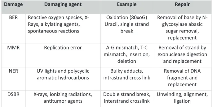
III. DNA repair mechanisms : overview 59
1. DNA Damage 59
2. DNA repair mechanisms 60
a. Base Excision Repair (BER) 61
b. Mismatch Repair (MMR) 61
c. Nucleotide Excision Repair (NER) 62
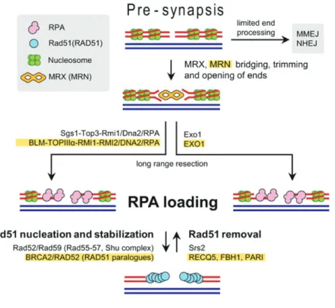
d. Double-Strand Break Repair 62
i. Homologous Recombination (HR) 62
ii. Non-homologous End-Joining (NHEJ) 64
3. DDR and viruses infection 65
a. The DNA damage response as an antiviral mechanism 66
b. The benefits of DNA damage response activation for virus replication 67
4. A role for the host DDR in Hepatitis B virus? 67
a. Integration of viral DNA 67
b. Viral dsL-DNA circularization 68
c. HBx and DDR 68
d. rcDNA to cccDNA conversion 69
IV. Chromatin 70
1. Chromatin template 70
a. The nucleosome 70
b. Structure of Core histones 72
a. Histone Post-translational Modifications 74
i. Methylation/Demethylation 75
ii. Acetylation/Deacetylation 75
iii. Phosphorylation/Dephosphorylation 76
b. Histone modifying enzymes 77
i. Histone acetylation 77
ii. Histone methylation 78
iii. Histone phosphorylation 78
c. ATP-dependent chromatin remodeling 79
d. Histone variants 80
i. H2A variants 80
ii. H3 variants 82
3. Nucleosome assembly pathway 83
a. Discovery and definition of histone chaperones 83
b. Involvement in distinct histone deposition pathways 85
i. DNA replication dependent 85
ii. DNA replication independent 87
c. Roles in transcription 87
d. HIRA, DNA damage recovering and chromatin dynamics 88
4. Viruses and chromatin organization 90
5. Chromatin analysis 92
a. Chromatin Immunoprecipitation (ChIP) techniques 93
b. Chromosome Conformation Capture (3C) techniques 93
V. Hepatitis B virus minichromosome: cccDNA 95
1. Background 95
2. cccDNA life span 96
a. Formation 96
From rcDNA to cccDNA : conceptual evidence for an involvement of host DNA repair 96
b. Regulation of cccDNA recycling 99
i. DHBV envelope proteins involved in cccDNA recycling 100 ii. HBV envelope protein involved in cccDNA recycling 101
iii. Regulation hypothesis 101
3. Activity 102
a. Minichromosome structure 102
b. cccDNA methylation 102
c. Histone’s modification: methylation and acetylation 103
d. Viral proteins associated to cccDNA 106
4. Fate of cccDNA 109
a. Persistence and reactivation 109
b. Natural clearance of cccDNA by host immune system 109
i. Hepatocyte lysis 110
ii. cccDNA dilution during mitosis 111
c. Therapeutic clearance 113
i. Curent available therapeutic strategies 113
ii. Investigated drugs in development 115
x By targeting the virus 115
C
CHAPTER II: RESEARCH PROJECT 121
II. Histone chaperone HIRA deposits histone H3.3 onto incoming PF-rcDNA and contributes to cccDNA
formation 123
III. Supplementary results 157
IV. Discussion 170
V. References 189
VI. Acknowledgements 220
VII. Appendices 227
1. Published article and comment 227
a. Analysis of intrahepatic virological events associated to chronic hepatitis B infection (article in
French 227
b. Antiviral activity of various interferons and pro-inflammatory cytokines in non-transformed cultured
hepatocytes infected with hepatitis B virus 232
Figure 1: Seroprevalence of Hepatitis B virus infection worldwide (adapted from CDC, 2016). ... 25
Figure 2: Genotype prevalence of Hepatitis B virus infection worldwide (from A. Valsamakis). ... 27
Figure 3: Viral components released by HBV-infected hepatocytes. ... 31
Figure 4: HBV genome organization (adapted from Liang et al., 2009). ... 33
Figure 5: The HBV genome, its regulatory elements and its messenger RNAs (adapted from Seeger and Mason, 2000). ... 35
Figure 6: Schematic representation of the ORFs of HBV genome, and the reported spliced variants. ... 36
Figure 7: The three forms of HBV envelop proteins (S, M and L). ... 38
Figure 8: Hepatitis B virus Pre-Core, Core and HBe proteins. ... 39
Figure 9 : Capside protein dimer structure within HBV capsid (from Alexander et al., 2013) ... 41
Figure 10: Domains of hepatitis B virus polymerase. ... 43
Figure 11: Formation of Hepatitis B Spliced Protein (HBSP) (adapted from Assrir et al., 2010). ... 46
Figure 12 : HBV replication cycle. ... 48
Figure 13: Schematic representation of the interaction between the polymerase and the HBV pgRNA in order to initiate the viral encapsidation step (Beck and Nassal, 2007). ... 51
Figure 14 : Translocation mechanism of the primer from the 5' end to the 3' end of the pgRNA allowing the initiation of reverse transcription (Beck and Nassal, 2007). ... 52
Figure 15: Representation of the different HBV replication steps (Beck and Nassal, 2007). ... 53
Figure 16: Different types of DNA damage. ... 60
Figure 17 : Homologous recombination, pre-synapsis step. ... 63
Figure 18 : Homologous recombination, synaptic step. ... 64
Figure 19 : Viral nucleic acid structures that provoke DNA damage response (adapted from Luftig, 2014) .. 66
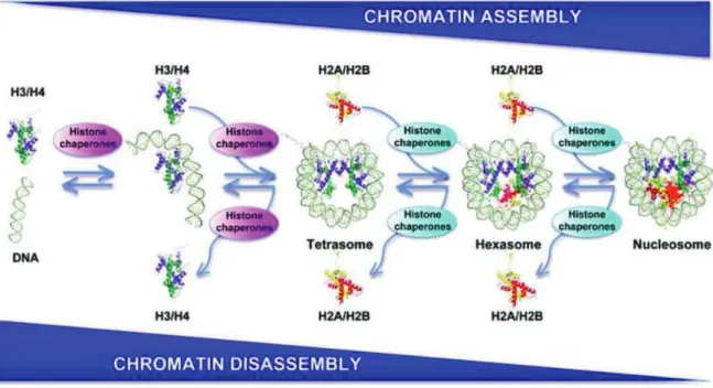
Figure 20 : Assembly and disassembly of nucleosomes. ... 71
Figure 21 : Overview of nucleosomes architecture... 72
Figure 22 : Histone post-translational modification perturbs histone-DNA interaction and allows better transcription factor binding (adapted from Bowman and Poirier, 2015). ... 74
Figure 23 : Crosstalk between histone modifications. Adapted from Rossetto et al., 2012. ... 76
Figure 24 : Post-translational histone modifications on N-terminal tails. ... 77
Figure 25 : Summary of histone modifications and their associated histone modifying enzymes. Adapted from Day and Sweatt, 2012. ... 79
Figure 26 : Schematic representation of human histones and their associated variants. From Henikoff and Smith... 81
Figure 27 : Genomic locations where histone variants are enriched. ... 84
Figure 28 : Histone variants and their associated histone chaperone (and associated factors). From Skene and Henikoff, 2013. ... 85
Figure 29 : Histone remodeling and chaperone’s involvment during DNA replication. From Hammond et al., 2017. ... 86
Figure 30 : Histone chaperones during UV-C DNA damage. From Gurard-Levin et al., 2014 ... 89
Figure 31 : The different steps leading to cccDNA formation from the rcDNA entering the hepatocyte. Adapted from Gomez-Moreno and Garaigorta, 2017. ... 97
Figure 32 : Epigenetic regulation of cccDNA (adaptated from Levrero et al., 2009). ... 104
Figure 33: HBV cccDNA epigenome in presence and in absence of HBx (From Hong et al., 2017) ... 108
Figure 34: Dilution model of cccDNA during hepatocyte division. ... 112
Figure 35 : The different treatment options for chronic hepatitis B (Zoulim, 2007) ... 114
Figure 36: RPA is required for cccDNA establishment ... 157
Figure 38: Modeling of the interaction between HBc and HIRA proteins. ... 161
Figure 39 : Table of predictive interface residues contact between HIRA (PDB 2i32) and HBc (PDB 1GQT). 162 Figure 40: Infection by HBV does not lead to HIRA relocalization to PML nuclear bodies. ... 164
Figure 41: Role of DAXX in cccDNA establishment ... 166
Figure 42: A role for HIRA in transcription regulation of cccDNA? (Preliminary results) ... 168
Figure 43: The different steps leading to cccDNA formation from the rcDNA entering the hepatocyte. Adapted from Gomez-Moreno and Garaigorta, 2017 ... 171
Figure 44 : HIRA is crucial for the early establishment of hepatitis b virus minichromosome ... 172
Figure 45: Role of HIRA in transcriptional regulation (From Nashun et al., 2015) ... 183
Figure 46: Model for DAXX-ATRX complex in the maintenance of heterochromatin. ... 186
Table 1: Characteristics of the in vivo systems available for studies with HBV and HBV-related viruses. ... 58 Table 2 : DNA repair mechanisms ... 61 Table 3: Outline of the main research findings on the methylation and acetylation mechanisms involved in HBV infection. From Kombi and Karayiannis, 2015. ... 105 Table 4 : cccDNA epigenetic modifications (adapted from Levrero, 2009). ... 107 Table 5: Current, in development, and possible therapies to treat hepatitis B chronic infection. ... 119
Abbreviations
3TC: Lamivudine
aa: amino acids
AAV: Adeno-Associated Virus
ADF: Adefovir Dipivoxil
ASF1: Anti-Silencing Function protein 1
ATRX: α-thalassemia/mental Retardation
X-linked Syndrome Protein
BCP: Basal Core Promoter
BER: Base Excision Repair
BER: Base Excision Repair
CAF1: Chromatin Assembly Factor 1
CBP: CREB Binding Protein
cccDNA: covalently closed circular DNA
CCL2: Chemokine Ligand 2
CCNE1: Cyclin E1
CHB: Chronic Hepatitis B
CHD: Chromodomain and Helicase-like
Domain
ChIP: Chromatin Immunoprecipitation
CHPKs: Conserved Herpesviruse Protein
Kinases
CMV: Cytomegalovirus
CTD: C-Terminal “protamine” Domain of
HBcAg
DAA: Direct Acting Agents
DAXX: Death domain-Associated protein 6
DCPD: Carboxypeptidase D
DDB1: DNA Damage Binding Protein
DDR: DNA Damage Response
DDX3: Dead box helicase 3
DHBV: Duck Hepatitis B Virus
DNA: Deoxyribose Nucleic Acid
DNMT: DNA methyltransferase
DR: Direct repeat
DSB: Double Strand Break
dsDNA: double stranded DNA
DsL-DNA: Double-stranded Linear DNA
EBV: Epstein-Barr virus
Enh: Enhancer
ER: Endoplasmic Reticulum
ETV: Entecavir
EZH2: Enhancer of Zeste Homolog 2
FACT: Facilitates Chromatin Transactions
HAP: Heteroaryldhydropyrimidine
HAT: Histone acetyltransferase
HBcAg: Hepatitis B core Antigen / capsid
protein
HBsAg: Hepatitis B surface Antigens / surface
proteins
HBSP: Hepatitis B Splicing Protein
HBV: Hepatitis B virus
HBxAg: Hepatitis B x Antigen / X protein
HCC: Hepatocellular Carcinoma
HCV: Hepatitis C virus
HDAC: Histone Deacetylase
HDM: Histone Demethylase
HFD: Histone Fold Domain
HIRA: Histone cell cyle Regulator A
HMT: Histone Methyltransferase
HPV: Human Papilloma Virus
HR: Homologous Recombination
Hsp90: Heat shock protein 90
HSPG: Heparan Sulfate Proteoglycans
HSV: Herpes Simplex Virus
IFN: Interferon
IP: Immunoprecipitation
ISG: Interferon Stimulated Gene
ISWI: imitation SWI
kDa: kiloDalton
KHSV: Kaposi’s sarcoma-associated virus
LTc: CD8 + lymphocytes; Cytotoxic T
Lymphocyte
MAPK1: Mitogen-Activated Protein Kinase 1
MCM: Minichromosome Maintenance helicase
miRNA: micro RNA
MLL4: Mixed-Lineage Leukemia 4
MLV: Moloney murine leukemia virus
MMR: Mismatch Repair
NA: Nucleoside Analogs
NAP1: Nucleosome Assembly Protein 1
ncRNA: non-coding RNA
NER: Nucleotide Excision Repair
NHEJ: Non Homologous End Joining
NK: Natural Killer
NLS: Nuclear Localization Signal
NTCP:Sodium Taurocholate Cotransporting
Polypeptide
NTD: N-Terminal Domain of HBcAg
NUC: Nucleos(t)ide Analogue
PDGFB: Platelet Derived Growth Factor
Subunit B
PF-rcDNA: Protein Free rcDNA
pgRNA: pregenomic RNA
PHH: Primary Human Hepatocyte
PLA: Proximity Ligation Assay
Pol: viral Polymerase
PolΚ: Polymerase Κ
PRC2: Polycomb Repressive Complex 2
PRR: Pathogen Recognition Receptor
PTM: Post-Translational Modification
rcDNA: relaxed circular DNA
RFC: Replication Factor C
RNA Pol II: RNA Polymerase II
RNA: Ribonucleic Acid
ROCK1: Rho-associated coiled-coil containing
protein kinase 1
ROS: Reactive Oxygen Species
RPA: Replication Protein A
RT: Reverse Transcriptase
SENP5: Sentrin-specific protease 5
Smc5/6: Structural maintenance of chromosome
SSB: Single Strand Break
ssDNA: single stranded DNA
SVP: subviral non-infectious particles
SWI/SNF: Switch/Sucrose nonfermentable
TDP2: Tyrosyl-DNA Phosphodiesterase 2
TERT: Telomerase Reverse Transcriptase
TLR: Toll-Like Receptor
TP: Terminal Protein
24
C
CHAPTER I: BACKGROUND
I. Overview
Hepatitis B virus (HBV) chronically infects 240 million people worldwide and is the major cause of hepatocellular carcinoma (HCC). HBV infection is not cytopathic, but the virus persists in infected cells as supercoiled DNA, termed covalently closed circular cccDNA. It acts as a reservoir and is therefore responsible for the persistence of infection during chronic hepatitis B, even during antiviral treatment. This cccDNA is comparable to an intrahepatocytic episomal "minichromosome", organized into a nucleosomal structure, which will serve as a template for viral transcription. In the nucleus, it is present in this form of minichromosome with a structure called "beads-on-a-string" (Bock et al., 1994).
While current treatments (mainly nucleos(t)ide analogues) can reduce viral load to undetectable levels, they do not have a direct effect on the intrahepatic cccDNA pool. Since these treatments do not influence the entry of the virus, they can not prevent the initial formation of cccDNA. In addition, the transcriptional activity of this minichromosome persists, thus allowing the production of messenger RNAs and pregenomic RNA. The ultimate and ideal goal of treatment against chronic infection with hepatitis B virus is thus treatment resulting in the complete disappearance of the cccDNA pool.
Upon entry into hepatocytes, the partially double stranded viral DNA is released into the nucleus, where it is repaired and wrapped up by host histone proteins to form an episomal chromatinized structure. The mechanisms leading to cccDNA formation and stabilization are still largely unknown and their elucidation would be extremely useful to identify new therapeutic targets to impair cccDNA stability.
25
III. Hepatitis B
1. Background
Hepatitis B virus (HBV), discovered in 1964 by Baruch Samuel Blumberg, chronically infects 240 million people worldwide (WHO, 2017). The persistence of viral replication is associated with a high morbidity and mortality, due to major complications. Indeed, it is estimated that acute and chronic HBV infections are responsible for around 800,000 deaths per year worldwide (i.e., ranked 15th in the Global Burden of Disease Study) (Locarnini et al., 2015). If acute hepatitis B can be resolved in 1-2 weeks, infected patients can develop chronic hepatitis B, which is defined by the persistence of HBsAg for more than 6 months. Untreated, chronic hepatitis B can lead to fibrosis, cirrhosis and ultimately hepatocellular carcinoma. Moreover, cirrhotic patients present an increased risk of liver decompensation, HCC and death (Locarnini et al., 2015).
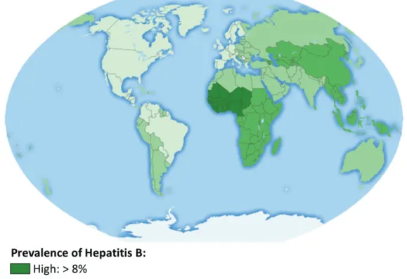
Figure 1: Seroprevalence of Hepatitis B virus infection worldwide (adapted from CDC, 2016).
Prevalence of Hepatits B is defined as the percentage of people infected with HBV categorized in four groups: low (<2%), low-intermediate (2-4%), high-intermediate (5-7,9%) and high (>8%).
26 Seroprevalence of HBV, as measured by HBsAg carriage, is globally of 3.61% (Chang and Nguyen, 2017). On this criteria, endemicity is categorized as low (<2%), low-intermediate (2-4.9%), high-intermediate (5-7.9%) and high (>8%), with the highest levels of 8.63% being found in Africa (Chang and Nguyen, 2017) (Figure 1).
aa. Genotypes
Before the genotypic classification of HBV strains, nine different serotypes noted ayw (1-4), ayr, adw2, adw4, adrq- and adrq + have been defined from sequences present in antigenic determinants located in the HBs envelope protein (Wagner et al., 2004).
Today, the genotypic classification of HBV is based on the comparison of nucleotide sequences of HBV strains. This classification takes into account the entire genome of HBV and currently identifies 8 genotypes, denoted A to J (Figure 2). The difference between two viral genotypes is defined by a variation of more than 8% of their entire nucleotide sequence and at least 4.1% in the surface gene (preS1, preS2, S). The correlation between serotypes and genotypes is far from perfect (Wagner et al., 2004). The genomes of different genotypes might have slightly different lengths. This is due to insertions or deletions in the regions encoding preS1 and core proteins. Correlations between genotypes, clinical progression and treatment response have been established, although it is not possible to attribute to each genotype a predictive score for the severity of the associated disease. Genotype C is nevertheless associated with an increased mutation frequency in the core protein promoter leading to an increased resistance to IFN therapies (Kao et al., 2000a).
Finally, genotypes also seem to influence the occurrence of HCC, since genotype C chronic infection are more susceptible to hepatocellular carcinoma development in respect to those of genotype B (Yu et al., 2005).
27
Figure 2: Genotype prevalence of Hepatitis B virus infection worldwide (from A. Valsamakis).
Major genotypes found in different regions of the world.
b
b. Routes of transmission
28 ¾ Vertical transmission (or perinatal transmission), from mother to infant, is frequent in highly endemic zones and is the cause of one third of chronic infection in low endemic countries (WHO, 2013). It occurs during the peripartum or during the initial months after birth. In addition, it is important to note that the earlier the individual is infected, the higher the risk of developing a chronic infection.
¾ Horizontal transmission occurs mainly by parenteral exposure to infected blood (blood transfusion, contaminated needles, on a razor blade for example (HBV can survive more than a week out of the body without losing its infectivity) (WHO, 2002)) or unprotected sexual contact with exchange of body fluids (Kidd-Ljunggren et al., 2006). Sexual transmission is the predominant mode of infection in low-endemic countries (de Franchis
et al., 2003).
2. HBV biology
aa. Viral classification
Hepatitis B virus belongs to the Baltimore class VII heparnavirus or Hepadnaviridae family (double-stranded DNA virus, with RNA replicative intermediate). The viruses belonging to this family, have been grouped together by four common features: hepatotropism, genetic organization, viral morphology and replication mechanisms (a retro-transcription step in their viral life cycle). Hepadnaviridae are divided into two genera: the genus orthohepadnavirus, including HBV along with other mammalians viruses (as woodchuck hepatitis virus (WHV), ground squirrel hepatitis virus, woolly monkey hepatitis virus and bat hepatitis virus), and the genus avihepadnavirus, including avian virus, as the duck hepatitis B virus (DHBV) and heron HBV (Schaefer, 2007).
b. Viral structure
i. Virions and subviral particles
Several types of particles are present in the patient's sera: Dane particles corresponding to infectious particles and two types of non-infectious subviral particles. These particles are not secreted following the same mechanism.
29 ¾ HBV, in its infectious form, is an enveloped virus of 42 nm in diameter (Marion and Robinson, 1983). Also called Dane particles (after their discoverer; Dane et al., 1970), the virion is composed of a single icosahedral nucleocapsid formed by the assembly of core protein (HBc). This capsid, formed of 90 or 120 HBc dimers depending on its symmetry, is surrounded by a complex envelope formed exclusively by the three viral envelope proteins (S-, M- and L-HBsAg in a ratio of 4:1:1) and contains the 3.2 kb viral genome consisting of circular relaxed double-stranded DNA (rcDNA) covalently linked to the viral polymerase (Knipe and Howley, 2013). In the serum of patients, viral titer can go up to 1010
genome-copies/ml.
¾ Subviral non-infectious particles (SVP) are only composed of envelop proteins. SVP are of, approximatively, 22 nm in diameter and circulate in the serum of patients. SVP are secreted by infected cells, in 1 000 to 10 000 fold excess compared to virions (Urban et al., 2014).
Two types of SVP are formed: spheres, composed mainly of S protein, with an octahedral symmetry, and filaments, which are asymmetrical with variable length and width. Filaments possess the same HBsAg ratio as Dane particles. The formation of SVP is related to the self-assembly ability of the HBs protein (Huovila et al., 1992) (Figure 3). Their role in HBV infection is still a matter of debate, but they are supposed to represent a decoy for the immune system.
In addition to these HBsAg containing particles, identified from patients’ sera, two other sub-viral forms have been described:
¾ Non-enveloped nucleocapsids, found to be secreted in vitro by HBV replicating hepatoma cell lines and suggested to play a role in virus spreading (Cooper and Shaul, 2006; Watanabe et al., 2007). They have also been shown to be released upon hepatocytes apoptosis (Arzberger et al., 2010). These nucleocapsids may contain viral DNA, but also all the intermediates between RNA encapsidation to final DNA retro-transcription (see viral life cycle). Indeed, it appears that HBV may secrete RNA-containing particles that have been found in the blood of infected patients (Jansen et
al., 2016). The nature of the HBV RNA present in the serum remains to be defined, as
30 the possibility for minus strand DNA-pgRNA hybrids (Hu and Liu., 2017) Their existence and role in vivo has still to be demonstrated;
¾ Enveloped empty capsids have been described in patients’ sera, but their relevance and function is still to be determined (Luckenbaugh et al., 2015).
31
Figure 3: Viral components released by HBV-infected hepatocytes.
The viral inoculum is composed of the Dane particles (infectious form of the virus, also called the virion), two subviral particles form (spheres and filaments), and secreted HBeAg. Upon apoptosis of hepatocytes, naked nucleocapsid can be released. In addition to Dane particles, other forms of enveloped nucleocapsid have been described in patients’ serum.
32
ii. Viral DNAs
rcDNA
With a 3.2 kb genome, HBV possess the smallest genome of all known DNA viruses. In an atypical manner, the genome of this virus, called relaxed circular DNA (rcDNA), is in the form of a partially double-stranded circular DNA consisting of a complete negative strand and an incomplete positive strand. It is found in secreted viral particles but also in mature capsid in the cytoplasm of infected hepatocytes. The complete negative strand is the template for transcription, and is linked to the viral polymerase at its 5’ extremity (Figure 4). The incomplete stand, as for it, presents a gap and a 3’ ending of variable length (Summers et al., 1975). It is also associated with a RNA oligomer in its 5’ end, which derives from the pre-genomic RNA (pgRNA) and serves as a primer for the synthesis of the positive strand.
In order to keep this DNA in circular form, a sequence of 200 nucleotides located 5 'of the positive strand and overlapping the 5’ and 3’ extremity of the negative strand is absolutely required (Gao and Hu, 2007). This overlapping region contains the direct repeat region (DR) 1 and 2, mandatory for viral replication (Knipe and Howley, 2013).
rcDNA contains four open reading frames (ORF), 4 promoters, 2 enhancers elements and 1 single polyadenilation signal. Because of its small genome size, HBV ORFs are overlapping; thus a mutation in one gene most likely has consequences on another HBV gene (Nassal, 2015).
HBV ORFs are all oriented in the same direction and led to the production of the 7 viral proteins: ¾ Polymerase ORF, the largest ORF, which represents 80% of HBV viral genome and codes
for the viral polymerase.
¾ Pre-S1/Pre-S2/S ORF is within the Polymerase ORF, has 3 in-phase start codons and is coding for the 3 different forms of HBV envelop protein (S-, M-, and L-HBsAg, for small, medium, and large HBsAg).
¾ Pre-Core/Core ORF presents 2 in-phase initiation codons and codes for secreted HBeAg and HBcAg (capsid protein).
33
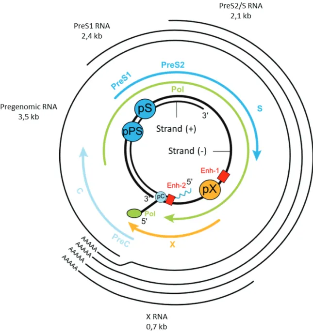
Figure 4: HBV genome organization (adapted from Liang et al., 2009).
Organization of HBV genome and its open reading frames. The rcDNA contains a complete negative strand and an incomplete positive strand. At the 5’ end of the negative strand is found the polymerase (pol). At both ends of the negative strand are the redundant sequences preventing the closure of this strand (direct repeats – DR). The RNA primer is located at the 5’ end of the positive strand.
34
dsL-DNA
Double-stranded Linear DNA (dsL-DNA) can be formed during viral replication. Indeed, in 10% of the time, the primer from the pgRNA is not translocated, thus leading to the formation of a linear double-stranded DNA (see later in figure 15) (Tu., 2017). This dsL-DNA can be converted to cccDNA, following a different pathway, via non homologous end joining (NHEJ). Generally, this conversion leads to defective cccDNA, which contains deletions in gap, due to the repair. This intermediate of replication leads to a major part of HBV DNA integration.
cccDNA
Covalently closed circular DNA is a form of HBV DNA that persists within infected hepatocytes, and its role will be described in the chapter “Viral minichromosome: cccDNA”.
iii. RNA transcripts
As for all viruses belonging to the hepadnaviridae family, HBV replication takes place through RNA intermediates. From the four ORFs, five messenger RNAs (mRNA) are generated by cellular RNA polymerase II, using cccDNA as template. All these mRNAs possess a 5’ cap and the same polyadenylation site/tail (Figure 5).
¾ The Pregenomic RNA (pgRNA) of 3.5 kb is synthesized from the preC/C promoter and represents the entire genome. It encodes the polymerase, the capsid HBc protein and the HBSP protein produced by alternative splicing. The polymerase and HBc are not in frame and the detection of the ORF of the polymerase is much less effective than that of HBc. Indeed, usually, 240 HBc proteins are translated for one or two polymerases (Seeger 2000). The pgRNA is also the template for de novo synthesis of rcDNA through reverse transcription.
¾ 3.5 kb Pre-Core mRNA (a few bases longer than pgRNA) that encodes soluble and secreted HBeAg.
¾ Two subgenomic mRNAs of 2.4 and 2.1 kb are produced from the preS1 and preS2/S promoters respectively. The 2.4 kb RNA codes for the large envelope protein labeled L and the other RNA codes for the M and S proteins.
35 Transcription of the 4 ORFs is monitored by 4 promoters, 2 enhancers and a cis-acting negative regulatory element. Regulatory elements are active only once rcDNA has been converted to cccDNA (Moolla et al., 2002). Further details of transcription regulation will be addressed in the cccDNA paragraph.
Figure 5: The HBV genome, its regulatory elements and its messenger RNAs (adapted from Seeger and Mason, 2000).
Schematic representation of HBV RNA transcripts with initiation codons and shared polyA tail. Four promoters regulate the expression of HBV genes, the preS1 promoter (pPS), the preS2 promoter (PS), the X promoter (pX) and the Core promoter (pC). The promoters are under the control of two enhancers, the enhancer 1 (Enh-1), and enhancer 2 (Enh-2).
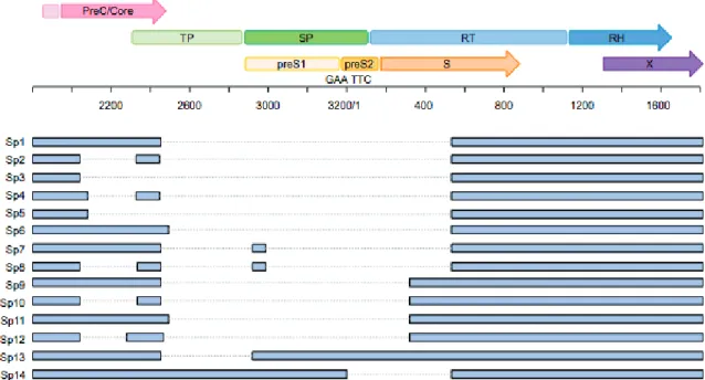
36 In addition to the different mRNAs encoded by the cccDNA, are found a series of spliced (SP) HBV RNAs. Those spliced form have been described in cell systems as well as in HBV-infected liver. The most frequently observed spliced variant is a 2.2 kb molecule, called SP1, which is formed through the removal of a 1.3 kb portion of the pgRNA (Figure 6). This variant accounts for up to 30% of pgRNAs (Bayliss et al., 2013). Spliced forms of HBV RNAs can be, as pgRNA, encapsidated and reverse transcribed into a defective HBV DNA, forming abnormal particles. The level of defective particles in the sera of patients with CHB has been shown to be correlated with liver disease, and to be enhanced prior the development of HCC. Furthermore, spliced variants can directly impact HBV replication. Indeed, the Sp10 variant, isolated from the serum of a patient with HCC, was reported to increase replication in vitro (Ma et al., 2009). Spliced variants are also associated with an impaired response to interferon therapy (Chen et
al., 2015). This is to be noted, as an increased replication is a major risk factor for the
development of hepatocarcinoma (Lupberger et al., 2007; Brechot et al., 2010).
Figure 6: Schematic representation of the ORFs of HBV genome, and the reported spliced variants.
Summary of 14 HBV splice variants. For each spliced variant, the removed region is indicated in grey. TP, terminal protein; SP, spacer; RT, reverse transcriptase; RH, RNase H; S, surface. From J Bayliss et al., 2013, adaptated from Sommer and Heise 2008.
37
iv. Viral proteins
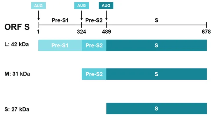
Surface proteins (HBsAg): small, medium and large
The three viral envelope proteins (S, M and L) are synthesized from two different mRNAs from the same ORF. The larger 2.4 kb mRNA encodes the large envelope protein L. The other 2.1 kb mRNA encodes the M or S proteins according to the translation initiation site used. Since the transcription start site of the mRNA coding for the HBs protein is more frequently recognized by the ribosomes, four times more S-HBs is synthesized with respect to the M protein (Sheu and Lo, 1992). The three HBV envelope proteins all contain the same carboxy terminal domain (Cter) coding for HBs, while the M and L proteins have the preS2 domain. Finally, the L protein is the only one to have the preS1 domain (Figure 7).
These surface proteins have four transmembrane domains and are synthesized in the endoplasmic reticulum and then matured in the Golgi apparatus. Moreover, they are all three modified by N- or O-glycosylation and the L protein is also myristylated on its second amino acid.
Domain S also has cysteine residues that allow disulfide bridges to be formed between envelope proteins to form virus particles (Wounderlich and Bruss, 1996). This domain also carries the determinant "a" (amino acids 124-147) which is the major target of the neutralizing antibodies.
M protein is not necessary for the formation or infectivity of viral particles (Bruss and Ganem, 1991).
38 The L protein is essential for both capsid envelopment during virion morphogenesis as well as viral entry via the binding to the sodium-taurocholate cotransporting polypeptide (NTCP) HBV receptor, which is expressed at the surface of hepatocytes (Schulze et al., 2010). During its synthesis, the L protein is inserted into the endoplasmic reticulum membrane by means of topogenic proximal signals of the S domain. Interestingly, the first transmembrane domain of the protein cannot be translocated and remains cytosolic. The L protein exhibiting this topology is called i-preS and allows interaction with the nucleocapsids during wrapping. Unknown translocation signals allow insertion of a first transmembrane domain into the membrane thus forming the e-preS form that promotes interaction with the HBV receptor (Prange, 2012).
Figure 7: The three forms of HBV envelop proteins (S, M and L).
Translation is initiated at different in-frame positions with the L form containing the 108 aa PreS1 domain, and the 55 aa PreS2 domain, the M-HBsAg contining the PreS2 and the shortest S form just the S protein. The three forms share the same C-terminus.
39
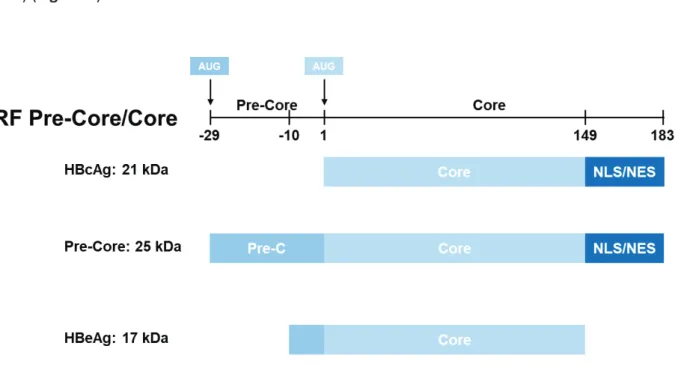
Capsid protein (HBcAg)
The core protein, also called capsid protein, is a 183 aa long, and 21 kDa protein that forms the viral icosahedral capsid of HBV. This protein is composed of two domains separated by a linker: the "core" domain (amino acids 1 to 140) and the "protamine" domain (residues 150-183) (Figure 8).
The "core" domain at the amino terminus (Nter), also called NTD (N-terminal domain) allows self-assembly of core protein into capsid (Birnbaum and Nassal, 1990). It also contains two predictive epitopes for the immune response whose immunodominant loop includes residues 78 to 83 (Steven et al., 2005). Indeed, HBc is recognized by the host's immune system when expressed on the surface of hepatocytes, inducing a T-cell mediated immune response against infected cells (Lee, 1997).
The "protamine"-rich domain, or CTD for C-terminal domain, has two functions. It contains a nuclear localization pattern (NLS) regulated by phosphorylation. Its exposure on the surface of nucleocapsids allows their transport to the nucleus of the cell. In addition, this domain has four arginine-rich clusters that bind nucleic acids and encapsidate pgRNA (Nassal, 1992).
Figure 8: Hepatitis B virus Pre-Core, Core and HBe proteins.
HBcAg, Pre-core and HBeAg all share the core domain, while only HBcAg and Pre-Core proteins contain the NLS/NES sequence. The NLS sequence as well as the main part of the Pre-Core domain is cleaved from Pre-Core protein to form the secreted HBeAg.
40 Phosphorylation level of CTD regulates capsid maturation. For instance, once core dephosphorylation is performed, conformational reorganisation of CTD, and finally DNA synthesis can be induced. This leads to the exposure of the binding sites, allowing the interaction with the envelop protein, and then to the assembly of mature virions (Mabit and Schaller, 2000; Perlman et al., 2005).
The core protein has a secondary structure in α helices and has the ability to assemble into dimers. The presence of disulfide bridges between each protein makes it possible to stabilize this structure. The association of an HBc dimer results in the formation of a composite peak by the union of four α-helices.
The oligomerization of the HBc dimers then leads to the formation of the viral capsid of T3 or T4 symmetry depending on the number of HBc dimers involved (90 or 120 respectively). The viral capsid of T4 symmetry of 36 nanometers in diameter is mostly found in virions (Steven et
al., 2005). Pores are present in the capsid probably for the entry and exit of nucleotides (Figure
9).
HBc protein is very variable in vivo (Chain and Myers, 2005). The in vitro individual mutation of 52 aa in the core domain of HBc determined which were important in the formation of capsids or virions (Ponsel and Bruss, 2003). It emerges from this study that the amino acids at position 17, 18, 60, 95, 96, 122, 127, 136, 137 and 139 are important for the enveloping of the capsids formed.
41
Figure 9 : Capside protein dimer structure within HBV capsid (from Alexander et al., 2013)
A) 3D representation of the capsid protein dimer with its secondary structural elements labelled. Helices (numbered from α1 to α5) are interacting, forming a protruding spike, and are surrounded by contact domains (orange and red). N and C represent the termini of one monomer. The disulfide link between C61 of each monomer is indicated in cyan.
B) Exterior surface of a T=4 capsid. Residues that perturb capsid formation when mutated are indicated.
42
Pre-Core protein and HBeAg
The HBe protein is a circulating peptide derived from the core protein. Contrary to HBc protein, it is translated from the 3.5 kb PreCore mRNA and contains the same domains (NTD and CTD) (Figure 8) as the core protein extended by a 29 aa at the N-terminus (Yeh et al., 1990). The initiator codon of the translation used is located 87 nucleotides upstream from that used for the synthesis of the HBc protein.
After two successive cleavages, the mature form of this protein is generated. First, the HBe protein is synthesized as a 25 kDa precursor. The latter, thanks to the presence of a hydrophobic sequence of 19 aa, is directed towards the endoplasmic reticulum. Subsequently, this hydrophobic sequence present in Nter is cleaved by a furine producing a 22 kDa protein (Messageot et al., 2003). This protein is then cleaved again in Cter in its arginine-rich domain to form the secreted HBe protein of 15 to 18 kDa (Ito et al., 2009).
This protein does not seem essential for the establishment of the viral cycle but is important for establishing persistent infections in vivo (Dandri and Locarnini, 2012). Moreover its presence in all Hepadnaviridae suggests a preponderant role of this protein for the virus. The HBe antigen has been shown to play an immunoregulatory role. Because of its mimicry of the HBc protein, HBe saturates the immune system receptors thus allowing the virus to escape an immune response (Lee, 1997). Indeed, HBe has been shown to decrease the secretion of IFN-γ produced by lymphocytes (Han et al., 2013).
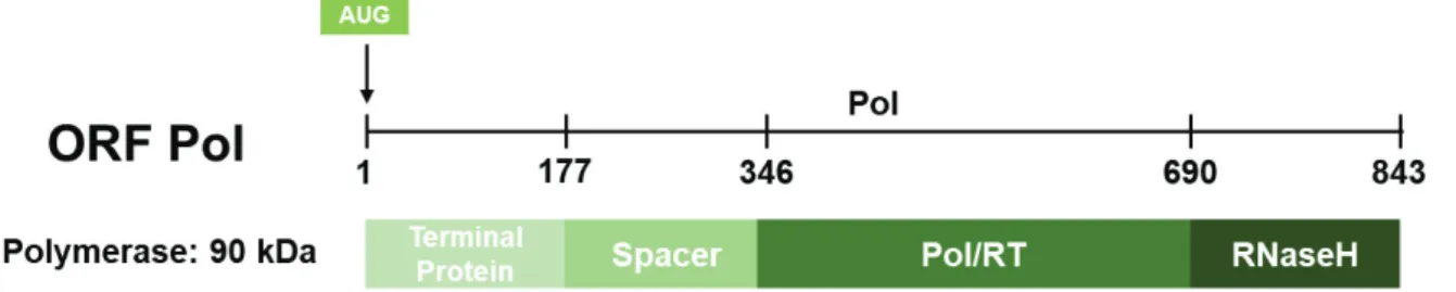
Polymerase (Pol)
HBV polymerase (845 aa; 90 kDa) is the only enzymatic protein coded by the HBV genome and contains four different domains (Figure 10):
- A first domain, the TP domain, (aa 1 to 180) corresponding to the terminal protein which connects the polymerase to the 5’ end of the negative strand through tyrosine Y63, (Lanford et al., 1997, Zoulim and Seeger, 1994) and which has a primase activity. This primase allows the initiation of reverse transcription by synthesizing a primer of 3 to 4 nucleotides.
43 Furthermore, this domain is necessary for the interaction of the polymerase with the « epsilon » ε, the signal leading to the packaging of pgRNA.
- A second domain (aa 181 to 349) called "spacer" which is genetically variable and whose role has not yet been clarified. This region overlaps the envelope domain and could allow flexibility of the envelope protein sequence that impacts on polymerase functions (Chen
et al., 2013).
- A third domain (aa 350 to 693) denoted "pol/RT" (polymerase/retrotranscriptase) comprises two functions. On one hand, it carries the RNA-polymerase-dependent activity, allowing the reverse transcription of the pgRNA into the DNA minus strand. On the other hand, it carries the dependent DNA polymerase activity which generates the positive strand.
- A fourth and last domain (aa 694 to 845) corresponding to the RNaseH domain. This domain allows the viral polymerase to degrade the pgRNA after the retrotranscription of the DNA minus strand (Seeger and Mason, 2000). RNAse-H inhibitors have been shown to block HBV replication and could therefore be a potential target for therapeutic options (Tavis and Lomonosova, 2015).
Figure 10: Domains of hepatitis B virus polymerase.
The four functional domains are represented from N- to C-terminus: Terminal protein, Reverse transcriptase and RNAse-H.
44
X protein (HBxAg)
The X protein, only present in Orthohepadnaviridae, is a non-structural protein, and the smallest coded by the HBV genome (154 aa; 17 kDa). This protein has been studied for a long time in a large number of different models giving rise to multiple functions for HBx. However, the effects associated with HBx vary according to the cellular context, the amount of HBx expressed and the model chosen (expression of HBx alone or in a context of HBV replication, for example). Numerous effects of HBx have been reported, not all well described or understood and can sometimes leave skeptical for the reasons outlined above. However, they can be classified into two broad categories, the influence on virus replication and the role of HBx in hepatocarcinogenesis.
Indeed, it is essential for establishing and maintaining the viral infection. In 1994, studying the X gene in groundhogs (WHx), Zoulim et al., demonstrated for the first time that Hepadnavirus X protein plays a vital role in viral replication. In 2007, Keasler et al., demonstrated in vitro, in a HepG2 cell model, that the absence of HBx could induce a 65% reduction in HBV replication and that trans-complementation of the HBx-mutated plasmid with wild-type HBx was restoring the replication. Later, it was confirmed that infection with an HBV mutant lacking the HBx protein is associated with repressed transcription from cccDNA (Lucifora et al., 2011). It turns out that the HBx protein plays a role of transcriptional activator of the cccDNA by recruiting histone acetylases resulting in an increase in the replication (Belloni et al., 2009). Furthermore, a recent study aiming at identifying the direct genomic targets of HBx by ChIP-Seq analysis, allowed to uncover new genes and ncRNAs targeted by HBx and involved in the positive regulation of cccDNA transcription and HBV replication (Guerrieri et al., 2017). This sequencing showed that HBx could activate several genes and miRNA, able to increase endocytosis and autophagy, thus favoring HBV replication. HBx is also able to repress miRNAs such as miR-224 or miR-138, which could potentially be involved in the inhibition of HBV replication, by targeting HBV pgRNA.
The HBx protein has many activities. In vitro, this protein is a transcriptional transactivator of many cellular and viral genes. Thus, this protein is involved in many cellular pathways such as cell cycle, apoptosis, or DNA repair. In addition, it interacts with different partners such as the proteasome, p53 (protein 53), and DDB1 (DNA damage-binding protein 1) (Benhenda et al.,
45 2009). Finally, more recently, a study has shown that X protein was able to interact with the DDB1/E3 ubiquitin ligase to degrade the Smc5/6 complex, implicated in the restriction of cccDNA (Decorsière et al., 2016).
It is important to note that the first demonstrations of HBx involvement in HBV replication have been conducted in transfection models, with a genome mutated for HBx. Thus meaning that HBx protein is necessary for HBV replication, but not for the establishment of the infection.
Other studies have shown a possible implication of the HBx protein in carcinogenesis. Indeed, HBx not only modifies transcription at the genetic level by interacting with other proteins but also act by modifying epigenetic levels of target genes. For example, HBx has been shown to induce DNA hypermethylation of host genes such as tumor-suppressor genes like p16 (INK4A) (Tian et al., 2013). In addition, the portion of the viral DNA encoding this protein is the most frequently found integrated into the genome of host cells (Unsal et al., 1994). Finally, integrated HBx is frequently mutated. Overall, integration of HBx and its mutations appear to be important in the process of HCC tumorigenesis (Yeh et al., 2000 ; Tu et al., 2001).
Hepatitis B splicing-regulated protein (HBSP)
The Hepatitis B Spliced protein (HBSP) was discovered in HBV chronically infected patient’s biopsies. HBSP is produced from a 2.2 kb spliced form of the pgRNA, called SP1, which is later translated into a 12 kDa protein of 93 aa (Figure 11) (Soussan et al., 2000). Translation of this protein starts from the polymerase AUG codon, and, therefore, the first 46 aa of HBSP and viral polymerase are identical. The following 47 aa are a new sequence generated by the new spliced RNA sequence. The biological functions of this protein are still under investigation. However, HBSP seems to play a role in the pathogenicity and/or persistence of HBV infection (Soussan et al., 2003).
46 Indeed, HBSP has been show to decrease liver inflammation during chronic HBV infection and could be promoting infection by down-regulating the immune responses (Pol et al., 2015). Indeed, Duriez et al., (2017) highlighted the ability of HBSP to control the recruitment of inflammatory monocyte/macrophages by downregulating the expression of C-C motif chemokine ligand 2 (CCL2). By decreasing CCL2, HBV, through HBSP could be escaping immune responses during liver pathogenesis.
Figure 11: Formation of Hepatitis B Spliced Protein (HBSP) (adapted from Assrir et al., 2010).
Schematic representation of pgRNA splicing to form the spliced mRNA of 2.2 kb later translated to form HBSP.
47
3. Viral life cycle
aa. Viral Entry
The envelope of HBV virions interacts with heparan sulfate proteoglycans (HSPG) in an electrostatic manner, a process that is necessary but not sufficient for the permissive entry of virions (Leistner et al., 2008; Schulze et al., 2007).
Until 2012, HBV entry receptor was still unknown. However, through a protein affinity purification approach, Yan et al., (2012) (Fu et al., 2017) discovered that NTCP (sodium taurocholate cotransporting polypeptide) interacted with the preS1 portion of envelope proteins (Figure 12, step 1). A peptide of 75 aa at the N-terminal sequence of Pre-S1 binds to NTCP to allow entry (Urban et al., 2014). This receptor allows the transport of bile acids. In addition, the extinction of this protein inhibits HBV infection and its expression is sufficient to make cells permissive to HBV (Yan et al., 2012). L-HBsAg is mandatory for viral entry into hepatocytes. Moreover, the antigenic loop of S-HBsAg also seems to play a role, as shown by a lack of infectivity when there is a modification of this antigen loop (Julithe et al., 2014; Le Duff et al., 2009). The steps of HBV internalization after binding to the receptor are not completely clear. A pH-independent endocytosis mechanism has been proposed, and both caveolin, on one side, and clathrin, on the other side, have been suggested to be involved (Huang et al., 2012; Macovei et
al., 2013). A recent study has highlighted the implication of glypican 5 as a co-entry factor of HBV