P
ROTEIN
T
RANSLATION AND
P
SYCHIATRIC
D
ISORDERS
Sophie Laguesse1,2 and Dorit Ron1 1
Department of Neurology, University of California San Francisco, San Francisco, CA, USA
2
GIGA-Neurosciences, GIGA-Stem Cells, University of Liège, Liège, Belgium
KEYWORDS: mRNA translation, protein translation, psychiatric disorders, major depressive disorder, bipolar disorder, schizophrenia, addiction
ABSTRACT
Although historically research has focused on transcription as the central governor of protein expression, protein translation is now increasingly being recognized as a major factor for determining protein levels within cells. The central nervous system relies on efficient updating of the protein landscape. Thus, coordinated regulation of mRNA localization, initiation, or termination of translation is essential for proper brain function. In particular, dendritic protein synthesis plays a key role in synaptic plasticity underlying learning and memory as well as cognitive processes. Increasing evidence suggests that impaired mRNA translation is a common feature found in numerous psychiatric disorders. In this review, we describe how malfunction of translation contributes to development of psychiatric diseases, including schizophrenia, major depression, bipolar disorder, and addiction.
Introduction
Precise and dynamic control of the translation of mRNA to proteins is essential for the proper function of all cell types. However, because of their extremely polarized morphology, neurons rely on the spatial localization of mRNA translation machinery. Since the first identification of synapse-associated polyribosome complexes (Steward and Levy 1982), it has been well established that in addition to the cell body, mRNAs are stored in dendritic compartments, where polyribosomes, translation factors, tRNAs, and tRNA synthetases are also present, forming a functional molecular translational machinery (Holt and Schuman 2013). Translational machinery is also present in axons and intra-axonal protein synthesis plays critical roles in axonal function, development and repair (Kar and others 2018; Sahoo and others 2018). However, axonal protein translation will not be discussed herein, as the review focuses on local translation in dendrites. Local synthesis of proteins requires the trafficking of mRNAs along microtubules, carried by molecular motors, and in order to spatially restrict protein synthesis, mRNAs are translationally repressed until they reach their final destination (Van Driesche and Martin 2018).
In 1963, Flexner and colleagues revealed that long-term memory requires protein synthesis (Flexner and others 1963). Nowadays, it is well established that synaptic plasticity underlying
learning and memory processes requires de novo local protein synthesis, and numerous studies have linked active translation of synaptic mRNAs in dendrites with synaptic plasticity (Buffington and others 2014). Furthermore, temporal control of mRNA translation is crucial for neurons to rapidly respond to environmental changes by modifying the composition of synaptic proteins (Holt and Schuman 2013). Local translation is also required for experience-dependent long-term modifications of synapses, which are hallmark of synaptic plasticity (Holt and Schuman 2013).
As proper functioning of synapses and neural circuits critically relies on tight spatial and temporal control of synaptic protein composition, it is not surprising that altered synaptic protein synthesis has been increasingly associated with pathophysiology of psychiatric disorders. In this review, we first give an overview of the translational machinery. We then discuss the implications of dysregulated local dendritic translation on psychiatric disorders, including schizophrenia, major depression, bipolar disorder, and addiction.
Overview of mRNA translation
INITIATION
CAP-DEPENDENT INITIATION OF TRANSLATION
Translation initiation is initiated by a set of reactions mediated by eukaryotic initiation factors (eIFs) that lead to the assembly of a functional 80S ribosome, in which the AUG start codon of the mRNA is base-paired with the anticodon of the initiator methionyl-tRNA (Met-tRNAi) in the ribosomal peptidyl-decoding site (Jackson and others 2010). Figure 1 depicts the sequence of events leading to the formation of an active translation initiation complex.
NON-CANONICAL INITIATION OF TRANSLATION
The translation initiation of most mRNAs involves a cap-dependent process and ribosomal scanning described in Figure 1. However, an increasing number of studies suggest that mRNAs are also being translated through non-canonical processes (James and Smyth 2018), and some short 5′UTR (untranslated region) mRNAs are efficiently being translated using a cap-dependent scanning-free process, which involves the translation initiation of short 5′UTR (TISU) element (Haimov and others 2015). Other mRNAs undergoing non-canonical translation bear structural elements called internal ribosomal entry sites (IRES), which allow the direct recruitment of the ribosome through a cap-independent mechanism (James and Smyth 2018).
ELONGATION
During the elongation phase, the ribosomal activity generates a polypeptide chain based on the codon sequence of the mRNA. This process requires the translocation of the ribosome along the mRNA, the decoding of mRNA codons, and the formation of a peptide bond to extend the newly synthesized polypeptide chain (Schuller and Green 2018). Figure 2 depicts the details of the elongation phase of translation.
TERMINATION
Translation termination takes place when a stop codon (UAA, UGA, or UAG) ending the coding sequence enters the A-site and involves the cooperation of the two eukaryotic release factors eRF1 and eRF3 (Hellen 2018). Figure 3 depicts the detailed events leading to translation termination.
Regulation of mRNA Translation
The initiation step is considered the rate-limiting step of translation, and as such, this step is tightly regulated. However, growing evidence suggests that control of translation elongation and termination also play a crucial role in the regulation of mRNA translation. The following section provides an overview of the main regulators of mRNA translation.
INITIATION
m
TORC1
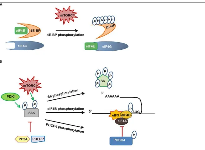
Mammalian target of rapamycin in complex 1 (mTORC1) is a complex that includes the serine/threonine protein kinase, mTOR, the regulatory protein associated with mTOR (RAPTOR), the mammalian lethal with Sec13 protein 8 (mLST8), as well as the two inhibitory subunits, proline-rich Akt substrate of 40 kDa (PRAS40) and DEP domain containing mTOR interacting protein (DEPTOR) (Saxton and Sabatini 2017). mTORC1 controls cell homeostasis through several anabolic and catabolic processes, integrating nutrients and growth factors to activate glycolysis, lipid, nucleotide, and ribosome biosynthesis, protein translation, and inhibition of autophagy (Saxton and Sabatini, 2017). mTORC1 regulates protein translation through its direct targets 4E-binding protein (4E-BP) and ribosomal S6 kinase (S6K) (Fig. 4). In addition to the control of global protein synthesis, mTORC1 was thought to preferentially promote the translation of a subset of mRNAs
containing pyrimidine-rich 5′ terminal oligopyrimidine (TOP) or TOP-like motifs, which are
typically encoded by genes involved in translation regulation (Nandagopal and Roux 2015; Thoreen and others 2012). However, a number of non-TOP mRNAs that are sensitive to mTORC1 activity have recently been identified (Gandin and others 2016; Liu and others 2017), and the identity of the pool of mTORC1-sensitive mRNAs is still uncertain (Meyuhas and Kahan 2015; Nandagopal and Roux 2015). mTORC1 plays a key role in local dendritic translation by promoting synaptic protein synthesis, and as such the kinase is involved in synaptic plasticity, learning, and memory (Lipton and Sahin 2014; Santini and others 2014). However, whereas mTORC1 activity is generally associated with increased mRNA translation, it can also reduce the local synthesis of specific proteins, particularly in diseased states exhibiting excessive mTORC1 activity (Auerbach and others 2011; Niere and others 2016; Raab-Graham and others 2006). In addition, as described below, accumulating evidence suggest that mTORC1 signaling is dysregulated not only in a number of neurological disorders, including neurodevelopmental and neurodegenerative diseases, but also in psychiatric disorders, including addiction (Lipton and Sahin 2014; Neasta and others 2014).
Figure 1. Initiation of translation
Legend. (A) Translation initiation starts with the assembly of the 43S pre-initiation complex (PIC), which is formed by the small 40S ribosomal subunit, the elongation initiation factors (eIFs) 1, 1A, 3, 5, and eIF2-GTP bound to the initiator Met-tRNAi. (B) At the same time, the eukaryotic initiation factor 4F (eIF4F) complex, composed of the RNA helicase eIF4A, the cap-binding protein eIF4E and the scaffold protein eIF4G, binds to the cap of the messenger RNA (mRNA), whereas the poly-A binding protein (PABP) binds to its poly-A tail, which activates and circularizes the mRNA. (C) The binding of the PIC to the 5 ′ end of m7
-G-capped mRNA is then facilitated by eIF4F complex and eIF3. (D) After attachment to the mRNA, the PIC scans the mRNA downstream from 5 ′ to 3 ′, inspecting each codon passing through the P-site of the ribosome, until it finds an appropriate AUG start codon. A perfect match triggers the arrest of scanning, initiated by the hydrolysis of eIF2-GTP to eIF2-GDP, which reduces the affinity of eIF2 for the Met-tRNAi. As a result, eIF2-GDP is displaced from the Met-tRNAi by eIF5B-GTP and eIF2-GDP, eIF1A and eIF3 dissociate from the 40S subunit. eIF2-GDP is then
cycled by the guanine exchange factor (GEF) eIF2B to eIF2-GTP. (E) The last step of the initiation process is the association of the 60S ribosomal subunit to the 48S, catalyzed by eIF5B-GTP, and the release of the other eIFs. Finally, the GTP hydrolysis releases eIF5B-GDP to yield the final 80S initiation complex, which synthesizes the first peptide bond after accepting the appropriate aminoacyl-tRNA into the A-site of the ribosome. eIF = eukaryotic initiation factors; GDP = guanosine diphosphate; GTP = guanosine triphosphate; PABP = poly-A binding protein; PIC = pre-initiation complex.
4E-BINDING PROTEIN
The eIF4E-binding protein family (4E-BP) comprises three members (4E-BP1-3). 4E-BP1 and 4E-BP2 undergo a hierarchical phosphorylation at Thr37 and Thr46, followed by the phosphorylation of four other sites in rodents (five in humans), whereas less is known about the regulation of 4E-BP3 (Qin and others 2016). 4E-BPs compete with the eukaryotic initiation factor 4G (eIF4G) for a shared binding site on eukaryotic initiation factor 4E (eIF4E) (Marcotrigiano and others 1999) (Fig. 4A). The interaction of non-phosphorylated 4E-BPs with eIF4E prevents its interaction with eIF4G, inhibiting the assembly of the translation initiation complex (Marcotrigiano and others 1999) (Fig. 4A). Upon activation, mTORC1 phosphorylates 4E-BP, resulting in the release of eIF4E and the recruitment of eIF4G, allowing translation to proceed (Lipton and Sahin 2014) (Fig. 4A). Besides mTORC1, several other kinases have been described to participate in 4E-BP phosphorylation, these include glycogen synthase kinase 3β (GSK3β), extracellular signal-regulated kinase (ERK), cyclindependent kinases (CDK), leucine-rich repeat serine/ threonine-protein kinase 2 (LRRK2), and ataxia-telangiectasia mutated kinase (ATM) (Qin and others 2016).
p
70 RIBOSOMAL S6 KINASE
p70 Ribosomal S6 kinases (S6K1 and S6K2) are members of the AGC family of serine/threonine kinases (Tavares and others 2015). Activation of S6Ks requires the phosphorylation of residue Thr229 by phosphoinositide-dependent kinase-1 (PDK1), as well as the mTORC1-dependent phosphorylation at Thr389 (Tavares and others 2015) (Fig. 4B). S6K phosphorylates several proteins that are associated with mRNA translation, the best known being the ribosomal protein S6 (S6), a component of the 40S ribosomal subunit (Fig. 4B). Phosphorylation of S6 by S6K occurs on five C-terminal residues in a sequential fashion (Biever and others 2015b). Phosphorylation of S6 is commonly used as a marker of translation activation; however, its biological significance remains controversial (Biever and others 2015b).
Legend. The elongation phase extends from the loading of the first aminoacyl-tRNA into the A-site until the ribosome reaches the termination codon. Elongation comprises three major steps: tRNA selection (A, B), peptide bond formation (C), and translocation (D, E). (A, B) the aminoacyl-tRNA bearing the appropriate anticodon is delivered to the ribosomal A-site by the GTPase eEF1A in a ternary complex with the GTP. On matching between codon and anticodon, eEF1A hydrolyses GTP to allow aminoacyl-tRNA loading. (C-E) Peptide bond is catalyzed and the growing peptide chain is transferred to the tRNA in the A-site. As the peptide bond forms, the ribosomal subunits rotate and trigger movement of the tRNAs into so-called hybrid P/E and A/P states, with the acceptor ends of the tRNAs in the E and P sites and the anticodon loop remaining in the P and A sites. Translocation of the tRNAs to the canonical E and P sites requires the GTPase activity of the elongation factor eEF2 in complex with GTP. A = acceptor site; aa-tRNA = aminoacyl-tRNA; E = exit site; eEF = eukaryotic elongation factor; P = peptidyl site.
Besides S6, other substrates of S6K participating in the translation process have been described (Tavares and others 2015) (Fig. 4B). For example, S6K-dependent phosphorylation of eukaryotic initiation factor 4B (eIF4B) enhances its interaction with eukaryotic initiation factor 3 (eIF3) and the recruitment of eukaryotic initiation factor 4A (eIF4A) at the translation initiation complex (Shahbazian and others 2006) (Fig. 4B). S6K1-mediated phosphorylation of the eIF4A inhibitor programmed cell death protein 4 (PDCD4) promotes its degradation and further enhances eIF4A activity (Dennis and others 2012). Finally, eukaryotic elongation factor 2 kinase (eEF2K), a negative regulator of protein synthesis, is phosphorylated by S6K1 (Tavares and others 2015) (Fig. 4B). S6K inhibition is mediated by the two phosphatases, protein phosphatase 2A (PP2A) and PH domain and
leucine-rich repeat protein phosphatase (PHLPP) (Liu and others, 2011; Peterson and others 1999).
Figure 3. Translation termination and recycling
Legend. (A, B) Termination takes place when the ribosome reaches the stop codon (UAA, UGA, or UAG) at the end of the coding sequence. Termination in eukaryotes is catalyzed by two collaborating factors, eRF1 and eRF3. eRF1 is responsible for the recognition of the stop codon and peptidyl-tRNA
hydrolysis, whereas eRF3 is a GTPase. (C, D) Once the polypeptide chain has been released, the ribosomal subunits must be dissociated and the mRNA and deacylated tRNA released recycled. The multifunctional ABC-family ATPase protein ABCE1 joins the post-termination complex and promotes ribosome subunits dissociation and recycling. ABCE1 = ATP-binding cassette sub-family E member 1; eRF = eukaryotic releasing factor.
Figure 4. 4E-BP and S6K regulation in translation initiation control
Legend. (A) The interaction of non-phosphorylated 4E-BP with eIF4E prevents its interaction with eIF4G and inhibits the assembly of the translation initiation complex. On activation, mTORC1 phosphorylates 4E-BP, resulting in the release of eIF4E and the recruitment of eIF4G, allowing cap-dependent translation to proceed. (B) Activation of S6K requires the PDK1-cap-dependent phosphorylation of residue Thr229, as well as the mTORC1-dependent phosphorylation at Thr389. S6K phosphorylates the ribosomal protein S6, which is commonly thought to activate translation process. S6K-dependent phosphorylation of eIF4B enhances its interaction with eIF3 and the recruitment of eIF4A to the translation initiation complex. S6K also phosphorylates the eIF4A inhibitor PDCD4 and promotes its degradation while enhancing eIF4A function. Inhibition of S6K is an active process mediated by phosphatases, and both PP2A and PHLPP have been shown to dephosphorylate S6K. 4E-BP = 4E-binding protein; eIF = eukaryotic initiation factor; mTORC1 = mammalian target of rapamycin in complex 1; P = phosphorylated residue; PDCD4, programmed cell death protein 4; PDK1, protein 3-phosphoinositide-dependent protein kinase-1; PHLPP, PH domain and leucine-rich repeat protein phosphatase; PP2A, protein phosphatase 2A.
Legend. eIF2 is an heterotrimer composed of α, β, and γ subunits, which binds to GTP and Met-tRNAi to form the PIC pre-initiation complex. After AUG codon recognition, GTP is hydrolyzed and eIF2-GDP is displaced from the Met-tRNAi (see Fig. 1). The recycling of inactive eIF2-GDP to active eIF2-GTP requires the guanine nucleotide exchange factor (GEF) activity of eIF2B. Phosphorylation of eIF2 on its α subunit (Ser51) by the kinases HRK, PKR, GCN2, or PERK converts eIF2 into a competitive inhibitor of eIF2B, which impairs the GDP/GTP exchange and results in a decrease in global translation. Inactivation of eIF2α involves dephosphorylation by protein phosphatase 1 (PP1) coupled to the protein phosphatase 1 regulatory subunit 15 (PPP1R15). eIF = eukaryotic initiation factor; GCN2 = general control non-derepressible-2; HRK = heme-regulated kinase; PERK = PKR-like endoplasmic reticulum resident protein kinase; PKR = double-stranded RNA activated protein kinase; PP1 = protein phosphatase 1; PPP1R15 = protein phosphatase 1 regulatory subunit 15.
EUKARYOTIC INITIATION FACTOR 2
αThe eukaryotic initiation factor 2 α (eIF2α) is another key player in the control of translation initiation (Jackson and others 2010). Following the scanning of mRNA by the 43S pre-initiation complex (PIC), eIF2-GTP is hydrolyzed and eIF2-GDP is released (Fig. 1). The recycling of inactive eIF2-GDP to active eIF2-GTP requires the guanine nucleotide exchange factor (GEF) activity of eIF2B (Fig. 5) (Jackson and others 2010). Phosphorylation of eIF2α converts eIF2 into a competitive inhibitor of eIF2B, which impairs the GDP/GTP exchange resulting in a decrease in global translation (Fig. 5) (Moon and others 2018). Activation of kinases phosphorylating eIF2α not only suppresses general translation but also stimulates the translation of specific mRNAs, mostly coding for proteins participating in endoplasmic reticulum (ER) stress responses (Fig. 5) (Bellato and Hajj 2016). However, the translational shift driven by eIF2α phosphorylation is also part of normal neuronal function in the absence of stress, and growing evidence shows that eIF2α is an important regulator of synaptic protein translation and synaptic plasticity (Bellato and Hajj 2016; Moon and others 2018).
micro
RNAS
microRNAs (miRNAs) are 20 to 22 nucleotides endogenous non-coding RNAs that control a broad array of biological processes (Eulalio and others 2008). The genesis of miRNAs occurs in the nucleus where the primary miRNA transcript is cleaved by the exonuclease III
Drosha to create a precursor miRNA product. The pre-miRNA is then exported to the cytoplasm where the RNase Dicer carries out a second processing step by excising the terminal loop, to create a mature miRNA duplex. One of the miRNA strands associates with Argonaute (AGO) and other proteins to form the RNA-Induced Silencing Complex (RISC) (Eulalio and others 2008). Most of miRNAs binding sites lie in the 3′UTR and are usually present in multiple copies. miRNA-RISC mediates two modes of gene silencing: mRNA decay and translational repression (Iwakawa and Tomari 2015). The general view is that the degree of complementarity between miRNA and mRNA is a key determinant of the regulatory mechanism: a perfect complementarity leads to the cleavage and degradation of the mRNA, whereas the presence of mismatches excludes cleavage and promotes repression of translation (Iwakawa and Tomari 2015). Several miRNAs have been identified in dendrites and were shown to play a role in local mRNA translation and synaptic plasticity (Weiss and others 2015); however, the regulation of miRNA expression at dendrites remains poorly understood. Recent evidence suggested that pre-miRNAs are transported and stored in dendrites, serving as a local source of miRNAs that can be processed locally into mature miRNA (Weiss and others 2015). This conclusion is reinforced by the demonstration that Dicer is localized at synapses and its catalytic activity is induced upon neuronal stimulation (Weiss and others 2015).
Figure 6. Translation elongation control
Legend. (A) eEF1A-GTP delivers the aminoacyl-tRNA to the A site of the ribosome (see Fig. 2), and following GTP hydrolysis, eEF1A-GDP is released. The recycling of eEF1A-GDP to eEF1A-GTP is catalyzed by eEF1B, which is a heteromer composed of at least four subunits: the structural eEF1γ, two exchange factors eEF1Bβ and eEF1Bδ, and a unique tRNA synthetase (Val-Rs). (B) eEF2K phosphorylates eEF2 at Thr56, inhibiting the binding of eEF2 to the ribosome and resulting in a
decrease in global translation. S6K and p90RSK phosphorylate eEF2K on the inhibitory residue Ser366, whereas PKA and AMPK activate eEF2K by phosphorylating Ser500 and Ser398, respectively. Dephosphorylation of eEF2 is mediated by phosphatases, including PP2A. A = acceptor site; AMPK = AMP-activated protein kinase; E = exit site; eEF = eukaryotic elongation factors; eEF2K = eukaryotic elongation factor kinase; GDP = guanine diphosphate; GTP = guanine triphosphate; P = peptidyl site; p90RSK = p90 ribosomal S6 kinase; PKA = protein kinase A; PP2A = protein phosphatase 2A; S6K = S6 kinase; Val-Rs = valine tRNA synthetase.
ELONGATION
EUKARYOTIC ELONGATION FACTORS
Eukaryotic elongation factor 1A (eEF1A) in its GTP-bound form delivers the aminoacyl-tRNA to the A site of the ribosome (Dever and Green 2012) (Fig. 2). Following GTP hydrolysis, eEF1A-GDP is released and is recycled to eEF1A-GTP by the GEF activity of the eukaryotic elongation factor 1B (eEF1B) (Fig. 6A). eEF1A and eEF1B are phosphorylated by a number of serine and threonine kinases (Migliaccio and others 2016). For example, protein kinase C (PKC) phosphorylates both eEF1A and eEF1B, causing an increase in the GDP/GTP exchange activity (Dever and Green 2012). S6 kinases, Rho-kinase, protein kinase A (PKA), casein kinase II, and CDKs have also been implicated in the phosphorylation-dependent regulation of eEF1A and eEF1B (Migliaccio and others 2016).
EUKARYOTIC ELONGATION FACTOR 2 KINASE
Eukaryotic elongation factor 2 kinase (eEF2K) is an atypical calcium calmodulin protein kinase (CaMK) (Proud 2015). eEF2K phosphorylates eEF2 at Thr56, thereby inhibiting the binding of eEF2 to the ribosome, resulting in a decrease in the elongation rate (Fig. 6B) (Proud 2015). S6K regulate eEF2K activity by phosphorylating the inhibitory site Ser366, which desensitizes eEF2K to its activation by calmodulin (Fig. 6B) (Wang and others 2001). p90 ribosomal S6 kinases (p90RSKs) also phosphorylate eEF2K at Ser366 (Fig. 6B) (Wang and others 2001). In addition, two other main pathways are known to activate eEF2K by phosphorylation: cAMP-dependent kinase PKA phosphorylates eEF2K at Ser500, which makes eEF2K activation less dependent on calcium (Diggle and others 2001), whereas the AMP-activated protein kinase (AMPK) stimulates eEF2K activity by phosphorylating Ser398 in order to slow down translation elongation when cells are in a low-energy status (Fig. 6B) (Browne and others 2004).
RIBOSOME TRANSLOCATION RATE, TRNA, AND CODON OPTIMALITY
An emerging idea is that modulation of ribosome translocation might be a crucial component in the regulation of translational elongation, as ribosome translocation rates may subtly influence protein expression (Hanson and Coller 2018; Yu and others 2015). In this view, tRNAs can be considered as critical regulators of the transcriptome as not all codons are decoded at the same rate (Yu and others 2015). In addition, codon optimality, which defines the differential recognition of codons by the translational machinery and reflects the balance between the availability of tRNA molecules and their demand during translation process, has been shown to be a powerful determinant of mRNA translation elongation (Hanson and Coller 2018).
TERMINATION
NONSENSE-MEDIATED DECAY
The nonsense-mediated mRNA decay (NMD) is a conserved pathway of mRNA degradation that recognizes and rapidly degrades select mRNAs (Raimondeau and others 2018). NMD was originally identified as a pathway that degrades mRNAs with a premature stop codon resulting from transcription errors, pausing, splicing, or damages (Raimondeau and others 2018). It is now recognized that the NMD also regulates the expression of specific genes by degrading mRNAs, and about 5% to 10% of the transcriptome appears to be sensitive to NMD (Raimondeau and others 2018). The three core NMD factors are the up-frameshift proteins UPF1-3, with UPF1 having RNA helicase activity and UPF2 and UPF3 being regulatory factors. Together with other factors, NMD recognizes and elicits the rapid degradation of abnormal mRNAs and a subset of normal mRNAs, affecting several physiological processes, including stress, immune responses, neurodevelopment, and neuronal functions (Raimondeau and others 2018).
RNA-BINDING PROTEINS
mRNA processing, transport, stability, degradation, and post-translation regulation are all processes carried out by a plethora of RNA-binding proteins (RBPs) (Brinegar and Cooper 2016). RBPs are essential effectors and regulators of mRNA alternative splicing and polyadenylation, which not only generate transcript diversity but also directly influence the stability, transport, localization, and translation efficiency of mRNAs (Brinegar and Cooper 2016). It is becoming increasingly clear that dysregulation of RBPs leads to abnormalities in RNA processing, which represent a common feature among many neurological disorders (Brinegar and Cooper 2016).
mRNA localization is highly dependent on RBPs. Specifically, mRNAs are transported along microtubules from the soma to dendritic and axonal synapses in ribonucleoprotein complexes (RNPs) that are formed by mRNAs and RBPs (Ravanidis and others 2018). RBPs within the RNP function as translational repressors to refrain the mRNA from being translated before reaching its final destination (Ravanidis and others 2018). The most well-characterized RBP involved in mRNA translation repression during mRNA transport is the Fragile X-linked mental retardation protein (FMRP), whose lack of expression causes the Fragile X syndrome (FXS), the most common form of inherited intellectual disability in boys (Chen and Joseph 2015). In addition, FMRP also regulates translation by interacting with eIF4E, cytoplasmic FMRP-interacting protein 1 (CYFIP1), and miRNAs, trapping target mRNAs in cytoplasmic granules, or by slowing or stalling ribosomes (Chen and Joseph 2015). Other RBPs involved in mRNA repression during transport, mRNA processing and stability, are the zip-code-binding protein 1 (ZBP1), Hu/ELAV-like, pumilio (PUM) and staufen (STAU) proteins (Brinegar and Cooper 2016; Goldstrohm and others 2018; Perrone-Bizzozero and Bird, 2013).
Dysregulation of mRNA Translation in Psychiatric
Disorders
Changes in spine number, morphology, and strength of synaptic connections, collectively termed as synaptic plasticity, mainly depend on rapid modification of the synaptic proteome, which is regulated by local dendritic translation of mRNAs (Forrest and others 2018). Aberrant dendritic spine morphology and function are associated with psychiatric disorders, including schizophrenia, depression, bipolar disorder, and addiction (Forrest and others 2018). Here we focus on the alterations of the regulatory mechanisms of mRNA translation associated with major psychiatric disorders (Fig. 7).
SCHIZOPHRENIA
Schizophrenia (SZ) is a severe disabling disorder characterized by disturbances in multiple brain functions, including cognitive and emotional processes that affects 1% of the population (Sarkar and others 2017). Intriguingly, although a strong genetic component exists in the etiology of SZ, no specific gene has been shown to be crucial for the development of the disorder, and accumulating evidence supports the involvement of synaptic proteins in the pathogenesis of the disease (Sarkar and others 2017). Below we describe examples of how disruption in mRNA translation contributes to the etiology of SZ.
m
TORC1 AND DISRUPTED IN SCHIZOPHRENIA 1 (DISC1)
DISC1 is a scaffolding protein that plays a role in dendritic mRNA targeting (Yerabham and others 2013). DISC1 knock-down in the hippocampal dentate gyrus (DG) of mice leads to deficits in cognitive performances and SZ-like behaviors (Zhou and others 2013). Interestingly, treatment of mice with the mTORC1 inhibitor, rapamycin, prevented the morphogenesis, excitability, and behavioral deficits resulting from DISC1 knockdown, suggesting that mTORC1 is a key contributor to the structural and behavioral deficits produced by DISC1 deficiency (Zhou and others 2013). In addition, mTORC1 has also been linked to SZ via the serotonin receptor 5-HT6. Specifically, Meffre and colleagues showed that rapamycin prevented deficits in social cognition and novel object discrimination induced by 5-HT6 agonists, suggesting that recruitment of mTORC1 by 5-HT6 receptors in the prefrontal cortex (PFC) contributes to the perturbed cognition in SZ (Meffre and others 2012). Meffre and coworkers also showed that 5-HT6 receptors physically interact with mTORC1 and increase mTORC1 signaling in the rodent PFC (Meffre and others 2012).
TRANSLATION INITIATION AND ELONGATION FACTORS
Whether translation initiation and elongation factors play a role in SZ has not been explored in great detail. However, English and colleagues used a proteomic profiling approach to identify protein synthesis pathways disrupted in SZ patient–derived olfactory neurospheres, and showed that several ribosomal proteins and eIF2α signaling-related proteins were robustly decreased in the SZ group compared with controls (English and others 2015). In addition, the authors conducted genome-wide association studies and found that
dysregulated eIF2α and eIF4 signaling pathways are correlated with the status of the disease (English and others 2015). Furthermore, Topol and colleagues revealed that the expression of translation factors and ribosomal proteins including eIF2B, eIF3, eIF4, eEF1 and eEF2, were increased in SZ patient– derived induced pluripotent stem cells (iPSC) (Topol and others, 2015). Together, these studies suggest of a global dysfunction of the translation machinery in SZ patient–derived cells.
mi
RNA
sThere is a growing body of literature showing that miRNAs play major roles in the pathophysiology of SZ (Gibbons and others 2018). Studies using postmortem SZ samples identified numerous miRNAs differentially expressed in the PFC (Gibbons and others 2018). Interestingly, many miRNAs whose expression is dysregulated in SZ are known to target synaptic proteins. In addition to alterations in miRNA expression, aberrations in miRNA biogenesis and processing have also been linked to SZ (Gibbons and others 2018).
RNA-BINDING PROTEINS
Dysregulation of several RBPs has been associated with SZ pathogenesis. In particular, the level of FMRP and its downstream targets are reduced in the superior frontal cortex of SZ patients (Fatemi and others 2017). In addition, large-scale genomic studies in SZ patients identified enrichment of uncommon mutations for predicted targets of FMRP (Leonenko and others 2017), and a single point mutation (SNP) in matrix metalloproteinase 9 (MMP-9), whose 3′UTR is targeted by FMRP, has been associated with SZ (Lepeta and others 2017).
Moreover, topoisomerase 3β (TOP3β), which encodes for a component of FMRP-containing
cytosolic messenger RNPs, has also been associated with SZ risk and cognitive impairment, highlighting the possible involvement of dysregulated FMRP-related signaling in the development of SZ (Stoll and others 2013). Another RBP possibly involved in SZ pathogenesis, is ELAV-4/ HuD, which has been shown to interact with the SZ-associated allelic variant of Dihydropyrimidinase like 2 (DPYSL2), characterized by a polymorphic
dinucleotide repeat (DNR) at the 5′UTR (Pham and others 2016). Interestingly, introduction
of the DPYSL2 DNR allele by CRISPR/Cas9 genome editing in human embryonic kidney (HEK) 293 cells resulted in a transcriptome signature that was highly overlapping with the results obtained by Chen and colleagues after knockdown of Zinc finger protein 804A
(ZNF804A) in neural progenitor cells (Chen and others 2015). ZNF804A was highlighted as a
top candidate gene for SZ in genome-wide association studies (Harrison 2017; O’Donovan
and others 2008). The precise role of ZNF804A in neuronal function is not fully understood,
but recent evidence suggest that the protein interacts with many RBPs and other proteins involved in mRNA translation, suggesting a role in translation (Harrison 2017). Finally, whole exome sequencing studies revealed that a nonsense mutation in RNA-binding-motif protein
12 (RBM12) is associated with psychosis and SZ (Steinberg and others 2017). This mutation results in the production of a truncated protein lacking one of the three predicted RNA-recognition motifs, which is likely to result in at least partial loss of function (Steinberg and others 2017), but further studies on the biological function of RBM12 are needed in order to understand the role of this protein in the pathogenesis of SZ.
MAJOR DEPRESSIVE DISORDER
Major depressive disorder (MDD) is a severe and highly debilitating psychiatric disease that affects nearly one-fifth of the U.S. population and is the leading cause of disability worldwide (Zuckerman and others 2018). In addition to depression, MDD is characterized by anhedonia, impairments in cognition, emotional state, memory, and motivation. The disorder is also often associated with low self-esteem, low energy, deficits in appetence and suicidal behaviors (Zuckerman and others 2018).
m
TORC1
Several lines of evidence suggest that activation of mTORC1 signaling is beneficial for the treatment of MDD (Raab-Graham and others 2016). Specifically, Li and coworkers demonstrated that the antidepressant, ketamine, activates mTORC1 in the PFC of rodents, increasing synaptic protein synthesis (Li and others 2010). The authors further showed that inhibition of mTORC1 activity in the PFC blocked the antidepressant effects of ketamine (Li and others 2010). Other studies confirmed that the antidepressant-like effect of N-methyl-d-aspartate receptors (NMDAR) antagonists requires mTORC1 and synaptic protein synthesis in PFC and hippocampal neurons (Workman and others 2013, 2018). These data raise an interesting hypothesis that reduction in mTORC1 activity contributes to the etiology of major depression. In line with this possibility, cyclosporine A-dependent inhibition of the calcium and calmodulin-dependent protein phosphatase, calcineurin, produced depressive-like behaviors by blocking the mTORC1 pathway in the rat PFC (Yu and others 2013), whereas the muscarinic receptor antagonist, scopolamine, increased mTORC1 signaling and synaptogenesis, producing rapid antidepressant behavioral responses (Voleti and others 2013). Moreover, the antidepressant effects of the selective serotonin reuptake inhibitor (SSRI), paroxetine, and the antipsychotic drug, levo-stepholidine, have also been linked to mTORC1 signaling (Xu and others 2018; Zhang and others 2017). Finally, Dwyer and colleagues demonstrated that increased expression of S6K in the PFC of rats produced antidepressant effects, whereas its inhibition resulted in depressive behaviors (Dwyer and others 2015). All those data converge to consolidate the hypothesis that malfunction in mTORC1 signaling in the PFC, and consequent impairment of protein synthesis contribute to the pathology of MDD.
TRANSLATION INITIATION AND ELONGATION FACTORS
Interestingly, ketamine-mediated blockade of NMDAR inhibits eEF2K, resulting in the reduction of eEF2 phosphorylation and the rapid synthesis of brain-derived neurotrophic factor (BDNF) in the hippocampus of mice, suggesting that inhibition of eEF2K contributes to the fast-acting behavioral antidepressant-like effects of ketamine (Adaikkan and others 2018; Monteggia and others 2013). In addition, the alkaloid berberine produces antidepressant effects in ovariectomized mice by modulating the eEF2-BDNF pathway in the hippocampus (Fan and others 2017), suggesting an important contribution of eEF2 signaling pathway to MDD. In contrast, Dagestad and colleagues showed that the SSRI fluoxetine increased the phosphorylation of eEF2 in the PFC, hippocampus and DG of rats (Dagestad and others 2006), suggesting that different antidepressants may differentially modulate eEF2 function. Besides eEF2, other translation initiation factors may also play a role in the development and/or maintenance of MDD. For example, two recent studies reported that eIF4E
phosphorylation modulates depression-like behaviors through selective mRNA translation and regulation of the inflammatory response (Aguilar-Valles and others 2018; Amorim and others, 2018). Specifically, the authors showed that ablation of eIF4E phosphorylation at Ser209 dysregulates the translation of a subset of mRNAs, exaggerating inflammatory responses and reducing serotonin levels, thereby producing depressive-like phenotypes (Aguilar-Valles and others 2018; Amorim and others, 2018). In addition, eIF4E phosphorylation was shown to mediate the antidepressant action of fluoxetine, providing a novel translational control mechanism underlying the pathogenesis of MDD (Amorim and others 2018).
mi
RNA
sSeveral studies have linked MDD with dysregulation of miRNAs (Dwivedi 2016). For instance, SNPs in the miRNA machinery have been associated with MDD, including SNPs within pre-miR-30e, AGO1, and DGCR8, the latter being part of the microprocessor complex and a component of miRNA biogenesis (He and others 2012; Xu and others 2010). Interestingly, a postmortem study revealed a significant down-regulation of 21 miRNAs in the dorsolateral PFC of depressed suicidal subjects (Smalheiser and others 2012). Smalheiser et al. also reported that the expression of several miRNAs is altered in the PFC of rats that exhibited learned helplessness, a behavioral paradigm that models depression (Smalheiser and others 2011). Furthermore, increased levels of miR-132 and miR-182 (both targeting BDNF) and decreased BDNF levels have been reported in the serum of depressive patients (Li and others 2013). Finally, miR-132 was suggested as an indicator for depression status and for monitoring the effectiveness of treatment (Fang and others 2018).
RNA-BINDING PROTEINS
Impaired function of RNA-binding proteins has also been associated with MDD. Specifically, recent genetic and brain-imaging studies suggested that bicaudal C homolog 1 (BICC1) may be associated with depression (Bermingham and others 2012). BICC1 encodes for an RNA-binding protein that plays a role in cytoskeletal organization, cell communication and regulation of Wnt signaling, a pathway involved in the physiopathology of depression (Voleti and others 2012). In addition, Ota et al. showed that BICC1 is up-regulated in the PFC and DG of MDD patients, as well as in the PFC and hippocampus of depressed rats (Ota and others 2015). The authors also reported that ketamine administration decreases BICC1 mRNA levels in the hippocampus and that hippocampal BICC1 knock-down attenuated rat depression-like phenotypes, identifying a role for BICC1 in the development of depressive behaviors (Ota and others 2015). Finally, a role for the ELAV-like RBPs, HuB and HuC, has been suggested in the modulation of emotional states, by using a rat model of depression induced by unpredictable chronic mild stress (Sanna and others 2018).
BIPOLAR DISORDER
Bipolar disorder (BD) is a mood disorder that affects around 2.4% of the global population, represents the sixth leading cause of disability worldwide and is characterized by chronic depressive and manic episodes (Harrison and others 2018).
m
TORC1
Several human studies suggested an involvement for the mTORC1 signaling pathway in BD pathophysiology. Specifically, decreased mTOR mRNA levels were detected in blood samples of short-term BD patients (Machado-Vieira and others 2015), and an SNP in raptor has been associated with BD in genetic studies (Rajkumar and others 2015).
Rodent studies revealed that mania is associated with increased activity of mTORC1. Specifically, ouabain, a specific NA/K-ATPase inhibitor, induces hyperactivity in a model of mania and increases mTORC1 activity and protein synthesis in prefrontal, cingulate, and orbitofrontal cortices (Kim and others 2013). In line with the latter finding, Flaisher-Grinberg and colleagues presented data to suggest that rapamycin possesses mood stabilizing properties and ameliorates manic-like behaviors in rodent models (Kara and others 2018), indicating that BD may be associated with excessive mTORC1 signaling in cortical brain regions. On another hand, Lee et al. analyzed the striatal transcriptome of Shank3-overexpressing transgenic mice, which exhibit manic-like behaviors, and observed a significant reduction in mTORC1 activity in the striatum, suggesting that an interaction between Shank3 and upstream regulators of mTORC1 might contribute to the induction of manic behaviors (Lee and others 2017). Altogether these studies suggest that mTORC1 signaling may be differentially dysregulated in cortical and striatal areas, but that both activation and inhibition of mTORC1 converge toward the development of mania-like behaviors.
TRANSLATION INITIATION AND ELONGATION FACTORS
Lithium chloride is universally accepted as the first-choice mood-stabilizer for maintenance treatment of BD. However, despite the extensive study of its molecular targets, how lithium alleviates BD pathologies is not fully understood. Using a proteomic approach, Karyo et al. identified eEF2 as a cellular target of lithium (Karyo and others 2010). Specifically, the authors show that eEF2 phosphorylation at Thr56 site, a key inhibitory site for eEF2 activity is reduced in cultured cells and mouse hippocampus, thus enhancing protein synthesis (Karyo and others 2010). This observation suggests that dysregulated protein synthesis may partly underlie the pathogenesis of BD. In addition, Pfaffenseller et al. observed an increased phosphorylation of eIF2α in leucocytes from control subjects after treatment with the ER stress activator tunacamycin, but not in BD patient–derived leucocytes, suggesting that dysfunction in the ER stress response and/or eIF2α signaling may also be associated with BD (Pfaffenseller and others 2014).
micro
RNA
The association of miRNAs with BD pathophysiology, risks, and treatments has been established by several studies, including analysis of miRNA expression in postmortem brain tissue (mostly PFC) and blood, as well as genetic studies. This has been nicely reviewed by Fries and colleagues in a recent publication (Fries and others 2018).
RNA-BINDING PROTEINS
Only a few lines of investigation have suggested a potential link between RBPs and BD. Fatemi et al. reported that BD individuals have reduced expression of FMRP in the lateral cerebellum, (Fatemi and others 2010). Another study showed that a nonsense mutation in
RBM12 was carried by 10 members of an Icelandic family suffering from psychosis, and two among them suffering from BD (Steinberg and others 2017). Moreover, the RNA-binding motif protein 3 (RBM3) levels were significantly higher in BD patient–derived neural progenitor cells that responded to the mood stabilizer lithium, compared with nonresponders, suggesting that increased levels of RBM3 might be required for positive response to lithium treatment (Papadima and others 2017).
ADDICTION
Addiction is a chronically relapsing disorder characterized by compulsive drug seeking and taking despite adverse consequences (Koob and Volkow 2016). Drug addiction affects approximately 10% of the population, and incurs significant societal costs, estimated to exceed $700 billion in the United States (Volkow and others 2016).
m
TORC1
Drugs of abuse are believed to usurp the molecular mechanisms and signaling pathways of learning and memory processes, leading to maladaptive formation of drug-dependent memories (Torregrossa and others 2011). mTORC1 is a central player in protein synthesis-dependent plasticity changes underlying learning and memory (Graber and others 2013), and growing evidence is now pointing toward mTORC1 also being a master regulator in synaptic neuroadaptations induced by drugs of abuse, leading to pathological drug taking and addiction (Neasta and others 2014; Ron and Barak 2016). The first evidence suggesting that mTORC1 plays a role in drug addiction came from a study by Narita and colleagues, which found that methamphetamine administration activates S6K in the nucleus accumbens (NAc) of rats and that rapamycin abolishes the development of methamphetamine sensitization (Narita and others 2005). Accordingly, another study reported that inhibition of mTORC1 in the hippocampus protects against amphetamine-induced seizures in rats (Renard and others 2016). However, Biever et al. showed that amphetamine administration increases the phosphorylation of S6 in the striatum but demonstrated that this increase was independent of mTORC1 and rather requires PKA activation (Biever and others 2015a). Interestingly, the authors showed that the increased S6 phosphorylation did not correlate with increased translation, contributing to the ongoing debate regarding the causal relationship between S6 phosphorylation and protein synthesis (Biever and others 2015a; Biever and others 2015b). Follow-up studies provided evidence for the association between mTORC1 signaling and cocaine addiction. Cocaine was shown to activate mTORC1 in reward-related brain regions and systemic administration of rapamycin was reported to suppress conditioned place preference (CPP) to cocaine in rodents (Bailey and others 2012; Wu and others 2011). In addition, the authors reported that cocaine administration leads to a transient increase in S6 phosphorylation in the NAc, dorsal striatum, PFC, and ventral tegmental area (VTA) of rats, which was abolished by rapamycin treatment (Bailey and others 2012; Wu and others 2011). In line with these results, mTOR deletion in the VTA of mice was shown to increase firing of GABAergic neurons, decrease dopamine release in the NAc, and attenuate cocaine-induced CPP (Liu and others 2018). Indeed, cocaine increases the extracellular levels of dopamine in the NAc, and we and others showed that activation of the dopamine D1 receptors (D1R) in the NAc of rodents activates mTORC1 signaling (Beckley and others 2016; Sutton and Caron 2015). The possibility that cocaine activates
mTORC1 in the NAc via D1R stimulation also stemmed from the finding that systemic administration of a D1R-antagonist blocked the cocaine-dependent activation of mTORC1 (Sutton and Caron 2015). Sutton and Carron further showed that deletion of mTOR or Raptor specifically in D1R neurons reduced cocaine-induced hyperlocomotion (Sutton and Caron 2015). Interestingly, another group reported that mTOR-dependent neural plasticity is also required for the process of drug relapse, by showing that mTORC1 inhibition in the NAc attenuated cue-induced reinstatement of cocaine seeking but had no effect on cocaine self-administration (Wang and others 2010). Moreover, James et al. showed that withdrawal from cocaine self-administration increases mTORC1 activity in the NAc, and that rapamycin attenuates the progressive ratio breakpoint in a cocaine self-administration paradigm suggesting a role for mTORC1 in the motivation to seek cocaine (James and others 2014; James and others 2016). In line with previous findings, inhibition of mTORC1 in the NAc also reduced the expression of incubated cocaine craving (Werner and others 2018). Together, these results highlight a central role for mTORC1 in stimulants-dependent neuroadaptations. The involvement of mTORC1 in drug-induced neuroadaptations also extends to opiates. Specifically, acute systemic rapamycin administration suppressed cue-induced drug craving in abstinent heroin addicts (Shi and others 2009), and prevented drug seeking via disrupting reconsolidation of morphine memories in rats (Lin and others 2014). In addition, mTORC1 activity was increased in the hippocampus of rats following morphine CPP, and intra-hippocampal administration of rapamycin prevented the CPP acquisition (Cui and others 2010). In addition, the kappa opioid receptor agonist U50,488H activates mTORC1 in the striatum and cortex of mice and rapamycin administration reduced kappa agonist–induced aversion (Liu and others 2019). Finally, Mazei-Robison and colleagues reported a long-lasting activation of mTORC1 signaling in the VTA following repeated morphine administration (Mazei-Robison and others 2011). mTORC1 signaling has also been reported
to be activated by cannabinoids. A single injection of tetrahydrocannabinol (THC) rapidly
increased mTORC1 activity in the hippocampus, striatum, PFC, and amygdala of mice (Puighermanal and others 2009; Puighermanal and others 2013). Furthermore, systemic administration of rapamycin reduced schizophrenia-like phenotypes produced by chronic THC exposure in mice. (Ibarra-Lecue and others 2018). Interestingly, a single administration of another cannabis-derived phytochemical, cannabidiol (CBD), produced rapid antidepressant-like effects and increased the expression of synaptic proteins in the PFC of rodents, which were reversed by intra-cerebroventricular injection of rapamycin (Sales and others 2019). These studies suggest an involvement of mTORC1 signaling in the actions of both THC and CBD.
Excessive alcohol drinking also activates mTORC1 in key brain regions of the cortico-striatal-limbic system of rodents. Specifically, we reported that repeated cycles of binge drinking and withdrawal activate mTORC1 in the NAc shell and orbitofrontal cortex (OFC), as evidenced by increased phosphorylation levels of its downstream targets 4E-BP1, S6K, and S6 (Laguesse and others 2017a; Neasta and others 2010). We further showed that administration of the specific mTORC1 inhibitors rapamycin and Rapalink-1 decreases rodent alcohol consumption and alcohol place preference (Beckley and others 2016; Morisot and others 2018; Neasta and others 2010). In addition, we reported that alcohol-dependent activation of mTORC1 in the NAc of mice initiates the translation of the synaptic proteins Arc, PSD-95, GLUA1, HOMER, the collapsing response mediator protein 2 (CRMP2), and
Prosap2-interacting protein 1 (Prosapip-1) (Beckley and others 2016; Laguesse and others 2017b; Liu and others 2017; Neasta and others 2010). We further provided evidence to suggest that the alcohol-dependent mTORC1-mediated synaptic protein synthesis plays a crucial role in molecular and cellular-adaptations that in turn drive alcohol drinking behaviors (Laguesse and others 2017b; Liu and others 2017). Specifically, we showed that the consequence of alcohol-dependent mTORC1-mediated increases in CRMP2 and Prosapip1 synthesis in the NAc were microtubules polymerization and F-actin assembly, respectively (Liu and others 2017; Laguesse and others 2017b). We further showed that the downstream consequences of Prosapip1-dependent F-actin assembly are spine maturation and the enhancement of activity of the GluA2-lacking AMPAR (Laguesse and others 2017b). Importantly, both CRMP2 and Prosapip1 in the NAc play a role in mechanisms that underlie heavy alcohol use (Laguesse and others 2017b; Liu and others 2017). In addition, we found that CRMP2 also contribute to mechanisms underlying alcohol-priming induced reinstatement of alcohol CPP (Ben Hamida and others 2018). Finally, we demonstrated that reconsolidation of alcohol-related memories activates mTORC1 signaling in the central amygdala (CeA) and several cortical regions, resulting in increased levels of synaptic proteins, and that systemic and intra-CeA administration of rapamycin disrupted alcohol-associated memories and reduced relapse to alcohol seeking (Barak and others 2013). Together, these findings suggest a central role for mTORC1-dependent neuroadaptations that underlie alcohol reward, seeking, drinking and relapse. In summary, mTORC1 is a convergent point of most if not all drugs of abuse (Fig 7).
TRANSLATION INITIATION AND ELONGATION FACTORS
Translational control by eIF2α kinases is critical for long-lasting plasticity of synaptic connections underlying memory and cognitive functions (Trinh and Klann 2013). Growing evidence suggest that phospho-eIF2α-mediated control of translation could also participate in the formation of drug-induced maladaptive memories underlying addiction.
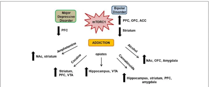
Figure 7. Central role of mTORC1 in psychiatric disorders.
Legend. Increasing evidence suggest a central role for mTORC1 in the development of major depressive disorder (MDD), bipolar disorder (BD), and addiction. Malfunction in mTORC1 signaling in the PFC and consequent impairment of synaptic plasticity contribute to the pathology of MDD. Clinical
studies and rodent models of BD have reported a dual involvement of mTORC1 signaling in BD pathophysiology, suggesting that mTORC1 signaling may be differentially dysregulated in striatal and cortical areas, but converging both toward the development of mania-like behaviors. Finally, mTORC1 appears as a focal point of distinct signaling cascades that govern drug-induced synaptic plasticity and consequent neuroadaptations in different reward-related brain regions. ACC = anterior cingulate cortex; NAc = nucleus accumbens; OFC = orbito-frontal cortex; PFC = prefrontal cortex; VTA = ventral tegmental area.
Specifically, Huang, Placzek, and colleagues showed that cocaine hijacks eIF2α-dependent translational control, and initiates synaptic potentiation in VTA neurons to induce addiction-related behaviors (Huang and others 2016; Placzek and others 2016b). The authors also reported that adult mice with reduced phospho-eIF2α levels in the VTA exhibited higher cocaine-induced long-term potentiation (LTP) (Huang and others 2016; Placzek and others 2016b). Furthermore, reduced eIF2α phosphorylation in the VTA was shown to increase cocaine CPP, whereas adolescent mice, exhibiting higher phospho-eIF2α levels, were more resilient to cocaine’s effects (Huang and others 2016; Placzek and others; 2016b). Interestingly, the same group found that reduced eIF2α phosphorylation may also enhance susceptibility to nicotine in humans and rodents, suggesting a common effect of both drugs on eIF2α signaling (Placzek and others 2016a). Moreover, re-exposure to a previous morphine- or cocaine-paired context increased eIF2α phosphorylation levels in the basolateral amygdala (BLA) of rats, whereas inhibition of eIF2α dephosphorylation disrupted the consolidation of morphine- and cocaine- induced CPP as well as heroin seeking behavior (Jian and others 2014). Together, these data suggest that eIF2α phosphorylation plays a major role in memory reconsolidation of drug-paired stimuli. Finally, Biever et al. reported that repeated amphetamine exposure enhances eIF2α phosphorylation in the striatum of mice, resulting in a global decrease in mRNA translation but also in a specific increase in Arc translation (Biever and others 2016), reinforcing the hypothesis that drug-induced dysregulation of eIF2α signaling activates the translation of specific synaptic mRNAs.
Although cellular and molecular evidence point to a major role played by eEF2 in mRNA translation underlying learning and memory (Taha and others 2013), only a few studies have implicated elongation factors in addiction. For example, Meyers and colleagues identified an association between frequency of alcohol consumption in humans and genetic variants in the mGluR-eEF2-AMPA pathway (Meyers and others 2015). Importantly, the authors identified a
hypermethylation of one CpG site in the 3′UTR of eEF2 in heavy alcohol users, suggesting
an involvement of eEF2-dependent regulation of translation in AUD (Meyers and others, 2015).
micro
RNAS
Over the past years, miRNAs have been heavily implicated in drug addiction (Smith and Kenny 2018). For instance, extended access to cocaine self-administration, increases the expression of 212 in the dorsomedial striatum, 101b, 137, 212 and miR-232 in the NAc shell, and miR-137 in the dorsolateral striatum (DLS) (Hollander and others 2010; Quinn and others 2018). Hollander and colleagues showed in a very elegant study that miR-212 negatively controls cocaine intake by suppressing the activity of Sprouty-related EVH1 domain-containing protein 1 (SPRED1), an endogenous inhibitor of rapidly accelerated
fibrosar-coma 1 (RAF1), amplifying the production of cAMP and the consequent activity of cAMP responsive element protein (CREB) (Hollander and others 2010). They thus identified a role for miR-212 in protection against development of compulsive cocaine taking (Hollander and others 2010). Furthermore, acute cocaine administration down-regulated miR-495 in the NAc, alleviating inhibition of its targets BDNF, CaMKIIa, and Arc, key players in synaptic plasticity underlying addiction (Bastle and others 2018). Conversely, overexpression of miR-495 in the NAc attenuated the upregulation of synaptic translation and reduced motivation to self-administer and seek cocaine (Bastle and others 2018). miR-9 was shown to be up-regulated in the mouse striatum following exposure to alcohol, mediating post-transcriptional
reorganization of BK channels by targeting the 3′UTR of specific splice variants that are
associated with increased sensitivity to alcohol (Pietrzykowski and others 2008). miR-124a, on the other hand, is down-regulated in the DLS following alcohol consumption and miR-124a overexpression enhanced alcohol drinking and alcohol CPP (Bahi and Dreyer 2013). We found that miR-30a-5p, a miRNA targeting BDNF, is upregulated in the PFC of mice consuming excessive amounts of alcohol and demonstrated that miR-30a-5p plays a role in the transition from moderate to excessive alcohol intake (Darcq and others 2015). Specifically, overexpression of miR-30a-5p in the PFC reduced BDNF expression and produced an escalation of alcohol intake, whereas miR-30a-5p inhibition restored BDNF levels and decreased excessive alcohol intake (Darcq and others 2015). Withdrawal from chronic alcohol exposure was accompanied by the upregulation of miR-206 which also targets BDNF, promoting escalation of alcohol self-administration (Tapocik and others 2014). Furthermore, miR-411, miR-203, miR-92a, and miR-137 were reduced in the PFC of mice following chronic alcohol drinking, and silencing of miR-411 decreased the maintenance but not the development of alcohol consumption in mice (Most and others 2018). miRNAs are also involved in opioids actions. For instance, morphine was shown to up-regulate let-7 expression, which led to the down-regulation of the mu opioid receptors and to the development of opioid tolerance (He and Wang 2012). Wang et al. showed that chronic morphine treatment decreased the expression of miR-219-5p in the spinal cord of rats, and that miR-219-5p overexpression prevented the development of morphine tolerance (Wang and others 2017).
RNA-BINDING PROTEINS
Over the past years, accumulating evidence suggested that RBPs may also influence neurobehavioral dysfunction underlying addiction (Bryant and Yazdani 2016). FMRP has been directly implicated in addiction to psychostimulants. Smith and colleagues reported that loss of FMRP impaired behavioral sensitization at moderate cocaine doses and enhanced the development of stereotypy at higher doses but had no effect on cocaine reward (Smith and others 2014). In addition, the authors showed that FMRP-deficient mice exhibited abnormalities in dendritic morphology and glutamatergic neurotransmission in the NAc following cocaine treatment (Smith and others 2014). Mutations in the FMRP-interacting protein CYFIP, which inhibits mRNA translation of synaptic proteins, have been shown to modulate sensitization induced by psychostimulants (Kumar and others 2013). Recent studies also linked FMRP to alcohol’s actions. Specifically, chronic alcohol exposure significantly increased FMRP phosphorylation in the hippocampus, and induced changes in NMDAR and Kv4.2 channel expression (Spencer and others 2016). Moreover, Wolfe and collaborators showed that alcohol promotes the synthesis of GABAB receptors (GABABR)
via FMRP (Wolfe and others 2016). Other RBPs have been less studied, but evidence showing their implication in drug of abuse-dependent dysregulation of local translation have started to emerge (Bryant and Yazdani 2016). For example, polypyrimidine tract binding protein 1 (PTBP1) and the RNA-binding protein, NOVA-1 bidirectionally regulate the short and long splice forms of the dopamine D2 receptor (Park and others 2011; Sasabe and others 2011). It has also been suggested that ELAV-like RBPs regulate the translation of synaptic plasticity-related mRNAs such as BDNF and induce behavioral adaptations in response to cocaine and other drugs of abuse (Bryant and Yazdani 2016; Tiruchinapalli and others 2008). For example, ELAV-4/HuD has been shown to regulate networks associated with addiction-like behaviors, including BDNF and CaMKII (Oliver and others 2018), and mice overexpressing HuD under the CaMKIIa promoter exhibited increased cocaine CPP compared with controls (Oliver and others 2018). Moreover, ectopic expression of ZBP1 in forebrain neurons impaired cocaine CPP (Lapidus and others 2012). Other RBPs have also been suggested to be involved in drug addiction and have been reviewed elsewhere (Bryant and Yazdani 2016).
Concluding Remarks
Aberrant synaptic plasticity is a common feature found in a number of psychiatric disorders, including addiction. As experience-dependent changes in synaptic strength are controlled in part by rapid local translation of mRNAs at synaptic sites, it is likely that impaired local translation is a central point for abnormal synaptic function and a common feature in the mechanisms underlying psychiatric disorders. For example, impairments in mTORC1 function have been associated with a variety of psychiatric diseases such as MDD, SZ, and BD and addiction, even though each of these disorders has different characteristics and symptoms (Fig. 7). Interestingly, mTORC1 signaling is also linked to the mechanism of action of diverse classes of drugs of abuse, that is, psychostimulants, opiates, alcohol, and cannabis (Fig. 7). It is therefore intriguing that different drugs of abuse, by activating different receptors and downstream signaling pathways, converge on mTORC1 to induce maladaptive synaptic plasticity to promote addiction-related behaviors. Although the precise mechanisms underlying drug-dependent modulation of mTORC1 activity and the consequent modifications in synaptic plasticity are only beginning to be understood, all these findings point toward mTORC1 as being a key effector of drug of abuse-dependent neuroadaptations. As mTORC1 is a master regulator of local dendritic translation and synaptic plasticity, it is not surprising that impaired synaptic plasticity through mTORC1, underlies the development of numerous psychiatric disorders (Fig. 7).
Apart from mTORC1, the initiation factor eIF2α and the elongation factor eEF2 also appear to be dysregulated in a variety of psychiatric disorders and adduction. eIF2α and eEF2 are also important players in the regulation of local translation and synaptic plasticity, and as such, their involvement in different psychiatric diseases is not surprising.
Furthermore, miRNAs which are responsible for mRNA degradation and repression of translation also play a critical role in the persistent neuroadaptations associated with psychiatric disorders and drug. Finally, RBPs are numerous and their functions have only
started to be elucidated, but accumulating evidence suggests an important role for RBPs in local translation defects and in the development of psychiatric disorders. Although common mechanisms for RBPs are still to be demonstrated, it is very likely that in the following years, their importance in local translation control underlying the development of psychiatric diseases will emerge. Together, the evidence described herein support the idea that malfunction of local protein synthesis is a common theme in the mechanisms underlying the pathophysiology of psychiatric disorders.
ACKNOWLEDGMENT
The authors thank Ellanor Whiteley for reviewing the manuscript.
DECLARATION OF CONFLICTING INTERESTS
The author(s) declared no potential conflicts of interest with respect to the research, authorship, and/or publication of this article.
FUNDING
The author(s) disclosed receipt of the following financial support for the research, authorship, and/or publication of this article: This research was supported by the National Institute of Alcohol Abuse and Alcoholism, NIH-NIAAA R01 AA027 (DR), U01AA023489 (DR), and R37AA01684 (DR), the European Union’s Horizon 2020 research and innovation program under the Marie Skłodowska-Curie grant agreement 839178 (SL), the F.R.S.-FNRS (SL), and the Foundation Leon Fredericq (SL).
References
Adaikkan C, Taha E, Barrera I, David O, Rosenblum K. 2018. Calcium/calmodulin-dependent protein kinase II and eukaryotic elongation factor 2 kinase pathways mediate the antidepressant action of ketamine. Biol Psychiatry 84:65–75.
Aguilar-Valles A, Haji N, De Gregorio D, Matta-Camacho E, Eslamizade MJ, Popic J, and others. 2018. Translational control of depression-like behavior via phosphorylation of eukaryotic translation initiation factor 4E. Nat Commun 9:2459.
Amorim IS, Kedia S, Kouloulia S, Simbriger K, Gantois I, Jafarnejad SM, and others. 2018. Loss of eIF4E phosphorylation engenders depression-like behaviors via selective mRNA translation. J Neurosci 38:2118–33.
Auerbach BD, Osterweil EK, Bear MF. 2011. Mutations causing syndromic autism define an axis of synaptic pathophysiology. Nature 480:63–8.
Bahi A, Dreyer JL. 2013. Striatal modulation of BDNF expression using microRNA124a-expressing lentiviral vectors impairs ethanol-induced conditioned-place preference and voluntary alcohol consumption. Eur J Neurosci 38: 2328–37.
Bailey J, Ma D, Szumlinski KK. 2012. Rapamycin attenuates the expression of cocaine-induced place preference and behavioral sensitization. Addict Biol 17:248–58.
Barak S, Liu F, Ben Hamida S, Yowell QV, Neasta J, Kharazia V, and others. 2013. Disruption of alcohol-related memories by mTORC1 inhibition prevents relapse. Nat Neurosci 16:1111–7.