HAL Id: dumas-02974016
https://dumas.ccsd.cnrs.fr/dumas-02974016
Submitted on 21 Oct 2020HAL is a multi-disciplinary open access
archive for the deposit and dissemination of sci-entific research documents, whether they are pub-lished or not. The documents may come from teaching and research institutions in France or abroad, or from public or private research centers.
L’archive ouverte pluridisciplinaire HAL, est destinée au dépôt et à la diffusion de documents scientifiques de niveau recherche, publiés ou non, émanant des établissements d’enseignement et de recherche français ou étrangers, des laboratoires publics ou privés.
Prévalence de l’atrophie des glandes meibomiennes chez
le kératocône
Camille Morice
To cite this version:
Camille Morice. Prévalence de l’atrophie des glandes meibomiennes chez le kératocône. Sciences du Vivant [q-bio]. 2020. �dumas-02974016�
Université de Bordeaux U.F.R DES SCIENCES MEDICALES
Année 2020 N°3077
Thèse pour l'obtention du
DIPLÔME D'ETAT DE DOCTEUR EN MEDECINE DISCIPLINE : OPHTALMOLOGIE
Présentée et soutenue publiquement le 25 septembre 2020 Par CAMILLE MORICE
Née le 05 mars 1992 à Brou sur Chantereine
PREVALENCE DE L'ATROPHIE DES GLANDES MEIBOMIENNES
DANS LE KERATOCONE
Directeur de thèse : Monsieur le Professeur David Touboul Rapporteur de thèse : Monsieur le Docteur Serge Doan Membres du jury :
Monsieur le Professeur Korobelnik Jean-François Madame le Professeur Delyfer Marie-Noëlle Monsieur le Professeur Fournié Pierre Monsieur le Professeur Touboul David Madame le Docteur Saunier Valentine
REMERCIEMENTS
Aux membres du jury :
Au président du jury, Monsieur le Professeur Jean-François Korobelnik, vous me faites l'honneur de présider ce jury et de juger cette thèse. Votre habilité chirurgicale, votre sens de l'humain, et votre dévotion pour le service d'ophtalmologie de Bordeaux sont un exemple pour moi. Soyez assuré de ma plus profonde reconnaissance.
A mon directeur de thèse, Monsieur le Professeur David Touboul, vous me faites l'honneur de de diriger ce travail. Je vous remercie infiniment de m'avoir guidée tout au long de ce travail et ces années d'apprentissage au sein du service de segment antérieur. J'admire votre expertise médicale comme chirurgicale au quotidien. Veuillez recevoir mes plus sincères remerciements et ma gratitude.
Au rapporteur de cette thèse, Monsieur le Docteur Serge Doan, je suis très reconnaissante du temps et de l'énergie que vous avez consacré à l'évaluation de ce travail. C'est un grand honneur pour moi de bénéficier de votre expertise dans le domaine de la surface oculaire. Soyez assuré de mon plus profond respect.
A Madame le Professeur Delyfer, merci pour tout. Merci pour votre expertise et votre pédagogie en matière de chirurgie, merci pour votre soutien et votre implication dans notre formation. Ce semestre passé à vos côtés dans le service de rétine m'a beaucoup appris et c'est grandement grâce à vous. Recevez le témoignage de mon plus grand respect.
A Valentine, merci de ta gentillesse et de partager ton savoir au quotidien avec autant d'humilité. Merci de m'avoir guidée pendant ce travail de thèse, malgré les quelques moments de découragement.
A mes maîtres d'internat :
A Cindy Puech, pour m'avoir fait découvrir l'ophtalmologie dans les meilleures conditions possibles dès mon premier semestre d'internat, et pour tout ce que tu as construit à Libourne.
A Wilfried Williamson, pour votre expérience chirurgicale et en randonnées de montagne. A Emilie Tournaire-Marquez, pour m'avoir tout appris du glaucome médical, des lentilles rigides, et pour avoir rendue claire la nébuleuse et passionnante neuro-ophtalmologie. Pour ta gentillesse et ta disponibilité tout au long de mon internat, merci.
A Cédric Schweitzer, pour votre expertise en glaucome chirurgical et votre pédagogie tout au long de cet internat. J'admire votre implication au quotidien pour la formation des internes, ainsi que votre sens relationnel.
A Marie-Bénédicte Rougier, pour votre connaissance en rétine médicale, votre dynamisme, votre humour et votre implication dans notre formation au quotidien.
A Valentine Coste, malgré le covid-19 ces quelques semaines avec toi au troisième étage m'ont appris beaucoup. Ta gentillesse, ton professionnalisme et ton amour de l'ophtalmo-pédiatrie ont rendu ce semestre en l'ophtalmo-pédiatrie inoubliable. Merci.
A toute l'équipe de neurochirurgie A, Mme le Professeur Liguoro, J-R Vignes, sans oublier Vincent et Edouard pour avoir fait de ce semestre un des meilleurs de mon internat, entre stress de poses de DVE nocturnes et victoires au billard. Merci aussi à Momo et Paul, mes acolytes pour tous ces bons moments passés à vos côtés.
Au cabinet Foch, à Valérie Dale, pour ton savoir, tes chaussettes chauffantes, et ta bonne humeur malgré les journées à rallonge, à Sylvie Simonpoli pour la planche de Parinaud, pour ton dynamisme, ta bonne humeur et tes conseils en running, à Patrick pour ton sens humain, ta gentillesse et pour prendre soin de belle mamie, et enfin à Bruno pour m'avoir beaucoup appris de l'ophtalmologie pédiatrique. Merci à tous pour ce semestre tout en bonne humeur et en efficacité.
A François Léger, pour m'avoir fait découvrir l'anatomopathologie et pour vos conseils voyages.
Aux chefs de cliniques et assistants qui m'ont formée : A Thomas, pour ces fous rires et ta gentillesse tout au long de ce semestre.
A Sarra, pour tes innombrables références bibliographiques, ta connaissance de NYC, et tes hoquets prémonitoires incessants.
A Cyril, pour m'avoir guidée lors de mon premier semestre d'ophtalmologie, pour ta rigueur, ta disponibilité et ta gentillesse.
A Hélène, pour ta douceur et ta disponibilité.
A Gabrielle, pour ce semestre passé en ta compagnie au troisième étage. Riche en émotions et en couleurs (et en éviscérations), il restera inoubliable pour moi. Malgré le stress des débuts, tu as toujours su garder ton sens pédagogique et a pris le temps de me montrer les rudiments de chirurgie oculo-palpébrale.
A Thibaut, entre questions d'anatomie et de physiopathologie, tu sais nous demander “pourquoi” sur tout ce qu'on prend pour acquis. Merci pour ta pédagogie et pour ton humour au quotidien.
A Caroline, merci de ta bonne humeur et de ton aide au sein du service de segment antérieur.
A Pierre, de mon premier à mon dernier semestre toujours présent et disponible.
Merci également à Antoine Beele, Antoine Robinet, et Quentin pour votre disponibilité et votre gentillesse tout au long de cet internat.
A tous mes co-internes et amis : A l'équipe des Marie-Chant :
A Elise, pour ton amitié sans faille, ton sourire, ta candeur et tes gaffes quotidiennes. A Hendrik, ton mari qui te rend plus heureuse de jour en jour.
A Sarah, pour ton amitié, ta détermination, pour ces moments inoubliables sur les rails d'un train au milieu du Pérou (tmtc) ou sur des quais de gare quelque part en Europe. Merci à Alex de te faire sourire au quotidien.
A Louchie, Jean-Michel bricolage en ces temps de confinement, toujours souriante et partante pour tout. Merci pour ces belles années d'amitié, et les nouvelles à venir.
A Marie, mi- tarte au Maroilles, mi- fallafel, pour cette année de colocation entre Pau et Bordeaux, ton dynamisme quotidien et ta bonne humeur.
A Arthur, pour ton amitié, ton humour et ta force de caractère. Merci de m'avoir fait connaître Sissy, “contraction volontaire” permanente, ça s'approche de J-Lo.
A Benoit le S, pour cette année de colocation, ton swag et ta sauce Maggi. Tu as eu la chance de trouver sur ton chemin ce petit bout de femme plein d'entrain qu'est Solweig (mais t'as pas intérêt à la laisser perdre au 6 qui prend).
A mes cointernes, Rabia, Paulin, Boris, Grégoire, Hélène, Camille, Caroline, Gobain, Marine, Moe, Anaïs, Diane, Maxime, Thomas, Julia, Nicolas, Nathalie, Guillaume, PH, Maud, Delphine, Anis, Emilie, Marion, Thibault, Antoine, Noémie, Eugénie et tous les autres, pour ces cinq ans passés dans l'entre-aide et la bonne humeur.
A Océane et David-Kamel-Ouali, inséparables depuis notre rencontre sur les bancs de Guist'hau. Tellement heureuse de l'arrivée du petit Ulysse. Merci pour votre amitié à toute épreuve, et pour les belles choses à venir.
A Margaux, pour ta spontanéité, ta peur de rien, ta joie de vivre et ton amitié inébranlable. A mes amis de Nantes et de Bordeaux, Priscille, Arthur, Camille, Camille, Paul, Iris, Romain, Philou, Rémi, Bilu, Gildas, Jules, Julien , Mathilde, Agathe, Floriane et tant d'autres. Tous ces moments et ces vacances passés ensemble resteront inoubliables.
A Jeanne, la distance et les années n'auront pas raison de notre amitié. Merci pour ta bonne humeur et pour ces belles vacances et soirées partagées avec Emma, Estelle, Loren,
Morgane, Sarah et les autres...
A mes amis d'enfance, de Longchamp et Talence, même si ce n'est pas toujours facile de rester en contact, vous resterez les amis de toujours : Louise et nos mercredi flûte-chorée-solfège-gerblé aux figues, Arthur et sa pétanque, Baptiste toujours là dans les bons moments comme dans les mauvais, Julie la belle fiancée, Anthony le cheminot toujours sourire, Gabin le basketteur, Bastien, et tous les autres...
Aux FDC, et a toutes les belles rencontres de ces dernières années : Godefroy le S première rencontre Bordelaise, le roi de la répartie et des blagues de daron merci de m'avoir fait rencontrer Antoine, après moult recherches tu as enfin trouvé Mme Aujay, et ça valait le coup d’attendre ; Elsa la reine des pelleteuses, Lucie la mélomane, Thomas le créole, Louis l'enthousiaste, Nicolas le Bijoune, Souz la chipoune, La Vinchatou, PL, Gui la P, Ismaël, Kev, Paul, Camille, Achille, ... sans oublier Francis, partenaire de tous les instants depuis quelques années.
Spéciale dédicace à Gilles, patron inébranlable de l'Espérance à Châtellerault. Un exemple vivant de ce que la chirurgie cornéenne peut changer dans votre vie.
A toute l'équipe du service d'ophtalmologie de Bordeaux : Aux cadres, Mme Pruvot, Mme Haidar et Mme Tubiana
Aux infirmières du service, Lydia, Annick, JB, Stéphanie, Léa, Sylvie, Céline, Astrid, Anthony, Anaïs, ...
Aux orthoptistes, Béatrice, Esperanza, Aline, Brigitte, Susan, Elodie, Marion, Axelle, Corinne. A toutes les élèves orthoptistes pour leur aide au quotidien.
Aux infirmier(e)s du bloc : Nathalie et Nathalie, Béatrice, Xavier, Marie-Pierre, Sylvie, Mélanie, Nicolas, Barbara, Tania.
Aux secrétaires : à Virginie, à Fanny, à Isabelle, à Florence, à Barbara et Marion.
A ma famille :
A Alexis, il est loin le temps du Lala. Tu as su supporté les longues après-midi de révisions de flûte, puis le silence complet obligatoire pendant la P1... Malgré la distance qui nous sépare, tu restes le meilleur frère:) Je suis heureuse de voir t'épanouir à Madrid et j'espère pouvoir passer plus de temps avec toi maintenant que ces études se terminent.
A papa et maman, pour être les meilleurs parents du monde. Sans vous, rien n'aurait été possible, merci de m'avoir supportée et soutenue durant toutes ces années.
A Papi, malgré ces derniers mois difficiles rien ne me rend plus heureuse que de t’entendre rire avant même de dire allô au moindre coup de téléphone. A ma petite mamie, la bretonne à la tête dure et aux confitures les plus gourmandes.
A Djédé, qui aurait été tellement fier d'être présent aujourd'hui, merci de m'avoir guidée sur la voie de la médecine.
accent qui me donne toujours le sourire, pour le meilleur couscous du monde, les zitounes et j’en passe. Pour être ma petite mouima tout simplement.
A Stef, pour les allers et retours sans compter pendant ces travaux sans fin. L’homme le plus heureux du monde accompagné de son frère, de curly, et d’une petite dodo. A Michèle, Benoît, Thomas et Clément, pour toutes ces années ou j’ai été une pissouse.
A Nadia, pour m'avoir initié aux rudiments du brushing et pour les meilleurs bricks du monde, à Farid pour ces vacances annuelles à Antibes qui resteront inoubliables. A Nadège, Rachid (t'es fouuu), Yanis, Leya, Elyas, Ronan, Soraya (sauveuse de statistiques), Gabriel et Adam.
A Catherine et Olivier, merci pour votre présence au quotidien.
Et enfin, merci à Antoine, je suis tellement heureuse de m’être trouvée sur ton chemin il y a quelques années de ça. Je ne doute pas que les nombreuses prochaines à venir seront encore plus merveilleuses à tes côtés.
TABLE DES MATIERES
REMERCIEMENTS...2
LISTE DES ABBREVIATIONS...8
INTRODUCTION...9
ARTICLE...12
ABSTRACT
MANUSCRIPT
MATERIAL AND METHODS
RESULTS
DISCUSSION
CONCLUSION
BIBLIOGRAPHY
TABLES ET FIGURES
ANNEXES...29
OSDI
SPEED Test
ANATOMIE ET PHYSIOPATHOLOGIE DES GLANDES DE MEIBOMIUS
REVUE DE LA LITTERATURE SUR LES FACTEURS DE RISQUES ASSOCIES A
L'ATROPHIE DES GLANDES DE MEIBOMIUS
BIBLIOGRAPHIE...44
LISTE DES ABBREVIATIONS
AGM : Atrophie des glandes de Meibomius D : Diopters
DGM : Dysfonction des glandes de Meibomius IL 6 : InterLeukine 6
KC : Keratoconus
LLT : Lipid Layer Thickness MG : Meibomian Gland
MGA : Meibomian Gland Atrophy MGD : Meibomian Gland Dysfunction MMP 9 : Matrix MetalloProteinase 9 NKC : Non Keratoconus
OCT : Optical Coherence Tomography OSDI : Ocular Surface Disease Index p : p-value
SPEED : Standardized Patient Evaluation of Eye Dryness T-BUT : Tear Break Up Time
INTRODUCTION
Le Kératocône (KC) est une dégénérescence cornéenne idiopathique caractérisée par un amincissement et une déformation progressive de la cornée. Sa prévalence est estimée à environ une personne sur 2000 habituellement mais serait plus proche d’une personne sur 400 selon les dernières études épidémiologiques (9). Cette déformation est responsable d’un astigmatisme irrégulier, d’une myopie, et de divers symptômes visuelsdont certains ne sont pas corrigeables par le port de lunettes.
Les travaux de recherche, le suivi, l'information et le traitement des patients atteints de Kératocône ont été grandement facilités par la création du centre de référence national du Kératocône (CRNK). Le CRNK fut labellisé en 2005 par le ministère de la santé dans le cadre du premier plan national "maladies rares", il est coordonné sur deux sites, au CHU de Toulouse et au CHU de Bordeaux. Son objectif est de structurer la filière de prise en charge, de fournir les dernières avancées technologiques aux patients, de diffuser la connaissance et de faire avancer la recherche dans le domaine du Kératocône. Également, 5 centres de compétences sont présents dans plusieurs CHU de France et œuvrent au quotidien pour une prise en charge optimisée des patients atteints de Kératocône.
Les causes de cette déformation sont probablement multifactorielles. Le rôle des frottements oculaires sur un terrain génétique prédisposé est aujourd'hui un facteur de progression reconnu de la maladie.
S’il est possible d’améliorer la qualité de vision par le port de lentilles de contact rigides ou la pose d'anneaux intra-cornéens, il parait tout aussi primordial de ralentir ou de stopper l'évolution de cette maladie qui se déclenche le plus souvent entre la quinzième et la vingt-cinquième année.
Il est ainsi primordial de déterminer les facteurs de risque afférents à cette progression. Comme nous l'avons rappelé plus haut, les frottements oculaires seraient un facteur clé dans la progression de la déformation oculaire. C'est pour cela qu'il est notamment conseillé de rechercher la présence d’atopie fréquemment associée aux frottements oculaires et retrouvée dans presque 50% des cas de KC.
De même, l'irritation induite par la sécheresse oculaire peut être à l'origine de frottements répétés, notamment lors de certaines activités récurrentes comme le travail intense sur écran, inhérente à l’immersion dans le monde numérique.
L’association entre la dysfonction des glandes meibomiennes (DGM), principale cause de syndrome sec par hyper évaporation et le Kératocône a récemment été suggérée dans quelques publications.
C'est pourquoi il nous a paru pertinent d’aller plus loin en étudiant la prévalence de l’atrophie des glandes meibomiennes (AGM), marqueur de la chronicité du DGM, dans notre
cohorte de Kératocônes.
Les résultats de cette étude nous permettrons de mieux cerner les facteurs participant à la progression du Kératocône, et donc de diminuer les risques d'apparition et de progression de la maladie chez les patients prédisposés.
ABSTRACT
Purpose: To assess the prevalence of meibomian gland atrophy (MGA) in keratoconus patients.
Setting: French Keratoconus national reference center, Bordeaux, France. Design: Observational, monocentric, comparative case control study.
Methods: Fifty-two Keratoconic eyes free of any surgical treatment (KC group) and fifty-two healthy eyes (NKC group) matched for age and gender, included from December 2018 to May 2019, underwent infrared meibography providing a meiboscore of MGA, scaled from 0 to 6. Lipid Layer Thickness (LLT) assessment using interferometry, Tear Break-Up Time (T-BUT) evaluated by a slit lamp examination and corneal OCT-based topography were also performed. Two different dry eye questionnaires (OSDI and SPEED) were filled in by the patients during the consultation. Additional data were collected on smoking, atopy, eye rubbing, contact lens wear, hormonal contraception and skin diseases.
Results: The MGA in Keratoconus group was significantly higher than in control group (p<0.0001) with a mean meiboscore of 2.33 ± 1.48 and 0.92 ± 1.04 respectively. The mean lower eyelid meiboscore was 1.15 in keratoconus group vs 0.56 in control eyes (p<0.0001) and the mean upper eyelid meiboscore was 1.17 in keratoconus group vs 0.36 in control group (p<0.0001). Although there was no difference between the two groups regarding the LLT and the T-BUT; OSDI and SPEED test scores were significantly higher in KC group.
Conclusion:
Keratoconus was associated with higher meibomian gland atrophy in this study. We emphasize that each Keratoconus patient should have a thorough examination of the ocular surface for signs of meibomian gland atrophy, as risk factors for dry eye, in a disease where eye rubbing was recognized as the main factor of progression. However, this preliminary study requires further studies with larger populations.
PREVALENCE OF MEIBOMIAN GLAND ATROPHY IN KERATOCONUS
INTRODUCTION
Meibomian glands (MG) are sebaceous glands located in the tarsus. Their role is to produce lipids that will participate in the formation of the tear film. This allows to decrease evaporation, maintaining the tear film integrity between two blinks. MG plays an important role in maintaining refractive power of the cornea and also in providing a biological barrier against germs (1).
Meibomian gland dysfunction (MGD) is the most common cause of dry eye syndrome with chronic micro-inflammation, neurotrophic impact, worsening cycle and in some cases can lead to corneal damage with severe visual impairment (2). MGD is thought to involve ductal hyperkeratinization with plugging and obstruction, resulting in an increased pressure within the gland (tortuosity and dilatation), and ultimately in a retrograde meibomian gland atrophy (MGA) (3).
Imaging the structure of the Meibomian glands can be achieved using a slit lamp, but more precisely by meibography (4). Further devices with infrared cameras can picture MG structure. Either with non-contact illumination screening the reflected infrared light from the reversed eyelid, either, more efficiently, using a contact probe for eyelid infrared light transillumination. It is thus possible to describe the percentage of MGA (glands dropout), the presence of dilatation of the central ductal and the tortuosity of the glands (glands dysfunction). Obstruction by itself is not visible with this imaging. An alternative method of evaluating the anatomy of the glands by applying a red filter on a slit-lamp has also recently been proposed by Sang-Mok Lee (5). The classification most used in the literature to quantify MGA is the “Arita’s meiboscore” graded from 0 to 3 per eyelid, the sum of the lower and upper eyelid being the total meiboscore (6). While the meibography is particularly useful for quantifying MGA, it is also highly valuable for educating patients.
Furthermore, it is possible to evaluate the functionality of the MG with a slit lamp by a calibrated mechanical squeezing of the eyelid and by observing both the quality of the meibum (transparency, viscosity, color) and the number of orifices expressing the meibum (obstruction). Moreover, it is possible to measure at a sub-micrometric scale, the thickness of the tear film lipid layer by optical interferometry (7). However, this examination remains poorly standardized, as the average normal thickness seems to vary a lot according to the studies. The blink dynamics are also more than contributive to evaluate the MG functionality (8).
Keratoconus is defined as a progressive corneal degenerative disorder, with nowadays, a prevalence from one in two thousand up to one in four hundred people worldwide (9). It is mainly a bilateral but asymmetrical disease with irregular corneal thinning and surface deformation. This causes a progressive irregular astigmatism and myopia and can be responsible for severe loss of vision. Eye rubbing has long been recognized as the major risk factor in the Keratoconus progression causing an excessive inflammation and biomechanical stress, mainly occurring in genetically predisposed populations (10).
A positive correlation between MGD and Keratoconus was recently reported by Mostovoy et al. (11). Therefore, we hypothesized that MGD can lead to eye rubbing, itself responsible for higher prevalence of KC in patients with MGA (12). The aim of our study was therefore to examine the MGA and MGD prevalence in a Keratoconus population, in comparison to a healthy control group.
METHODS
Participants: in this case-control study, we prospectively recruited fifty-two patients among our Keratoconus patients routinely followed at the French National Reference Center for Keratoconus in Bordeaux (KC group). A non-Keratoconus group (NKC) of fifty-two healthy volunteers working in our hospital was randomly recruited from December 2018 to May 2019. All patients were over eighteen years old and were matched for age and gender. The control subjects should not have any known or infraclinical Keratoconus (ruled out by performing a corneal topography with pachymetry measurement), nor any known ocular disease. The discovery of blepharitis or symptoms of dry eye after inclusion was not an exclusion criterion. The use of drugs or the presence of dermatological disease that could affect meibum production was carefully noted in each group but was not an exclusion factor. The diagnosis of Keratoconus was relying on topographical results and approved by the corneal specialist of our center (DT). Keratoconus patients should not have been treated by corneal collagen cross-linking (CXL) or any ocular surgery. Contact lens wearers were not excluded but we asked both groups to stop wearing contact lenses at least two days before the examination. All patients gave written consent to participate after receiving oral and written information. The study was conducted in accordance with the principles of the Declaration of Helsinki.
Corneal topography: all patients received a Swept-Source OCT corneal topography (Tomey CASIA 2, Japan) with measurement of the anterior steepest keratometry (Ks), the anterior flattest keratometry (Kf), the anterior mean keratometry (Km) and the corneal thinnest point (CTP), in order to rule out the presence of an unknown Keratoconus in the NKC group and to classify the Keratoconus severity in the KC group, according to the Amsler Krumeich
classification (13).
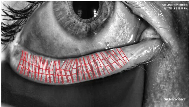
Meibomian gland atrophy (MGA): a meibography imaging with photography of the upper and lower eyelids was performed using LipiView (Tearscience, USA). We arbitrarily divided each gland into three segments (upper, middle, lower), each part being based on the size of the longest gland in that eyelid, taking into account the curvature and exposure of the eyelid. The cutting of the portions was performed semi-automatically after downloading the images of the meibography into the "Paint" software (Microsoft). Two examiners were then asked to blindly determine the number of atrophic portions in each eyelid. Atrophic portions (fully =1 and partly =0,5) were added together and then divided by the number of total portions of that eyelid. Then, we converted this score to a percentage. The meiboscore, calculated as described by Arita et al. in literature, was between 0 and 3 for each eyelid: grade 0 (no loss of MGs); grade 1 (loss <33% of the total surface area of MGs); grade 2 (loss between 33% and 67% of the total surface area of MGs); grade 3 (loss > 67% of the total surface area of MGs). The scores of the upper and lower eyelids were added together to obtain a “total meiboscore” rated between 0 and 6 for each eye. See figures 1 to 3.
Blinking dynamics: by capturing a 20-second video of the eye, the LipiView system allowed the examiner to observe the blinking dynamics and calculate the blinking rate per video, as well as the uncompleted blinking ratio per video.
Lipid Layer Thickness (LLT): we also measured the lipid layer thickness (LLT) with the Lipiview interferometer using the principle of white light interferometry for each eye. It was measured after ensuring the patient had not used any eye drop during the last two hours, and before any eyelids manipulation with the meibographer transilluminator probe or slit lamp testing.
Slit-lamp Examination: we instilled one drop of fluorescein and recorded the Tear BreakUp Time (T-BUT) under blue light illumination. We therefore noted the fluorescein staining for dry eye screening. Finally, we observed the quality of meibomian glands content, in particular the consistency of the expressed meibum by squeezing the eyelid with a dedicated and calibrated tool (Meibomian Gland Evaluator – Tear science). This clinical examination was carried out by the same physician (CM).
Patients “dry eyes questionnaires”: two different questionnaires were consecutively filled by each patient: the Ocular Surface Disease Index (OSDI) questionnaire and the Standardized
Patient Evaluation of eye dryness (SPEED test). The OSDI is a 12-questions questionnaire on
symptoms of eye discomfort over the past week, rated on a scale of 0-100, with higher scores representing greater disability. The second is aiming to assess the dry eye symptoms over the past three months, rated on a scale of 0 to 28. It was implemented with complementary items such as the mention of contact lenses wear (soft, rigid, orthokeratology), intensive and repeated eye rubbing habits (yes/no), presence of dermatological conditions that can
influence meibum production (rosacea, seborrheic dermatitis), atopy (defined by the presence of atopic dermatitis, allergic conjunctivitis, allergic rhinitis, or allergic asthma), smoking and past use of isotretinoin and/or hormonal contraception as drugs known to affect the MG production (14).
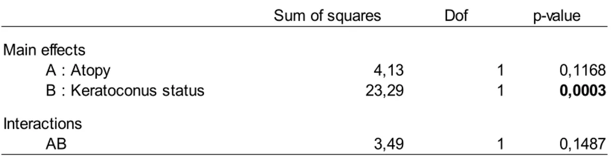
Statistical Analysis: Only the right eye was recruited per patient for statistical analysis, to avoid intra-individual bias and the risk of pseudo-repetition of interdependent variables. Since some of the variables studied were binary (e.g. presence of keratitis), it did not seem relevant to us to compare the means of both eyes of each patient. Each case was matched, by gender and age, to a control and compared using SAS®9.3 software program. The Student-test was used to compare quantitative data between the KC and NKC groups. The Chi2 / Fischer-test (based on n > or < 5) was used when the data for comparing KC and NKC were qualitative (yes/no). The interaction between atopy and the presence of a KC on the Meiboscore was studied using multivariate analysis (ANOVA). The Spearman correlation-test was used to detect the correlation between variables.
RESULTS
Population characteristics: both the KC group as the NKC group consisted of 52 eyes each, they were matched by age and sex during inclusion. The mean age was 30.17 ± 9.17 years old in KC group versus 30.13 ± 8.47 years old in NKC group (p>0.05). Male ratio was 48% (25/52) in both groups. See Table 3. The mean keratometry in KC group was 45.92 ± 2.33 D compared to 42.14 ± 1.78 D in NKC group. The reported pachymetry in KC group was 452 ± 37.7 µm compared to 532 ± 20.7 µm in NKC group. See Table 1. The KC group had fewer soft contact lens wearers (p=0.0052) ; but there was no statistical difference for rigid gas permeable lens wear (p=0.6176) and orthokeratology user (p=0.1177), the number of smokers (p=0.8269); the isotretinoin use (p=0.4878); history of rosacea (p=0.5867); nor seborrheic dermatitis (p=0.5255) between the two groups. See Table 3. We statistically found more atopic patients in KC group (22; 42.31%) than in NKC group (10; 19.23%; p=0.0107).
Meibomian gland atrophy (MGA): the mean glands number visible in our patients were 23 in the upper eyelid and 20 in the lower, which is consistent with the literature (15). After a segmentation of 3 portions for each gland, each upper eyelid was therefore segmented in 69 parts, and 60 parts in the lower eyelid. As MGA was our main criteria of analysis, the most represented meiboscore was stage 2 in KC group (18 eyes; 34.62%) and stage 0 and 1 in NKC group (20 eyes; 38.5% each). See Table 2. Total meiboscore was higher (p<0.0001) in KC group than in NKC group with a mean of 2.33 ± 1.48 and 0.92 ± 1.04 respectively. Superior meiboscore was higher (p<0.0001) in KC group than in NKC group with 1.17 ± 0.92 and 0.36 ± 0.62 respectively. Inferior meiboscore was higher (p<0.0001) in KC group than in NKC group
with a mean of 1.15 ± 0.71 and 0.56 ± 0.74 respectively. See Table 3. After multivariate analysis, no significant interaction was found between keratoconus status and atopy on the meiboscore (p=0.1487). See Table 4
We did not find statistical differences in the Meiboscore of the KC group as a function of Keratoconus stage (p=0.405), nor between males and females in the total sample of participants (p=0.572). We found statistically greater lower tortuosity in KC group (p=0.0459) but no difference in upper tortuosity (p=0.8356).
After a subgroup analysis, we also found a strong positive correlation between age and meiboscore in the healthy population (r=0.36 p=0.0005), but this correlation disappeared in the Keratoconus group (r=0.03 p=0.79) an in total sample of population (r=0,14 p=0,14). See
Table 5.
Blinking dynamics: the blinking rate per video and the incomplete blinks ratio were comparable between the two groups (p=0.36 and p=0.6296 respectively).
Lipid Layer Thickness: was similar in KC group (60.4µm ± 23.9) and NKC group (55.73µm ± 19.44 ; p=0.28). No significant correlation was found with meiboscore and LLT in total sample of participants (r=0,002 p=0,98).
Slit lamp examination: keratitis (fluorescein staining) was more common in KC group (p=0.0199). No difference was observed for the T-BUT (p=0.1935). The proportion of physiological meibum (clear and liquid) was significantly higher in the NKC group (p=0.0379). T-BUT (r= -0.21 p=0.028) was significantly correlated with MGA.
Dry eye questionnaires: OSDI and SPEED test scores were significantly higher in KC group (both p<0.0001). Excessive eye rubbing habits were more frequent in KC group (34; 65.4%) compared to the NKC group (10; 19.23%; p<0.0001). OSDI (r=0.29 p=0.002), SPEED test (r=0.19 p=0.046) were significantly correlated with MGA.
DISCUSSION
As the main result, our study showed a higher prevalence of MGA in the KC group. Meibum was clearer in KC group, but we did not fin any difference in tear film stability. Nevertheless, MGD evaluation is more challenging to standardize than meibography-based MGA evaluation.
As mentioned before, an association between MGD and KC has previously been reported in the literature. In a recent study, Mostovoy and al. studied the ocular surface of 50 KC patients compared to healthy controls. This study showed a higher prevalence of blepharitis, MGD
and dry eye symptoms. They conclude by advising a systematic search for signs of blepharitis, as this could be responsible for a progression of the disease (11).
Mc Monnies and al. also described an increase in corneal temperature, epithelial thinning, increased concentration of inflammatory mediators, and slippage between collagen fibrils at the corneal apex in response to persistent eye rubbing associated with heat. He advised topographical monitoring when massage with warm compresses is prescribed (16).
We do know that MGD can lead to MGA, nevertheless, there is to date, only one published study that investigated the correlation between MGA and KC (17). In this case-control study conducted in Egypt in 2019, by Mostafa et al, 300 eyes of KC patients were compared to 100 healthy eyes. The author found a non-significant trend towards greater MGA in KC, while dry eye symptoms increased and T-BUT decreased. We can however note that this lack of significance could be explained by the young mean age of KC patients involved in that study (mean of 19 years old versus 30 in our study). Yet we know that MGA is an evolving process that can be delayed for years after MGD and symptoms started. In the same study, the authors also found a positive correlation of the meiboscore with age and sex, similarly to several studies which have shown the same (18,19). However, signs of atrophy were still present in adolescents and even in children in another study that explored a pediatric population (20).
Our study did not find a positive correlation between age and MGA in the total sample of participants, but a subgroup analysis found this association in NKC group. This shows that Keratoconus-associated MGA disrupts the normal chronological course of age-related atrophy. We found no significant difference in MGA between men and women. Although meibum production is partly under hormonal control and upregulated by androgens, a 2017 study also found no significant difference between men and women (1,21). Only the subgroup analysis showed a higher MGA in hormonal Birth Control users (22). The authors also found greater dry eye symptoms in women than in men in a KC population (23).
Our study included 48% men and 52% women, which allows us to have an unbiased study of the link between KC and MGA. It is for all these reasons that we felt it was important to match cases and controls on age and sex of patients.
Numerous studies have demonstrated the role of hyperkeratinization and ductal obstruction on the pathogenesis of MGD. Additionally, there is also evidence that the gland can undergo a degenerative atrophic process (24).
If MGD occurs before the appearance of MGA, then it is interesting to ask ourself how MGD can promote the occurrence of KC. First of all, the rational could involve the impact of excessive stress applied at MG level. These repeated and intensive frictions can probably be responsible for a progression of corneal deformation by decreasing keratocyte density (25). It is also possible that the mechanical effect generated by the action of the fingers on the eyelid tissue may be at the origin of cellular metaplasia, with destruction of the meibomian glands, as can be found in other tissues of the human body following chronic mechanical
irritation (26). Indeed, a procedure as trivial as eye rubbing may seem benign, but it has been shown, for example, that it can cause a spectacular increase of intraocular pressure, up to 300 mmHg, demonstrating the adverse consequences of this frequent behavior (27).
Furthermore, it is well documented that chronic blepharitis and dry eyes are common diseases of the ocular surface, causing repeated and intensive rubbing of the eyes (28). Indeed, our study showed more intensive and repeated eye rubbing behavior in Keratoconus patients. This is consistent with the literature, showing that excessive eye rubbing is a fairly common activity in KC patients, triggered by emotional stress, fatigue, eye irritation, screen working and also a frequent spontaneous behavior before going to sleep and upon waking (29,30).
It is also interesting that high levels of inflammatory factors such as IL6, MMP9 and TNF alpha have been found in the tears of KC patients, like in chronic cases of dry eye disease (31,32).
KC patients seemed to suffer greater symptoms of dry eye in our study, as more keratitis was found in KC group but with comparable T-BUT and LLT between the two groups. These results are consistent with data from two case-control studies that studied the relationship between keratoconus and dry eye (33).
This higher prevalence of dry eye in KC could be related to atopy, as proven risk factor for KC, which is known to be associated with a loss of goblet cells and a decrease in BUT and in secreted and membrane-associated mucins (mainly MUC5AC) (34, 35,36). In fact, we have found a higher frequency of atopy in our KC patients, which is consistent with data in the literature (37). Several studies have shown a link between distortion of the MGs and allergic conjunctivitis, as well as allergic keratoconjunctivitis (38). Arita offers two explanations for this: inflammatory changes in the connective tissue could induce pressure on the meibomian glands in a stiff, inextensible tarsus; and that eye rubbing secondary to the allergy could be the cause of this distortion, all the more so as it is more frequent in the upper eyelid. However, to our knowledge, there is no study that found a positive association between MGA and allergic conjunctivitis. Our study also found no interaction of the allergy on the link between meiboscore and KC after multivariate analysis.
An itchy eye can easily lead to excessive eye rubbing in atopic patients, inducing corneal deformation and additionally, MG distortion. That MG distortion during eye rubbing could be the cause of a poorer evacuation of the meibum due to a more sinuous path of the ducts. This also may change the force vectors during the daily blinking, inexorably leading to obstruction and progressive MGA. Histological examination of the tortuous and atrophied glands may be necessary to see whether they are simply different levels of the same disease or two distinct responses to different inflammatory or mechanical processes. Confocal in vivo microscopy, allowing real-time visualization on a microscopic scale could also be of interest here. It would enable the evaluation of the density and diameter of the acini, as well as the reflectivity of the meibum, or the presence of demodex.
Surprisingly, we found comparable LLT between KC and healthy subjects, and even a trend to a thicker LLT in KC group. However, although the literature reports a negative correlation
between LLT and dry eye symptoms, (39) a Japanese study found higher LLT in patients suffering from allergic conjunctivitis (36). As far as our KC group included a higher ratio of atopic patients than the healthy control group, that could explain the unexpected results. This tendency of a thicker LLT may also be artefactual, resulting from uneventful eye rubbing prior to interferometric examination (40).
In addition, it is important to note that the LipiView has an upper limit of 100 nm, which does not allow us to report higher numbers, thus presenting a bias in the calculation of averages. However, these results should not divert us from measuring LLT, which remains a relatively constant examination over time, and positively correlated to the clinical BUT, as a German study reminds us (41).
To our knowledge, our study is the first to have used the LipiView meibograph in a Keratoconus survey. Systematically studying the lower and the upper eyelids, Lipiview is currently the reference machine for imaging the meibomian glands structure, and which also allows the visualization of the blinking dynamics and the simultaneous calculation of the LLT. One of the strengths of our study is the presence of a control group, matched on age and sex, that are potential confounding factors, as discussed above.
A possible bias in our study could be caused by having fewer lens wearers in KC group. This is because a large proportion of the recruited patients were visiting a referral center for the first time, and therefore had not yet benefited from a specific contact lens fitting consultation. However, it has been shown in the literature that prolonged contact lens wear was a risk factor for MGA (42). Nevertheless, some more recent studies did not find this association. Arita et al. only found, in 2012, a correlation between contact lens wear and gland distortion (according to the author, due to contact lens allergy) but not with MGA. However, this bias is probably more at risk of underestimation of the link between KC and MGA than of its overestimation (38). Also, the slit lamp examination of the ocular surface was succinct in our study, it would have been interesting to note also the aspect of the conjunctiva, the quantitative expression of the meibomian glands, or the presence of telangiectasias.
Another weakness of the study is the lack of a standardized computerized measure of gland atrophy. We have tried to reduce that bias by the blind reading of all photographs, as well as by implementing semi-automated segmentation with double reading.
Finally, due to the exclusion of KC patients that have benefited from cross-linking or eye surgery, we removed from our sample the most advanced KC stages, which should be associated with even more severe MGA.
Our results urge the need of a systematic structural and functional MG assessment in the routine examination of the ocular surface, especially for the youngest patients who still are able to develop KC or post laser surgery ectasia in case of refractive surgery. Furthermore, in order to standardize our daily practice and design future studies, it would be advisable to benefit from an intelligence software integrated in meibographers. It would improve and enable automatic quantification of the MGA, the degree of tortuosity and even the dilatation of the MG ducts that could represent the earliest biomarkers of MGA (43). Further studies,
specifically longitudinal, are needed to investigate the chronological link between MGD, MGA and eye rubbing in KC, taking into account the main confounding factors such as age, sex, atopy and contact lens wear.
CONCLUSION
To our knowledge, this study is the first to find a statistically higher prevalence of MGA in KC compared with a healthy population. Our results emphasize that young patients should have a thorough examination of the ocular surface looking for signs of meibomian gland atrophy, as potential indirect risk factor to develop a KC. However, this preliminary study requires further studies with larger populations.
BIBLIOGRAPHY
1. Knop E, Knop N, Millar T, Obata H, Sullivan DA. The International Workshop on Meibomian Gland Dysfunction: Report of the Subcommittee on Anatomy, Physiology, and Pathophysiology of the Meibomian Gland. Invest Ophthalmol Vis Sci. mars 2011;52(4):1938-78.
2. Baudouin C, Messmer EM, Aragona P, et al. Revisiting the vicious circle of dry eye disease: a focus on the pathophysiology of meibomian gland dysfunction. Br J Ophthalmol. 2016;100(3):300-306. doi:10.1136/bjophthalmol-2015-307415
3. Bron A, Tiffany JM. The Contribution of Meibomian Disease to Dry Eye. The ocular surface. 1 mai 2004;2:149-65.
4. Obata H. Anatomy and histopathology of human meibomian gland. Cornea. oct 2002;21(7 Suppl):S70-74.
5. Lee S-M, Park I, Goo YH, Choi D, Shin MC, Kim EC, et al. Validation of Alternative Methods for Detecting Meibomian Gland Dropout Without an Infrared Light System: Red Filter for Simple and Effective Meibography. Cornea. mai 2019;38(5):574-80.
6. Arita R, Itoh K, Maeda S, Maeda K, Furuta A, Fukuoka S, et al. Proposed diagnostic criteria for obstructive meibomian gland dysfunction. Ophthalmology. nov 2009;116(11):2058-2063.e1.
7. Kim JS, Lee H, Choi S, Kim EK, Seo KY, Kim T-I. Assessment of the Tear Film Lipid Layer Thickness after Cataract Surgery. Semin Ophthalmol. 2018;33(2):231-6.
8. Bai Y, Nichols JJ. In vivo thickness measurement of the lipid layer and the overall tear film by interferometry. Opt Lett. 15 mai 2019;44(10):2410-3.
9. Godefrooij DA, de Wit GA, Uiterwaal CS, Imhof SM, Wisse RPL. Age-specific Incidence and Prevalence of Keratoconus: A Nationwide Registration Study. Am J Ophthalmol. mars 2017;175:169-72.
10. Eye Rubbing, a Sine Qua Non for Keratoconus? International Journal of Keratoconus and Ectatic Corneal Diseases. avr 2016;5(1):6-12.
11. Mostovoy D, Vinker S, Mimouni M, Goldich Y, Levartovsky S, Kaiserman I. The association of keratoconus with blepharitis. Clin Exp Optom. 2018;101(3):339-44. 12. Balasubramanian SA, Pye DC, Willcox MDP. Effects of eye rubbing on the levels of
protease, protease activity and cytokines in tears: relevance in keratoconus. Clin Exp Optom. mars 2013;96(2):214-8.
13. Rabinowitz YS, McDonnell PJ. Computer-assisted corneal topography in keratoconus. Refract Corneal Surg. déc 1989;5(6):400-8.
14. Moy A, McNamara NA, Lin MC. Effects of Isotretinoin on Meibomian Glands. Optom Vis Sci. sept 2015;92(9):925-30.
15. Knop N, Knop E. [Meibomian glands. Part I: anatomy, embryology and histology of the Meibomian glands]. Ophthalmologe. oct 2009;106(10):872-83.
16. McMonnies CW, Korb DR, Blackie CA. The role of heat in rubbing and massage-related corneal deformation. Cont Lens Anterior Eye. 2012;35(4):148-154. doi:10.1016/j.clae.2012.01.001
17. Mohamed Mostafa E, Abdellah MM, Elhawary AM, Mounir A. Noncontact Meibography in Patients with Keratoconus. J Ophthalmol. 2019;2019:2965872.
18. Arita R, Itoh K, Inoue K, Amano S. Noncontact infrared meibography to document age-related changes of the meibomian glands in a normal population. Ophthalmology. mai 2008;115(5):911-5.
19. Ban Y, Shimazaki-Den S, Tsubota K, Shimazaki J. Morphological Evaluation of Meibomian Glands Using Noncontact Infrared Meibography. The Ocular Surface. 1 janv 2013;11(1):47-53.
20. Gupta PK, Stevens MN, Kashyap N, Priestley Y. Prevalence of Meibomian Gland Atrophy in a Pediatric Population. Cornea. avr 2018;37(4):426-30.
21. Schirra F, Suzuki T, Richards SM, Jensen RV, Liu M, Lombardi MJ, et al. Androgen control of gene expression in the mouse meibomian gland. Invest Ophthalmol Vis Sci. oct 2005;46(10):3666-75.
22. Yeh TN, Lin MC. Risk factors for severe Meibomian gland atrophy in a young adult population: A cross-sectional study. PLoS ONE. 2017;12(9):e0185603.
23. Fink BA, Wagner H, Steger-May K, Rosenstiel C, Roediger T, McMahon TT, et al. Differences in keratoconus as a function of gender. Am J Ophthalmol. sept 2005;140(3):459-68.
24. Gutgesell VJ, Stern GA, Hood CI. Histopathology of meibomian gland dysfunction. Am J Ophthalmol. sept 1982;94(3):383-7.
25. Kallinikos P, Efron N. On the etiology of keratocyte loss during contact lens wear. Invest Ophthalmol Vis Sci. sept 2004;45(9):3011-20.
26. Herfs M, Hubert P, Delvenne P. Epithelial metaplasia: adult stem cell reprogramming and (pre)neoplastic transformation mediated by inflammation? Trends Mol Med. juin 2009;15(6):245-53.
27. Turner DC, Girkin CA, Downs JC. The Magnitude of IOP Elevation Associated with Eye Rubbing. Ophthalmology. janv 2019;126(1):171-2.
28. Viso E, Gude F, Rodríguez-Ares MT. The association of meibomian gland dysfunction and other common ocular diseases with dry eye: a population-based study in Spain. Cornea. janv 2011;30(1):1-6.
29. Najmi H, Mobarki Y, Mania K, Altowairqi B, Basehi M, Mahfouz MS, et al. The correlation between keratoconus and eye rubbing: a review. Int J Ophthalmol. 2019;12(11):1775-81.
30. Shetty R, Sureka S, Kusumgar P, Sethu S, Sainani K. Allergen-specific exposure associated with high immunoglobulin E and eye rubbing predisposes to progression of keratoconus. Indian J Ophthalmol. mai 2017;65(5):399-402.
31. Galvis V, Sherwin T, Tello A, Merayo J, Barrera R, Acera A. Keratoconus: an inflammatory disorder? Eye (Lond). juill 2015;29(7):843-59.
32. Pflugfelder SC, Bian F, Paiva CD. Matrix metalloproteinase-9 in the pathophysiology and diagnosis of dry eye syndrome. Vol. 4, Metalloproteinases In Medicine. Dove Press; 2017. p. 37-46.
33. Dienes L, Kiss HJ, Perényi K, Nagy ZZ, Acosta MC, Gallar J, et al. Corneal Sensitivity and Dry Eye Symptoms in Patients with Keratoconus. PLoS ONE. 2015;10(10):e0141621. 34. Dogru M, Matsumoto Y, Okada N, Igarashi A, Fukagawa K, Shimazaki J, et al. Alterations
of the ocular surface epithelial MUC16 and goblet cell MUC5AC in patients with atopic keratoconjunctivitis. Allergy. oct 2008;63(10):1324-34.
35. Toda I, Shimazaki J, Tsubota K. Dry Eye with Only Decreased Tear Break-up Time is Sometimes Associated with Allergic Conjunctivitis. Ophthalmology. 1 févr 1995;102(2):302-9.
36. Suzuki S, Goto E, Dogru M, Asano-Kato N, Matsumoto Y, Hara Y, et al. Tear Film Lipid Layer Alterations in Allergic Conjunctivitis. Cornea. avr 2006;25(3):277–280.
37. Galvis V, Tello A, Carreño NI, Berrospi RD, Niño CA. Risk Factors for Keratoconus: Atopy and Eye Rubbing. Cornea. 2017;36(1):e1.
38. Arita R, Itoh K, Maeda S, Maeda K, Furuta A, Tomidokoro A, et al. Meibomian gland duct distortion in patients with perennial allergic conjunctivitis. Cornea. août 2010;29(8):858-60.
39. Blackie C, Solomon J, Scaffidi R, Greiner J, Lemp M, Korb D. The Relationship Between Dry Eye Symptoms and Lipid Layer Thickness. Cornea. 1 août 2009;28:789-94.
40. Craig JP, Blades K, Patel S. Tear lipid layer structure and stability following expression of the meibomian glands. Ophthalmic Physiol Opt. nov 1995;15(6):569-74.
41. Finis D, Pischel N, Borrelli M, Schrader S, Geerling G. [Factors influencing the measurement of tear film lipid layer thickness with interferometry]. Klin Monbl Augenheilkd. juin 2014;231(6):603-10.
42. Arita R, Itoh K, Inoue K, Kuchiba A, Yamaguchi T, Amano S. Contact lens wear is associated with decrease of meibomian glands. Ophthalmology. mars 2009;116(3):379-84.
43. Wang J, Yeh TN, Chakraborty R, Yu SX, Lin MC. A Deep Learning Approach for Meibomian Gland Atrophy Evaluation in Meibography Images. Transl Vis Sci Technol. nov 2019;8(6):37.
FIGURES
FIGURE 1 : Grade 0: Meiboscore in lower eyelid. 0/60 atrophic portions (no atrophy).
TABLES
Table 1. Distribution of meiboscore between KC (n=52) and NKC (n=52)
Table 2. Demographic and topographic characteristics KC (n=52) vs NKC (n=52)
KC (n=52) NKC (n=52) mean SD mean SD p* Km (D) 45,92 2,33 42,14 1,78 <0,0001 Ks (D) 47,41 2,66 42,58 1,84 <0,0001 Kf (D) 44,32 2,28 41,65 1,7 <0,0001 Pachymetry (µm) 452,28 37,71 532,77 20,7 <0,0001 Age 30,17 9,17 30,13 8,47 0,94 n n p** Sex (M/F) 25/27 25/27 1
* : t test student ** : Chi2
Meiboscore KC (n=52) NKC (n=52) n % n % 0 5 9,61 20 38,46 1 10 19,23 20 38,46 2 18 34,62 11 21,15 3 8 15,38 0 0 4 6 11,54 0 0 5 3 5,78 0 0 6 2 3,84 1 1,93
Table 3. Demographic characteristics in KC(n=52) vs NKC (n=52)
KC (n=52) NKC (n=52)
n mean SD Min-max n mean SD Min-max p*
Total Meiboscore 52 2.33 1.48 0-6 52 0.92 1.04 0-6 <0.0001
Superior Meiboscore 52 1.17 0.92 0-3 52 0.36 0.62 0-3 <0.0001
Inferior Meiboscore 52 1.15 0.71 0-3 52 0.56 0.74 0-3 <0.0001
N blinks (per video) 52 3.59 2.74 0-10 52 4.11 3.03 0-13 0.36
Lipid layer thickness (nm) 52 60.4 23.9 23-100 52 55.73 19.44 26-100 0.28
OSDI 52 26.51 16.62 0-61 52 11.39 8.73 0.33 <0.0001
SPEED 52 6.94 4.82 0-21 52 3.3 2.71 0-10 <0.0001
BUT (s) 52 6.47 3.22 1-15 52 6.79 2.79 2-17 0.1935
Incomplete blinking rate 52 0.5 0.4 0-1 52 0.5 0.4 0-1 0.6296
*t test n % n % p** Superior tortuosity of MG 35 67.31 34 65.4 0.8356 17 32.69 18 34.6 Inferior tortuosity 8 15.38 2 3.84 0.0459 44 84.62 50 96.16 Clear Meibum 42 80.77 49 94.23 0.0379 10 19.23 3 5.77 Keratitis 17 32.7 7 13.46 0.0199 35 67.3 45 86.64
Soft Contact Lens wear 6 11.54 18 34.62 0.0052
46 88.46 34 65.38 RGP Lens wear 3 5.77 1 1.92 0.6176 49 94.23 51 98.08 Orthokeratology 0 0 4 7.77 0.1177 52 100 48 92.23 Tobacco use 15 28.84 14 26.92 0.8269 37 71.16 38 73.08 Eye Rubbing 34 65.4 10 19.23 <0.0001 18 34.6 42 80.77 Roaccutane use 3 5.77 6 11.54 0.4878 49 94.23 46 88.46 Seborrheic dermatitis 7 13.46 4 7.69 0.5255 45 86.54 48 92.31 Rosacea 9 17.31 7 13.46 0.5867 43 82.69 45 86.64 Atopy 22 42.31 10 19.23 0.0107 30 57.69 42 80.77
T
able 4. Multivariate analysis model (ANOVA) studying interaction between keratoconus status and atopy on Meiboscore.
Table 5. Correlation between Meiboscore and other factors (n=104)
Meiboscore r p Age 0,14 0,14 OSDI 0,29 0,002 SPEED 0,19 0,046 BUT -0,21 0,028 LLT (µm) 0,002 0,98
p : Spearman correlation test
Sum of squares Dof p-value Main effects
A : Atopy 4,13 1 0,1168
B : Keratoconus status 23,29 1 0,0003
Interactions
AB 3,49 1 0,1487
ANNEXE 1
ANNEXE 2
ANNEXE 3
ANATOMIE ET PHYSIOPATHOLOGIE DES GLANDES DE MEIBOMIUS I) ANATOMIE NORMALE
A- Généralités
Les glandes de Meibomius (GM) sont des glandes sébacées localisées dans le tarse. Leur rôle est de produire des lipides qui participeront à la formation du film lacrymal. Leur contribution est importante pour :
la diminution de la tension de surface nécessaire au bon étalement du film lacrymal la diminution de l'évaporation pour le maintien du film lacrymal entre deux
clignements et à la stabilité réfractive
la barrière biologique, limitant le contact avec les germes.
Les GM ont été décrites pour la première fois par Galen, un anatomiste grec, mais c'est Heinrich Meibum, un anatomiste allemand, qui leur attribua leur nom en 1666.
Les paupières humaines contiennent environ 25 GM au niveau de la paupière supérieure et 20 GM dans la paupière inférieure (44). Leur volume est deux fois supérieur en paupière supérieure (26 µl) qu'en paupière inférieure (13 µl) (TFOS International Workshop On Meibomian Gland Dysfunction). Cependant leur part respective dans la participation dans la formation du film lipidique n'est toujours pas élucidée.
Les GM partagent plusieurs similarités embryologiques avec l'unité pilo-sébacée du cil. Histologiquement, il s'agit d'un canal central kératinisé semblant à de l'épithélium cutané, néanmoins exempt de la couche dite « granulaire ».
Elles sont constituées de nombreux acini sécréteurs contenant des meibocytes, se drainant latéralement au niveau multiples canaux excréteurs, convergent vers un canal excréteur central puis terminal. Celui-ci s'abouche au niveau du bord libre palpébral juste en avant de la ligne de Marx (15).
Cf Figure 1
Le mode de sécrétions des lipides se fait sur le mode holocrine : ainsi les cellules basales des acini vont se différencier de façon centripète puis se disloquer sous la forme de meibum (augmentation de la taille des cellules et lipogénèse). Les glandes de Meibomius sécrètent un mélange lipidique (meibum) qui existe sous forme liquide à la température corporelle, avec un intervalle de fusion compris entre 19,5 et 32,9 °C selon Tiffany (45), ou entre 10 et 40 °C selon Butovich et al (46).
Elles produisent des lipides polaires et apolaires par des mécanismes complexes.
C'est la contraction des muscles (muscle orbiculaire et muscle de Riolan) lors du clignement qui permettra à ces lipides de s'aboucher au niveau du bord libre puis de s'étaler sur la surface oculaire, soulignant l'importance de la dynamique de clignement.
L'innervation, principalement sous contrôle parasympathique (avec des neuropeptides tels que le calcitonin gene-related peptide (CGRP), la substance P et le vasoactive intestinal polypeptide (VIP)) permet de coordonner la sécrétion lipidique et est constituée d'un dense réseau nerveux autour de la membrane basale des acini et des canaux excréteurs. Leur fonctionnement est également sous contrôle de facteurs de croissance et hormonaux : avec une action positive des androgènes sur la sécrétion et une action négative des œstrogènes. B- Méthodes d'exploration
1) Imagerie de la structure
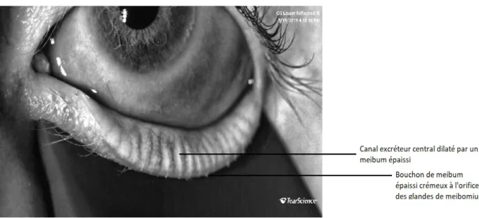
L’examen à la lampe à fente nous permet d’analyser de manière frugale les GM mais la meibographie permet une analyse plus précise (4). Celle-ci utilise une méthode photographique en lumière infra-rouge directement réfléchie ou de manière plus efficace obtenue par trans-illumination. Il est ainsi possible de décrire le pourcentage de perte, la présence d'une dilatation du canal central ainsi que la tortuosité des glandes. Cette méthode est particulièrement utile pour quantifier la trophicité du tissu et éduquer les patients. Il a également récemment été proposé par Sang-Mok Lee une méthode alternative d'évaluation de l'anatomie des glandes lacrymales par l'application d'un filtre rouge (5).
Cf figure2.
La microscopie confocale in vivo a également été utilisée pour visualiser les GM. On a retrouvé, chez des patients souffrant de DGM, une diminution en densité et une augmentation de diamètre des acini, ainsi qu'une inflammation péri-glandulaire (47).
L'OCT SS, présente également un intérêt dans l'évaluation de l'anatomie meibomienne, car contrairement à l'analyse par meibographie utilisant des rayons de type infra-rouge, celui-ci n'est pas dépendant de l'épaisseur de la conjonctive palpébrale. La technologie Swept-Source, associée à une profondeur d'imagerie plus grande permettrait donc une analyse plus fine et plus complète de la morphologie meibomienne avec une analyse de la couche superficielle et de la couche profonde. Cependant l'analyse en OCT couplée à la 3D reste pour le moment trop chronophage pour la pratique quotidienne. Néanmoins les progrès technologiques à venir nous permettront le développement de nouveaux systèmes performants pour l'analyse des glandes de Meibomius.
2) Évaluation de la fonctionnalité
Il est possible d'évaluer la fonctionnalité des glandes de Meibomius à la lampe à fente, par l'expression de celles-ci au niveau du bord libre en exerçant une pression avec deux doigts au niveau de la paupière. Il a également été proposé par Donald Korb et al un outil permettant une pression standardisée du bord libre afin d'uniformiser notre pratique clinique : le MGE (Meibomian Gland Evaluator) (48). La cotation de l'expression est divisée en trois segments supérieurs et inférieurs, cotant à la fois la qualité du meibum (transparence, viscosité), ainsi que la quantité d'orifice exprimant le meibum.
Il est également possible de mesurer sans contact l'épaisseur sub-micrométrique du film lipidique, par interférométrie optique (LIPIVIEW- Johnson&Johnson). Cependant, cet examen reste peu standardisé, l’épaisseur normale moyenne semblant varier selon les études : 42 nm pour King Smith et al, 87 nm pour Korb et al. La qualité de la sécrétion aqueuse intervient également dans la mesure, comme la fréquence et la complétude du clignement, possiblement mesurable (8).
Cf figure 3.
On peut également directement identifier les lipides meibomiens après prélèvement, par spectrométrie de masse, pour étudier les changements en composants lipidiques chez les patients souffrant de DGM. Cette documentation est notamment intéressante lors d'essais cliniques.
II) DYSFONCTIONNEMENT ET ATROPHIE DES GLANDES DE MEIBOMIUS A- PHYSIOPATHOLOGIE
La définition du dysfonctionnement des glandes de Meibomius a été précisée dans le rapport du TFOS International Workshop On Meibomian Gland Dysfunction. : il s'agit d'une anomalie chronique et diffuse des glandes de Meibomius, communément caractérisée par une obstruction du canal terminal et/ou des changements qualitatifs/quantitatifs de la sécrétion glandulaire. Ceci pouvant être à l'origine d'altération du film lacrymal, de symptômes d'irritation oculaire, d'inflammation clinique, et pathologies de la surface oculaire.
Obata a décrit en 2002 les principales étapes des changements pathologiques de la structure des glandes de Meibomius au niveau histologique : (4)
1) La dilatation des acini et des canaux, probablement due à une hyperkératinisation du canal glandulaire et à l’obstruction de l’orifice.
2) L'atrophie des acini avec une diminution de la taille et de la régularité.
3) L'épaississement de la membrane basale des acini, le plus souvent retrouvé en cas d’atrophie des acini. Elle pourrait être à l'origine d'une moins bonne oxygénation par augmentation de la barrière entre cellule glandulaire et vascularisation périacinaire, et donc favoriser l’atrophie.
4) L'apparition d’un tissu de granulation avec mise en évidence d'inflammation, de prolifération fibroblastique et d'angiogénèse au niveau d’une perte en GM.
5) L'inflammation lipogranulomateuse avec de nombreux lymphocytes et cellule épithélioïdes avec cellules géantes multi nucléés, plus communément appelé le chalazion. Le syndrome sec par hyperévaporation est le mécanisme le plus fréquent dans la genèse du syndrome sec (58% selon Tong et al) (49). Le DGM est une maladie fréquente, probablement sous-estimée. Une étude récente retrouve une prévalence de 8,6% de DGM symptomatique
et de 21,9% de DGM asymptomatique (50).
Chez l'enfant, on a retrouvé une prévalence de 12 à 15% dans des études de cohorte indienne et américaine (51).
Nous retiendrons deux types de dysfonctionnement des glandes de Meibomius : une augmentation de la production de Meibum ou, beaucoup plus fréquemment, une diminution de la production (modes hyposécrétoire ou obstructif).
L'hypothèse principale de la première cause de DGM serait que l'obstruction du canal excréteur par un Meibum épaissi et par les débris cellulaires kératinisés seraient à l'origine d'une dilatation du canal qui mènerait à une atrophie des acini. Cependant la chronologie de ces événements reste difficile à préciser en l'absence d'étude prospective longitudinale. D'autres hypothèses concernant la physiopathologie de l'atrophie des acini ont été proposées (augmentation de l'épaisseur de la membrane basale, trouble d'oxygénation des acini, ...).
L'augmentation de la température de fusion du Meibum, serait à l'origine d'une augmentation de sa viscosité et donc d'une stagnation et d'une obstruction des glandes. Ce serait un déséquilibre dans la répartition des différents composants lipidiques qui serait à l'origine de cette augmentation de la température de fusion meibomienne (52).
Pour ce qui est de la séborrhée meibomienne, elle serait à l'origine d'une augmentation des peroxydes d'acides gras, pouvant être à l'origine d'une kératinisation glandulaire et donc à terme, d'une obstruction des glandes de Meibomius.
Il également été étudié le rôle de certains agents infectieux dans la physiopathologie du DGM. Le Demodex, acarien commensal du bord libre palpébral, présent en trop grande quantité pourrait être à l'origine d'une réaction inflammatoire lymphocytaire (53). Son rôle dans la pathogénie du DGM et donc l'intérêt du traitement par Ivermectine visant à le détruire est cependant toujours discuté. Il a également été montré une augmentation de la flore microbienne commensale (Staphylococcus aureus, Staphylococcus epidermidis, Propionibacterium acnes) chez les patients atteins de blépharite (54). Or ces bactéries possèdent une activité lipasique à l'origine de changements dans la composition lipidique du meibum, à l'origine d'une inflammation oculaire et d'un environnement plus propice à la prolifération bactérienne (cercle vicieux) (55).
On sait également que la kératinisation de la partie distale des canaux excréteurs aggrave le DGM obstructif et mène à une dilatation des canaux puis à une atrophie progressive et rétrograde des glandes de Meibomius au cours du temps (24) . Ce processus pourrait être favorisé par les lipides cytotoxiques présents dans le meibum pathologique ainsi que par l'inflammation de la surface oculaire.
Il est également démontré que l'hyperosmolarité lacrymale peut induire une kératinisation de l'épithélium cornéen, (56) il pourrait également être à l'origine d'un mécanisme semblable au niveau des méats meibomiens.
Plusieurs études démontrent le lien entre l'hyperkératinisation et l'obstruction des glandes de Meibomius, à l'origine du DGM obstructif, pathologie la plus fréquente de la glande de Meibomius. Or, il existe également des preuves que les glandes peuvent subir un processus atrophique dégénératif avec destruction progressive du tissu glandulaire (4). La dégénérescence atrophique peut s'expliquer par l'augmentation de la pression intra-glandulaire due à la stase du sérum produit en continu. La stase peut également favoriser la croissance des bactéries commensales, augmentant l'activité lipasique et la libération de médiateurs toxiques. Ces facteurs peuvent, en retour, agir de façon rétroactive (tel un cercle vicieux) aggravant alors l'hyperkératinisation primaire et perturbant la composition du meibum, conduisant à un DGM progressif. Il est également prouvé que l'atrophie se produit avec l'âge, comme dans d'autres organes du corps, par diminution des capacités de multiplication cellulaire (diminution des mitoses cellulaires avec l'index de prolifération ki67) (57).
En effet, le processus naturel du vieillissement sera à l'origine de changements architecturaux du bord libre palpébral, mais aussi d'une atrophie des glandes de Meibomius, et également d'une diminution de l'activité glandulaire comme l'ont montré Norn et al dans cette étude de 1987 qui retrouve une diminution d'activité holocrine cellulaire de moitié entre l'âge de 20 et 80 ans (58).
L'atrophie alors retrouvée dans cette dégénérescence liée à l'âge n'est pas associée à une dilatation parallèle de tous les canaux contrairement à l'atrophie pathologique du DGM obstructif.
Les inflammations oculaires, comme on peut l'observer dans la conjonctivite gigantopapillaires, peuvent également entraîner une AGM (59). On peut donc supposer que les médiateurs inflammatoires altèrent le fonctionnement des GM à travers le tissu conjonctival, causant en l'absence de traitement, la destruction irréversible du tissu
glandulaire (60).
Cependant, la chronologie de cette cascade menant à l'atrophie reste floue en l'absence de grandes études longitudinales.
Il existe également des causes congénitales d'atrophie des glandes de Meibomius. Les dysplasies ectodermiques sont liées à un défaut de développement de l'ectoderme. Elles sont souvent à l'origine de troubles de la sudation, d'anomalies dentaires et des phanères. L'atteinte oculaire des dysplasies ectodermiques associe une atrophie des glandes de Meibomius à une hypotrophie des cils et des sourcils (61).
Les comorbidités oculaires associées au DGM retenues par la Tear Film and Ocular Surface society sont : la présence d'une blépharite antérieure, le port de lentille de contact, la présence de Demodex folliculorum, et l'existence d'un syndrome sec.