TRANSPORT PHYSIOLOGY
Acute parathyroid hormone differentially regulates renal
brush border membrane phosphate cotransporters
Nicolas Picard&Paola Capuano&Gerti Stange&
Marija Mihailova&Brigitte Kaissling&Heini Murer&
Jürg Biber&Carsten A. Wagner
Received: 12 February 2010 / Revised: 16 March 2010 / Accepted: 13 April 2010 / Published online: 5 June 2010 # Springer-Verlag 2010
Abstract Renal phosphate reabsorption across the brush border membrane (BBM) in the proximal tubule is mediated by at least three transporters, NaPi-IIa (SLC34A1), NaPi-IIc (SLC34A3), and Pit-2 (SLC20A2). Parathyroid hormone (PTH) is a potent phosphaturic factor exerting an acute and chronic reduction in proximal tubule phosphate reabsorp-tion. PTH acutely induces NaPi-IIa internalization from the BBM and lysosomal degradation, but its effects on NaPi-IIc and Pit-2 are unknown. In rats adapted to low phosphate diet, acute (30 and 60 min) application of PTH decreased BBM phosphate transport rates both in the absence and the presence of phosphonoformic acid, an inhibitor of SLC34 but not SLC20 transporters. Immunohistochemistry showed NaPi-IIa expression in the S1 to the S3 segment of superficial and juxtamedullary nephrons; NaPi-IIc was only detectable in S1 segments and Pit-2 in S1 and weakly in S2 segments of superficial and juxtamedullary nephrons. PTH reduced NaPi-IIa staining in the BBM with increased intracellular and lysosomal appearance. NaPi-IIa internali-zation was most prominent in S1 segments of superficial nephrons. We did not detect changes in NaPi-IIc and Pit-2 staining over this time period. Blockade of lysosomal protein degradation with leupeptin revealed NaPi-IIa accumulation
in lysosomes, but no lysosomal staining for NaPi-IIc or Pit-2 could be detected. Immunoblotting of BBM confirmed the reduction in NaPi-IIa abundance and the absence of any effect on NaPi-IIc expression. Pit-2 protein abundance was also significantly reduced by PTH. Thus, function and expression of BBM phosphate cotransporters are differen-tially regulated allowing for fine-tuning of renal phosphate reabsorption.
Keywords Phosphonoformic acid . Internalization . Type II and type III phosphate transporter
Introduction
Renal phosphate excretion is mostly determined by the rate of reabsorption of filtered phosphate along the proximal tubule. To date, at least three distinct sodium-dependent phosphate transporters have been identified to be expressed in the brush border membrane (BBM) of the proximal tubule: NaPi-IIa (SLC34A1), NaPi-IIc, and more recently Pit-2 [7,37,43]. However, these transporters have distinct transport properties with respect to ion coupling, preferred species of phosphate, voltage sensitivity, and pH depen-dence [31, 32, 36, 45]. NaPi-IIa and Pit-2 mediate the electrogenic transport of inorganic phosphate coupled to three and two sodium ions, respectively, whereas NaPi-IIc transports inorganic phosphate together with two sodium ions in an electroneutral fashion [31,32,36,45]. Moreover, NaPi-IIa and NaPi-IIc prefer divalent inorganic phosphate (HPO4
2−). In contrast, Pit transporters preferentially trans-port monovalent phosphate (H2PO4−) [31,32,36,45].
A plethora of factors affects renal phosphate reabsorp-tion, among them parathyroid hormone (PTH), dopamine, dietary phosphate intake, glucocorticoids, acid–base status,
Nicolas Picard and Paola Capuano contributed equally to this manuscript and therefore share first authorship.
N. Picard
:
B. KaisslingInstitute of Anatomy, University of Zurich, Zurich, Switzerland
P. Capuano
:
G. Stange:
M. Mihailova:
H. Murer:
J. Biber:
C. A. Wagner (*)Institute of Physiology and Zurich Center for Integrative Human Physiology (ZIHP), University of Zurich,
Winterthurerstrasse 190, CH-8057 Zurich, Switzerland e-mail: [email protected] DOI 10.1007/s00424-010-0841-1
growth factors, insulin, and FGF-23 [1,3,6,7,32,33]. The hormonal regulation of the NaPi-IIa cotransporter has been studied in much detail demonstrating that hormones such as PTH, dopamine, FGF-23, sFRP4, MEPE, glucocorticoids cause a reduction of NaPi-IIa expression [7]. Much less is known about the hormonal regulation of NaPi-IIc with only few studies showing that PTH and FGF-23 cause lower expression [24,38]. In contrast, no hormonal regulators of Pit-2 have been reported to date. Dietary phosphate intake or potassium depletion is the only known regulatory factor of Pit-2 [10,43].
PTH is one of the most potent acute phosphaturic factors inducing urinary phosphate excretion within 15 to 45 min after application [15, 19]. We and others have shown that PTH causes phosphate excretion at least in part by inducing the internalization of the NaPi-IIa phosphate cotransporter from the brush border membrane of proximal tubules in rats and mice [2,3,7, 12, 31,51]. NaPi-IIa is then routed via clathrin-coated pits and early endosomes to lysosomes and subsequently undergoes degradation [3, 22, 35, 41]. Interestingly, the time course of renal phosphaturia and brush border membrane expression of NaPi-IIa do not strictly correlate following an acute PTH bolus [15]. Furthermore, Tenenhouse and colleagues observed that in mice lacking the NaPi-IIa transporter (Slc34a1−/−), PTH injections in vivo (2 h) did not reduce phosphate uptake rates in isolated brush border membrane vesicles (BBMV) and had no effect on phosphaturia [51]. Indeed, Segawa et al. reported PTH-reduced brush border membrane NaPi-IIc expression only 8 h after injection [38]. Taken together, these observations strongly suggest that acute PTH may differentially affect expression and/or function of brush border membrane phosphate transporters.
Therefore, we examined the acute effects of PTH on the expression and function of all three brush border membrane sodium-dependent phosphate cotransporters in rat kidney. Our data demonstrate that PTH differentially regulates NaPi-IIa, NaPi-IIc, and Pit-2 allowing for the fine-tuning of renal phosphate reabsorption.
Materials and methods Animals
The experiments were performed with male Wistar rats (120–150 g, Charles River, Germany). The rats were kept on a low-phosphate diet (0.1% Pi content; Kliba AG, Switzerland) for 3 days to upregulate renal phosphate reabsorption in the BBM of all proximal tubular segments as described previously [3]. The protein abundance of all three known phosphate transporters, NaPi-IIa, NaPi-IIc, and Pit-2, is increased by low phosphate diets [27,34,36,37].
Moreover, all rats received 4% CaCl2 in their drinking water 24 h prior to experiments to suppress endogenous PTH levels [3]. On the day of the experiment, all animals at 8 a.m. received either saline as control or PTH (PTH 1–34 fragment, 10μg/100 g body weight (Sigma, St Louis, MO, USA)) injections as a bolus into the tail vein. Some rats were killed after 30 or 60 min to collect kidneys for the preparation of brush border membrane vesicles. The remaining rats were fixed by perfusion at each time point (30, 60 min). Application of leupeptine (12 mg/ml phosphate-buffered saline (PBS)) 15 min before injection of PTH was also performed as described previously [3].
All animal studies were according to Swiss Animal Welfare laws and approved by the local Cantonal Veterinary Authority of Zurich.
Fixation
After treatment, all animals were fixed by vascular perfusion via the abdominal aorta, at a pressure of 0.4 bar, as described previously [3]. Rats were anesthetized with thiopental (Pentothal, 100 mg/kg body weight) injected intraperitoneally, their abdominal cavity was opened, and the aorta and vena cava were exposed. The fixative consisted of 3% paraformaldehyde, 0.1% glutaraldehyde, and 0.05% picric acid in 0.6 M cacodylate buffer (pH 7.4; containing 3 mM MgCl2and adjusted to 300 mosmol/l with sucrose) and 4% hydroxyethyl starch in saline (HAES steril; Fresenius, Stans, Switzerland). After 5 min, the fixative was washed out by perfusion with hydrostatic pressure of 70 cm for 5 min with cacodylate buffer. Kidneys were removed.
Immunohistochemistry
Slices of fixed kidneys were frozen in liquid propane and cooled with liquid nitrogen. Serial sections, 4-μm thick, were cut at −22°C on a cryomicrotome, mounted on chromalum/gelatine-coated glass slides, thawed, and kept in cold PBS until use. Before immunofluorescence staining, sections were pretreated with blocking solution (Normal Goat Serum 5% in PBS with 0.1% bovine serum albumin) for 60 min. After blocking, serial sections were incubated overnight at 4°C either with a rabbit anti-rat antiserum against the NaPi-IIa protein [13] diluted 1:1,000 or with an immunopurified rabbit anti-mouse NaPi-IIc [33] diluted 1:1,500 or a rabbit anti-rat Pit-2 [43] diluted 1:250. We also tested two commercially available anti-Pit-2 antibodies (Alpha Diagnostics, San Antonio, TX, USA, and Santa Cruz Biotechnologies, Santa Cruz, CA, USA) but could not obtain specific signals by immunohistochemistry of kidneys or immunoblotting of brush border membranes from rats with phosphate depletion. All primary antibodies were
diluted in PBS with 0.1% bovine serum albumin. Sections were then rinsed three times with PBS and covered for 1 h at 4°C with the secondary antibodies: Alexa Fluor 555 goat–anti-rabbit IgG (1:500, Invitrogen), FITC–phalloidin (Molecular probes, Eugene, OR, USA, 1:50), and 4,6-diamidino-2-phenylindole (DAPI; Sigma, St Louis, MO), dilution 1:1,000). Finally, the sections were rinsed three times with PBS, coverslipped using DAKO-Glycergel (Dakopatts) containing 2.5% 1,4-diazabicyclo[2.2.2]octane (Sigma) as a fading retardant.
Immunohistochemistry images were acquired with a Leica DFC490 charged-coupled device camera attached to a Leica DM 6000 fluorescence microscope (Leica, Wetzlar, Germany) using equivalent camera parameters for kidneys sections stained with the same primary antibody. Pictures were processed using Adobe Photoshop (overlays). Brush border membrane vesicle preparation and phosphate uptake experiments
BBMV were prepared from rat kidney cortex and outer medulla using the Mg2+precipitation technique as described previously [8,9]. The phosphate transport rate into BBMV was measured in freshly prepared BBMV at 25°C in the presence of inward gradients of 100 mM NaCl or 100 mM KCl and 0.1 mM K-phosphate. The substrate Piwas made with 0.125 mM K2HPO4and32P (1μCi/ml) to give a final concentration 0.1 mM close to the expected apparent KmPi for Na+-dependent transport in renal BBMV. The stop solution contained 100 mM Mannitol, 5 mM Tris–HCl pH 7.4, 150 mM NaCl, 5 mM Pi. Na+-dependence was established by incubating BBMV in solutions in which KCl replaced NaCl equimolarly. Phosphate uptake was determined after 60 s, representing initial linear conditions, and after 120 min, to determine the equilibrium values. In order to distinguish between Na+-dependent Pi uptake mediated by SLC34 family members (e.g., NaPi-IIa and NaPi-IIc) and other Na+-dependent phosphate transporters such as SLC20 family members (e.g., Pit-1 and Pit-2), we used Trisodium phosphonoformic acid (PFA, final concen-trations 0.1–6 mM) added to the same solution with 107 mM NaCl. PFA has previously been shown to have a higher selectivity for SLC34 than SLC20 phosphate trans-porters at these concentrations [11, 36, 43, 44]. Total protein concentration was measured using the Bio-Rad Protein Assay kit, Bio-Rad, Hercules, CA, USA. BBMV were stored at−80°C until further use.
Immunoblotting
Ten micrograms of renal brush border membrane proteins were solubilized in loading buffer containing DTT and separated on 8% polyacrylamide gels. For immunoblotting,
the proteins were transferred electrophoretically to poly-vinylidene fluoride membranes (Immobilon-P, Millipore Corp., Bedford, MA, USA). After blocking with 5% milk powder in Tris-buffered saline/0.1% Tween-20 for 60 min, the blots were incubated with the primary antibodies: rabbit polyclonal anti-NaPi-IIa (1:6,000) [13], rabbit polyclonal anti-NaPi-IIc (1:10,000) [33], rabbit polyclonal anti-Pit-2 (1:3,000) [43], and mouse monoclonal β-actin anti-body (42 kD; Sigma, St. Louis, MO; 1:5,000) either for 2 h at room temperature or overnight at 4°C. Membranes were then incubated for 1 h at room temperature with secondary goat anti-rabbit or donkey anti-mouse antibodies 1:5,000 linked to alkaline phosphatase (Promega, Madison, WI, USA) or HRP (Amersham Life Science, Little Chalfont Buckinghamshire, UK). The protein signal was detected with the appropriate substrates (Millipore Corp, Bedford, MA, USA) using the DIANA III-chemiluminescence detection system (Raytest, Straubenhardt, Germany). All images were analyzed using the software Advanced Image Data Analyser AIDA (Raytest, Straubenhardt, Germany) to calculate the protein of interest/β-actin ratio.
Statistical analysis
All data are summarized as mean ± SE and were analyzed using the unpaired Student’s t test with only p values ≤0.05 considered as statistically significant.
Results
Parathyroid hormone decreases at least two types of sodium-dependent phosphate transport activities
BBMV were prepared from kidneys of rats adapted to low phosphate diet for 3 days and injected either with saline (0.9% NaCl) or PTH. Sodium-dependent phosphate uptake was measured in the absence and presence of 6 mM PFA. PFA has been known to inhibit BBMV phosphate uptake for almost three decades [39, 40] and has more recently been shown to affect only type II (i.e., NaPi-IIa and NaPi-IIc) but not type III (i.e., Pit-2) mediated phosphate transport at this concentration[43,44]. Thirty minutes after PTH injec-tions, sodium-dependent phosphate uptake was reduced from 3,570 ± 294 pmol/mg protein to 2,987 ± 316 pmol/mg protein (p = 0.1, non-significant). After 60 min, the uptake was reduced from 4,244 ± 241 pmol/mg protein to 3,095 ± 403 pmol/mg protein (p = 0.015, significant;
Fig.1a). In the presence of PFA, however, the total
sodium-dependent phosphate uptake was reduced by about 70% indicating that type II phosphate transporters are responsi-ble for about 70% of the total sodium-dependent phosphate transport activity in chronically low phosphate fed rats.
PFA-resistant phosphate uptake was further decreased from 1,091 ± 50 pmol/mg protein to 767 ± 46 pmol/mg protein after 30 min PTH (p = 0.0007, significant) and from 1,264 ± 116 pmol/mg protein to 808 ± 77 pmol/mg protein after 60 min PTH (p = 0.004, significant) (Fig. 1b). We further characterized the inhibition of sodium-dependent phosphate uptake into brush border membrane vesicles from rats injected either with saline (control) or PTH (30 min) using increasing concentrations of PFA in the range of 0.1 mM to 6 mM (Fig. 1c). Increasing concen-trations decreased Na+-dependent phosphate uptake pro-gressively. We did not use concentrations higher than 6 mM since preliminary experiments suggested unspecific effects on other transport systems such as for glucose (data not shown). We also estimated the apparent half-maximal concentration (EC50) of PFA needed to inhibit Na+
-dependent phosphate uptake and obtained values of approximately 3 mM both in saline and PTH-treated brush border membranes (Fig.1d). Thus, PTH decreases both the PFA-sensitive and the PFA-resistant sodium-dependent phosphate transport activities suggesting that type II and type III transporter(s) are downregulated.
PTH induces internalization and lysosomal degradation of the type II NaPi-IIa cotransporters
Acute application of PTH leads to the rapid internalization of the NaPi-IIa cotransporter from the brush border membrane and its subsequent routing to the lysosome and degradation
[3, 22, 35, 41]. We used immunohistochemistry on
consecutive sections of rat kidneys to examine the localiza-tion of NaPi-IIa, NaPi-IIc, and Pit-2 after applicalocaliza-tion of
2000 2500 3000 3500 4000 4500 5000 0 500 1000 1500 2000 + 6 mM PFA PTH 30 min PTH 60 min Control 30 min Control 60 min PTH 30 min PTH 60 min Control 30 min Control 60 min * *** ** pmol 32 Pi-uptake / mg pr otein pmol 32 Pi-uptake / mg pr otein pmol 32 Pi-uptake / mg pr otein 0 200 400 600 800 1000 1200 1400 0 0.1 mM 0.3 mM 1 mM 3 mM 6 mM control PTH (30 min) PFA concentration Control, EC50= 2.96 ± 1.14 mM PTH, EC50= 3.12 ± 1.16 mM -3 -2 -1 0 1 0 50 100 150 PFA mM (log) Nor malized 32 Pi uptak e activity (%)
a
b
c
d
Fig. 1 Parathyroid hormone (PTH) decreases brush border membrane sodium-dependent phosphate uptake in the absence and presence of phosphonoformic acid (PFA). Rats were injected either with saline (control) or PTH, and kidneys were harvested 30 or 60 min after injections. Brush border membrane vesicles were prepared, and sodium-dependent phosphate fluxes were measured in the absence (a) or presence of 6 mM PFA (b), an inhibitor of type II but not type III phosphate cotransporters (n = 5 each group and time point). PTH decreased the total and PFA-resistant phosphate uptake
indicating that type II and type III transporters are regulated by PTH. c Sodium-dependent phosphate uptake rates in the presence of increasing concentrations of PFA (0.1–6 mM) into brush border membrane vesicles prepared from rats injected with saline (control) or PTH (30 min). d Phosphate transport rates were normalized against rates in the absence of PFA and the apparent half-maximal PFA concentration calculated (EC50) needed to inhibit phosphate
uptake (n = 4 each group and time point). *p < 0.05, ** p < 0.01, *** p < 0.001
Pit2 Control 30 min. 60 min. 60 min. leupeptin NaPilla NaPillc
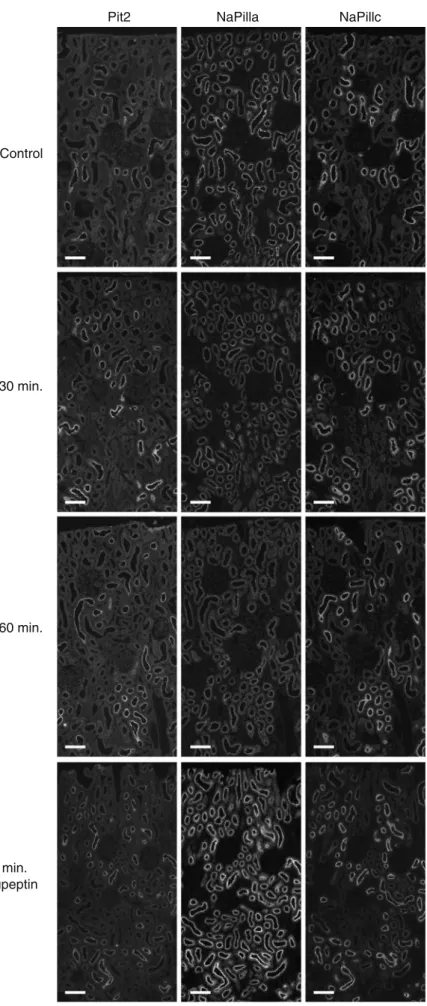
Fig. 2 Effect of acute PTH on NaPi-IIa, NaPi-IIc, and Pit-2 immunostaining. Rats were injected with NaCl (control), or PTH (30 and 60 min). Some rats received also leupeptin prior to PTH to prevent lysosomal degradation (n = 5 animals per group). Cryostat serial sections were stained for Pit-2, NaPi-IIa, and NaPi-IIc. In control kidneys, NaPi-IIa-related staining was seen along the entire length of the proximal tubule (S1–S3 segments) and in both super-ficial and juxtamedullary nephrons. In contrast, NaPi-IIc and Pit-2 staining was mostly observed in early convoluted proximal tubules (S1 segments) with similar intensities in super-ficial and juxtamedullary nephrons. After PTH injection, NaPi-IIa labeling intensity was reduced in kidneys 30 and 60 min after PTH application whereas Pit-2- and NaPi-IIc-related labeling intensity remained unchanged. Inhibition of lysosomal activity with leupeptin increased the intensity of NaPi-IIa staining in PTH injected rats; the staining for NaPi-IIc and Pit-2 appeared unaffected. Bar size∼40 μm
PTH or saline, respectively. In saline-treated animals, no differences in the segmental and subcellular localization of NaPi-IIa, NaPi-IIc, and Pit-2 between control and after 30 or 60 min PTH were observed (Fig.2). In control kidneys from animals on a low phosphate diet, NaPi-IIa-related staining was seen in superficial and juxtamedullary nephrons along the entire length of the proximal tubule (S1–S3 segments). In contrast, NaPi-IIc and Pit-2 staining was mostly observed in early convoluted proximal tubules (S1 segments) with similar intensities in superficial and juxtamedullary nephrons. After PTH injection, NaPi-IIa labeling intensity was reduced in kidneys at both time points (30 and 60 min), whereas Pit-2- and NaPi-IIc-related labeling intensity appeared unchanged (Fig.2). Segmental differences in the response to PTH were observed. The strongest effect of PTH was seen in the early proximal tubules of superficial nephrons (S1 segments) where PTH induced a strong
decrease of NaPi-IIa-related staining in the brush border membrane and the intracellular appearance of staining (Fig. 3). We have previously shown that these intracellular structures include clathrin-coated pits, early endosomes, and lysosomes [3]. In the S2 segment of superficial nephrons, NaPi-IIa internalization was also observed; however, strong residual NaPi-IIa staining in the brush border membrane was seen (Fig. 4). In juxtamedullary nephrons, almost no difference in NaPi-IIa staining between control, saline-, and PTH-treated kidneys could be observed (data not shown). In contrast to NaPi-IIa staining, no changes in NaPi-IIc- or Pit-2-related staining (both apparent intensity and subcellular localization) could be detected on the level of light microscopy at any time point in superficial and juxtamedullary proximal tubules (Figs. 3and 4).
In order to examine if NaPi-IIc or Pit-2 may be rapidly routed to lysosomes and degraded, we pretreated rats with
leupteptin
Fig. 3 Acute PTH induces internalization and lysosomal degradation of NaPi-IIa but not NaPi-IIc and Pit-2 in S1 proximal tubules. S1 segments of superficial nephrons stained with antibodies against Pit-2, NaPi-IIa, or NaPi-IIc (green label) and with rhodamine– phalloidin againstβ-actin fila-ments (red) to mark brush border membranes. Serial sec-tions are shown. Under control conditions, Pit-2, NaPi-IIa, and NaPi-IIc stainings are predominantly localized in the BBM. Thirty minutes after PTH injection, the intensity of the NaPi-IIa staining in the BBM is reduced whereas intracellular NaPi-IIa staining (green) is strongly increased. Pit-2 and NaPi-IIc staining is only detectable in the BBM at all time points, and its intensity remained unaltered. Blockade of lysosomal degradation with leupeptin caused accumulation of NaPi-IIa staining in lysosomes whereas no NaPi-IIc- and Pit-2-related staining could be observed. N = 5 animals per group. Bar size∼10 μm
leupeptin to inhibit lysosomal degradation of proteins. Leupeptin treatment enhanced the signal for NaPi-IIa after 30 and 60 min in lysosomes whereas no staining for NaPi-IIc and Pit-2 could be found (Figs.3and4).
PTH decreases brush border membrane expression of NaPi-IIa and Pit2 but not NaPi-IIc
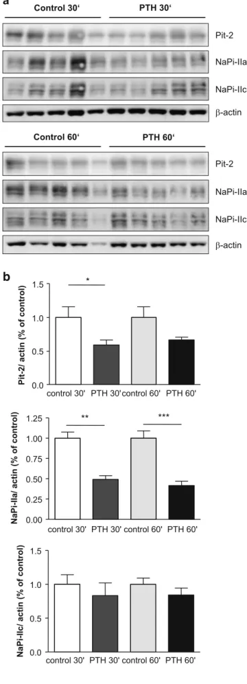
In a next step, we prepared brush border membranes for immunoblotting to examine the relative abundance of NaPi-IIa, NaPi-IIc, and Pit-2 after 30 and 60 min PTH application. Thirty minutes after PTH application, the relative abundance of NaPi-IIa and Pit-2 was significantly reduced in the brush border membrane, whereas NaPi-IIc abundance remained unchanged (Fig. 5a, b). Similarly, 60 min after PTH application, NaPi-IIa abundance was significantly decreased. Pit-2 abundance was lower than in
the respective control group but did not reach anymore statistical significance (p = 0.07; Fig. 5a, b). Thus, PTH treatment acutely decreases expression of NaPi-IIa and Pit-2 in the brush border membrane.
Discussion
The acute effects of PTH on the activity and localization of the renal sodium-phosphate cotransporter NaPi-IIa has been studied in great detail using mouse and rat models, isolated perfused proximal tubules as well as the OK cell model
[3, 20, 21, 26, 29, 42, 48]. Taken together, these studies
demonstrated that PTH binds to receptors localized at the basolateral membrane and in the brush border membrane
[12, 30, 42]. Both cAMP- and PKC-dependent pathways
may at least in part converge in ERK1/2 and induce the Pit2 Control 30 min. 60 min. 60 min. leupeptin NaPilla NaPillc
Fig. 4 Weak effect of PTH in S2 segments on NaPi-IIa internalization. S2 segments of superficial nephrons were stained with antibodies against Pit-2, NaPi-IIa, or NaPi-IIc (shown in green) and with rhodamine–phalloidin against β-actin filaments (red) to mark brush border membranes. Serial sections are shown. In kidneys from control animals, NaPi-IIa mostly resided in the brush border membrane. Pit-2 showed a weaker signal than in S1 segments whereas NaPi-IIc was barely detectable in superficial S2 proximal tubules. Thirty minutes after PTH injection, the intensity of the intracellular NaPi-IIa staining (green) was increased whereas Pit-2 was only detectable at the BBM. A pool of NaPi-IIa was still present in the BBM. Pretreat-ment with leupeptin caused a more intensive intracellular lysosomal staining for NaPi-IIa; no intracellular or lysosomal signal could be detected for NaPi-IIc and Pit-2. N=5 animals per group. Bar size∼10 μm
internalization of NaPi-IIa [4]. NaPi-IIa localized in the brush border membrane forms part of a multiprotein complex and interacts via its PDZ-binding motif with the scaffolding protein NHERF1 [16, 18, 25, 49]. PTH enhances NHERF1 phosphorylation leading to dissociation of the NaPi-IIa–NHERF1 complex and enabling NaPi-IIa routing to clathrin-coated pits, early endosomes, and ultimately to lysosomes [14, 48]. The mechanisms and signals governing the internalization or regulation of NaPi-IIc and the recently described Pit-2 are not known to date. Here, we examined the acute effects of PTH on all three known isoforms of renal brush border membrane sodium-phosphate cotransporters using three complimentary approaches. We demonstrate that acute application of PTH causes within 30 min a reduction in total sodium-dependent phosphate transport in isolated brush border membrane vesicles that became significant after 60 min. Importantly, the use of PFA allowed distinguishing at least two types of phosphate transport activities. Dousa and colleagues had demonstrated more than 20 years ago that brush border membrane phosphate transport could be partly blocked by PFA and related substances [40]. We and others have shown that PFA acts on type II phosphate transporters (NaPi-IIa, NaPi-IIb, and NaPi-IIc) expressed heterologously in Xenopus oocytes and that concentrations in the range of 3–6 mM block about 80–90% of activity [11, 44]. Importantly, type III phosphate transporters (Pit-1 and Pit-2) appear to be much less sensitive to this class of molecules and show only mild inhibition at concentrations higher than 10 mM [36, 43]. The fact that the PFA-resistant sodium-dependent uptake was also reduced by PTH strongly suggests that PTH acts also on the BBM abundance and/or activity of non-type II phosphate trans-porters. To further evaluate the inhibition of phosphate transport, we estimated the apparent half-maximal concen-tration of PFA required to inhibit phosphate transport in brush border membrane vesicles obtained from control and PTH injected rats. Our data demonstrate that PTH affected only the maximal transport rate of phosphate and the PFA inhibitable fraction but did not alter the apparent EC50 for PFA which remained in the range of about 3 mM. These data are in perfect agreement with previous data from rat
a
b
Fig. 5 Parathyroid hormone (PTH) decreases the abundance of NaPi-IIa and Pit-2 in the brush border membrane. Brush border membranes were prepared from kidneys of PTH and saline (control) injected rats (n = 5 animals per condition) and 10 μg loaded per lane for immunoblotting. a Membranes were tested for NaPi-IIa, NaPi-IIc, and Pit-2 and stripped to reprobe forβ-actin to control for loading. b Densitometric analysis of membranes was performed, and the bands for the transporters of interest were normalized againstβ-actin and the respective control groups. PTH decreased NaPi-IIa abundance significantly 30 and 60 min after PTH injections. Pit-2 abundance was reduced significantly 30 min after PTH, whereas NaPi-IIc abundance was unaltered at both time points. *p < 0.05, **p < 0.01
brush border membrane vesicles where binding of radio-labelled PFA and inhibition of phosphate transport were studied and an apparent Km for binding of∼3.6 mM was found [39]. Also in agreement with the work of Dousa and colleagues, we find that about 25–30% of overall sodium-dependent phosphate uptake at the highest concentration of PFA (6 mM) is resistant to the inhibitory effect. Higher concentrations were not used since PFA starts to exert unspecific effects at concentrations of 10 mM and higher (unpublished results). Thus, these data suggest that PTH acts on at least two different populations of phosphate transporters, PFA-sensitive and PFA-insensitive ones.
Consistent with the PFA data, we observed that PTH reduced NaPi-IIa and Pit-2 abundance in isolated brush border membranes as detected by immunoblotting. NaPi-IIc expression was not altered by PTH in agreement with an earlier report showing that NaPi-IIc abundance decreased only approximately 8 h after PTH injection [38]. Immuno-histochemistry confirmed that PTH reduced brush border-localized NaPi-IIa-related staining as described previously
[3,29]. Intracellular NaPi-IIa staining increased after PTH
injection, and blockade of lysosomal degradation with leupeptin showed accumulation of intracellular NaPi-IIa in structures identified previously as lysosomes [3, 29]. In contrast to the findings for NaPi-IIa and the reduced abundance of Pit-2 as shown by immunoblotting, no changes in NaPi-IIc- and Pit-2-related stainings could be detected at the level of light microscopy. This discrepancy for Pit-2 might be due to our antibodies that might detect Pit-2 in immunohistochemistry only in a specific confor-mation. Alternatively, the binding site for the antibody might be blocked by an interacting protein that associates with Pit-2 upon PTH application. We also tested two commercially available antibodies against Pit-2 but failed to detect specific signals preventing a further investigation of this question. Clearly, the fate of Pit-2 following PTH application will require further studies.
The distinct transport properties and differential regulation by hormones and other factors with different time courses allow for a better and finer adaptation of renal phosphate reabsorption under various conditions. Differential regulation of NaPi-IIa, NaPi-IIc, and Pit-2 occurs not only in response to PTH. During dietary potassium restriction and hypokalemia, increased expression of NaPi-IIa has been observed despite phosphaturia [50]. Phosphaturia may be explained by the downregulation of NaPi-IIc and Pit-2, providing another example for differential regulation of BBM phosphate transporters [10]. However, the relative contribution of these transporters to overall renal phosphate handling and the integration of several signals on the level of the proximal under these circumstances remain an open question. PTH application elicits a rapid phosphaturic response both in humans and rodents [15,19]. In contrast to rodents, several
lines of evidence suggest that NaPi-IIa may not be the major type II phosphate transporters in human kidney. Missense and even truncating mutations in NaPi-IIa (SLC34A1) do not cause phosphaturia in humans [23,46], whereas mutations in NaPi-IIc (SLC34A3) are associated with massive phos-phaturia, hypophosphatemia, rickets, and hypercalcuria suggesting that NaPi-IIc is an important contributor to renal phosphate reabsorption [5,28]. The expression of Pit-2 and the relevance of a PFA-sensitive phosphate transport activity in the brush border membrane have not been studied in human kidney and remain unknown to date. If NaPi-IIa does not play a major role in human kidney and if NaPi-IIc does internalize or inactivate only with a delayed time course (as indicated by studies in rodents [38]), then other phosphate transporters must mediate the acute phosphaturic effect of PTH in humans [19]. Whether Pit-2 could be a candidate for this transport activity remains to be investigated in humans and human cell lines. Differential regulation of the three known phosphate transporters in the brush border membrane would allow to fine-tune phosphate excretion not only to PTH status but also to other regulating factors such as dietary phosphate intake, acid–base status, vitamin D3, and others. In fact, renal phosphate excretion is almost refractory to PTH during growth or if systemic phosphate depletion exists [17,47]. How this fine-tuning is achieved remains to be further examined.
In summary, we demonstrate that acute application of PTH affects the brush border membrane expression of NaPi-IIa and Pit-2 in rat kidney. PTH also reduces PFA-sensitive and PFA-insensitive phosphate transport activities. There-fore, our data suggest that renal brush border membrane phosphate transporters are differentially regulated by acute application of PTH.
Acknowledgments The anti-Pit-2 antibody was kindly provided by Dr. Victor Sorribas, University of Zaragoza, Zaragoza, Spain. This study was supported by grants from the Swiss National Science Foundation (SNF; 31-065397/1) to H. Murer and (3100A0-122217) to C.A. Wagner, and the EU 6th Framework program EuReGene to H. Murer, J. Biber, and C.A. Wagner. We also acknowledge the use of the Zurich Center for Integrative Human Physiology (ZIHP) Core Facility for Rodent Phenotyping.
References
1. Bacic D, Capuano P, Baum M, Zhang J, Stange G, Biber J, Kaissling B, Moe OW, Wagner CA, Murer H (2005) Activation of dopamine D1-like receptors induces acute internalization of the renal Na+/phosphate cotransporter NaPi-IIa in mouse kidney and OK cells. Am J Physiol Renal Physiol 288:F740–F747
2. Bacic D, Capuano P, Gisler SM, Pribanic S, Christensen EI, Biber J, Loffing J, Kaissling B, Wagner CA, Murer H (2003) Impaired PTH-induced endocytotic down-regulation of the renal type IIa Na+/Pi-cotransporter in RAP deficient mice with reduced megalin
3. Bacic D, Lehir M, Biber J, Kaissling B, Murer H, Wagner CA (2006) The renal Na+/phosphate cotransporter NaPi-IIa is inter-nalized via the receptor-mediated endocytic route in response to parathyroid hormone. Kidney Int 69:495–503
4. Bacic D, Schulz N, Biber J, Kaissling B, Murer H, Wagner CA (2003) Involvement of the MAPK-kinase pathway in the PTH mediated regulation of the proximal tubule type IIa Na+/Pi
cotransporter in mouse kidney. Pflügers Arch 446:52–60 5. Bergwitz C, Roslin NM, Tieder M, Loredo-Osti JC, Bastepe M,
Abu-Zahra H, Frappier D, Burkett K, Carpenter TO, Anderson D, Garabedian M, Sermet I, Fujiwara TM, Morgan K, Tenenhouse HS, Juppner H (2006) SLC34A3 mutations in patients with hereditary hypophosphatemic rickets with hypercalciuria predict a key role for the sodium-phosphate cotransporter NaP(i)-IIc in maintaining phosphate homeostasis. Am J Hum Genet 78:179– 192
6. Berndt TJ, Bielesz B, Craig TA, Tebben PJ, Bacic D, Wagner CA, O’Brien S, Schiavi S, Biber J, Murer H, Kumar R (2006) Secreted frizzled-related protein-4 reduces sodium-phosphate co-transporter abundance and activity in proximal tubule cells. Pflugers Arch 451:579–587
7. Biber J, Hernando N, Forster I, Murer H (2009) Regulation of phosphate transport in proximal tubules. Pflugers Arch 458:39–52 8. Biber J, Stieger B, Stange G, Murer H (2007) Isolation of renal proximal tubular brush-border membranes. Nat Protoc 2:1356– 1359
9. Biber J, Stieger B, Haase W, Murer H (1981) A high yield preparation for rat kidney brush border membranes. Different behaviour of lysosomal markers. Biochim Biophys Acta 647:169– 176
10. Breusegem SY, Takahashi H, Giral-Arnal H, Wang X, Jiang T, Verlander JW, Wilson P, Miyazaki-Anzai S, Sutherland E, Caldas Y, Blaine JT, Segawa H, Miyamoto K, Barry NP, Levi M (2009) Differential regulation of the renal sodium-phosphate cotransporters NaPi-IIa, NaPi-IIc, and PiT-2 in dietary potassium deficiency. Am J Physiol Renal Physiol 297:F350–F361
11. Busch AE, Wagner CA, Schuster A, Waldegger S, Biber J, Murer H, Lang F (1995) Properties of electrogenic Pi transport by a
human renal brush border Na+/Pitransporter. J Am Soc Nephrol
6:1547–1551
12. Capuano P, Bacic D, Roos M, Gisler SM, Stange G, Biber J, Kaissling B, Weinman EJ, Shenolikar S, Wagner CA, Murer H (2007) Defective coupling of apical PTH receptors to phospho-lipase C prevents internalization of the Na+/phosphate
cotrans-porter NaPi-IIa in NHERF1 deficient mice. Am J Physiol Cell Physiol 292:C927–C934
13. Custer M, Lötscher M, Biber J, Murer H, Kaissling B (1994) Expression of Na-Picotransport in rat kidney: localization by
RT-PCR and immunohistochemistry. Am J Physiol 266:F767–F774 14. Deliot N, Hernando N, Horst-Liu Z, Capuano P, Bacic D, Wagner
CA, O’Brien S, Biber J, Murer H (2005) PTH treatment induces dissociation of NaPi-IIa/NHERF1 complexes. Am J Physiol Cell Physiol 289:C159–C167
15. Friedlaender MM, Wald H, Dranitzky-Elhalel M, Levi M, Popovtzer MM (2004) Recovery of renal tubule phosphate reabsorption despite reduced levels of sodium-phosphate trans-porter. Eur J Endocrinol 151:797–801
16. Gisler SM, Stagljar I, Traebert M, Bacic D, Biber J, Murer H (2001) Interaction of the type IIa Na/Picotransporter with PDZ
proteins. J Biol Chem 276:9206–9213
17. Gloor HJ, Bonjour JP, Caverzasio J, Fleisch H (1979) Resistance to the phosphaturic and calcemic actions of parathyroid hormone during phosphate depletion. Prevention by 1,25-dihydroxyvitamin D3. J Clin Invest 63:371–377
18. Hernando N, Deliot N, Gisler SM, Lederer E, Weinman EJ, Biber J, Murer H (2002) PDZ-domain interactions and apical expression
of type IIa Na/P(i) cotransporters. Proc Natl Acad Sci USA 99:11957–11962
19. Kaminsky NI, Broadus AE, Hardman JG, Jones DJ Jr, Ball JH, Sutherland EW, Liddle GW (1970) Effects of parathyroid hormone on plasma and urinary adenosine 3′,5′-monophosphate in man. J Clin Invest 49:2387–2395
20. Karim-Jimenez Z, Hernando N, Biber J, Murer H (2000) A dibasic motif involved in parathyroid hormone-induced down-regulation of the type IIa NaPi cotransporter. Proc Natl Acad Sci USA 97:12896–12901
21. Kempson SA, Lotscher M, Kaissling B, Biber J, Murer H, Levi M (1995) Parathyroid hormone action on phosphate transporter mRNA and protein in rat renal proximal tubules. Am J Physiol 268:F784–F791
22. Keusch I, Traebert M, Lötscher M, Kaissling B, Murer H, Biber J (1998) Parathyroid hormone and dietary phosphate provoke a lysosomal routing of the proximal tubular Na/Pi-cotransporter type II. Kidney Int 54:1224–1232
23. Lapointe J-Y, Tessier J, Paquette Y, Wallendorff B, Coady M, Pichette V, Bonnardeaux A (2006) NPT2a gene variation in calcium nephrolithiasis with renal phosphate leak. Kidney International 69:2261–2267
24. Larsson T, Marsell R, Schipani E, Ohlsson C, Ljunggren O, Tenenhouse HS, Juppner H, Jonsson KB (2004) Transgenic mice expressing fibroblast growth factor 23 under the control of the alpha1 (I) collagen promoter exhibit growth retardation, osteomalacia, and disturbed phosphate homeostasis. Endocrinology 145:3087–3094 25. Lederer ED, Khundmiri SJ, Weinman EJ (2003) Role of NHERF-1
in regulation of the activity of Na-K ATPase and sodium-phosphate co-transport in epithelial cells. J Am Soc Nephrol 14:1711–1719 26. Lederer ED, Sohi SS, Mathiesen JM, Klein JB (1998) Regulation
of expression of type II sodium-phosphate cotransporters by protein kinases A and C. Am J Physiol 275:F270–F277 27. Levi M, Lötscher M, Sorribas V, Custer M, Arar M, Kaissling B,
Murer H, Biber J (1994) Cellular mechanisms of acute and chronic adaptation of rat renal Pi transporter to alterations in
dietary Pi. Am J Physiol 267:F900–F908
28. Lorenz-Depiereux B, Benet-Pages A, Eckstein G, Tenenbaum-Rakover Y, Wagenstaller J, Tiosano D, Gershoni-Baruch R, Albers N, Lichtner P, Schnabel D, Hochberg Z, Strom TM (2006) Hereditary hypophosphatemic rickets with hypercalciuria is caused by mutations in the sodium-phosphate cotransporter gene SLC34A3. Am J Hum Genet 78:193–201
29. Lötscher M, Scarpetta Y, Levi M, Halaihel N, Wang H, Zajicek HK, Biber J, Murer H, Kaissling B (1999) Rapid downregulation of rat renal Na/Pi cotransporter in response to parathyroid
hormone involves microtubule rearrangement. J Clin Invest 104:483–494
30. Muff R, Fischer JA, Biber J, Murer H (1992) Parathyroid hormone receptors in control of proximal tubule function. Annu Rev Physiol 54:67–79
31. Murer H, Forster I, Biber J (2004) The sodium phosphate cotransporter family SLC34. Pflugers Arch 447:763–767 32. Murer H, Hernando N, Forster I, Biber J (2000) Proximal tubular
phosphate reabsorption: molecular mechanisms. Physiol Rev 80:1373–1409
33. Nowik M, Picard N, Stange G, Capuano P, Tenenhouse HS, Biber J, Murer H, Wagner CA (2008) Renal phosphaturia during metabolic acidosis revisited: molecular mechanisms for decreased renal phosphate reabsorption. Pflugers Arch 457:539–549 34. Ohkido I, Segawa H, Yanagida R, Nakamura M, Miyamoto K
(2003) Cloning, gene structure and dietary regulation of the type-IIc Na/Pi cotransporter in the mouse kidney. Pflugers Arch 446:106– 115
35. Pfister MF, Ruf I, Stange G, Ziegler U, Lederer E, Biber J, Murer H (1998) Parathyroid hormone leads to the lysosomal degradation
of the renal type II Na/Pi cotransporter. Proc Natl Acad Sci USA 95:1909–1914
36. Ravera S, Virkki LV, Murer H, Forster IC (2007) Deciphering PiT transport kinetics and substrate specificity using electrophysiology and flux measurements. Am J Physiol Cell Physiol 293:C606–C620 37. Segawa H, Kaneko I, Takahashi A, Kuwahata M, Ito M, Ohkido I, Tatsumi S, Miyamoto K (2002) Growth-related renal type II Na/Pi cotransporter. J Biol Chem 277:19665–19672
38. Segawa H, Yamanaka S, Onitsuka A, Tomoe Y, Kuwahata M, Ito M, Taketani Y, Miyamoto K (2007) Parathyroid hormone-dependent endocytosis of renal type IIc Na-Pi cotransporter. Am J Physiol Renal Physiol 292:F395–F403
39. Szczepanska-Konkel M, Yusufi AN, Dousa TP (1987) Interactions of [14C]phosphonoformic acid with renal cortical brush-border membranes. Relationship to the Na+-phosphate co-transporter. J Biol Chem 262:8000–8010
40. Szczepanska-Konkel M, Yusufi AN, VanScoy M, Webster SK, Dousa TP (1986) Phosphonocarboxylic acids as specific inhibitors of Na+-dependent transport of phosphate across renal brush border membrane. J Biol Chem 261:6375–6383
41. Traebert M, Roth J, Biber J, Murer H, Kaissling B (2000) Internalization of proximal tubular type II Na-Picotransporter by
PTH: immunogold electron microscopy. Am J Physiol Renal Physiol 278:F148–F154
42. Traebert M, Völkl H, Biber J, Murer H, Kaissling B (2000) Luminal and contraluminal action of 1-34 and 3-34 PTH peptides on renal type IIa Na-P(i) cotransporter. Am J Physiol Renal Physiol 278:F792–F798
43. Villa-Bellosta R, Ravera S, Sorribas V, Stange G, Levi M, Murer H, Biber J, Forster IC (2009) The Na+-Pi cotransporter PiT-2 (SLC20A2) is expressed in the apical membrane of rat renal
proximal tubules and regulated by dietary Pi. Am J Physiol Renal Physiol 296:F691–F699
44. Villa-Bellosta R, Sorribas V (2008) Role of rat sodium/phosphate cotransporters in the cell membrane transport of arsenate. Toxicol Appl Pharmacol 232:125–134
45. Virkki LV, Biber J, Murer H, Forster IC (2007) Phosphate transporters: a tale of two solute carrier families. Am J Physiol Renal Physiol 293:F643–F654
46. Virkki LV, Forster IC, Hernando N, Biber J, Murer H (2003) Functional characterization of two naturally occurring mutations in the human sodium-phosphate cotransporter type IIa. J Bone Miner Res 18:2135–2141
47. Webster SK, Haramati A (1985) Developmental changes in the phosphaturic response to parathyroid hormone in the rat. Am J Physiol 249:F251–F255
48. Weinman EJ, Biswas RS, Peng G, Shen L, Turner CLEX, Steplock D, Shenolikar S, Cunningham R (2007) Parathyroid hormone inhibits renal phosphate transport by phosphorylation of serine 77 of sodium-hydrogen exchanger regulatory factor-1. J Clin Invest 117:3412–3420
49. Weinman EJ, Boddeti A, Cunningham R, Akom M, Wang F, Wang Y, Liu J, Steplock D, Shenolikar S, Wade JB (2003) NHERF-1 is required for renal adaptation to a low-phosphate diet. Am J Physiol Renal Physiol 285:F1225–F1232
50. Zajicek HK, Wang H, Puttaparthi K, Halaihel N, Markovich D, Shayman J, Beliveau R, Wilson P, Rogers T, Levi M (2001) Glycosphingolipids modulate renal phosphate transport in potas-sium deficiency. Kidney Int 60:694–704
51. Zhao N, Tenenhouse HS (2000) Npt2 gene disruption confers resistance to the inhibitory action of parathyroid hormone on renal sodium-phosphate cotransport. Endocrinology 141:2159–2165