Académie Universitaire Wallonie - Europe
Université de Liège
Faculté de Médecine Vétérinaire
Département des Maladies Infectieuses et Parasitaires
Service d’Immunologie et de Vaccinologie
Thèse présentée en vue de l’obtention du grade
de Docteur en Sciences Vétérinaires
Année académique 2011-2012
Guillaume FOURNIER
Study of the portals of entry of Cyprinid herpesvirus 3
in Cyprinus carpio
Etude des portes d’entrée de l’Herpèsvirus cyprin 3
chez Cyprinus carpio
Académie Universitaire Wallonie - Europe Université de Liège Faculté de Médecine Vétérinaire Département des Maladies Infectieuses et Parasitaires Service d’Immunologie et de Vaccinologie
Thèse présentée en vue de l’obtention du grade de Docteur en Sciences Vétérinaires
Année académique 2011-2012
Guillaume FOURNIER
Study of the portals of entry of Cyprinid herpesvirus 3
in Cyprinus carpio
Promoteur : Prof. Alain Vanderplasschen
Etude des portes d’entrée de l’Herpèsvirus cyprin 3
chez Cyprinus carpio
« La science progresse en indiquant l'immensité de l'ignoré. »
Liège, le 15 février 2012
L’accomplissement d’une thèse est un long et palpitant voyage en océan où se mélangent la curiosité, le doute, la persévérance, et la confiance… en soi bien sûr, mais surtout envers toutes les personnes qui, par leurs conseils, leur aide, leur soutien m’ont permis de mener cette thèse à bien. Je tiens ici à remercier mes collègues, amis et famille qui ont été tantôt les phares, tantôt les boussoles, toujours les fidèles compagnons de cette aventure.
Je commencerais par adresser mes plus sincères remerciements à mon promoteur, le Professeur Alain Vanderplasschen, qui m’avait déjà remarqué en amphithéâtre pour ma curiosité, à moins que ce ne soit pour mon irrésistible coiffure... Toujours est-il qu’il a accepté, en capitaine, de me prendre à son bord dès que j’ai manifesté mon intérêt pour l’immunologie et la recherche. Alain, tu m’as fait confiance et tu as su me guider pendant ces quatre années. Tu m’as permis un épanouissement scientifique grâce à la diversité des thématiques abordées. Tu as su me communiquer ta passion pour la recherche. J’admire ton esprit de synthèse, ta clairvoyance, ton sens du challenge et ton éternel optimisme. Ces qualités se sont révélées essentielles pour braver les tempêtes essuyées. Sans toi, ce travail n'aurait jamais vu le jour.
Je tiens également à remercier Bérénice Costes, qui m’a initié aux recherches sur le « CyHV-3 » au laboratoire et qui est à l’origine de nombreux outils présents dans ce manuscrit. Bérénice, tu m’as appris la rigueur scientifique dans les manips, tu as toujours été présente pour solutionner mes problèmes et bienveillante à mon égard. Merci de m’avoir encadré pendant ces trois années.
Je remercie également les membres de mon comité d’accompagnement, le Professeur Jean-Christophe Renauld et le Dr François Lieffrig pour leurs conseils et encouragements.
Cette thèse est également le fruit de plusieurs collaborations. Je souhaite ici adresser mes remerciements au Professeur Frédéric Farnir pour sa disponibilité et son aide dans les analyses statistiques, le Professeur Jan Balzarini, pour l’étude de l’activité des gènes TK et TmpK, le Professeur Ruddy Wattiez et le Dr Baptiste Leroy pour leur accueil et leur aide dans l’analyse du mucus de carpe, le Dr Jan Mast pour ses analyses en microscopie électronique, le Dr Dominique Peeters pour l’endoscopie de la carpe, la Professeur Nadine Antoine pour l’étude histologique des tissus de carpe, mais aussi le Dr Charles Mélard, le Professeur Eric Parmentier, et le Professeur Pierre Vanderwalle pour leur expertise dans le domaine des poissons.
Durant cette épopée, le laboratoire est devenu comme une deuxième maison où l’on passe le plus clair de son temps. J’ai eu le bonheur d’y rencontrer de nombreuses personnes qui m’ont guidé, conseillé, aidé, soutenu. Aussi, je souhaiterais commencer par les plus anciens matelots, les plus sages ? Merci à Benjamin Dewals pour ses conseils avisés, merci à Laurent Gillet pour ses conseils et ses espiègleries.
Je tiens également à remercier tous les techniciens, pour leur aide précieuse sur les grosses manips, mais aussi pour leur maintenance quotidienne du laboratoire, des cellules et des animaleries. Merci Cédric alias « Mc Gyver » pour les nombreux aquariums montés, démontés et remontés, pour les Southern-Blot, Very low copy… et bien sûr pour ton amitié. Merci également à Lorène, la reine des cellules, Christine l’agent triple, François le mauve, Antoine le rouge et Nathalie. Merci encore aux anciens techniciens, Charles et l’inimitable Dominique « de mal ampi ».
J’aimerais remercier les compagnons de bords que je n’ai pas encore cités et qui m’ont accompagné dans cette aventure. Tout d’abord l’ « immuno », terre d’accueil au cours de mon stage. Je remercie les anciens : Nicolas Markine pour son encadrement pendant mon stage, Christelle Boudry, pour son énergie débordante, Christophe et Sophie. Merci aussi à Céline et Bénédicte, qui ont partagé les bancs de la Fac avant de me « poursuivre » en immuno, à Sarah « body blondy », Sylvie, Bérengère et Bilal qui apporte un peu d’exotisme à l’immuno!
Il y a également l’immuno-bis, mes quartiers durant près de quatre ans… J’y ai rencontré de nombreux moussaillons de différents horizons. Lorsque l’on fait escale, certains descendent : Virginie, Muriel. Certains hésitent… n’est-ce pas Michaël. D’autres partagent une étape : c’est le cas d’Hélène, Stalin et Benjamin. Stalin from India, thank you for your kindness and the long discussion about your culture! Benjamin, merci pour ton humour et ta bonne humeur… il faut que tu m’apprennes « champagne » en wallon. Enfin, il y a ceux qui montent à bord : Maygane, Maxime, Ping, Anca, Robert, Krzysztof. Un sacré brassage ethnique, avec cinq nationalités pour six matelots... Je leur souhaite également un beau périple. Merci à tous pour vos conseils et votre amitié.
Je n’oublie pas l’immuno-ter, merci à Leonor pour les discussions passionnées et parfois loin de la science, merci également à Françoise et Océane.
Merci aussi à toute la virologie qui partage le bâtiment : le Professeur Etienne Thiry, Alexandra, Benoit, Angélique, Elisabeth, Julien, Axel, Ana, William, Damien, Fabiana et Ludo, l’espion venu d’épidémio.
Je suis également reconnaissant envers Jessica pour son art du dessin informatique et Christina la championne de l’orthographe, pour son écoute et ses relectures attentives de ma prose.
mes parents, qui m’ont toujours soutenu dans mes études, mes entreprises et m’ont toujours voué une profonde tendresse. Merci à mon frère et ma sœur, qui m’ont offert plus d’affection qu’un frère ne peut recevoir. Merci aussi à toute ma belle-famille pour leur gentillesse et leurs marques de soutien.
Je souhaite également remercier mes précieux amis : Mathias « le passionné », qui m’a poussé dans cette aventure, Nat et Phil qui ont toujours été là pour moi dans les moments difficiles, Daniel et Angelina également indéfectibles compagnons. Mais aussi, Maria, Marie, Bertrand, Julien, Hugues, Gregory, Benoît, Claire et David, Hélène et Nadav… Vous tous m’avez soutenu pour mener cette aventure à bon port.
Enfin, il y a Sophie… celle qui malgré la distance, m’a toujours poussé tel l’alizé, m’incitant à prendre le large, celle qui a toujours brillé dans le ciel pour m’orienter la nuit, celle que j’aime. Sophie, merci pour ta patience, ta tendresse et ton amour. Maintenant que ce voyage est terminé, je souhaite du fond du cœur que nos chemins se rejoignent et ne s’écartent plus.
Ce travail a été financé par le Fonds pour la formation à la Recherche dans l’Industrie et l’Agriculture (F.R.I.A.). Je remercie cette institution pour la confiance qu’elle m’a accordée.
Abbreviations
List of abbreviations
α gene : immediate early gene
Aa : amino acid
AbHV-1 abalone herpesvirus 1
AcHV atlantic cod herpesvirus
AciHV-1 : acipenserid herpesvirus 1
AciHV-2 : acipenserid herpesvirus 2
ADN : acide désoxyribonucléique
AlHV-1 : alcelaphine herpesvirus 1
Amp : ampicillin
AngHV-1 : anguillid herpesvirus 1
Ap : anterior pharynx
AtHV-3 : ateline herpesvirus 3
ATPase adenosine triphosphatase
Au : goldfish fin cell
Aw : abdominal wall
β gene : early gene
Ba : branchial arch
BAC : bacterial artificial chromosome
Bc : body cavity (2nd study)
Bc : buccal cavity (4th study)
BoHV-1 : bovine herpesvirus 1
BoHV-2 : bovine herpesvirus 2
BoHV-4 : bovine herpesvirus 4
BoHV-5 : bovine herpesvirus 5
Bp base pair
Ca caudal fin
CaF-2 : carp fin cell (=CFC)
CCB : Cyprinus carpio brain cell
CCG : Cyprinus carpio gill cell
CCO : channel catfish ovary cell
CCV : channel catfish virus (=IcHV-1)
cDNA : complementary DNA
CEFRA : centre de formation et de recherche en aquaculture
CeHV-2 : cercopithecine herpesvirus 2
CeHV-9 : cercopithecine herpesvirus 9
CER : centre d’économie rurale
CFC : carp fin cell
CHSE-214 : chinook salmon embryo cell
CHV : carp herpesvirus (=CyHV-1)
CMC : carboxymethylcellulose
CME : clarified mucus extract
CNGV : carp interstitial nephritis and gill necrosis virus
Cp : chewing pad
CPE : cytopathic effect
CS : clinical signs
Cy : cytosol
CyHV-1 : cyprinid herpesvirus 1 (CHV)
CyHV-2 : cyprinid herpesvirus 2
CyHV-3 : cyprinid herpesvirus 3
∆ : deleted
Da : dalton
DMEM : dulbecco’s modified essential medium
dpi : days post-infection
dpt : days post-transfection
dsDNA : double-stranded DNA
Do : dorsal fin
E : esophagus
E gene : early gene
EBV : epstein-barr virus (=HHV-4)
EEDV : epizootic epitheliotrope disease virus (=SalHV-3)
EGFP : enhanced green fluorescent protein
EHV-1 : equid herpesvirus 1
EHV-4 : equid herpesvirus 4
ELISA : enzyme-linked immunosorbent assay
EM : electron microscopy
emPAI : exponentially modified protein abundance index
EPC : epitheliuma papulosum cyprinid cell
ER : external repeat
ER : endoplasmic reticulum
FAO : food and agriculture organisation
FCS : fetal calf serum
FHM : fathead minnow cell
FL strain : strain isolated by François Lieffrig
FNRS : fonds national de la recherche scientifique
FRFC : fonds de la recherche fondamentale collective
FRIA : fonds pour la formation à la recherche dans l’industrie et l’agriculture
FV-4 : frog virus 4 (=RaHV-2)
Fw : forward
galK : galactokinase
GaHV-1 : gallid herpesvirus 1
GaHV-2 : gallid herpesvirus 2
GaHV-3 : gallid herpesvirus 3
GAM : goat anti-mouse
γ gene : late gene
gB, gC, gD, gG glycoproteins B, C, D, G
GC : guanidine chloride
GFHNV : goldfish hematopoietic necrosis virus
Gi : gills
GPCR : G-protein coupled receptor
Gr : gill raker HCMV : human cytomegalovirus (=HHV-5) HHV-1 : human herpesvirus 1 HHV-2 : human herpesvirus 2 HHV-3 : human herpesvirus 3 HHV-4 : human herpesvirus 4 HHV-5 : human herpesvirus 5 HHV-6 : human herpesvirus 6 HHV-7 : human herpesvirus 7 HHV-8 : human herpesvirus 8
hpi : hours post-infection
HVA : herpesvirus angillae (=AngHV-1)
I : intestine
ICCD : intensified charge coupled device camera
IcHV-1 : ictalurid herpesvirus 1
Abbreviations
IcmHV : Ictalurus melas herpesvirus (=IcHV-2)
ICTV : international committee on taxonomy of viruses
IE gene : immediate early gene
IFN : interferon
Ig : immunoglobulin
IL-10 : interleukin 10
INRA : Institut national de la recherche agronomique
IP : intraperitoneal
IR (L, S) : internal repeat (large, small)
IVIS : in vivo imaging system
Kana : kanamycin
Kb (p) : kilobase (pairs)
KCF-1 : koi caudal fin-1 cell
kDa : kilo Dalton
KFC : koi fin cell
KF-1 : koi fin cell
KHV (I, J, U) koi herpesvirus (Israel, Japan, USA)
KSHV : kaposi’s sarcoma-associated herpesvirus
L gene : late gene
LAMP : loop-mediated isothermal amplification
LAT : latency transcript
Lba : left branchial arch
Lo : left operculum
Lp : lingual process
LR : left region
LTHV : lucké tumor herpesvirus (=RaHV-1)
LTR : left terminal repeat
LUC : luciferase gene
MC : mortality in carp during cohabitation
MCMV : murine cytomegalovirus
MEM : minimum essential medium
MOI : multiplicity of infection
MS : mass spectrometry
MuHV-1 : murine herpesvirus 1
MuHV-2 : murine herpesvirus 2
MuHV-4 : murine herpesvirus 4
MYA : million years ago
OGP : N-octyl β-D-glucopyranoside
ORF : open reading frame
OsHV-1 : ostreid herpesvirus 1
PBS : phosphate-buffered saline
p.f.u. : plaque forming unit
pi : post-infection
pORF : protein coded by the open reading frame
RaHV-1 : ranid herpesvirus 1
RaHV-2 : ranid herpesvirus 2
rpm : rotations per minute
RR : right region
RT : reverse transcription
RTG-2 : rainbow trout gonad cell
RTR : right terminal repeat
RTp : room temperature
SaHV-1 : saimiriine herpesvirus 1
SalHV-2 : salmonid herpesvirus 2
SalHV-3 : salmonid herpesvirus 3
SDS : sodium dodecyl sulfate
TK : thymidine kinase
TmpK : thymidylate kinase
TNFR : tumor necrosis factor receptor
Tol/FL : silver carp fin cell
TX : triton X-100
UL long unique sequence
US short unique sequence
v/v : volume/volume
Table of contents
Preamble ……… 1
Introduction ………... 3
1st chapter: 4
“The order Herpesvirales”
Introduction 5
Virus structure 5
Genomic features 5
Common biological properties 6
Biological cycle 6
Lytic infection 6
Latent infection 8
Classification of the order Herpesvirales 9
The Herpesviridae family 9
The Malacoherpesviridae family 9
The Alloherpesviridae family 10
References 11
2nd chapter: 14
“Cyprinid herpesvirus 3: an interesting virus for applied and fundamental research”
Abstract 15 Introduction 16 Characterization of CyHV-3 16 Viral classification 16 Structural characterization 16 Molecular characterization 17 Viral genome 17
Viral structural proteome 18
In vitro replication 18
Resistance to environmental factors 19
Disease caused by CyHV-3 19
Epidemiological history 19
Host range 20
Pathogenesis 20 Transmission 21 Clinical signs 21 Histopathology 22 Diagnosis of CyHV-3 22 Viral isolation 22
Polymerase chain reaction 22
Loop-mediated isothermal amplification 23
Enzyme-linked immunosorbent assay 23
Sero-neutralisation 23
Detection of CyHV-3 in environmental water 24
Immune response against CyHV-3 24
Prophylaxis and control of CyHV-3 24
Selection of CyHV-3 resistant carp 25
Vaccination of carp against CyHV-3 25
Conclusion 26 Acknowledgments 27 References 28 Objectives ………... 33 Experimental section ……… 36 Preamble 37 1st chapter: 38
“Cloning of the koi herpesvirus genome as an infectious bacterial artificial chromosome demonstrates that disruption of the Thymidine kinase locus induces partial attenuation in Cyprinus carpio koi”
Preamble 49
2nd chapter: 50
“The major portal of entry of koi herpesvirus in Cyprinus carpio is the skin”
Preamble 63
3rd chapter: 64
Table of contents
Preamble 74
4th chapter: 75
“Feeding Cyprinus carpio with infectious materials mediates cyprinid herpes virus 3 entry through infection of pharyngeal periodontal mucosa”
Discussion et perspectives ………. 86
Summary – Résumé ……….. 97
Preamble
The common carp, cultivated for human consumption, is one of the most important freshwater species in aquaculture with a world production of 3.2 million metric tons per year (estimation from the FAO for 2009). While the common carp is a cheap source of animal proteins, its coloured subspecies koi is grown for personal pleasure and competitive exhibitions and can be sold for thousands of Euros per animal. In the 1990s, a highly contagious and fatal disease started to cause severe economic losses in these two carp industries worldwide. The causative agent of the disease was initially called koi herpesvirus (KHV). It has been recently renamed cyprinid herpesvirus 3 (CyHV-3) and classified in the Alloherpesviridae family of the Herpesvirales order.
The structure of this manuscript is as follows. It starts with an introduction devoted to the Herpesvirales order and to CyHV-3. The objectives of the thesis are then briefly exposed followed by the result section organised into four chapters corresponding to four published or in press publications. In the last section of this manuscript “Discussion et perspectives”, the main results are discussed and potential perspectives are presented. According to the rules applied to PhD theses in Veterinary sciences, this last section is in French.
Introduction: The order Herpesvirales
Introduction
1
st
chapter:
The order Herpesvirales
Introduction
At the border of living and non-living, viruses are submicroscopic biological agents consisting of nucleic acid and protein shell which may be multilayered. They can’t replicate in the extracellular medium and reproduce as obligate intracellular parasites in the host organism. Since the description of the tobacco mosaic virus at the end of the 19th century, thousands of viruses were described in every ecosystem. They infect bacteria, plants and animals (Dimmock et al., 2007). The International Committee on Taxonomy of viruses (ICTV) developed universal systems for classifying viruses. In the current ICTV taxonomy, six orders have been established, the Caudovirales, the Herpesvirales, the Mononegavirales, the Nidovirales, the Picornavirales and the Tymovirales (King et al., 2012).
Members of the order Herpesvirales are enveloped viruses with a linear double-stranded DNA (dsDNA) genome. They share an identical structure consisting in a densely packed DNA core in an icosahedral capsid. The capsid is embedded in a complex proteinaceous layer called the tegument. A lipid envelope containing numerous viral glycoproteins forms the outermost structure of the viral particle (McGeoch et al., 2008). Most of the members of the order Herpesvirales have been shown to realize two distinct phases in their life cycle: lytic replication characterized by a transcription program where immediate-early (IE), early (E), and late (L) genes are expressed successively; and latency, consisting of the maintenance of the viral genome as a non-integrated episome and the expression of a limited number of viral genes and microRNAs (Roizman & Pellet, 2007). Upon reactivation, latency reverses to a lytic replication.
The origin of the order Herpesvirales has been estimated at several hundred million years ago (Davison, 2002). So far, approximately 135 members have been isolated from oyster, fish, amphibian, reptile, bird and mammal species, including human(Davison et al., 2009). Herpesviruses have mainly co-evolved with their host and in most cases are well adapted to them. This adaption is demonstrate that the ability of most herpesviruses to persist in the host species without inducing lethal infection.
The order Herpesvirales contains three families, the Herpesviridae (infecting reptiles, birds and mammals), the Alloherpesviridae (infecting fish and amphibians) and the Malacoherpesviridae families. Below, we will first provide a general and brief description of the structure, the genome, the common biological properties and the replication cycle of the members of the order Herpesvirales. Next, we will discuss briefly the biological specificities of the three families.
Virus structure
Every virus classified in the order Herpesvirales possesses an identical structure (Ackermann, 2004). Their genome is protected by an icosahedral capsid with diameter of approximately 100 nm. The capsid is composed of 162 capsomers (150 hexons and 12 pentons) (Figure 1). This nucleocapsid is surrounded by an amorphous layer of proteins termed tegument, which contains proteins mainly
F
A
LTR RTRD
IR U ER S ULE
B
C
R4 R3 R2 R1 1 1 1 2 4 1 UL b’ c’ US c an b a’n an Isomers HHV-6 AlHV-1 BoHV-4 HHV-4 BoHV-1 HHV-1 MuHV-1 Examples CyHV-3Figure 2. The order Herpesvirales regroups 6 classes of genome. Horizontal lines represent unique regions.
Rectangles represent left and right terminal repeats (LTR and RTR, respectively) for A group; internal repeats R1 to R4 of the C group and internal and external repeats (IR and ER) for the D group. Terminal repeats of the E group are constituted by two parts. One is composed by n copies of the a sequence near the larger b sequence. The other one is composed by the repeated a sequences followed by a c sequence. Terminal sequences anb and can are inversed and are separated by long (UL) and short (US) unique sequences. In the B group, terminal sequences are repeated a variable number of times at each extremity. In the D group, UScan be inverted compared to the ULgiving two different isomers. In the E group, ULand US regions can also be inverted generating four different isomers. Terminal repeats were not described in the F group. Human herpesvirus 1 (HHV-1), 4 (HHV-4) and 6 (HHV-6), Alcelaphin herpesvirus 1 (AlHV-1),
Bovin herpesvirus 1 (BoHV-1) and 4 (BoHV-4), Murin herpesvirus 1 (MuHV-1) and Cyprinid herpesvirus 3
(CyHV-3) were chosen as examples (adapted from Roizman et al., 2007).
Figure 1. Herpesvirus structure. Schematic representation and electron microscopy picture of a viral particle. Glycoproteins
Envelope
Genome Capsid Tegument
involved in gene expression regulation. Finally, a lipid envelope bearing viral glycoproteins is covering the elements listed above to form a spherical particle of approximately 150 to 300 nm in diameter (Figure 1).
Genomic features
Herpesvirus genome is a long dsDNA molecule, linear in the capsid, but circular once it penetrates the nucleus of the host cell (Roizman & Pellet, 2007). Depending of the virus species, the guanine plus cytosine (G+C) percentage varies from 31 to 75% while the genome length varies from 120 to 295 kilobase pairs (kb) (Aoki et al., 2007; Roizman & Pellet, 2007). The genome contains variable internal and terminal repeated sequences. Based on the arrangement of these sequences, herpesvirus genomes have been classified in 6 different groups (Figure 2) (Roizman & Pellet, 2007). All herpesvirus genomes contain at their termini conserved signals for packaging of the DNA into capsids (Roizman & Pellet, 2007).
Common biological properties
Herpesviruses seem to share 4 important biological properties (Ackermann, 2004). Firstly, they encode their own enzymes for nucleic acid synthesis. Secondly, both viral DNA replication and assembly of the nucleocapsid take place in the nucleus of the infected cell. Thirdly, production of progeny viral particles leads to the lysis of the infected cell. Finally, even if this is not firmly demonstrated for the Alloherpesviridae and Malacoherpesviridae families, all studied herpesviruses are able to establish a latent infection in their natural host.
Biological cycle
Herpesviruses have two distinct phases in their life cycle: lytic and latent infection. The characterization of these two phases is based on the study of the members of the Herpesviridae family.
Lytic infection
The herpesvirus multiplication cycle is illustrated in Figure 3. It starts with the virion attachment on the host cell surface mediated by the interaction of viral glycoproteins with their cellular receptors. For example, human herpesvirus 1 (HHV-1) first binds to the cells through interaction of glycoproteins gC and gB with some cellular proteoglycans such as heparan sulfate (Spear, 2004). A stronger attachment is then mediated by the interaction of gD to its specific cellular receptor (Spear, 2004).
After fusion of the viral envelope with the plasma membrane (or eventually endocytic vesicles), the nucleocapsid and tegument proteins are delivered in the cytoplasm where microtubules bring the nucleocapsid surrounded by the tegument close to the nucleus (Figure 3)(Sodeik et al.,
Attachment of the virion on the cell surface and fusion of the envelope with the plasma membrane
Viral protein expression and viral DNA replication Viral egress Immediate Early Early Late α β γ Cytoplasm Nucleus Concatemers Enveloppement-deenveloppement model Luminal model
Figure 3. Schematic representation of the lytic infection of herpesviruses (adapted from Flint et al., 2000).
Viral gene expression α genes β genes Time DNA synthesis γ1 genes γ2 genes Virion production
1997). The genome is then released and enters the nucleus through a pore of the nuclear membrane. As soon as the genome enters in the nucleus, the viral DNA circularizes prior to viral protein synthesis (Garber et al., 1993). This circularization is realized by direct ligation of single unpaired 3’ end nucleotides present at both ends of the genome (Davison, 1984). Tegument proteins migrate with genome into the nucleus where they regulate virus and cellular gene expression.
Herpesvirus gene expression is characterized by a transcription program where immediate-early (IE or α), immediate-early (E or β), and late (L or γ) genes are expressed successively (Figures 3 and 4) (Honess & Roizman, 1974; 1975; Jones & Roizman, 1979). IE gene expression is initiated by tegument proteins which interact with cellular transcriptional proteins, such as RNA polymerase II, to activate the transcription. IE genes encode mainly for transcription factors which inhibit IE gene expression and promote E gene expression. The maximum of E gene expression is usually observed between 4 and 8 hours post-infection (Figure 4). They are mainly coding for enzymes involved in nucleotide metabolism and viral DNA replication (Figure 3). Similarly as IE genes, E genes down regulate their own expression while stimulating the expression of L genes. Maximum L gene expression occurs after virus DNA replication (Figures 3 and 4). L genes are further divided in L1 (or γ1) and L2 (or γ2) subclasses. L1 gene expression is increased by viral DNA synthesis genes while L2 gene expression starts only after the synthesis of the viral genome (Figure 4) (Wagner et al., 1998). Most of the L genes code for the proteins incorporated in mature virions; these proteins are called structural proteins. The structural proteome of a virus is defined as all the proteins which enter in the virion composition. Produced capsid proteins encoded by L genes are assembled in the nucleus to form the nucleocapsid containing newly synthesized viral DNA (Figure 3).
The replication of the viral genome is initiated from one or several origins of replication. Specific viral proteins are involved in viral DNA synthesis through a rolling-circle mechanism (Ackermann, 2004; Jacob et al., 1979). This process generates concatemers consisting of complexe structure of high molecular weight made of several genomic units linked head-to-tail (Figure 3). A viral protein complex brings concatemers close to the portal complex of a capsid through which a single genomic unit is internalized and cleaved from the concatemer(Mettenleiter et al., 2009).
Different models were proposed for the egress of the nucleocapsid from the nucleus to the extracellular space (Granzow et al., 2001; Johnson & Spear, 1982; Wild et al., 2005). In the envelopment-deenvelopment model (Figure 3), the temporary enveloped virus in the peri-nuclear space fuses with the external nuclear membrane to deliver the naked capsid in the cytoplasm. Tegument proteins are associated with the capsid before it buds into trans-golgi vesicles to form the envelope (Browne et al., 1996; Granzow et al., 2001; Masse et al., 1999; Smith, 1980). The virion is finally released from the cell by exocytosis or cell lysis (Figure 3) (Flint et al., 2000; Mettenleiter, 2004; Mettenleiter et al., 2009). In the luminal model, the capsids bud in the internal nuclear membrane then migrate in the endoplasmic reticulum (ER). The enveloped virions are then (i) incorporated in a transport vesicle and delivered in the golgi apparatus (vesicular model) or
Figure 5. Acquisition process of herpesvirus envelope. (A) Primary enveloped virions in the perinuclear
space. The electron-dense sharply bordered layer of tegument underlying the envelope and the absence of envelope glycoprotein spikes is noteworthy. (B) After translocation into the cytosol, capsids of HSV-1, PrV and BoHV-4 appear “naked”, whereas those of HCMV and KHV are covered with a visible layer of “inner” tegument. (C) Secondary envelopment and (D) presence of enveloped virions within a cellular vesicle during transport to the plasma membrane. The same stages can be observed for members of the Herpesviridae family and KHV, a member of the Alloherpesviridae family. HSV-1: Herpes simplex type 1; PrV: Pseudorabies virus; HCMV: Human cytomegalovirus; BHV-4: Bovin herpesvirus 4; KHV: Koi herpesvirus. Bars represent 100 nm. Reproduced from Mettenleiter et al. (2009).
(ii) reach the golgi apparatus through connexions between the latter and the ER (intra-cisternal model). Independently of these models, the enveloped virions are released by exocytosis (Darlington & Moss, 1968; Johnson & Spear, 1982). Recently, a new model was described for BoHV-1 where capsids present in the nucleus are able to reach the cytoplasm trough enlarged nuclear pore (Wild et al., 2005). The capsids, once in the cytoplasm, bud with golgi-derived vesicles before egress from the host cell by exocytosis.
A recent study by electron microscopy on the morphogenesis of different herpesviruses belonging to the Herpesviridae and Alloherpesviridae families, concludes that the nucleocapsids follow the envelopment-deenvelopment model before being released in the extracellular space by exocytosis (Figure 5) (Mettenleiter et al., 2009).
Latent infection
Latency is observed in all members of the Herpesviridae. It consists in the virus maintenance in the host cell without production of viral particles. The mechanisms that induce latency are still poorly understood (Roizman & Pellet, 2007). Latency is supposed to occur when the virus infected specific cell types. The virus can then persist in the host even after the onset of an adaptive immune response able to clear cells supporting a replicative infection. Only few viral genes are expressed during latency. During latency, the genome is maintained as non integrated episome in the nucleus. When the latent infected cells divide (if they do so), the viral episome is replicated with the cellular genomic DNA. Copies of this episome are then distributed between daughter cells. The latent infection can be interrupted by exogenous stimulus and switched to lytic infection. Latency has been studied mainly in the family Herpesviridae. Regulation of latency seems to be mediated mainly by transcripts (LATs for latency associated transcripts) in alphaherpesviruses (Jones, 2003) while in beta- and gammaherpesviruses latency proteins are expressed (Ballestas & Kaye, 2001; Cardin et al., 2009; Lee et al., 1999).
Recent studies described the presence of microRNAs (miRNA) in the genome of different herpesviruses of the Herpesviridae family (Pfeffer et al., 2005). Ever since, several studies demonstrated miRNA productions amongst the latency transcripts (alphaherpesvirus LATs). They seem to play an important role in cooperation with the beta- and gammaherpesvirus proteins during the viral biological cycle and essentially during the latency where they can modulate cell apoptosis and immune pathways, as well as the viral lytic cycle (Burnside et al., 2006; Cai et al., 2005; Lu et al., 2008; Umbach et al., 2008; Wang et al., 2008).
Introduction: The order Herpesvirales
Classification of the order Herpesvirales
The International Committee on Taxonomy of Viruses (ICTV) has classified the order Herpesvirales according to viruses encoding the putative ATPase subunit of the terminase (a complex that is responsible for packaging virus DNA into progeny capsids) (Davison, 1992; 2002; Waltzek et al., 2009). This protein is specific to herpesviruses; however, it is also conserved to a lesser degree in the T4-like bacteriophages of the family Myoviridae (Davison et al., 2009). The Herpesvirales order is subdivided in three families: the Herpesviridae, the Alloherpesviridae and the Malacoherpesviridae (Davison et al., 2009; Roizmann et al., 1992).
The Herpesviridae family
The family Herpesviridae is highly studied and is divided in three sub-families: Alpha-, Beta-, and Gammaherpesvirinae (Davison et al., 2009; Roizman & Pellet, 2007). It regroups herpesviruses infecting reptiles, birds and mammals, including humans.
The alphaherpesviruses have a variable host range, a relatively short reproduction cycle, a rapid spread in culture, an efficient destruction of infected cells, and a capacity to establish latent infection in sensory neurons. As example, this subfamily contains the human herpesvirus 1 (HHV-1 or HSV-1) and 3 (HHV-3 or VZV), belonging to the genera Simplexvirus and Varicellovirus, respectively.
In contrast to alphaherpesviruses, betaherpesviruses have a restricted host range. The reproductive cycle is relatively long, and the infection progresses slowly in cell culture. Infected cells frequently become enlarged (cytomegalia). Their latency is established mainly in secretary glands. As example, this subfamily contains the human herpesvirus 5 (HHV-5 or HCMV) and the murid herpesvirus 1 (MuHV-1 or MCHV), belonging to the genera Cytomegalovirus and the Muromegalovirus, respectively.
Gammaherpesviruses have usually a host range restricted to the family or the order of their natural host. In vitro, all members replicate in lymphoblastoid cells, and some also cause lytic infections in some types of epithelioid and fibroblastic cells. Viruses in this group are usually specific for either T or B lymphocytes. Latent virus is frequently demonstrated in lymphoid tissue. As example, this subfamily contains the human herpesvirus 4 (HHV-4 or EBV) and 8 (HHV-8 or KSHV), belonging to the genera Lymphocryptovirus and Rhadinovirus, respectively.
The Malacoherpesviridae family
Until recently, this family consisted in a single virus (Davison et al., 2005): the Ostreid herpesvirus 1 (OsHV-1) infecting the Japanese oyster (Crassostrea gigas). Its genome contains 207 kb and is composed of two unique regions (UL and US; 168 kb and 3 kb, respectively), each flanked by an
inverted repeat (TRL/IRL and TRS/IRS of 7 kb and 10 kb, respectively). The presence of 124 ORFs are
Virus name
(abbreviation) Clade
Common name
(abbreviation) Host(s) Disease
Anguillid HV 1
(AngHV1) 1
HV anguillae (HVA)
Japanese eel Anguilla japonica
and European eel A. Anguilla Haemorrhages of skin, fins, gills, liver Cyprinid HV 1
(CyHV1) 1
HV cyprini, carp pox HV, carp HV(CHV)
Common carp
Cyprinus carpio
High losses in fry- exophthalmia haemorrhages, survivors have papilloma Cyprinid HV 2 (CyHV2) 1 Goldfish hematopoietic necrosis virus (GFHNV) Goldfish Carassius auratus
High mortality at all ages. Necrosis of hematopoietic tissue, spleen, pancreas,
intestine Cyprinid HV 3
(CyHV3) 1
Koi HV (KHV), carp nephritis and gill
necrosis virus (CNGV)
Common carp
Gill inflammation, hyperplasia, and necrosis, hematopoietic tissue necrosis.
High mortality at all ages Ictalurid HV 1
(IcHV1) 2
Channel catfish virus (CCV), Channel catfish herpesvirus
Channel catfish
Ictalurus punctatus
Kidney, liver and intestinal necrosis, haemorrhages, high mortality in young
subjects Ictalurid HV 2 (IcHV2) 2 Ictalurus melas HV (IcmHV) Black bullhead Ameiurus melas
Kidney necrosis, haemorrhages, high mortality at all ages Acipenserid HV
1 (AciHV1) 2 White sturgeon HV 1
White sturgeon
Acipenser transmontanus diffuse dermatitis, high losses in juveniles
Acipenserid HV
2 (AciHV2) 2 White sturgeon HV 2 White sturgeon Epithelial hyperplasia Salmonid HV 1 (SalHV1) 2 HV salmonis (HPV) Steelhead herpesvirus (SHV) Rainbow trout Oncorhynchus mykiss
Mild disease associated with low losses at 10 °C. Adults: female shed virus in ovarian
fluid. Asymptomatic infection Salmonid HV 2
(SalHV2) 2
Oncorhynchus masou virus (OMV)
Cherry salmon O. masou, coho salmon O. kisutch, sockeye salmon O. nerka, coho salmon
O. keta, rainbow trout,
Viremia, external haemorrhages exophthalmia, hepatic necrosis. High mortality in young subjects. Survivors have oral papilloma. Infected female shed virus in
ovarian fluid Salmonid HV 3 (SalHV3) 2 Epizootic epitheliotropic disease virus (EEDV)
Lake trout Salvelinus
namaycush, lake trout × brook
trout S. fontinalis hybrids
Epithelial hyperplasia, hypertrophy, haemorrhages on eye and jaw. High mortality in juveniles at 6–15 °C Gadid herpesvirus 1 (GaHV1) 2 Atlantic cod herpesvirus (ACHV) Atlantic cod Gadus morhua
Hypertrophy of cells in gills. High mortality in adults. Ranid HV 1 (RaHV1) 2 Lucké tumor HV (LTHV) Leopard frog
Rana pipiens Renal adenocarcinoma
Ranid HV 2
(RaHV2) 2
Frog virus 4
(FV-4) Leopard frog No known disease Pilchard HV 2 Australian pilchard
Sardinops sagax
Gill inflammation associated with epithelial hyperplasia and hypertrophy. High mortality Tilapia HV Possible Herpesviridae Tilapia larvae encephalitis virus (TLEV) Blue tilapia
Oreochromis aureus Encephalitis in larvae. High mortality
Percid HV 1 (PeHV1)
HV vitreum, walleye HV
Walleye
Stizostedion vitreum Diffuse epidermal hyperplasia
Table 1. Herpesviruses of fish and amphibians (adapted from Hallon et al. 2011).
Introduction: The order Herpesvirales
described whose 12 are duplicated in inverted repeats. Interestingly, among all these genes, 38 belong to 12 families of related genes (Davison et al., 2005). Recently, a neurotropic herpesvirus infecting the gastropod abalone (Haliotis spp) was described (Savin et al., 2010). Based on the homology existing between Abalone Herpesvirus (AbHV) and OsHV-1, it has been proposed to include the AbHV-1 in the Malacoherpesviridae family (Savin et al., 2010). Despite the lack of similarity with the capsid proteins encoded by other herpesviruses, electron microscopy analysis demonstrates that OsHV-1and AbHV-1 have a capsid morphology comparable to that of HHV-1 and IcHV-1 (Davison et al., 2005; Savin et al., 2010).
The Alloherpesviridae family
The Alloherpesviridae encompasses viruses infecting fish and amphibians. So far, this family regroups 13 viruses infecting teleostei fish, 2 viruses of chondrostei fish and 2 viruses infecting amphibians (Hanson et al., 2011) (Table 1). Phylogenetic studies based on the DNA polymerase and the terminase genes led the subdivision of the Alloherpesviridae family into two clades: the first clade comprises large linear dsDNA viruses (245-295 kb) as Anguillid and Cyprinid herpesviruses; the second clade comprises viruses with smaller genome (134-235 kb) as Ictalurid, Salmonid, Acipenserid and Ranid herpesviruses (Davison & Stow, 2005; Waltzek et al., 2009). The genomes of several Alloherpesviridae have been sequenced: Ictalurid herpesvirus 1 (IcHV-1), Cyprinid herpesvirus 3 (CyHV-3), Anguillid herpesvirus 1 (AngHV-1); the Ranid herpesvirus 1 (RaHV-1) and 2 (RaHV-2). Based on these sequences, 12 conserved genes have been identified in the Alloherpesviridae family (Aoki et al., 2007; van Beurden et al., 2010).
Even though Alloherpesviridae are distantly related to Herpesviridae, there are similarities in the way they infect, replicate and persist in the host (Table 1). (i) They display a high level of host specificity, causing disease in only one species or in closely related members of the same genus. (ii) Some alloherpesviruses have been evaluated for long-term latent infections (persistence of viral DNA in survivors without production of infectious particles). Latency has been demonstrated in CyHV-1, CyHV-3, SalHV-2 and IcHV-1 (Hanson et al., 2011). Much of our knowledge on the biology of Alloherpesviridae is derived from research on two models of infection: IcHV-1 for clade 2 and CyHV-3 for clade 1. CyHV-3 being the subject of this thesis, the remaining part of this introduction has been devoted to this virus.
References
Ackermann, M. (2004). Herpesviruses: a brief overview. Methods Mol Biol 256, 199-219.
Aoki, T., Hirono, I., Kurokawa, K., Fukuda, H., Nahary, R., Eldar, A., Davison, A. J.,
Waltzek, T. B., Bercovier, H. & Hedrick, R. P. (2007). Genome sequences of three
koi herpesvirus isolates representing the expanding distribution of an emerging disease
threatening koi and common carp worldwide. J Virol 81, 5058-5065.
Ballestas, M. E. & Kaye, K. M. (2001). Kaposi's sarcoma-associated herpesvirus
latency-associated nuclear antigen 1 mediates episome persistence through cis-acting terminal
repeat (TR) sequence and specifically binds TR DNA. J Virol 75, 3250-3258.
Browne, H., Bell, S., Minson, T. & Wilson, D. W. (1996). An endoplasmic
reticulum-retained herpes simplex virus glycoprotein H is absent from secreted virions: evidence
for reenvelopment during egress. J Virol 70, 4311-4316.
Burnside, J., Bernberg, E., Anderson, A., Lu, C., Meyers, B. C., Green, P. J., Jain, N.,
Isaacs, G. & Morgan, R. W. (2006). Marek's disease virus encodes MicroRNAs that
map to meq and the latency-associated transcript. J Virol 80, 8778-8786.
Cai, X., Lu, S., Zhang, Z., Gonzalez, C. M., Damania, B. & Cullen, B. R. (2005). Kaposi's
sarcoma-associated herpesvirus expresses an array of viral microRNAs in latently
infected cells. Proc Natl Acad Sci U S A 102, 5570-5575.
Cardin, R. D., Schaefer, G. C., Allen, J. R., Davis-Poynter, N. J. & Farrell, H. E. (2009).
The M33 chemokine receptor homolog of murine cytomegalovirus exhibits a
differential tissue-specific role during in vivo replication and latency. J Virol 83,
7590-7601.
Darlington, R. W. & Moss, L. H., 3rd (1968). Herpesvirus envelopment. J Virol 2, 48-55.
Davison, A. J. (1984). Structure of the genome termini of varicella-zoster virus. J Gen Virol
65 ( Pt 11), 1969-1977.
Davison, A. J. (1992). Channel catfish virus: a new type of herpesvirus. Virology 186, 9-14.
Davison, A. J. (2002). Evolution of the herpesviruses. Vet Microbiol 86, 69-88.
Davison, A. J., Eberle, R., Ehlers, B., Hayward, G. S., McGeoch, D. J., Minson, A. C.,
Pellett, P. E., Roizman, B., Studdert, M. J. & Thiry, E. (2009). The order
Herpesvirales. Arch Virol 154, 171-177.
Davison, A. J. & Stow, N. D. (2005). New genes from old: redeployment of dUTPase by
herpesviruses. J Virol 79, 12880-12892.
Davison, A. J., Trus, B. L., Cheng, N., Steven, A. C., Watson, M. S., Cunningham, C., Le
Deuff, R. M. & Renault, T. (2005). A novel class of herpesvirus with bivalve hosts. J
Gen Virol 86, 41-53.
Dimmock, N. J., Easton, A. J. & Leppard, K. N. (2007). Towards a definition of a virus. In
Introduction to modern virology, 6 edn, pp. 3 - 17. Edited by N. J. Dimmock, A. J.
Easton & K. N. Leppard. Oxford: Wiley-Blackwel.
Flint, S., Racaniello, V. R., Enquist, L. W., Skalka, A. M. & Krug, R. M. (2000). Virology
: molecular biology, pathogenesis, and control. Washington, D. C: ASM press.
Garber, D. A., Beverley, S. M. & Coen, D. M. (1993). Demonstration of circularization of
herpes simplex virus DNA following infection using pulsed field gel electrophoresis.
Virology 197, 459-462.
Granzow, H., Klupp, B. G., Fuchs, W., Veits, J., Osterrieder, N. & Mettenleiter, T. C.
(2001). Egress of alphaherpesviruses: comparative ultrastructural study. J Virol 75,
3675-3684.
Hanson, L., Dishon, A. & Kotler, M. (2011). Herpesviruses that infect fish. Viruses 3,
2160-2191.
Introduction: The order Herpesvirales
Honess, R. W. & Roizman, B. (1974). Regulation of herpesvirus macromolecular synthesis.
I. Cascade regulation of the synthesis of three groups of viral proteins. J Virol 14,
8-19.
Honess, R. W. & Roizman, B. (1975). Regulation of herpesvirus macromolecular synthesis:
sequential transition of polypeptide synthesis requires functional viral polypeptides.
Proc Natl Acad Sci U S A 72, 1276-1280.
Jacob, R. J., Morse, L. S. & Roizman, B. (1979). Anatomy of herpes simplex virus DNA.
XII. Accumulation of head-to-tail concatemers in nuclei of infected cells and their role
in the generation of the four isomeric arrangements of viral DNA. J Virol 29, 448-457.
Johnson, D. C. & Spear, P. G. (1982). Monensin inhibits the processing of herpes simplex
virus glycoproteins, their transport to the cell surface, and the egress of virions from
infected cells. J Virol 43, 1102-1112.
Jones, C. (2003). Herpes simplex virus type 1 and bovine herpesvirus 1 latency. Clin
Microbiol Rev 16, 79-95.
Jones, P. C. & Roizman, B. (1979). Regulation of herpesvirus macromolecular synthesis.
VIII. The transcription program consists of three phases during which both extent of
transcription and accumulation of RNA in the cytoplasm are regulated. J Virol 31,
299-314.
King, A., Lefkowitz, E., Adams, M. & Carstens, E. (2012). Order of presentation of virus
taxonomic descriptions. In Virus Taxonomy : Ninth Report of the International
Committee on Taxonomy of Viruses, pp. 23-36. Edited by A. King, E. Lefkowitz, M.
Adams & E. Carstens. London: Elsevier.
Lee, M. A., Diamond, M. E. & Yates, J. L. (1999). Genetic evidence that EBNA-1 is
needed for efficient, stable latent infection by Epstein-Barr virus. J Virol 73,
2974-2982.
Lu, F., Weidmer, A., Liu, C. G., Volinia, S., Croce, C. M. & Lieberman, P. M. (2008).
Epstein-Barr virus-induced miR-155 attenuates NF-kappaB signaling and stabilizes
latent virus persistence. J Virol 82, 10436-10443.
Masse, M. J., Jons, A., Dijkstra, J. M., Mettenleiter, T. C. & Flamand, A. (1999).
Glycoproteins gM and gN of pseudorabies virus are dispensable for viral penetration
and propagation in the nervous systems of adult mice. J Virol 73, 10503-10507.
McGeoch, D. J., Davison, A. J., Dolan, A., Gatherer, D. & Sevilla-Reyes, E. E. (2008).
Molecular evolution of the Herpesvirales. In Origin and evolution of viruses, 2 edn,
pp. 447-476. Edited by E. Domingo, C. R. Parrish & J. J. Holland. Amsterdam:
Elsevier.
Mettenleiter, T. C. (2004). Budding events in herpesvirus morphogenesis. Virus Res 106,
167-180.
Mettenleiter, T. C., Klupp, B. G. & Granzow, H. (2009). Herpesvirus assembly: an update.
Virus Res 143, 222-234.
Pfeffer, S., Sewer, A., Lagos-Quintana, M., Sheridan, R., Sander, C., Grasser, F. A., van
Dyk, L. F., Ho, C. K., Shuman, S., Chien, M., Russo, J. J., Ju, J., Randall, G.,
Lindenbach, B. D., Rice, C. M., Simon, V., Ho, D. D., Zavolan, M. & Tuschl, T.
(2005). Identification of microRNAs of the herpesvirus family. Nat Methods 2,
269-276.
Roizman, B. & Pellet, P. E. (2007). The Family: Herpesviridae a brief introduction. In Fields
virology, 5 edn, pp. 2479-2499. Edited by D. M. Knipe & P. M. Howley. Philadelphia:
Lippincott Williams and Wilkins.
Roizmann, B., Desrosiers, R. C., Fleckenstein, B., Lopez, C., Minson, A. C. & Studdert,
M. J. (1992). The family Herpesviridae: an update. The Herpesvirus Study Group of
the International Committee on Taxonomy of Viruses. Arch Virol 123, 425-449.
Savin, K. W., Cocks, B. G., Wong, F., Sawbridge, T., Cogan, N., Savage, D. & Warner,
S. (2010). A neurotropic herpesvirus infecting the gastropod, abalone, shares ancestry
with oyster herpesvirus and a herpesvirus associated with the amphioxus genome.
Virol J 7, 308.
Smith, J. D. (1980). An additional role for the outer nuclear membrane in the morphogenesis
of herpes simplex virus. Intervirology 13, 312-316.
Sodeik, B., Ebersold, M. W. & Helenius, A. (1997). Microtubule-mediated transport of
incoming herpes simplex virus 1 capsids to the nucleus. J Cell Biol 136, 1007-1021.
Spear, P. G. (2004). Herpes simplex virus: receptors and ligands for cell entry. Cell
Microbiol 6, 401-410.
Umbach, J. L., Kramer, M. F., Jurak, I., Karnowski, H. W., Coen, D. M. & Cullen, B. R.
(2008). MicroRNAs expressed by herpes simplex virus 1 during latent infection
regulate viral mRNAs. Nature 454, 780-783.
van Beurden, S. J., Bossers, A., Voorbergen-Laarman, M. H., Haenen, O. L., Peters, S.,
Abma-Henkens, M. H., Peeters, B. P., Rottier, P. J. & Engelsma, M. Y. (2010).
Complete genome sequence and taxonomic position of anguillid herpesvirus 1. J Gen
Virol 91, 880-887.
Wagner, E. K., Petroski, M. D., Pande, N. T., Lieu, P. T. & Rice, M. (1998). Analysis of
factors influencing kinetics of herpes simplex virus transcription utilizing recombinant
virus. Methods 16, 105-116.
Waltzek, T. B., Kelley, G. O., Alfaro, M. E., Kurobe, T., Davison, A. J. & Hedrick, R. P.
(2009). Phylogenetic relationships in the family Alloherpesviridae. Dis Aquat Organ
84, 179-194.
Wang, F. Z., Weber, F., Croce, C., Liu, C. G., Liao, X. & Pellett, P. E. (2008). Human
cytomegalovirus infection alters the expression of cellular microRNA species that
affect its replication. J Virol 82, 9065-9074.
Wild, P., Engels, M., Senn, C., Tobler, K., Ziegler, U., Schraner, E. M., Loepfe, E.,
Ackermann, M., Mueller, M. & Walther, P. (2005). Impairment of nuclear pores in
bovine herpesvirus 1-infected MDBK cells. J Virol 79, 1071-1083.
Introduction: The Cyprinid herpesvirus 3
Introduction
2
nd
chapter:
Cyprinid herpesvirus 3: an interesting virus for applied
and fundamental research
Emerging Infectious Diseases (2010)16, 1835-1843
B. Michel, G. Fournier, F. Lieffrig, B. Costes and A. Vanderplasschen
Note: Most of the text of this introduction has been copy-pasted from a recent review on
CyHV-3. This introduction has been updated with the data published after the publication of
the review. To avoid confusion of the readers, the data generated during the present thesis
were not included in this introduction.
Abstract
The koi herpesvirus recently designated cyprinid herpesvirus 3 (CyHV-3) is an emerging agent that causes fatal disease in common and koi carp. Since its emergence in the late 1990s, this highly contagious pathogen has caused severe financial losses in koi and common carp culture industries worldwide. In addition to its economical importance, recent studies suggest that CyHV-3 is an interesting object for fundamental research: CyHV-3 has the largest genome amongst the order Herpesvirales, and serves as an extreme model for mutagenesis of large DNA viruses. The effect of temperature on viral replication suggests that the body temperature of its poikilotherm host could regulate the outcome of the infection (replicative versus non replicative). In this review, we summarize the recent advances in CyHV-3 fundamental and applied research.
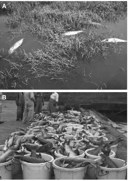
A
B
Figure 1. Mass mortality of common carp caused by CyHV-3 disease in Lake Biwa, Japan, in 2004. (A) The
death of wild common carp was observed throughout the lake. (B) More than 100,000 dead carp were collected from this lake in 2004 alone. It is estimated that 2–3 times as many carp died but were not collected from the lake. Reproduced with permission from Matsui et al. (2008).
Introduction
Common carp (Cyprinus carpio carpio) is a freshwater fish and one of the most important species in aquaculture, with a world production of 3.2 million metric tons per year (2009, www.fao.org). In addition to common carp, which is cultivated for human consumption, koi (Cyprinus carpio koi), an often-colourful subspecies, is grown for personal pleasure and competitive exhibitions. In the late 1990s, a highly contagious and virulent disease began to cause severe economic losses in these two carp industries worldwide (Figure 1)(Haenen, 2004; Matsui et al., 2008). The observed rapid worldwide spread of the disease has been attributed to the international fish trade and koi shows that occur around the world (Hedrick, 2000). The causative agent of the disease was initially called koi herpesvirus (KHV) according to its morphological resemblance to viruses belonging to the order Herpesvirales (Hedrick, 2000). Later, the virus was also known as carp interstitial nephritis and gill necrosis virus (CNGV), because of the associated lesions (Ronen et al., 2003). Recently, the virus was renamed cyprinid herpesvirus 3 (CyHV-3; species Cyprinid herpesvirus 3, genus Cyprinivirus, family Alloherpesviridae, order Herpesvirales) based on the homology of its genome with that of previously described cyprinid herpesviruses (Waltzek et al., 2005).
Because of its economical importance, once isolated, CyHV-3 rapidly became an important subject for applied research. However, recent studies have demonstrated that CyHV-3 is also an interesting fundamental research object. In the present review, we summarize recent advances made in CyHV-3 applied and fundamental research.
Characterization of CyHV-3
Viral classification
CyHV-3 is a member of the newly designated Alloherpesviridae family of the order Herpesvirales (Figure 2A) (Davison, 2002; Waltzek et al., 2009). The family Alloherpesviridae comprises viruses that infect fish and amphibians. The common ancestor of this family is thought to have diverged from the common ancestor of the Herpesviridae family (herpesviruses infecting reptiles, birds and mammals) some 450 million years ago (MYA) (Davison, 2002). The Alloherpesviridae family appears to be subdivided into two clades according to phylogenetic analysis of specific genes (the DNA polymerase and the terminase genes) (Figure 2B) (Waltzek et al., 2009). The first clade comprises anguillid and cyprinid herpesviruses that possess the largest genomes in the order Herpesvirales (245–295 kilobase pairs [kb]). The second clade comprises ictalurid, salmonid, acipenserid, and ranid herpesviruses with smaller DNA genomes (134–235 kb) (Hanson et al., 2011).
Structural characterization
The CyHV-3 structure is typical of the order Herpesvirales. An icosahedral capsid contains the genome, which consists of a single linear, double-stranded DNA (dsDNA) molecule. The capsid is
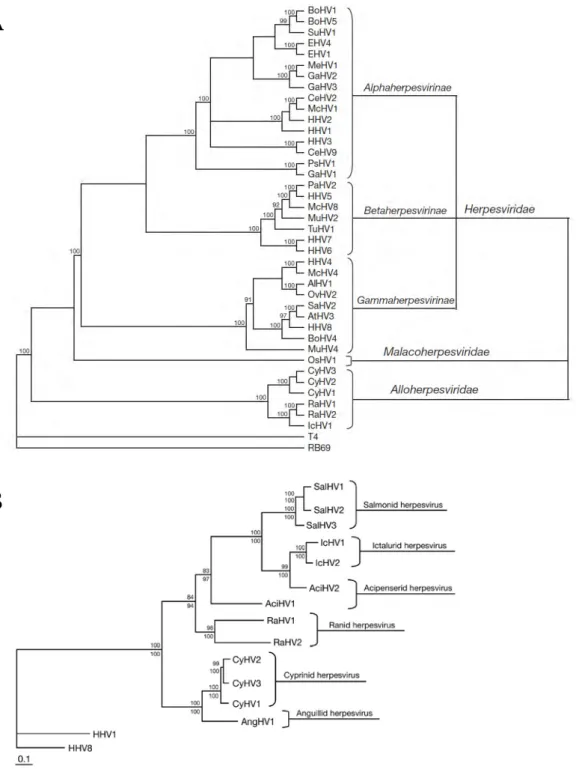
A
B
Figure 2. (A) Cladogram depicting relationships among viruses in the order Herpesvirales, based on the
conserved regions of the terminase gene. The Bayesian maximum likelihood tree was rooted using bacteriophages T4 and RB69. Numbers at each node represent the posterior probabilities (values > 90 shown) of the Bayesian analysis. (B) Phylogenetic tree depicting the evolution of fish and amphibian herpesviruses, based on sequences of the DNA polymerase and terminase genes. The maximum likelihood tree was rooted with two mammalian herpesviruses (HHV-1 and HHV-8). Maximum likelihood values (>80 are shown) and Bayesian values (>90 are shown) are indicated above and below each node, respectively. Branch lengths are based on the number of inferred substitutions, as indicated by the scale bar. AlHV-1: alcelaphine herpesvirus 1; AtHV-3: ateline herpesvirus 3; BoHV-1, -4, -5: bovine herpesvirus 1, 4, 5; CeHV-2, -9: cercopithecine herpesvirus 2, 9; CyHV-1, -2: cyprinid herpesvirus 1, 2; EHV-1, -4: equid herpesvirus 1, 4; GaHV-1, -2, -3: gallid herpesvirus 1, 2, 3; HHV-1, -2, -3, -4, -5, -6, -7, -8: human herpesvirus 1, 2, 3, 4, 5, 6, 7, 8; IcHV-1: ictalurid herpesvirus 1; McHV-1, -4, -8: macacine herpesvirus 1, 4, 8; MeHV-1: meleagrid herpesvirus 1; MuHV-2, -4: murid herpesvirus 2, 4; OsHV-1: ostreid herpesvirus 1; OvHV-2: ovine herpesvirus 2; PaHV-1: panine herpesvirus 1; PsHV-1: psittacid herpesvirus 1; RaHV-1, -2: ranid herpesvirus 1, 2; SaHV-2: saimiriine herpesvirus 2; SuHV-1: suid herpesvirus 1; TuHV-1: tupaiid herpesvirus 1. Reproduced with permission from Waltzek et al. (2009).
covered by a proteinaceous matrix called the tegument, which is surrounded by a lipid envelope derived from host cell trans-golgi membrane (Figure 3) (Mettenleiter et al., 2009; Miyazaki et al., 2008). The envelope contains viral glycoproteins (Hedrick, 2000). The entire CyHV-3 particle has a diameter of approximately 170–200 nm (Hedrick, 2000; Miyazaki et al., 2008; Neukirch & Kunz, 2001).
Molecular characterization
Viral genome. The genome of CyHV-3 is a 295 kb linear dsDNA molecule consisting of a large central portion flanked by two 22 kb repeat regions called the left repeat (LR) and the right repeat (RR) (Figure 4)(Aoki et al., 2007). The genome size is similar to that of CyHV-1 (Waltzek et al., 2005), but is larger than that of the other members of the order Herpesvirales, which generally range from 125 to 240 kb. The GC content of the genome is relatively high approaching 59.2% (Aoki et al., 2007).
The CyHV-3 genome encodes 156 potential protein-coding open reading frames (ORFs) including eight ORFs encoded by the repeat regions. These eight ORFs are consequently present as two copies in the genome (Aoki et al., 2007). Among them, 3 genes (ORF1, -3 and -6) are potential immediate early genes (Dr Aoki, personal communication) and one is coding for a tumor necrosis factor receptor (TNFR; ORF4) (Aoki et al., 2007). The terminal repeats are supposed to be involved in the capsid packaging of a single copy of the genome coming from the concatemer during the viral replication. Five families of related genes have been described in the CyHV-3 genome: the ORF2, TNFR, ORF22, ORF25, and RING families. The ORF25 family consists of six ORFs (ORF25, 26, -27, -65, -148, and -149) encoding related, potential membrane glycoproteins. The expression products of four of the sequences were detected in mature virions (ORF25, -65, -148, and -149) (Michel et al., 2010). CyHV-3 encodes several genes that could be involved in immune evasion processes, such as ORF16, which codes for a potential G-protein coupled receptor (GPCR); ORF134, which codes for an IL-10 homologue; and ORF12, which codes for a TNFR homologue.
Within the Alloherpesviridae family, anguillid herpesvirus 1 (AngHV-1) is the closest relative of CyHV-3 sequenced to date (Doszpoly et al., 2011; van Beurden et al., 2010). The two viruses possess 40 ORFs exhibiting similarity. It is likely that the sequencing of CyHV-1 and -2 should reveal even more CyHV-3 gene homologues. The putative products of most ORFs in the CyHV-3 genome lack obvious relatives in other organisms. Indeed, 110 ORFs fall into this class. Six ORFs encode proteins whose closest relatives are found in virus families such as the Poxviridae and Iridoviridae (Aoki et al., 2007; Waltzek et al., 2005). For example, CyHV-3 genes such as B22R (ORF139), thymidylate kinase ([TmpK] ORF140), thymidine kinase ([TK] ORF55), and the subunits of ribonucleotide reductase (ORF23 and -141) appear to have evolved from poxvirus genes (Aoki et al., 2007). Interestingly, neither TmpK nor B22R has been identified previously in a member of the order Herpesvirales.
Figure 3. Electron microscopy examination of CyHV-3 virion. Bar represents 100 nm. Adapted with
permission from Mettenleiter et al. (2009).
Figure 4. Genomic organisation of the CyHV-3. Potential ORF and their orientations are shown by coloured
arrows and numbered from 1 to 156. Introns are depicted as narrow bars. Left (LR) and right (RR) repeats are represented by grey rectangles. Red arrows represent conserved ORF among the Alloherpesviridae CyHV-3, IcHV-1, AngHV-1, RaHV-1 and -2. Reproduced from Aoki et al. (2007) and van Beurden et al. (2010).
Cell lines Cytopathic effect
Cyprinus carpio brain cells (CCB) Yes (Neukirch & Kunz, 2001,15)
Cyprinus carpio gill cells (CCG) Yes (Neukirch & Kunz, 2001)
Epithelioma papulosum cyprinid cells (EPC) No (Hedrick et al., 2000, Ronen et al., 2003), Davidovich et al., 2007, Oh et al.,
2001) / Yes (Neukirch & Kunz, 2001)
Koi fin cells (KFC, KF-1) Yes (Hedrick et al., 2000, Ronen et al., 2003), Davidovich et al., 2007, Pikarsky
et al., 2004)
Carp fin cells (CFC, CaF-2) Yes (Neukirch & Kunz, 2001)
Fathead minnow cells (FHM) No (Hedrick et al., 2000, Davidovich et al., 2007) / Yes (Oh et al., 2001)
Chinook salmon embryo cells (CHSE-214) No (Oh et al., 2001)
Rainbow trout gonad cells (RTG-2) No (Oh et al., 2001)
Glodfish fin cells (Au) Yes (Davidovich et al., 2007)
Channel catfish ovary cells (CCO) No (Davidovich et al., 2007)
Silver carp fin cells (Tol/FL) Yes (Davidovich et al., 2007)
Koi caudal fin cells (KCF-1) Yes (Dong et al., 2011)
Three strains of CyHV-3, isolated in Israel (CyHV-3 I), Japan (CyHV-3 J), and the USA (CyHV-3 U), have been fully sequenced (Aoki et al., 2007). Despite their distant geographical origin, these strains exhibit highly homologous sequences. A low diversity of sequences among strains seems to be a characteristic of the CyHV-3 species. Despite this low diversity, molecular markers allowing for discrimination among nine genotypes (seven European and two Asian) have been identified (Kurita et al., 2009). Analyses of variable number of tandem repeat (VNTR) proved to be useful to investigate CyHV-3 genetic diversity. These analyses revealed a relatively slow genetic evolution of CyHV-3 in vitro (Avarre et al., 2011). However, prolonged CyHV-3 cultivation in vitro leads to the spontaneous attenuation of the virus (Ronen et al., 2003).
Viral structural proteome. The structural proteome of CyHV-3 was characterized recently using liquid chromatography tandem mass spectrometry (Michel et al., 2010). Forty structural proteins, comprising three capsid, 13 envelope, two tegument, and 22 unclassified proteins, were described. The genome of CyHV-3 possesses 30 potential transmembrane protein-coding ORFs (Aoki et al., 2007). One cannot exclude that some low abundant envelope proteins have been overlooked during proteome analysis With the exception of ORF81, which encodes a type 3 membrane protein expressed on the 3 envelope (Michel et al., 2010; Rosenkranz et al., 2008), none of the CyHV-3 structural proteins have been studied to date. ORF81 is thought to be one of the most immunogenic (major) membrane proteins of CyHV-3 (Rosenkranz et al., 2008). A recent study of the structural proteome of AngHV-1 revealed that its viral particle contains a number of proteins comparable to CyHV-3 (van Beurden et al., 2011).
In vitro replication
CyHV-3 is widely cultivated in cell lines derived from koi fin (KFC), Cyprinus carpio brain (CCB), and Cyprinus carpio gill (Table 1) (Davidovich et al., 2007; Hedrick, 2000; Neukirch & Kunz, 2001; Oh, 2001; Pikarsky et al., 2004; Ronen et al., 2003). Other cell lines have been tested but few showed cytopathic effect following CyHV-3 infection (Table 1). For cell lines that did not support viral replication, it is not known whether these cell lines are nevertheless sensitive to the infection.
The CyHV-3 replication cycle was studied recently by electron microscopy (Mettenleiter et al., 2009). The morphological stages observed suggest that it replicates in a manner similar to that of the family Herpesviridae that utilizes an envelopment–deenvelopment mechanism to acquire the viral envelope (Mettenleiter et al., 2009; Miwa et al., 2007; Miyazaki et al., 2008). Following this theory, capsids leave the nucleus by a first budding event at the inner nuclear membrane resulting in the formation of primary enveloped virions in the perinuclear space. The primary envelope then fuses with the outer leaflet of the nuclear membrane thereby releasing nucleocapsids into the cytoplasm. Final envelopment occurs by budding into trans-golgi vesicles.
Figure 5. Effect of temperature on CyHV-3 replication. CCB cells were infected with CyHV-3. Following
infection, the cells were either kept at 22 °C (B) or shifted to 30 °C. At 24 hours post-infection (hpi) (C) or 48 hpi (D), the cells were returned to 22 °C. At 9 days post-infection (dpi), the cells were fixed, stained, and photographed. (A) Noninfected control. (E) Infected cells kept at 30 °C after infection. Magnification: 20x. Reproduced with permission from Dishon et al. (2007).