HAL Id: hal-01909474
https://hal.archives-ouvertes.fr/hal-01909474
Submitted on 25 May 2021
HAL is a multi-disciplinary open access
archive for the deposit and dissemination of
sci-entific research documents, whether they are
pub-lished or not. The documents may come from
teaching and research institutions in France or
abroad, or from public or private research centers.
L’archive ouverte pluridisciplinaire HAL, est
destinée au dépôt et à la diffusion de documents
scientifiques de niveau recherche, publiés ou non,
émanant des établissements d’enseignement et de
recherche français ou étrangers, des laboratoires
publics ou privés.
Distributed under a Creative Commons Attribution - NonCommercial| 4.0 International
License
A Case of Miliary Tuberculosis Presenting with Whitlow
of the Thumb
Romaric Larcher, Albert Sotto, Jean-Marc Mauboussin, Jean-Philippe
Lavigne, François-Xavier Blanc, Didier Laureillard
To cite this version:
Romaric Larcher, Albert Sotto, Jean-Marc Mauboussin, Jean-Philippe Lavigne, François-Xavier
Blanc, et al.. A Case of Miliary Tuberculosis Presenting with Whitlow of the Thumb. Acta
Dermato-Venereologica, Society for Publication of Acta Dermato-Dermato-Venereologica, 2016, 96 (4), pp.560 - 561.
�10.2340/00015555-2285�. �hal-01909474�
Acta Derm Venereol 96
Acta Derm Venereol 2016; 96: 560–561
© 2016 The Authors. doi: 10.2340/00015555-2285 Journal Compilation © 2016 Acta Dermato-Venereologica. ISSN 0001-5555
SHORT COMMUNICATION
Tuberculosis remains a major public health concern,
accounting for millions of cases and deaths worldwide.
In 2013, an estimated 9.0 million people developed
tuberculosis and 1.5 million died from the disease (1).
Among the 5.4 million new notified cases, 2.6 million had
bacteriologically confirmed pulmonary tuberculosis, 2.0
million had clinically diagnosed pulmonary tuberculosis,
and 0.8 million had extra-pulmonary tuberculosis (1).
Extra-pulmonary tuberculosis can affect any organ of the
body, and has a broad spectrum of manifestations (2).
Cutaneous tuberculosis is an uncommon manifestation of
tuberculosis, accounting for 0.5–2% of all
extra-pulmo-nary tuberculosis (3). Cutaneous tuberculosis is caused
mainly by infection with Mycobacterium tuberculosis
complex and occasionally by infection with M. bovis
or BCG vaccine. The clinical appearance of cutaneous
tuberculosis can vary, depending on the exogenous or
endogenous origin of the infection.
We report here an original case of disseminated
tuberculosis, revealed by a whitlow, and describe our
diagnostic approach, treatment and outcome. The
pa-tient provided written consent for publication of this
case report.
CASE REPORT
A 75-year-old woman with a medical history of autoimmune thyroiditis treated with L-thyroxin was admitted to the emer-gency department with pain, oedema, erythema of the left thumb, and fever for 3 days. Her general practitioner diagnosed a whitlow and started pristinamycin with no improvement. The patient reported back pain that had started 6 months previously, associated with a vertebral fracture of T11–T12, which had been treated with corticosteroids for 3 months.
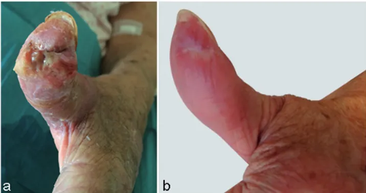
At admission, physical examination revealed an inflammatory swelling of the left thumb. The skin was tender from the base of the thumb to the thenar region. Extension of the left thumb was limited. Diagnosis of a sheath phlegmon affecting the flexor of the left thumb was established. Surgery with drainage, and co-amoxiclav treatment were performed immediately. The patient was then transferred to the infectious diseases department. Re-sults of intra-operative samples of pus and initial blood cultures were negative. Despite 10 days of intravenous co-amoxiclav, followed by 15 days of piperacillin-tazobactam, evolution of the skin wound was unfavourable (Fig. 1a).
Because of a mild dyspnoea with normal auscultation of the lungs and tenderness of the thoracic spine at T10–T11–T12 levels, additional investigations were performed. Arterial blood gas showed mild hypoxia (PO2 66 mmHg) associated with
respiratory alkalosis (pH 7.52 and bicarbonates 25 mmol/l) and significant hypocapnia (PCO2 27 mmHg). A chest X-ray
revealed a miliary. The white blood cell count was 5,700/mm3
and lymphocytes were at 720/mm3. C-reactive protein (CRP)
was 166 mg/l. Blood electrolytes and liver function tests were normal. Radiography of the spine was suggestive of a T10–T11 spondylodiscitis. Thoracic computed tomography (CT)-scan revealed uniformly distributed 1–3 mm pulmonary nodules, strongly suggesting infectious miliary. Magnetic resonance imaging confirmed the diagnosis of spondylodiscitis, showing STIR hypersignal with irregularity and erosion of the endplate of the T10–T11–T12 vertebral bodies, STIR hypersignal of discs, epidural and pre-vertebral collections, and local compres-sion of the spinal cord without T2 hypersignal.
The association of a pulmonary miliary and a spondylodiscitis led us to consider the diagnosis of disseminated tuberculosis. CT-scan-guided discovertebral biopsies of T12 were acid-fast bacilli (AFB) smear-positive. Antibiotic failure on the thumb wound led us to perform a M. tuberculosis complex PCR from the skin biopsy, which was positive. Cultures from discoverte-bral biopsies, skin biopsy and gastric aspiration proved positive for drug-sensitive M. tuberculosis. The HIV test was negative. After 2 weeks of standard anti-tuberculosis treatment (isoniazid, rifampicin, ethambutol and pyrazinamide), fever, pain and dyspnoea all improved. Evolution of the left thumb wound was also favourable. Clinical and biological tolerance of the treatment was good. According to drug susceptibility test results, ethambutol was stopped after 1.5 months of anti-tuberculosis treatment. Isoniazid, rifampicin and pyrazinamide were prescribed for a total of 3 months, while both isoniazid and rifampicin were continued for a further 9 months. Anti-tuberculosis therapy resulted in a favourable outcome with complete resolution of the skin lesion (Fig. 1b).
DISCUSSION
In the present case, the diagnosis of disseminated
tuber-culosis was established because of the association of a
pulmonary miliary, Pott’s disease, and a phlegmon of the
flexor sheath of the thumb. Tuberculosis explains the fact
A Case of Miliary Tuberculosis Presenting with Whitlow of the Thumb
Romaric Larcher1, Albert Sotto1*, Jean-Marc Mauboussin1, Jean-Philippe Lavigne2, François-Xavier Blanc3 and Didier Laureillard1
1Infectious Disease Department, 2Department of Microbiology, University Hospital Caremeau, Place du Professeur Robert Debré, FR-0029 Nîmes Cedex 09, and 3L’Institut du Thorax, Respiratory Medicine Department, University Hospital, Nantes, France. *E-mail: albert.sotto@chu-nimes.fr
Accepted Nov 10, 2015; Epub ahead of print Nov 11, 2015
Fig. 1. (a) Sheath phlegmon of the left thumb at admission to the infectious diseases department; and (b) evolution of the left thumb after 9 months of tuberculosis treatment.
561
Short communication
that the sheath phlegmon evolved unfavourably despite
rapid surgery and conventional antibiotic treatment.
Cutaneous tuberculosis can have various clinical
manifestations. Infection can be acquired through
exo-genous routes from direct inoculation of M.
tuberculo-sis into the skin. With this route of infection, clinical
manifestations are tuberculous chancre, tuberculosis
verrucosa cutis, and in some cases, lupus vulgaris.
These clinical manifestations are commonly observed
in healthcare workers and are difficult to diagnose
be-cause of their paucibacillary nature, leading to a high
frequency of negative cultures. Cutaneous tuberculosis
can also result from an endogenous infection, secondary
to a pre-existing primary focus. Tuberculosis cutis
orifi-cialis, scrofuloderma, and most cases of lupus vulgaris,
result from contiguous dissemination, acute miliary
tuberculosis and tuberculous abscess from
lymphohe-matogenous dissemination (3). Tuberculous abscesses,
or gumma, has been described in individuals with acute
miliary tuberculosis and may infrequently affect
immu-nocompetent adults (4). These are non-tender fluctuant
subcutaneous nodules. The nodules eventually penetrate
the skin, resulting in the development of ulcers and
draining sinuses (3, 5). Lesions may occur at any skin
site, but frequently develop on the extremities. Satellite
regional lymph nodes are usually not present (3, 5).
Tuberculous gumma have a poor prognostic value in
immunocompetent individuals and may persist for years
if untreated (3, 5). The confirmation of the diagnosis of
tuberculous gumma is based on identification of bacilli
by AFB stains, or culture or PCR that demonstrates the
presence of M. tuberculosis complex (6, 7), and requires
aspiration or skin biopsy (8).
In our case, PCR and culture from the skin biopsy
were positive and explained the unfavourable outcome
of the wound with standard antibiotic treatment.
How-ever, the diagnosis of disseminated tuberculosis was
based initially on the positive AFB-smear of gastric
aspiration and of vertebral biopsies of T12, later
con-firmed by PCR and culture. In general, manifestations
of miliary pulmonary can be acute, but are more likely
to be sub-acute or chronic (9). Patients often report
dyspnoea or cough, and hypoxemia is common. In
the present case, the patient had a febrile dyspnoea
with mild hypoxemia related to a sub-acute or chronic
miliary tuberculosis. Pathogenesis is likely to be a
lympho-haematogenous dissemination of M.
tuber-culosis from a pulmonary focus with embolization to
vertebral bones then to the left thumb joint and the skin.
This dissemination was favoured by the corticosteroids
given to treat the back pain.
Treatment of cutaneous tuberculosis is the same as for
systemic tuberculosis (6, 7). The therapeutic response
of cutaneous lesions can be assessed clinically. Surgical
treatment is not usual for the management of cutaneous
tuberculosis, but is sometimes required to manage
extensive or recalcitrant tuberculous skin lesions (6,
7). The treatment duration is usually 6 months. In our
case, the treatment duration was dependent on the bone
involvement. We opted for a 12-month regimen (10).
In conclusion, disseminated tuberculosis can be
revealed by an atypical cutaneous lesion mimicking
a whitlow and can be observed in non-HIV-infected
patients. This diagnosis should be considered when the
outcome is unfavourable despite surgery and antibiotic
treatment, notably in the context of
immunosuppres-sion.
ACKNOWLEDGEMENT
The authors would like to thank Emma Rubenstein for her as-sistance with the manuscript preparation.
The authors declare no conflicts of interest.
REFERENCES
1. Global tuberculosis report 2014. [Cited 2015 Feb 25]. Available from: http://apps.who.int/iris/bitstre am/10665/137094/1/9789241564809_eng.pdf?ua=1. 2. Zumla A, Raviglione M, Hafner R, Fordham von Reyn C.
Tuberculosis. N Engl J Med 2013; 368: 745–755. 3. Santos JB, Figueiredo AR, Ferraz CE, Oliveira MH, da
Silva PG, Medeiros VLS. Cutaneous tuberculosis: epide-miologic, etiopathogenic and clinical aspects – part I. An Bras Dermatol 2014; 89: 219–228.
4. Almagro M, Del Pozo J, Rodríguez-Lozano J, Silva JG, Yebra-Pimentel MT, Fonseca E. Metastatic tuberculous abscesses in an immunocompetent patient. Clin Exp Der-matol 2005; 30: 247–249.
5. MacGregor RR. Cutaneous tuberculosis. Clin Dermatol 1995; 13: 245–255.
6. Dias MFRG, Bernardes Filho F, Quaresma MV, do Nasci-mento LV, Nery JA da C, Azulay DR. Update on cutaneous tuberculosis. An Bras Dermatol 2014; 89: 925–938. 7. Santos JB, Figueiredo AR, Ferraz CE, Oliveira MH, da
Silva PG, Medeiros VLS. Cutaneous tuberculosis: diag-nosis, histopathology and treatment – part II. An Bras Dermatol 2014; 89: 545–555.
8. Sharma SK, Mohan A. Extrapulmonary tuberculosis. Indian J Med Res 2004; 120: 316–353.
9. Sharma SK, Mohan A, Sharma A, Mitra DK. Miliary tu-berculosis: new insights into an old disease. Lancet Infect Dis 2005; 5: 415–430.
10. Ramachandran S, Clifton IJ, Collyns TA, Watson JP, Pear-son SB. The treatment of spinal tuberculosis: a retrospective study. Int J Tuberc Lung Dis Off J Int Union Tuberc Lung Dis 2005; 9: 541–544.