Journal of Small Animal Practice • © 2018 British Small Animal Veterinary Association 1
Journal of Small Animal Practice (2018) DOI: 10.1111/jsap.12933
Accepted: 8 August 2018
Comparison of two minimally invasive
enilconazole perendoscopic infusion
protocols for the treatment of canine
sinonasal aspergillosis
E. Vangrinsven1,* , M. Girod*, D. Goossens*, L. Desquilbet†, C. Clercx* and F. Billen* *Department of Clinical Sciences, Faculty of Veterinary Medicine, University of Liège, 4000 Liège, Belgium
†Clinical Epidemiology and Biostatistics Unit, National Veterinary School of Alfort, University of Paris-Est, 94704 Paris, France 1Corresponding author email: emiliepm@hotmail.com
Objectives: To compare two minimally invasive enilconazole infusion protocols for the treatment of
canine sinonasal aspergillosis and evaluate the importance of complete endoscopic debridement in determining first treatment success rate.
Materialsand MethOds: Data for 48 dogs with confirmed sinonasal aspergillosis treated with endoscopic
debridement followed by per-endoscopic enilconazole infusion were collected. Twenty-four dogs were treated according to the previously published 1-hour infusion protocol and 24 dogs underwent a simpli-fied 15-minute infusion protocol. Completeness of debridement, evaluated as partial or complete at the end of the procedure and outcome after one or several treatments were assessed in all dogs. Multi-variable analysis was performed to derive odds ratios and 95% confidence intervals.
results: The median duration of the simplified protocol – 92∙3 minutes (range 40 to 140) – was
sub-stantially shorter than the duration of the previous protocol – 201∙3 minutes (range 120 to 265). First treatment success rates were 58 and 62∙5% for the previous and simplified protocol, respectively. Overall treatment success rate was similar in both groups (96%). Complete debridement was associ-ated with an improved first treatment success rate compared to partial debridement.
clinical significance: The simplified protocol is a valid alternative approach to the treatment of sinonasal
aspergillosis. Completeness of endoscopic debridement before infusion is an important step for the success of treatment in canine sinonasal aspergillosis.
M. Girod’s current address is Internal Medicine Department, Centre Hospitalier Vétérinaire Frégis, Arcueil, France
Paper presented as an oral abstract at the 25th ECVIM congress in Lisbon.
INTRODUCTION
In dogs, mycotic rhinosinusitis accounts for 7 to 11% of chronic nasal diseases (Meler et al. 2008, Tasker et al. 1999, Lobetti 2009) and is most often caused by Aspergillus fumigatus (Peeters & Clercx 2007). Treatment of canine sinonasal aspergillosis (SNA) remains a challenge and no gold standard procedure has been
established. Oral treatments with antifungal drugs (azole deriva-tives) have generally poor to moderate efficacy, require prolonged administration and are commonly associated with adverse effects (Peeters & Clercx 2007). However, improved clinical remission rates were recently reported for combination treatment with posaconazole and terbinafine (Stewart & Bianco 2017). Treat-ment with enilconazole or clotrimazole infusion administered topically through catheters placed surgically, blindly or endo-scopically have been associated with higher success rates (Peeters & Clercx 2007, Billen & Peeters 2017).
http://www
.bsa
v
Minimally invasive protocols involve a thorough endoscopic debridement of the affected areas, followed by a 1-hour infusion of 1% clotrimazole or 1 to 2% enilconazole solutions through catheters placed either blindly into each nasal cavity (Mathews
et al. 1998, Zonderland et al. 2002, Saunders et al. 2003, Schuller
& Clercx 2007, Greci et al. 2008, Pomrantz & Johnson 2010, Sharman et al. 2010) or endoscopically into the affected frontal sinuses and/or nasal cavities (Mathews et al. 1996, McCullough
et al. 1998, Saunders et al. 2003, Schuller & Clercx 2007). In
some cases, further addition of 1% bifonazole cream instilled into the infusion catheter of the affected nasal cavity has been performed (Billen et al. 2010).
Treatment outcomes are difficult to compare due to the lack of a standardised definition of successful treatment (Billen & Peeters 2017). In studies in which dogs were reassessed endoscopically, minimally invasive protocols showed positive outcomes in 46∙6 to 85∙7% of cases after the first treatment and 89∙5 to 100% after multiple treatments (Zonderland et al. 2002, Saunders et al. 2003, Schuller & Clercx 2007, Greci et al. 2008, Billen et al. 2010).
Endoscopic therapeutic procedures are minimally invasive but are also laborious, time-consuming, require prolonged general anaesthesia and dogs are frequently hospitalised overnight fol-lowing the procedure. Complications associated with the per-endoscopic infusion of antifungal medication mainly include leakage of drug around infusion catheters with few negative con-sequences (Zonderland et al. 2002) and, on rare occasions, bleed-ing after withdrawal of the catheters (Zonderland et al. 2002, Mathews & Sharp 2006) or neurological signs due to leakage of antifungal solution into the cerebral cavity secondary to cribri-form plate destruction (Peeters & Clercx 2007).
For protocols using surgically placed catheters, antifungal agents are instilled through a trephination site in the affected frontal sinuses (Mathews et al. 1998, Friend et al. 2002, Sissener
et al. 2006, Pomrantz & Johnson 2010, Hazuchova et al. 2017).
Such procedures may be necessary when the frontal sinus cannot be reached endoscopically. Frontal sinus trephination is a quicker but more invasive technique and does not improve treatment outcome compared to endoscopic procedures alone (Hazuchova
et al. 2017).
Although previous minimally invasive protocols offer satisfac-tory results, one of their most important drawbacks is the dura-tion of the procedure, mainly related to the length of soak. As the soak time had been arbitrarily selected we first considered that a shorter soak period would be as efficacious. Moreover, incom-plete debridement before application of topical treatment has been suggested as one of the reasons for treatment failure (Claeys
et al. 2006, Billen et al. 2010, Sharman et al. 2010, Sharman et al. 2012, Sharman & Mansfield 2012). However, the impact
of completeness of endoscopic debridement on outcome has not been previously reported.
Therefore, the first aim of this study was to investigate the effectiveness of a shorter infusion protocol (SP=simplified pro-tocol) compared with the previously described 1-hour infusion protocol (PP=previous 1-hour protocol) (Billen et al. 2010). The second objective was to assess the usefulness of complete
debride-ment as part of the treatdebride-ment (the benefit of complete endoscopic debridement) on first treatment success rate.
MATERIALS AND METHODS
Animals
Dogs presented to the institution between October 2006 and February 2015 with a definitive diagnosis of SNA and treated with a minimally invasive enilconazole infusion protocol were included in this study. In December 2011 it was decided to adopt a simplified form of the previously described infusion protocol. Consequently, from then on, all dogs with a first diagnosis of SNA were treated with the SP. Dogs treated before December 2011 with the PP were used retrospectively as a control group. Dogs were included in the study if they had a confirmed diag-nosis of SNA based on the presence of compatible clinical signs and per-endoscopic identification of fungal plaques with turbi-nate destruction. Additional diagnostic procedures consisted of CT, histopathology, microscopic examination and fungal cul-ture. Age, gender, breed, bodyweight and duration of clinical signs were recorded for each case. Presence or absence of systemic clinical signs was also noted when the information was available.
Anaesthesia
Dogs were pre-oxygenated and then premedicated with medeto-midine (Sedator; Eurovet) intravenously (iv) alone, or a combina-tion of medetomidine iv and butorphanol (Butomidor; Ecuphar) iv, or a combination of midazolam (Midazolam Mylan; Mylan) iv, methadone (Comfortan; Dechra) iv. and propofol (Diprivan; AstraZeneca) iv was used for induction, after which they were intubated and anaesthetic monitoring was implemented. Anaes-thesia was maintained with isoflurane (Iso-Vet; Eurovet) (in oxygen). If necessary, atipamezole (Atipazole; Prodivet Pharma-ceuticals) intramuscularly was used to reverse medetomidine at the end of the protocol.
Rhinoscopy
Under general anaesthesia, dogs were placed in sternal recum-bency and rhinosinuscopy was performed using a paediatric bronchoscope (Paediatric bronchoscope Fujinon EB-4105; Onys SA) and/or a rigid endoscope (Cystoscope K Storz SL 30°, Ref BA 3059308; Karl-Storz-Endoscopy) appropriately to access to the nasal cavities and frontal sinuses depending on the size of the patient and the area to be scoped.
Debridement
In all dogs, debridement of the sinonasal cavities was performed using per-endoscopic forceps. Aspiration was performed using a suction device and a sterile saline solution was used for lavage. This procedure was performed by the same experienced operators throughout the study (FB and CC) and completeness of debride-ment was systematically docudebride-mented in the medical records as complete or partial. Debridement was considered complete if no macroscopic fungal plaques were visible at the end of the proce-dure. Debridement was defined as partial when small amounts of
fungal material could not be removed due to limited endoscopic access to the sinus, limited visibility due to excessive bleeding or when removal of remaining fungal plaques was considered haz-ardous for the endoscope, due to the presence of sharp splinters of turbinates.
Original (previous) protocol (before December 2011)
After debridement, the infusion procedure was performed as pre-viously described by Billen et al. (2010). During this procedure the dog was placed in dorsal recumbency. A 24-Fr Foley catheter (Balloon Catheter; Teleflex Medical) was first inserted through the mouth into the nasopharynx with right-angle forceps and the 30 to 50 mL balloon was inflated, with gauze sponges placed behind the Foley catheter in the pharynx. A 12-Fr fenestrated infusion catheter (Suction Catheter; Servoprax GmbH) was then placed in both sinonasal cavities. If the frontal sinus was involved, the infusion catheter was placed endoscopically in the caudal part of the affected frontal sinus. If the fungal infection was restricted to one nasal cavity or both nasal cavities, the infusion catheter was placed blindly dorso-medially up to the level of the medial canthus of the eye. This was also the case in the unaffected con-tralateral nasal cavity in case of unilateral involvement. Both nos-trils were then occluded using separate 12-Fr Foley catheters. The infusion catheters were filled with the antifungal solution and connected using a T-shape connecting piece to a 60-mL infusion syringe. All Foley catheters were closed with clamps. Approxi-mately 120 mL (60 mL in each nasal cavity) of a 2% enilconazole (Imaverol; Janssen-Cilag SA) solution was slowly infused manu-ally in order to soak both sinonasal cavities for 60 minutes.
To maximise drug contact with all sinonasal surfaces, the dog was rotated every 15 minutes by 90°. At the end of the procedure the head was tilted downwards at an angle of 30° and infusion solution was allowed to drain through the nostrils for 10 min-utes after the nasal Foley catheters were removed. Finally, a dose of 15 g 1% bifonazole cream (Canestene; Bayer) was adminis-tered through the infusion catheter into the frontal sinus of the affected side(s) with the nose in upright position (head posi-tioned at an angle of 90°) for at least 10 minutes. The infusion catheters, the nasopharyngeal Foley catheter and gauze sponges were then removed.
Simplified protocol (since December 2011)
After debridement, placement of the infusion catheters was simi-lar to the PP but the dog was placed in dorsal recumbency with the head positioned at an angle of 90° (Fig. 1); Foley catheters were not needed for nostril occlusion. A dose of approximately 120 mL (60 mL in each nasal cavity) of 2% enilconazole solution was slowly infused manually, until the solution was seen at the nostrils, in order to soak both sinonasal cavities for 15 minutes. Bifonazole cream was not administered.
At the end of the procedure, antibiotic therapy was started in all dogs with a dose of 20 mg/kg cefazoline (Cefazoline Sandoz; Sandoz) iv followed by oral treatment dose of 20 mg/kg cefal-exine (Rilcefal-exine; Virbac) orally every 12 hours for 5 days. All dogs received oral antifungal treatment with a dose of 5 mg/kg itraconazole (Sporanox; Janssen-Cilag SA) orally every 12 hours
FIG 1. Position of the dog during the simplified protocol. The dog is placed in dorsal recumbency with the head positioned at 90°. A Foley catheter was introduced into the nasopharynx (black arrowhead). Nasal infusion catheters (red arrowheads) were filled with 2% enilconazole solution until the solution appears at the edge of the nostrils (red arrow)
until re-evaluation of treatment success. The duration of anaes-thesia was recorded in each case.
Outcome
Dogs were re-examined and rhinoscopy was repeated 3 to 5 weeks after treatment. If there was a resolution of clinical signs and absence of fungal plaques at follow-up rhinoscopy, treatment was deemed successful and no more treatment was administered. If fungal plaques were detected, treatment was considered to have failed. In that case, debridement and a second identical infusion protocol were performed and a new follow-up rhinoscopy was scheduled 3 to 5 weeks later. In dogs in which endoscopic follow-up was not performed 3 to 5 weeks after treatment, treatment success was based on the presence/absence of compatible clinical signs (established by telephone consultation).
Statistical analysis
Analyses were performed using logistic regression models where the outcome was first treatment failure. Crude odds ratios (OR) were calculated to quantify the associations between the outcome and both the type of protocol (PP/SP) and complete debride-ment (yes/no). Separate bivariate logistic regression models were performed in order to remove potential confounding factors including the exposure of interest (protocol type or debridement) and one exposure among the following: age, sex, bodyweight, duration of clinical signs and presence of systemic signs. Time of anaesthesia between the two groups were compared using a Mann-Whitney U test. For all analysis, a threshold of P<0∙05 was considered statistically significant. Data were analysed using
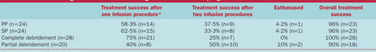
Table 2. Short- and long-term outcome of 48 dogs with sinonasal aspergillosis
Treatment success after one infusion procedure*
Treatment success after two infusion procedures
Euthanased Overall treatment success
PP (n = 24) 58∙3% (n=14) 37∙5% (n=9) 4∙2% (n=1) 96% (n=23) SP (n=24) 62∙5% (n=15) 33∙3% (n=8) 4∙2% (n=1) 96% (n=23) Complete debridement (n=28) 75% (n=21) 25% (n=7) 0% 100% (n=28) Partial debridement (n=20) 40% (n=8) 50% (n=10) 10% (n=2) 90% (n=18)
PP previous protocol, SP simplified protocol
*Short-term assessment by rhinoscopy or long-term assessment through telephone consultation in six dogs (40 to 127 months after infusion protocol)
RESULTS
Animals
Forty-eight dogs were included. During the study period, 35 additional dogs with a presumptive or definitive diagnosis of canine SNA were presented to the institution. These cases did not meet the inclusion criteria (unconfirmed diagnosis, surgical rhinotomy or infusion refused by the owners for financial con-cerns) and were not included.
The median age of the dogs at initial presentation was 5∙5 years (range 9 months to 12 years), with 33 males (11 neutered) and 15 (six neutered) females. The most frequently represented breeds included the rottweiler (n=9), golden retriever (n=6), beauceron (n=3), Labrador retriever (n=3) and Border collie (n=3). The median bodyweight was 29∙2 kg (range 13 to 55 kg). Reported duration of clinical signs before treatment ranged from 1 to 40 weeks (median 9∙5 weeks). Distributions of age, gender, body-weight and duration of clinical signs were similar between the two groups (Table 1). At the time of diagnosis, 50% (23/46) of dogs presented systemic clinical signs including lethargy, reduced appetite, weight loss and/or mild hyperthermia. This informa-tion was lacking in two dogs (one in each group). Local clini-cal features included sneezing (n=25) or reverse sneezing (n=4), uni- or bilateral nasal discharge (n=42), uni- or bilateral epistaxis (n=9), hyperkeratosis (n=9), signs of nasal pain (n=14), nasal pla-num depigmentation (n=12) and nasal plapla-num erosion (n=7).
Rhinoscopy and CT
CT was performed in 31% (n=15) of cases, with no evidence of perforation of the cribriform plate in any case. Nasal turbinate destruction and fungal plaques were endoscopically observed in all dogs. Unilateral SNA was diagnosed in 35 cases (73%), bilat-eral SNA in five (10%), unilatbilat-eral nasal aspergillosis (i.e. fungal
Table 1. Signalment, presence of systemic signs and duration of clinical signs in dogs treated with the PP and the SP
PP (n=24) SP (n=24)
Age (years) 5∙5 (1∙5 to 12) 5∙5 (0∙8 to 11) Bodyweight (kg) 29∙5 (14∙5 to 55) 29∙2 (16∙8 to 52) Gender Nine females and
15 males
Six females and 18 males Presence of systemic signs 10 13 Duration of clinical signs
before treatment (weeks)
11 (3 to 40)* 9 (1 to 24)*
PP previous protocol, SP simplified protocol *Median (ranges) or absolute values
infection restricted to the nasal cavity) in three (6%) and bilateral nasal aspergillosis in four (8%) of dogs. In one case, the infection was sinonasal on one side with an extension to the nasal cavity on the contralateral side through destruction of the nasal septum.
Treatment and outcome
Twenty-four dogs were treated with the SP and 24 dogs with the PP. The sinus was accessible and the infusion catheter(s) could be placed endoscopically in all but two dogs (n=39; 81%) with SNA, both belonging to the SP group. Of the 48 dogs, 42 dogs were re-examined – including a recheck rhinoscopy 3 to 5 weeks after treatment. Two dogs, one in each group, were euthana-sed after failure of the first treatment and the owner’s decision not to pursue further treatment (Table 2). For six dogs (three
per group), recheck rhinoscopy was declined by the owner for
financial or personal reasons although in all six cases there was a reported resolution of clinical signs. For the six dogs in which recheck rhinoscopy could not be performed at 3 to 5 weeks, long-term follow-up through telephone consultation, between 40 and 127 months after their infusion protocol, demonstrated that they did not show recurrence of clinical signs of nasal disease. In these dogs, the infusion protocol was considered successful after one treatment. Systemic antifungal treatment was stopped in all cases either at time of recheck endoscopy, considered to be successful treatment, or 3 to 5 weeks after the infusion protocol in the six dogs in which recheck endoscopy could not be performed.
Median duration of anaesthesia was 200 minutes (range 120 to 265) for the PP and 85 minutes (range 40 to 140) for the SP (P<0∙0001). With the SP, recovery from anaesthesia was shorter and all dogs were ready for discharge on the same day. The SP was well tolerated in all dogs and did not cause any significant adverse effects.
Overall first treatment success rate was 60% (n=29), and over-all treatment success rate was 96% (n=46). No dog received more than two infusion protocols. The first treatment success rate in the PP group was 58% (n=14) compared to 62∙5% (n=15) for the SP group (Table 2). The overall treatment success rate (after one/two infusion protocols) was 96% (n=23) in both groups. The first treatment failure rate was not significantly different between treatment groups (SP: 37∙5% versus PP: 42%, OR 0∙84, CI 0∙26 to 2∙68, P=0∙77). In separated bivariate models, after adjustment for each of the potential confounders (age, body-weight, gender, presence of systemic signs or duration of clinical signs), the treatment remained not significantly associated with the outcome, with adjusted OR comparing SP dogs versus PP dogs always lower than 1.
The effect of completeness of endoscopic debridement on first treatment success rate was evaluated for all 48 dogs. Endoscopic debridement was complete in 60% (n=28) of dogs. First treat-ment failure rate was lower in dogs with complete debridetreat-ment (25%) compared to dogs with partial debridement (60%) (OR 0∙24, CI 0∙07 to 0∙85, P=0∙03). In separated bivariate models, adjustment for each of the dog exposures (age, bodyweight, gen-der, presence of systemic signs or duration of clinical signs) still resulted in a significant effect of completeness of debridement.
DISCUSSION
Despite a reduced contact time and removal of the use of bifon-azole cream for the SP, the results revealed a similar treatment success rate – 96% – and similar first treatment success rates – ~60% – for the two techniques. These results are similar to previ-ously reported first treatment success rates of 46∙6 to 76∙6% for SNA (Schuller & Clercx 2007, Greci et al. 2008).
The primary objective of this study was to determine whether a shorter procedure could be performed without compromis-ing treatment efficacy. At this institution a minimally invasive approach has been preferred over surgical trephination for some years due to reduced trauma to patient and because it has been associated with fewer complications (subcutaneous emphy-sema, infection) (Mathews et al. 1998, McCullough et al. 1998, Zonderland et al. 2002, Peeters & Clercx 2007, Sharman & Mansfield 2012). A recent study (Hazuchova et al. 2017) showed that trephination did not influence the number of treatments necessary for resolution of the disease among dogs with success-ful outcomes.
The major disadvantages associated with a minimally invasive approach have been prolonged anaesthesia leading, potentially, to increased anaesthetic risk, prolonged recovery and patient cool-ing. Duration of anaesthesia was substantially shorter (mean dif-ference of 109 minutes) in the SP compared to that in the PP. This time reduction was mainly achieved by simplifying the set-up of the procedure and by reducing the drug contact time from 1 hour to 15 minutes. Preparation of the patient for the antifungal infusion was easier and faster in the SP compared to that in the PP. Foley catheters for nostril occlusion were not needed in the SP and nasal Foley catheter adjustment was frequently required during the PP infusion due to leakage of the antifungal solution through the nostrils. Additionally, in the SP the dog remained in dorsal recumbency throughout the infusion and bifonazole cream was not administered. It has been reported previously that the addition of bifonazole cream did not seem to improve first treatment success rate (Billen et al. 2010).
For the SP, the dog was maintained in dorsal recumbency with the head angled upwards at 90°. Mathews et al. (1996) per-formed a study in which dogs were similarly positioned in dorsal recumbency and the head rotated successively to the right and left side for 15 minutes per side. Postinfusion CT images con-firmed infusion solution throughout all areas of both frontal sinuses and nasal cavities. Although there are differences with the positioning used in this study (absence of rotation of the head)
we expect to have reached a similar drug distribution with this approach. Ideal contact times for successful therapy have not yet been established (Sharman & Mansfield 2012), and we decided to maintain the 15 minutes contact period with each mucosal surface described in the previous protocol. Since enilconazole was infused until it appeared at the nostrils, we were confident that the solution completely filled all areas of the sinonasal cavities ensuring contact of the drug with all mucosal surfaces during the 15 minutes of infusion.
The second objective of our study was to address the impor-tance of completeness of the debridement. In people, non-inva-sive forms of fungal rhinosinusitis can be managed with surgical debridement as a sole treatment (Uri et al. 2003). In dogs, exten-sive rhinoscopic debridement before infusion is considered an important element determining therapeutic success. How-ever, to our knowledge, it has not previously been investigated (Zonderland et al. 2002, Sharman et al. 2010, Sharman et al. 2012). In the present study, we showed a significant improve-ment in first treatimprove-ment success rate when complete debrideimprove-ment was achieved. The results of this study strongly support complete debridement being a critical point for therapeutic success. How-ever, since this was an observational study, residual confounding bias or reverse causality cannot be ruled out and causal inference must be made with caution – for instance, less severe cases may be easier to debride.
In this study, when debridement was reported as being com-plete, the clinician was confident that no residual fungal or necrotic material had been missed as the entirety of the nasal cav-ity (uni- or bilaterally) and sinus(es) could be explored. However, in a study by Sharman et al. (2010), CT demonstrated significant residual disease even if debridement was considered complete on visual assessment by rhinoscopy, which makes the degree of accu-racy of the latter questionable. Completeness of debridement was not determined by CT in this study, consequently, in dogs with complete debridement first treatment failure could potentially have been due to residual disease not detected during rhinoscopic evaluation. CT could have been an interesting tool to assign dif-ferent levels for partial debridement and evaluate the potential association between outcome and debridement level.
Association between younger age and treatment success was the only significant factor described to influence outcome in a prospective multi-centre study (Sharman et al. 2010). In our study the important factors we identified remained significant after adjustment for potential confounding factors including age.
Finally, a substantial variability and asymmetry in frontal sinus and nasal cavity anatomy between and within individual dogs has been demonstrated (Burrow et al. 2011) which may influence distribution and retention of antifungal agents and, therefore, outcome (Sharman & Mansfield 2012). This could be another explanation for treatment failure in dogs in which debridement was considered to be complete in our study. The infusion cath-eter localisation (blindly in the nasal cavity or endoscopically in the affected frontal sinus) is unlikely to have affected the results because the frontal sinus was accessible and the catheter was placed endoscopically within the sinus in almost all dogs (except two in the SP group) with unilateral or bilateral SNA. A prospective
cohort study involving more dogs and designed to examine all potential confounding factors is warranted to confirm our pre-liminary results.
Antibiotic therapy was prescribed in all dogs following treat-ment. This may be considered controversial, because some reports describing treatment of SNA advise against the use of antibiotics as part of good antimicrobial stewardship. However, the extensive local necrosis, lysis and bleeding might represent an increased risk for secondary bacterial infection. A study by De Lorenzi et al. (2006) found neutrophilic inflammation, free bacteria and phagocytosed bacteria in 100% of nasal cytology preparations from dogs with SNA, suggesting secondary bacterial infection. Furthermore, as antimicrobial treatment was part of the previously described protocol (Billen et al. 2010), we decided to maintain the same treatment after infusion for the SP group to facilitate comparison of these two protocols. The prescription of oral antifungal medication after infusion protocol in all dogs was based on the assumption that it could contribute to better control of the disease (Claeys et al. 2006).
It has been shown that Aspergillus plaques still can be found in nasal cavities/sinuses at follow-up rhinoscopy, despite the absence of overt clinical signs (Zonderland et al. 2002). A limitation of the present study was the absence of follow-up rhinoscopy in a small number of dogs. In six cases the owners declined a recheck endoscopy. However, the absence of clinical signs of nasal disease for 40 to 127 months was strongly suggestive of treatment success in these animals.
In conclusion, the SP seems a promising alternative approach for the treatment of canine SNA. Complete debridement of the fungal plaques before infusion of antifungal medication appears critical for therapeutic success in canine SNA.
Acknowledgements
This study not supported by a grant or otherwise.
Conflict of interest
No conflicts of interest.
References
Billen, F. & Peeters, D. (2017) Aspergillosis – canine. In: Textbook of Veterinary Internal Medicine. 8th edn. Eds S. J. Ettinger, E. C. Feldman and E. Côté. Else-vier, St Louis, MO, USA. pp 1035-1039
Billen, F., Guieu, L.-V., Bernaerts, F., et al. (2010) Efficacy of intrasinusal adminis-tration of bifonazole cream alone or in combination with enilconazole irrigation in canine sino-nasal aspergillosis: 17 cases. Canadian Veterinary Journal 51, 164-168
Burrow, R., McCarroll, D., Baker, M., et al. (2011) Frontal sinus depth at four land-marks in breeds of dog typically affected by sinonasal aspergillosis. Veterinary
Record 170, 20
Claeys, S., Lefebvre, J., Schuller, S., et al. (2006) Surgical treatment of canine nasal aspergillosis by rhinotomy combined with enilconazole infusion and oral itraconazole. Journal of Small Animal Practice 47, 320-324
De Lorenzi, D., Bonfanti, U., Masserdotti, C., et al. (2006) Diagnosis of canine nasal aspergillosis by cytological examination: a comparison of four different collection techniques. Journal of Small Animal Practice 47, 316-319
Friend, E., White, R. & Williams, J. (2002) Invasive treatment of canine nasal aspergillosis with topical clotrimazole. Veterinary Record 151, 298-299 Greci, V., Mortellaro, C. M., Romussi, S., et al. (2008) Retrospective study on one
hour intranasal infusion of 1% clotrimazole solution for treatment of sinonasal aspergillosis in dogs: 47 cases (February 1998–October 2007). Proceedings of the British Small Animal Veterinary Association, Birmingham, UK. April 3 to 6. Hazuchova, K., Neiger, R. & Stengel, C. (2017) Topical treatment of mycotic
rhi-nitis-rhinosinusitis in dogs with meticulous debridement and 1% clotrimazole cream: 64 cases (2007-2014). Journal of the American Veterinary Medical
Asso-ciation 250, 309-315
Lobetti, R. (2009) A retrospective study of chronic nasal disease in 75 dogs.
Jour-nal of the South African Veterinary Association 80, 224-228
Mathews, K. G. & Sharp, N. J. (2006) Canine nasal aspergillosis-penicilliosis. In: Infectious Diseases of the Dog and Cat. 3rd edn. Ed C. E. Greene. Elsevier, St Louis, MO, USA. pp 613-620
Mathews, K., Koblik, P., Richardson, E., et al. (1996) Computed tomographic assessment of noninvasive intranasal infusions in dogs with fungal rhinitis.
Veterinary Surgery 25, 309-319
Mathews, K. G., Davidson, A. P., Koblik, P. D., et al. (1998) Comparison of topical administration of clotrimazole through surgically placed versus nonsurgically placed catheters for treatment of nasal aspergillosis in dogs: 60 cases (1990-1996). Journal of the American Veterinary Medical Association 213, 501-506 McCullough, S. M., McKiernan, B. C. & Grodsky, B. S. (1998) Endoscopically
placed tubes for administration of enilconazole for treatment of nasal aspergil-losis in dogs. Journal of the American Veterinary Medical Association 212, 67-69 Meler, E., Dunn, M. & Lecuyer, M. (2008) A retrospective study of canine persistent
nasal disease: 80 cases (1998-2003). Canadian Veterinary Journal 49, 71-76 Peeters, D. & Clercx, C. (2007) Update on canine sinonasal aspergillosis.
Veteri-nary Clinics of North America: Small Animal Practice 37, 901-916
Pomrantz, J. S. & Johnson, L. R. (2010) Repeated rhinoscopic and serologic assessment of the effectiveness of intranasally administered clotrimazole for the treatment of nasal aspergillosis in dogs. Journal of the American Veterinary
Medical Association 236, 757-762
Saunders, J. H., Duchateau, L., Stork, C., et al. (2003) Use of computed tomog-raphy to predict the outcome of a noninvasive intranasal infusion in dogs with nasal aspergillosis. Canadian Veterinary Journal 44, 305-311
Schuller, S. & Clercx, C. (2007) Long-term outcomes in dogs with sinonasal asper-gillosis treated with intranasal infusions of enilconazole. Journal of the
Ameri-can Veterinary Medical Association 43, 33-38
Sharman, M. J. & Mansfield, C. S. (2012) Sinonasal aspergillosis in dogs: a review. Journal of Small Animal Practice 53, 434-444
Sharman, M., Paul, A., Davies, D., et al. (2010) Multi-centre assessment of mycotic rhinosinusitis in dogs: a retrospective study of initial treatment success (1998 to 2008). Journal of Small Animal Practice 51, 423-427
Sharman, M. J., Lenard, Z., Hosgood, G., et al. (2012) Clotrimazole and enilcon-azole distribution within the frontal sinuses and nasal cavity of nine dogs with sinonasal aspergillosis. Journal of Small Animal Practice 53, 161-167 Sissener, T. R., Bacon, N. J., Friend, E., et al. (2006) Combined clotrimazole
irriga-tion and depot therapy for canine nasal aspergillosis. Journal of Small Animal
Practice 47, 312-315
Stewart, J. & Bianco, D. (2017) Treatment of refractory sino-nasal aspergillosis with posaconazole and terbinafine in 10 dogs. Journal of Small Animal Practice 89, 504-509
Tasker, S., Knotenbelt, C. M., Munro, E. A., et al. (1999) Aetiology and diagnosis of persistent nasal disease in the dog: a retrospective study of 42 cases. Journal
of Small Animal Practice 40, 473-478
Uri, N., Cohen-Kerem, R., Elmalah, I., et al. (2003) Classification of fungal sinus-itis in immunocompetent patients. Otolaryngology-Head and Neck Surgery 129, 372-378
Zonderland, J. L., Störk, C. K., Saunders, J. H., et al. (2002) Intranasal infusion of enilconazole for treatment of sinonasal aspergillosis in dogs. Journal of the