Université de Sherbrooke
Photodynamic Therapy: Enhancement of phthalocyanine targeting
from modifications on the macrocycle to the use of protein
delivery vehicles
By
Cynthia M. Allen
Department ofNuclear Medicine and Radiobiology, Faculty of Medicine, University of Sherbrooke,
Sherbrooke, Québec, Canada, Jl H 5N4
Thèse présentée à la Faculté de Médecine en vue de l'obtention du grade de Philosophiae Doctor (Ph.D.) en radiobiologie
l+I
National Libraryof Canada Bibliothèque nationale du Canada Acquisitions et services bibliographiques 395 Wellington Street attawaON K1AON4 Canada 395, rue Wellington Ottawa ON K1A ON4 Canada
The author
bas
gnmted a
non-exclusive licence allowing the
National Librw:y of Canada
toreproduœ, loan, distnbute
or sell
copies of this thesis in microform,
paper or
electronic formats.
The
author retains
ownership
of the
copyright
in tbis thesis. Neither the
thesis nor
substantial
ex.tracts from it
may be
printed or otherwise
reproduœd without the
author'
s
pe:mnssmn.
L'auteur
a accordé une licence non
exclusive permettant
à
la
Bibliothèque nationale du Canada de
reproduire, prêter, distribuer ou
vendre
des copies de
cette
thèse sous
la
forme
de
microfiche/film, de
reproduction
sur
papier ou sur format
électronique.
L'auteur
conserve
la
propriété du
droit d'auteur qui protège cette
thèse.
Ni la
thèse
ni des extraits substantiels
de celle-ci ne doivent être imprimés
ou autrement reproduits sans son
autorisation.
/t is not what we know that is important, it is what we do not know.
Charles F. Kettering
A task without a vision is drudgery. A vision without a task is a dream. A task with a vision is victory.
Table of Contents
List of illustrations
Figures ... i
Tables ... ii
List of Abbreviations and Symbols ... iii
Abstract ... v
Chapter 1 - Introduction ... 1
1.1-Historical perspective ... 3
1.2-Mechanisms of photosensitization ... .4
1.3-First generation photosensitizers: Hematoporphyrin derivatives ... 6
1. 4-Second generation photosensitizers: Phthalocyanines ... 9
1.4.1-Phthalocyanine induced apoptosis or necrosis ... 12
1. 4 .2-Phthalocyanine delivery systems ... 15
1. 5-Research objectives ... 18
Chapter 2 Photodynamic properties of amphiphilic derivatives of aluminum tetrasulfo phthalocyanine ... 20
Chapter 3 Low density lipoprotein-bound aluminum sulfophthalocyanine: Targeting tumor cells for photodynamic therapy ... 30
Chapter 4 Photodynamic therapy: Tumor targeting with adenoviral proteins ... 38
Chapter 5 Attenuation of photodynamically induced apoptosis by an RGD containing peptide ... 51
Chapter 6 - Discussion ... 61 6.1-Amphiphilic phthalocyanines ... 62 6.2-Protein carriers ... 63 6.3-Serum Proteins ... 64 6.3.1-Albumin ... 65 6.3.2-Lipoproteins ... 66 6.4-Antibody conjugates ... 71 6.4. l-Phthalocyanines ... 71
6.5-Epiderrnal growth factor ... 74
6.6-Adenoviral carriers ... 75
6.7-Advances toward peptidic carriers ... 77
6.8-Conclusions ... 80
Chapter 7 - Acknowledgements ... 84
Chapter 8 - Bibliograpby ... 85
Appendices I-Role of Activated Oxygen Species in Photodynamic Therapy ... 117
II-Photodynamic Therapeutics: Basic Priniciples and clinical applications .... 143
III-Current Status of Phthalocyanines in the Photodynamic Therapy of Cancer ... 155
List of Figures
1.1 Type I and Type II photosensitized oxidation reactions ... 6
2.1 AlPcS4(C0 ) structure and absorption spectrum ... 22
2.2 AlPcS4(C8) absprtion spectra in PBS, methanol, l % CRM ... 24
2.3 A549 cell membrane adsorption of AlPcS4 derivatives ... 24
2.4 Effect of LDL on Pc uptake in A549 cells ... 25
2.5 A549 photocytotoxicity using AlPcS4 derivatives ... 25
3.1 Diagram depicting AlPcS4 coupling to LDL. ... 32
3.2 Effect of light dose cell survival with LDL coupled AlPcS4 ... 34
3.3 Effect of AlPcS4-LDL concentration on cell survival ... 34
4.1 PAGE and Western analysis of Ad proteins coupled to a Pc ... 42
4.2 AlPcS4A1 and AlPcS4A2 structure ... 42
4.3 A549 cell adsorption of AlPcS4 coupled to adenoviral proteins ... 43
4.4 A549 photocytotoxicity using AlPcS4-Ad2 proteins ... 44
5.1 Effect ofRGD on AlPcS2a<lj phototoxicity ... 54
5.2 Effect oftemperature on RGD attenuation of apoptosis ... 55
5.3 Effect ofrinsing cells post peptide prior to PDT. ... 55
5.4 Effect of overnight incubation with RGD on cell survival post PDT ... 56
5.5 RGD effect on nucleosomal laddering induced by PDT and UV-C ... 57
List of Tables
2.1 L-Tryptophan photooxidation by AlPcS4 derivatives ... 23
2.2 Phototoxic activity of AlPcS4 derivatives versus retention times ... 25
2.3 EMT-6 tumor response in vivo using AlPcS4 derivatives ... 26
3.1 Photoinactivation ofEMT-6 and A549 with LDL-Pc ... 35
3.2 EMT-6 tumor response post PDT with LDL-Pc ... 35
4.1 Labeling efficiency of viral proteins with AlPcS4 ... 44
4.2 Phototoxic activity of AlPcS4-Ad2 protein conjugates ... 44
4.3 EMT-6 tumor response using AlPcS4-Ad2 protein conjugates ... 47
6.1 Summary of photosensitizer serum-based vehicles ... 70
A549 cells Ad2wt AlPcS AlPcS2adj AlPcS4 AlPcS4(Cn) AlPcS4A1 AlPcS4A2 Bmax ATP BPD BSA
CRM
DDM DMEM DMF ECM EDTA EGF EMT-6 cells ERK FAK FCS HSA HEP-2 cells HpD HPPIAbbreviations and Symbols
adenocarcinoma lung cancer cellswildtype adenovirus serotype 2 aluminum sulfophthalocyanine aluminum disulfophthalocyanine aluminum tetrasulfophthalocyanine
aluminum mono-(alkylaminosulfonyl)-tetrasulfophthalocyanine
aluminum mono( 6-carboxypentylaminosulfony l)tetrasulfophthalocyanine aluminum di( 6-carboxypentylaminosulfony l)tetrasulfophthalocyanine receptor density
adenosine triphosphate benzoporphyrin derivative bovine serum albumin Cremophor EL
aspartate-aspartate-methionine peptidic sequence Dulbecco' s modified Eagle' s medium
N ,N-dimethy lformamide extracellular matrix
ethylenediamine tetraacetic acid epidermal growth factor
murine mammary tumor cells epsilon, molar extinction coefficient extracellular regulating kinase focal adhesion kinase
fetal calf serum human serum albumin
human epidermoid laryngeal carcinoma hematoporphyrin derivative
IC ISC LDL MOI MTT O.D. PAGE PBS PDT PEG PFU <!> PI-3 kinase PLA RGD ROS Rt So
s,
SDS 102
Sens* Sf9 îT T1 TAE TE TFA TNF internai conversion intersystem crossing low-density lipoproteinmaximum absorption wavelength multiplicity of infection
3-(4,5-dimethylthiazol-2-yl)-2,5-(diphenyltetrazolium bromide) optical density
polyacrylamide gel electrophoresis phosphate-buffered saline
photodynamic therapy polyethylene glycol plaque forming unit quantum yield
phosphatidylinositol 3-kinase poly(D,L-lactic acid)
arginine-glycine-aspartic acid peptidic sequence reactive oxygen species
retention time
ground state of sensitizer
singlet excited state of sensitizer sodium dodecyl sulfate
singlet oxygen
excited state of sensitizer
Spodoptera frugiperda
lifetime of triplet state of sensitizer triplet excited state of sensitizer Tris-acetate EDT A
10 mM Tris pH 7.5, l mM EDTA trifluoroacetic acid
Abstract-
Photodynamic therapy is a promising treatment modality that involves the localization of a light-sensitive drug or photosensitizer in the target tissue prior to illumination with visible light. In the presence of molecular oxygen, cytotoxic agents are generated upon illumination and trigger a cascade of biochemical responses that may eradicate both malignant and non-malignant conditions. Phthalocyanine derivatives are second generation photosensitizers with enhanced photophysical, photochemical and photobiological properties as compared to the clinically accepted hematoporphyrin derivative Photofrin®. The current study aims at improving the targeting of these photosensitizers to cancerous tissues either through modifications on the phthalocyanine macrocycle or through protein carriers. AlPcS4 derivatives substituted with long aliphatic chains of varying lengths were photodynamically active in vitro against a lung adenocarcinoma cell line, A549 cells. Activity varied directly with the degree of lipophilicity: AlPcS4(Cl6) > AlPcS4(Cl2) > AlPcS4(C8) > AlPcS4(C4). In vivo studies using the EMT-6 murine model were promising, requiring 0.2 µmol kg-1 and 400 J cm-2 to completely ablate the tumor. LDL receptor expression is often upregulated in cancer cell membranes. Therefore, in order to enhance Pc targeting, LDL was loaded with AlPcS by two different approaches. The AIPcS4(Cl2) derivative was non-covalently inserted in the phospholipid bilayer of LDL particles. Conversely, AIPcS4 substituted with two caproic acid spacer chains were covalently attached to the LDL protein moiety. While the latter had only marginal success, the former proved to be photodynamically active both in vitro and in vivo. To further exploit cell surface receptors as potential targets in PDT, adeno-viral proteins were covalently labeled with AIPcS4. Adenoviruses penetrate the cell via receptor mediated endocytosis. The Ad penton base contains 5 repeats of the peptidic RGD binding sequence. This motif binds with high affinity to several members of the integrin receptor family. Integrin receptors play a major role in tumorigenesis and metastases with altered expression being a factor. The class of integrin receptors with high affinity for the RGD sequence is expressed in high numbers on A549 cells. Cell uptake of the AIPcS4-adenovirus protein conjugates was enhanced over the parental Pc; however, photocytotoxicity results were unfavorable. In vivo studies using the same rodent model as that of the LDL study were encouraging. The AlPcS4-adenovirus protein conjugates caused complete tumor regression at a dose of 0.5µmol kg·' and 400 J cm·2• The role of RGD on PDT induced cell death was also
investigated. It was anticipated that soluble RGD would have a synergistic effect on PDT induced apoptosis yet the opposite was found. RGD enhanced cell survival following PDT in a dose dependent manner in both a receptor negative (EMT-6) and a receptor positive (A549) ceU line. This was an unexpected phenomenon and warrants further investigation. Once understood, the possibility of designing peptidic carriers for photosensitizers or adjunct therapies using the RGD peptide are within the realm of possibility. Nonetheless, specific targeting of AlPcS4 derivatives to cancer cells resulted in enhanced photodynamic activity as compared to the parental molecule, AlPcS4 .
Chapter 1.
Introduction
1.
Introduction
Cancer is one of the leading causes of death and disease in the Western World. Over 1.2 million new cases of cancer were diagnosed in the United States in 1999 (American Cancer Society) while approximately one third of the population of Western Europe can expect to be touched by the disease within their lifetime (Brown et al., 1999). Cancer is an unregulated proliferation of transformed cells displaying an altered phenotype, distorted differentiation and genetic instability. These cells usually spread to and infiltrate distant sites of the body, giving rise to metastases (Fingert et al, 1997). Traditional cancer treatment modalities such as surgery, chemotherapy and radiation therapy aim at locoregional or systemic control of the malignancy and involve a delicate balance between removing or destroying diseased tissue while sparing neighboring healthy cells. These conventional therapies produce serious side effects caused by the loss of normal cell function as a result of their relatively indiscriminate cytotoxic effects. Furthermore, despite operating at near optimal efficiencies, these treatment protocols often are not sufficiently effective, frequently leading to important recurrences. While therapeutic improvements are continually being made, there have been no substantial enhancements in survival for a number of major malignancies over the past decade (Brown et al., 1999). Consequently, the search for new and improved treatment protocols that exhibit more selectivity for diseased tissue and less morbidity remains unabated.
Photodynamic therapy (PDT) is a promising new treatment modality that has recently been accepted in clinic as a curative and/or palliative therapy for cancer and also for non-malignant disorders. Photodynamic therapy entails a classic binary system which combines a light-activated drug (photosensitizer) with light of the appropriate wavelength
that is consistent with the absorption spectrum of the given photosensitizer. Illumination of the photosensitizer typically with visible light triggers a sequence of photophysical, photochemical and photobiological processes that cause irreversible photodamage to the target tissue. Neither the photosensitizer nor the light are harmful by themselves and only produce cytotoxic species when combined in the presence of rnolecular oxygen. Like all new cancer therapies, PDT prirnarily airns at destroying diseased tissue without causing inordinate injury to surrounding healthy cells. This targeting of diseased tissue is aided by a number of factors unique to PDT. The dual selectivity of PDT is produced by both a preferential uptake and/or prolonged retention of the photosensitizer by the diseased tissue along with the ability to confine activation of the photosensitizer to the target volume. This is accomplished by restricting the illumination to that specific volume, often using lasers and fiber optics. Hence, PDT allows for the selective destruction of tumors, providing the potential for minimal morbidity and large therapeutic indices.
1.1 Historkal perspective
The therapeutic benefits of light have been known since the ancient world with early Greek, Egyptian, Roman and Indian civilizations ail recognizing the healing powers of the sun. In India, as early as 1400 B.C., extracts of Psoralea corylifolia were given
orally, followed by exposure to sunlight, in order to treat vitiligo (Daniell and Hill, 1991; Bonnert, 1999). Modern photodynamic therapy however can trace its origins to the beginning of the 20th century when a rnedical student, Oscar Raab, working under the direction of Dr. von Tappeiner in Munich, noticed the lethal effects oflight on Paramecia treated with acridine dyes (Raab, 1900). This phenomenon was more extensively studied
by von Tappeiner and Jodlbauer who coined the term "photodynamic action" (von Tappeiner and Jodlbauer, 1907). It was noted that other compounds such as eosin were able to induce rapid cell kill in the presence of light. It was established that dissolved molecular oxygen is needed for the reaction to occur. The first attempt at using PDT as a cancer treatment dates to 1903 where topically applied eosin was used in conjunction with sunlight to treat skin cancer (von Tappeiner and Jesionek, 1903). The relative specificity of POT was clearly demonstrated by Lipson in 1961 when the use of hematoporphyrin derivative (HpD) for the fluorescent detection of tumor tissue was reported (Lipson et al., 1961 ). In 1975, Dougherty first reported the eradication of transplanted animal tumors with HpD and red light without excessive damage to surrounding skin (Dougherty et al., 1975). This was soon followed by the first systemic clinical trials in humans and since then the field of PDT has rapidly expanded (Dougherty et al., 1978). Due to the clinical acceptance of Photofrin®, verteporfin and the prodrug 5-aminolaevulinic acid (ALA), PDT has evolved into a useful treatment modality.
1.2 Mechanisms of photosensitization
Upon illumination, the photosensitizer is excited from its electronic ground state
(So) to its first excited state (S1). This excited singlet state has a lifetime in the nanosecond range (Phillips, 1997), which is far too short for effective interaction with surrounding biological molecules. The S1 state can dissipate its excitation energy via radiative fluorescence or non-radiative internai conversion (IC). The most important of these processes in terms of PDT, however, is intersystem crossing (ISC) to the first excited triple state (Ti) with the corresponding change in electron spin. The lifetime of the T1 state is typically in the µs to ms range since the return T1 S0 is spin-forbidden
(Ochsner, 1997). The large increase in lifetime allows for the excited photosensitizer to internet with its environment and it is generally accepted that the triplet state is responsible for the generation of the cytotoxic species produced during POT in the majority of cases (Sharman et al., 2000).
This excited triplet state can internet in one of two ways, defined as Type I and Type II (Figure 1 ). The T 1 state of the excited photosensitizer can be quenched via a hydrogen atom extraction or an electron transfer reaction with neighboring substrates. These reactions termed a Type I mechanism, lead to the formation of radicals and radical ions. These radical species react efficiently with molecular oxygen, leading to important oxidative damage and the production of reactive oxygen species such as superoxide anion, hydrogen peroxide and the hydroxyl radical. The following radical chain reactions cause important biological lesions. On the other hand, the T1 state can transfer its energy to molecular oxygen via a triplet-triplet annihilation reaction, generating singlet oxygen (Sharman et al., 2000). This Type II reaction mechanism has been shown to be predominant during POT. Singlet oxygen, the most important cytotoxic agent generated during POT, is highly reactive due to its zwitterionic character and can readily react with a number of biologically important substrates including amino acids, phospholipids, and nucleic acids (Singh, 1982). This leads to disruption of cellular and mitochondrial membranes, increased cellular permeability, the loss of vital protein and enzyme fonction and irreparable ONA damage, all of which can be lethal to the cell. It is generally accepted that the Type II mechanism prevails during POT and that singlet oxygen is the most important cytotoxic species produced (Sharman et al., 2000; Weishaupt et al., 1976; Valenzeno, 1987; Fuchs and Thiele, 1998). The range of singlet oxygen in cellular
medium is limited to approximately 45 nm (Moan and Boye, 1981 and Moan, 1990). With the diameter of human cells ranging from 104 to 105 nm, the site of primary generation of singlet oxygen consequently determines which subcellular target is attacked, either initiating an apoptotic or necrotic response. Appendix I outlines the importance of singlet oxygen in photodynamic therapy. However, while it is known that Type II processes predominate during PDT, Type I mechanisms become important at low oxygen concentrations and in more polar environments (Ochsner, 1997; Sharman et al., 2000). These Type I mechanisms are no doubt involved in most photodynamic processes to a certain degree. Irrespective of the initial reaction, both Type I and Type II mechanisms lead to similar oxidative damage and comparable free radical chain reactions. The two processes, Type I and Type II are outlined in figure 1.
Oxygemted Oxygemted
Prodœts
Sem
Prodœts1
1
Typel îYpeII
Radicals or
Sem*
10:2
Radical iorn Stbstrate or
Solveri
Sernox+Oi-Figure 1. Diagramatic presentation of Type I and Type II photosensitized oxidation reactions (Adapted from C. S. Foote, 1991)
1.3 First generation photosensitizer: Hematoporphyrin derivative
The first photosensitizers accepted for clinical use by governmental regulatory agencies are the first generation hematoporphyrin derivatives such as Photofrin®.
Photofrin®, an enriched sample of the more active dimeric and oligomeric components, has been accepted in several countries for the treatment of early- and late-stage lung cancer, superficial and advanced oesophageal cancer, bladder cancer, superficial and early-stage gastric cancer, early stage cervical cancer and cervical dysplasia. In addition, Photofrin® is being investigated as a possible therapy for a number of other malignant and non-malignant conditions. Other hematoporphyrin derivatives are also being used, with Photoheme accepted for clinical use in Russia for easily accessible cancers including skin, breast, oropharyngeal, lung, larynx and gastrointestinal cancers along with psoriasis and recurrent blindness (Sharman et al., 1999; Sobolev and Stranadko, 1997; Stranadko et al., 1996; Sokolov et al., 1994).
Despite the clinical success achieved usmg Photofrin®, this first generation photosensitizer has several important drawbacks. It is a complex chemical mixture produced by the reaction of hematoporphyrin with 5% sulfuric acid in acetic acid followed by treatment with aqueous base and neutralization (Bonnett, 1995). The resulting dimers and oligomers, linked primarily with ester and ether bonds, may vary with different preparations and storage times and make structure-activity relationships impossible to determine (Brown and Truscott, 1993). It absorbs very weakly at the therapeutic wavelength of 630 nm, where tissue penetration of light is minimal, limiting treatment to tumor depths of no more than 5 mm (Wohrle et al., 1998). While suitable for more superficial lesions presently being treated, absorption at longer wavelengths is necessary to treat more deep-seated or larger tumors. In addition, hematoporphyrin derivatives have proven to be ineffective for such cancers as pigmented melanoma due to overlapping absorption spectra of the photosensitizer and melanin in the malignant tissue
(Peeva et al., 1999). Most importantly, hematoporphyrin derivatives exhibit an extended retention in cutaneous tissue for up to 10 weeks post-injection (Phillips, 1997). This results in a prolonged skin photosensitivity, an obvious disadvantage especially for patients with late stage malignancies. Nonetheless, the prolonged skin sensitivity induced by PDT with Photofrin ® is only a mild inconvenience as compared to the severe adverse effects of standard chemotherapeutic regimens. While these disadvantages have not prevented Photofrin® from becoming a useful drug against cancer and other conditions, the search for new second generation photosensitizers with improved physical, chemical and spectral properties remains an important goal (Sharman et al., 1999; Ali and van Lier, 1999).
Ideal photosensitizers for PDT must meet certain criteria, (Bonnett, 1999; Bonnett, 1995; Wôhrle et al., 1998; van Lier, 1988; MacRoberts et al., 1989) namely:
0 It should be chemically pure and maintain a constant composition throughout
treatment, undergoing minimal photobleaching.
0 It should have minimal dark toxicity.
@ The photosensitizer should be preferentially retained by the target tissue, whether malignant tumour cells or viral components, so as to induce only marginal toxicity to surrounding healthy biological matter. In addition, the excess dye should be rapidly excreted from the body exhibiting low systernic toxicity.
0 The dye should have high photochemical reactivity with a high quantum yield of
long-lived triplet-states energetic enough to produce singlet oxygen.
• The sensitizer should have a large absorption coefficient at a long wavelength ( 600-800 nm) where there is optimal tissue penetration by light with a low degree of
attenuation by hemoglobin. Furthermore, cheaper diode lasers can be used in this range, thus increasing the potential utility of PDT in a clinical setting.
While no photosensitizer can be expected to fulfill ail of these parameters, numerous second generation photosensitizers have been investigated that can overcome the shortcomings of hematoporphyrin derivatives and take advantage of their more highly suited properties in order to treat a number of conditions. For instance, the use of 5-aminolaevulinic acid (ALA)-induced endogenous photosensitizers has gained clinical acceptance for the treatment of actinie keratoses of the face and scalp. In the meanwhile verteporfin (benzoporphyrin derivative monoacid ring A) has received market approval in several countries including Switzerland, the United States and Canada, under the trade name Visudyne® as a therapy for wet age-related macular degeneration (AMD) a leading cause of blindness in people over the age of 50. Overall, the photosensitizer used to treat a given condition will greatly depend on the characteristics of the compound and the condition to be treated, with a wide range of different photosensitizers being used in clinic, each for its own particular use. Appendix II outlines the applications of several photosensitizers in pre-clinical and clinical trials.
1.4 Second generation photosensitizers: Phthalocyanines
Among the more promising second generation photosensitizers are phthalocyanines. The term phthalocyanine finds its origin in the Greek word "naptha" which means rock oil and "cyanine" which means dark blue. It was first used to describe this class of macrocyclic compounds by Sir Reginald Linstead in 1933 during his pioneering work on the subject (Linstead, 1934 ). In addition to the immediate applications as dyes and colorants, it was realized that such compounds would be of great
academic interest. As such, starting in 1929, Linstead and his group began their work on phthalocyanines which lead to the determination of their structure in the early l 930's (Linstead and Lowe, 1934; Dent et al., 1934; Linstead and Robertson, 1936). The synthesis of Pcs and their relationship to porphyrins, along with further examination into the intricate structure of Pcs, their planar nature, their complexes with metal ions and their stability were subsequently studied (Byrne et al., 1934; Linstead and Lowe, 1934; Dent and Linstead, 1934; Dent, 1938; Barrett et al., 1936; Barrett et al., 1938). The structure of phthalocyanines was later confirmed by Roberston via x-ray crystallography in a series of classic papers (Robertson, 1935; Robertson, 1936; Robertson and Woodard, 1937; Roberston and Woodard, 1940). In the seventy years to follow this initial work, a plethora of information emerged concerning diverse synthetic routes, Pc photochemical and photophysical properties and potential applications.
Of the utmost importance is their potential role in photodynamic therapy. These azaporphyrin derivatives have stronger absorbances at longer wavelengths than do porphyrins. Pcs have favorable photophysical and chemical properties. In addition, these properties can be altered through the addition of substituents to the periphery of the macrocycle or axial ligands to the chelated central metal ion. This makes them interesting photosensitizing agents.
Phthalocyanine derivatives have attractive photophysical and photochemical properties as compared to hematoporphyrin derivatives. Monomeric Pcs have a characteristic absorption spectra with a strong Soret absorption peak at approximately 350 nm, a weak maximum around 600 nm and a narrow, very strong, absoption peak in the far red region of the visible spectra (Pc Àmax;;:; 680 nm; HpD Àmax;;:; 630 nm) where
tissue penetration by visible light is supenor. In addition to the red-shift of phthalocyanine
Q
band absoption maxima, Pcs have an improved capacity to absorb light, by two orders of magnitude over that of the highest Q band absorption ofHpDs (Pc molar extinction coefficient (E) in the 105 M-lcm-1 range; HpD E M-lcm-1) (van Lier et al., 1988; van Lier and Spikes, 1989). These qualities therefore lead to enhanced photophysical and photochemical properties.The nature of the central metal ion influences the Pc photophysical properties (triplet quantum yield and lifetime). Complexation of Pcs with open shell or paramagnetic metal ions such as cu2+, Co2+, Fe2+, Nï2+, vo2+, Cr3+ and Pd2+ give dyes with shortened triplet lifetimes (nanosecond range) due to increased intersystem crossing back to the ground state, which renders the dye photoinactive (Chan et al., 1987). Pcs containing closed d shell or dimagnetic metal ions, such as zn2+, At3+ and Ga3+, are dyes producing high triplet yields C<l>T > 0.4) with long lifetimes (tT > 200 µs) (Darwent, 1982). These triplet M-Pcs vary in energy from 110-126 kJ/mol which is ample energy to generate singlet oxygen (94.5 kJ/mol) with high quantum yields of approximately 0.3-0.5 (Wagner et al., 1987). Singlet oxygen yields are affected by ring substituents and axial ligands. Phthalocyanines tend to aggregate or dimerize which in tum determines the MPcSn activity (Wagner et al., 1987). Aggregation is easily detected spectroscopically as absorption occurs at shorter wavelengths in the Soret region and there is a 30-50 nm blue-shift of the Q band absorption peak which appears more broad and less intense. Disaggregation to form monomeric dyes can be accomplished by adding detergents, serum or plasma proteins or organic solvents (van Lier and Spikes, 1989; Ben Hur et al., 1987).
1.4.1 Phthalocyanine induced apoptosis or necrosis
Phthalocyanines are very potent photosensitizers for use in PDT. Since first reported, several differently substituted Pc molecules have been synthesized and their PDT potential evaluated. Preliminary PDT results were promising using Pc derivatives, thus prompting more in depth studies regarding mode of cell death.
Apoptosis is a notable mode of cell death in response to PDT usmg phthalocyanines (Oleinick, 1998). A promising phthalocyanine receiving a great deal of attention is the silicon Pc, denoted as Pc4, bearing a long chain amino axial ligand (HOSiPcOSi(CH3)2(CH2)3N(CH3) 2). This silicon phthalocyanine has shown promising results both in vitro and in vivo. Axial ligated Pcs have an important advantage over Pcs substituted on the periphery. Not only does the axial ligand impart greater solubility and prevent aggregation, these compounds do not have isomers, thus simplifying the preparation of pure samples.
Pc4 has been shown to initiate an apoptotic response m several cell lines including L5 l 78Y-R cells. DNA fragmentation, a haUmark of apoptosis, was observed (He et al., 1997). This same research team demonstrated the importance of cell membrane damage with respect to apoptosis induction. Photodamaging the cell membrane initiates a cascade of events such as phospholipase C activation, release of secondary messengers and an increase in intracellular calcium concentration, all leading to cell suicide (Agarwal et al., 1993). Furthermore, an increase in intracellular calcium concentration also leads to the formation of nitric oxide (NO), an important bioactive signaling molecule. It has been demonstrated that NO is involved in PDT-mediated apoptosis (Gupta et al., l 998). In addition to NO, other well documented apoptotic
pathways are initiated by Pc4-PDT. Cytochrome c is released into the cytosol and subsequent activation of proteases, (Vames et al., 1999) now known as caspases, resulting in the cleavage of various proteins including poly(ADP-ribose)polymerase have been documented (He et al., 1998). Cell cycle arrest in the GO/G 1 phase of the cell cycle as a result of p2 l induction has been observed following Pc4 therapy (Agarwal et al., 1993). In addition, the participation of retinoblastoma protein has been studied following phototherapy (Ahmad et al., 1998). Several other apoptotic pathways and proteins involved in PDT mediated apoptosis are under investigation.
Furthermore, enhanced apoptotic responses to photodynamic therapy with AlPc was observed after bcl-2 transfection (Choi Kim et al., 1999). Bcl-2 is a known inhibitor of apoptosis via antagonizing the release of mitochrondrial cytochrome c. In this case, using immortalized MCFlOA cells, overexpression of bcl-2 and bax was revealed and PDT caused selective destruction of bcl-2, leaving bax unaffected. As such, the greater apoptotic response towards PDT in this case is probably attributed to the higher bax:bcl-2 ratio after photodamage. On the other hand, CHO-KI cells transfected with Bcl-2 exhibited a partial resistance to PDT-induced cell death, either via apoptosis or non-apoptotic mechanisms (He et al., 1996).
Subsequently, it was demonstrated that the mode of cell death is not only dependant on cell type (i.e. fibroblasts do not die via organized DNA fragmentation (He et al., 1997)) but also on the photosensitizer, its intracellular localization and the drug and energy dose administered. In addition, it has recently been shown that the mode of cell death also varies with cell density due to the bystander effect (Dahle et al., 1999). Using AlPc and lower PDT doses resulted in the induction of apoptosis as opposed to necrosis.
Organelles such as the mitochondria and lysosymes were mainly affected. At higher PDT doses, cells underwent severe membrane damage resulting in necrosis (Luo and Kessel, l 997). Interestingly, Dellinger reported a mixed response when using Photofrin® and CV-1 cells (Dellinger, 1996). At higher irradiation doses, the cells lysed instead of exhibiting fragmentation associated with apoptosis. However, morphological changes typical of apoptosis were observed initially, such as plasma membrane blebbing, condensation of the cytoplasm and nuclear fragmentation. These cells appear to enter an apoptosis-like death, yet this pathway is stopped by plasma membrane lysis and ultimately, the cells undergo necrosis. ln vivo studies using the murine MS-2 fibrosarcoma tumor model demonstrated random necrosis of tumor cells post ZnPc-PDT. Interestingly, apoptotic pathways were also detected, suggesting different localization of photosensitizers within the cell upon illumination (Jori and Fabris, 1998).
Photodynamic therapy using phthalocyanines is known to induce a number of responses that ultimately lead to apoptosis. For instance, PDT using Pc4 induces an oxidative stress associated with increased ceramide generation (Separovic et al., 1997), which has been associated with apoptosis in several malignant and non-malignant cell lines. The increased production of ceramide is due to the stress-induced activation of sphingolyelinase (SMase ). It has been shown that normal human lymphoblasts accumulate ceramide and undergo apoptosis following Pc4-PDT. In contrast, Niemann-Pick disease lymphoblasts, which are deficient in acid sphingomyelinase, fail to respond to Pc4-PDT via ceramide accumulation and apoptosis (Separovic et al., 1999). However, addition of exogenous bacterial Smase during Pc4-PDT induced a significant apoptotic response. This lead to the hypothesis that SMase may be an important proapoptotic
factor determining responsiveness of cells to POT. Previous work showed that Pc4-POT was able to induce apoptosis in numerous cell lines. Three cell lines were studied in vitro revealing the accumulation of ceramide post POT and induction of apoptosis. Interestingly, there were increased levels of ceramide in RIF-1 cells post POT with no evidence of apoptosis. Ceramide clearly is associated with POT-mediated cell death either by inducing apoptosis or necrosis (Separovic et al., 1998).
1.4.2 Phthalocyanine delivery systems
Targeting of photosensitizers is imperative, as singlet oxygen is extremely reactive thus limiting POT action to the site of singlet oxygen generation. As previously stated, its lifetime in biological systems has been estimated to be 200 ns with a diffusion range of 45 nm (Ochsner, 1997). Mammalian cells are 104 to 105 nm in diameter thus the importance of PS distribution within the target tissue is evident. Hydrophobie sensitizers have been shown to be biologically beneficial as compared to hydrophilic compounds. The former exhibits greater photocytotoxicity and tumor eradication, most probably because these hydrophobie dyes localize in more photosensitive cellular compartments. However in aqueous solutions, photosensitizers, either non-substituted or substituted with hydrophilic groups, tend to aggregate and dimerize which affects their cell penetrating properties and results in a loss of photochemical activity. Increased photodynamic action has been attributed to increased monomerization of the photosensitizer.
Lipophilic photosensitizers require biologically compatible delivery systems. Nanoparticles have received attention as potential delivery vehicles for drug targeting. A hexadecafluoro zinc phthalocyanine was formulated in polyethylene-glycol-coated poly(lactic acid) nanoparticles and evaluated for POT e:fficiency. Using the EMT-6
tumor model, improved tumor response was observed using the nanoparticle formulation as compared to the same dye solubilized in Cremophor EL (Allémann et al., 1996). Nanoparticle formulation allows for increased serum levels of the dye as compared to Cremophor EL (CRM) formulation. Increased blood levels and enhanced tumor uptake lead to higher tumor-to-organ concentration ratios enhancing the overall dye efficacy (Allémann et al., 1995). Likewise pH-responsive, polymeric micelles consisting of random co-polymers of N-isopropylacrylamide, methacrylic acid and octadecyl acrylate have been loaded with water-insoluble AlPc and shown to exhibit higher phototoxicity against EMT-6 cells in vitro than the control CRM preparation (Taillerfer et al., 2000).
ln addition to emulsifiers such as Cremophor EL, Solutol HS or lipid-based vehicles such as liposomes, a amount of research has focused on proteinic carriers for improved targeting of malignant cells. Serum lipoproteins can incorporate hydrophobie sensitizers into their lipid portion. Frequently, neoplastic cells have increased LDL receptors on the cell surface as increased proliferation requires cholesterol for membrane synthesis (Vitols, 1991). Subsequently, LDL are known to deliver photosensitizers to neoplastic cells through a receptor intemalization mechanism (Hamblin and Newman, 1994; Mazière et al., 1991; Allison et al., 1994). Jori and associates have do ne extensive studies using ZnPc incorporated into liposomes of varying composition. As will be discussed in greater detail later, Jori et al. have investigated the rote of serum lipoproteins and found them to be effective delivery vehicles for phthalocyanines (Polo et al., 1995; Reddi, 1997; Reddi et al., 1990; Valduga et al., 1999).
Recently, a series of publications has reported the use of a modified aluminum tetrasulfophthalocyanine covalently coupled to various proteins via one or two caproic
acid spacer chains. Brasseur et. al. targeted the scavenger receptor of macrophages using a maleylated bovine serum albumin covalently labeled with AlPcS4. Relative photocytotoxicities were reported using a receptor positive and a receptor negative cell line where their lethal effects correlated with receptor affinity for the BSA-Pc conjugate (Brasseur et al., 1999).
Using the same phthalocyanine derivative, it was found that covalently labeling a monoclonal antibody for the CD3+ antigen in a similar manner did not hinder receptor recognition. There was increased uptake of the antibody-Pc conjugate in a receptor positive cell line as compared to the free phthalocyanine and antibody-Pc uptake was less in a cell line void of receptors (Ménard, 1998). However, the Pc-antibody conjugates were photoinactive, most likely due to inefficient subcellular targeting. Similarly, AlPcS4 was covalently coupled to monoclonal antibodies directed against carcinoembryonic antigen (CEA) (Carcenac et al., 1999). In vivo studies using nude mice bearing human colon carcinoma xenografts (T380) demonstrated increased uptake of the antibody-Pc conjugate as compared to the free Pc. In vitro results were encouraging using this antibody complex.
1.5 Research objectives
Over the past 20 years, several photosensitizers have been synthesized and tested both in vitro and in vivo with varying results. Initially, this approach to POT was sufficient. However, more elaborate research is now underway to improve efficiency by targeting the photosensitizer (PS) to various tissue types. A number of second generation photosensitizers such as phthalocyanines are effective drugs for POT. However, to improve tumor uptake and preferential tumor-to-normal tissue ratios, third generation photosensitizers are being designed where the chromophore is tagged with a targeting system in order to improve these characteristics.
The current research is sub-divided into four categories as represented by four research papers that have been published or submitted for publication as part of the present study. Chapter 2 presents results of a study employing a series of AlPc derivatives substituted with aliphatic chains of varying lengths on the Pc macrocycle imparting different lipophilicity. Amphiphilicity plays a major role in determining the usefulness of a Pc for POT. The first study investigates the POT potential of a series of amphiphilic AlPc derivatives as a function of lipophilicity. This study tests the hypothesis that amphiphilic phthalocyanines have improved POT potential.
Subsequently, the role of low-density lipoproteins (LOL) as vehicles for AlPcS delivery to target tumor cells was evaluated and reported in chapter 3. Parallel studies using both non-covalently labeled and covalently labeled LOL to target human adenocarcinoma lung cancer cells (A549) were carried out. The amphiphilic AlPcS4 molecule bearing a twelve-carbon alkyl side chain on the Pc macrocycle was noncovalently inserted in the LOL phospholipid layers. AlPcS4 was covalently coupled to
the apolipoprotein moiety via two sulfonamide five-carbon spacer chains. The two fold question addressed was whether or not LDL molecules are suitable vehicles for Pc delivery to the target tissue and whether Pc attachment to the protein or the lipid portion of the LDL affects PDT.
Using the same principle, adenoviral proteins were employed to target lung cancer cells rich in the appropriate class of integrin receptors. Adenoviruses have been receiving a great deal of attention as gene therapy vectors. These viral particles gain entrance into the cells via receptor mediated endocytosis. Two receptors are required, the first for attachment and the other mediating intemalization. lntemalization of the virus particle is via integrin receptors able to bind with high affinity to the RGD binding motif found in the penton base protein of adenovirus. Chapter 4 outlines studies using the adenovirus capsid proteins covalently labeled with AlPcS4 derivatives. Is Pc delivery to cells improved and is PDT more efficient as a result of this?
Tumor targeting using protein vehicles may have serious limitations invoking adverse immune responses. The search for small peptidic vectors was the rational behind the fourth objective of this thesis as described in chapter 5. The RGD peptide sequence is currently being investigated as a targeting vehicle for conventional chemotherapeutics. The present study evaluated the effects of RGD on cell survival following AlPcS2adj photodynamic treatment of both a receptor negative and a receptor positive cell line. Does RGD enhance Pc-PDT?
Chapter 2.
Photodynamic properties of amphiphilic derivatives of aluminum
tetrasulfophthalocyanine
Cynthia M. Allen, Réjean Langlois, Wesley M. Sharman, Carole La Madeleine and Johan E. van Lier (2002) Photochem. Photobiol. 76, 208-216.
Pour l’article complet voir la copie papier à la Bibliothèque des sciences de la santé Section Monographie WB 480 A57 2001
Chapter 3.
Low Density Lipoprotein-Bound Aluminum Sulfophthalocyanine: Targeting
Tumor Cells for Photodynamic Therapy
Pascale Urizzi, Cynthia M. Allen, Réjean Langlois, René Ouellet, Carole La Madeleine and Johan E. van Lier (2001) J Porphyrins and Phthalocyanines 5, 154-160.
Pour l’article complet voir la copie papier à la Bibliothèque des sciences de la santé Section Monographie WB 480 A57 2001
Chapter 4.
S ymposium-in-Print
Photodynamic therapy: Tumor targeting with adenoviral proteins
Cynthia M. Allen, Wesley M. Sharman, Carole La Madeleine, Joseph M. Weber, Réjean Langlois, René Ouellet and Johan E. van Lier ( 1999) Phot oc hem. Photobiol. 70, 512-523.
Pour l’article complet voir la copie papier à la Bibliothèque des sciences de la santé Section Monographie WB 480 A57 2001
Chapter 5.
Attenuation of photodynamically induced apoptosis by an RGD containing
peptide
Cynthia M. Allen, Wesley M. Sharman, Carole La Madeleine, Johan E. van Lier and Joseph M. Weber (2002) Photochem. Photobiol. Sci.1, 246-254.
Pour l’article complet voir la copie papier à la Bibliothèque des sciences de la santé Section Monographie WB 480 A57 2001
Chapter 6.
Discussion
6. Discussion
Second generation photosensitizers have several advantages over hematoporphyrin derivatives. However even the most potent chemotherapeutic agents are of no use if they don't target the desired tissue and can have deleterious effects. This alone outlines the importance of drug targeting.
6.1 Amphiphilic phthalocyanines
Substituents on the phthalocyanine framework have included aliphatic chains and higher order aromatics to improve both the solubility and potential usefulness of the Pc. The current study uses AlPcS4 substituted with aliphatic chains of increasing length. In general, amphiphilic molecules have been found to be more photodynamically active. The hydrophobie chain portion can slip into the cell membrane and anchor the Pc while their charged sulfonate groups interact with the aqueous layer outside the cell. Following 48 hours incubation, the highest cellular concentration was observed with the more lipophilic AlPcS4(Cl6) derivative followed by the Cl2 < C8 < C4. The more hydrophilic compounds accumulated to a less extent. The AlPcS2adj, having a similar retention time on the HPLC column as the AlPcS4(Cl2), also had comparable A549 cell uptake properties. It is important to stipulate cell type as different cell types may exhibit different membrane permeation of the different Pc molecules. In addition, decreasing aggregation of the phthalocyanine enhances cell uptake of the sensitizer. In the present study the AlPcS4(Cl6) was initially solubilized in methanol then further diluted in PBS. The methanol was evaporated off leaving only trace arnounts. Studies where the AlPcS4(Cl6) derivative was dissolved directly in PBS resulted in different uptake by the
cells of the photosensitizer and the phototoxicity of the sensitizer was altered. This is most likely due to increased sensitizer aggregation.
To circumvent such problems as aggregation but to retain the amphiphilic nature of the sensitizer, a series of silicon phthalocyanines with charge axial ligands was synthesized and tested on the EMT-6 cell line (Murphy et al., 2001). Axial ligated Pcs have an important advantage over Pcs substituted on the periphery. Not only does the axial ligand impart greater solubility and prevent aggregation these compounds do not have isomers, thus simplifying the preparation of pure samples. The SiPc in this study, with the longest axial ligand thus the more amphiphilic, was most phototoxic. This is comparable to the present study where the AlPcS4(C16) molecule was most photoactive. This increased length of the axial substituent presumably helps the dye to better partition into the membrane. A promising amphiphilic phthalocyanine receiving a great deal of attention is the silicon Pc, denoted as Pc4, bearing a long chain amino axial ligand (HOSiPcOSi(CH3)i(CH2)JN(CH3)i). Clinical trials are scheduled to begin in 2001 (persona! communication).
6.2 Protein carriers
Site-specific drug delivery is an integrated part of the intellectual and experimental design of a therapeutic system. Enhanced delivery systems can access previously inaccessible cellular compartments thus reducing the amount of drug administered and protecting the body from unwanted accumulation and toxicity. The dye must be exclusively delivered to the pharmacological site of action at an appropriate dose to effectively combat the disease and improve the therapeutic index. As the pathophysiology of various diseases becomes known, researchers can exploit cellular
systems to increase dye uptake. Receptor-mediated endocytosis allows for the uptake and intracellular processing of macromolecules that bind to the cell surface receptors. Endocytosis of the plasma membrane is a continuous activity in many cell types occurring at a constant rate regardless of extemal stimuli. In this way, cell surface receptors are regularly re-distributed to the plasma membrane. Receptor intemalization can also be stimulated by ligand binding. It has been estimated that 80% of the epidermal growth factor (EGF) receptors intemalize within a half time of 2.5 minutes upon binding saturation (Hopkins, 1986). In view of this, drug trafficking across the cell plasma membrane is feasible. Additionally, protein carriers specifically targeting a cellular population will decrease liver entrapment of the photosensitizers as the drug package is strictly addressed. Several protein/receptor systems have been explored for the purpose of drug delivery.
We report that the presence of 4 µg/mL of LDL slightly enhances the phototoxicity of the AlPcS4 derivatives substituted with long chains. Naturally, the next
phase in this study was to incorporate these lipophilic sensitizers with LDL prior to administration. This was accomplished either via covalent or non-covalent coupling of the Pc to the LDL. Extensive studies have investigated serum proteins as drug carriers and merit acknowledging.
6.3 Seru.m proteins
Serum proteins are predominantly responsible for the transport of photosensitizers throughout the body. Serum albumin generally transports hydrophilic PS whereas more hydrophobie PS localize preferentially in lipoproteins, leading to enhanced intracellular accumulation of the dye via receptor mediated endocytosis (Table 6.1) (Rosenthal, 1991;
Reddi, 1997; Hamblin and Newman, 1994a). To further benefit from this, coupling of photosensitizers with albumin and low-density lipoproteins (LDL) prior to administration has been investigated as a means to deliver PS with higher target specificity.
6.3.1 Albumin
Initial studies involved the non-covalent binding of PS to bovine serum albumin (BSA) prior to intravenous administration (Larrocque et al., 1996). Using two in vivo tumor models, murine mammary carcinoma and human colon carcinoma, tumor regression was observed at ZnPc doses of0.5 and 2.0 µmol per kg respectively.
Alternatively, covalent conjugates were prepared using BSA and vanous photosensitizers. These biomolecules can enter the cell either via passive diffusion or via receptor mediated endocytosis. Tumor-associated macrophages accumulate higher levels of photosensitizer as compared to neighboring tumor cells (a nine fold increase in the case of porphyrins) (Hamblin and Newman, 1994b). It has been estimated that in several cancers over 50% of the tumor mass is of the macrophage lineage. Macrophages express high numbers of the scavenger receptor which is able to bind a wide range of ligands, shuttling them to endosomes and lysosomal compartments. This receptor readily binds oxidized LDL and maleylated bovine serum albumin but not the corresponding native proteins (Krieger et al., 1993 ). Thus by covalently attaching photosensitizers to the BSA molecule, specific targeting of this receptor ensues.
Brasseur et al. reported that maleylated-BSA was covalently coupled to AlPcS4
via one or two sulfonamide hexanoic amide spacer chains. The photosensitizers used in the preparation of these bioconjugates is the same as that used in the covalent labeling of the adenoviral proteins (chapter 4) and the LDL molecules (chapter 3). The Pc-conjugate
was readily taken up by the scavenger receptors on J774 cells. Mal-BSA-Pc was more photocytotoxic against this receptor-bearing cell line compared to a negative control (Brasseur et al., 1999). Similarly, rnal-BSA coupled to chlorin e6 exhibited increased PS accumulation and phototoxicity when using J774 cells as compared to non-target OVCAR-5 cells (Hamblin et al., 2000a; 2000b). Oye uptake was decreased after incubating the rnal-BSA-Ce6 with the cells at 4°C, indicating an endocytotic route of entry.
6.3.2 Lipoproteins
The current study evaluates low density lipoproteins (LDL) as potential dye carriers. LDL are particles that are naturally produced by the liver. Their primary role is to transport cholesterol and other lipids to various cells throughout the body. They possess a receptor-binding motif, apolipoprotein B-1 OO, which binds with high affinity to a LDL cell surface receptor and is subsequently internalized. Cancer cells naturally express greater numbers of these receptors, rnaking LDL particles attractive vehicles for drug targeting (Gueddari et al., 1993).
Early in vivo studies showed that very hydrophobie photosensitizers could only be administered following inclusion in lipid material (Reddi et al., 1990). Therefore ZnPc was enclosed in various liposomes. Several studies have investigated the interactions of LOL and liposomes containing ZnPc to determine whether the ZnPc becomes incorporated in the LDL (Valduga et al., 1999; Versluis et al., 1994; Rensen et al., 1994). Following in vitro incubation of the LDL and liposomes, the LDL particles acquired a net negative charge as seen using electrophoretic mobilities. This is indicative of ZnPc loading into the LOL. Consequently, intravenous injection of liposomal Pc showed that
LDL particles were indeed involved in Pc transport. Therefore, studies in which the ZnPc was non-covalently complexed with LDL prior to injection showed a two-fold greater tumor uptake of the Pc than when the Pc was enclosed in DPPC liposomes (Reddi et al., 1990). Accordingly, the use of the LDL receptor pathway as a tool for enhancing the selectivity of PS delivery to tumor cells has been extensively studied (Mazière et al.,
1991; lori et al., 1993).
AlPc was non-covalently incorporated into oxidized LDL and the photodynamic efficiency evaluated. The oxidized LDL-Pc complex was taken up by the scavenger receptor after incubation with macrophages. Incubation of this Pc-LDL complex in serum revealed that it was quite stable, showing very little re-distribution amongst the other serum components (de Vries et al., 1999). In addition, LDL were non-covalently labeled with hematoporphyrin. Selectivity of tumor targeting was prominent. Electron microscopy demonstrated that the Hp-LDL conjugate induced direct tumor cell kill as opposed to the indirect effect seen when free Hp is administered. This suggested that targeting was through the LDL receptor pathway as the Hp-LDL seemed to be taken up by the cells via receptor-mediated endocytosis (Zhou et al., 1988).
Chapter 3 outlines the study whereby AlPcS4 with a twelve carbon alkyl chain (C12) was non-covalently inserted into the phospholipid moiety of the LDL (Figure 3.1 ).
ln vitro PDT studies on adenocarcinoma lung cancer cells (A549 cells), which have
increased LDL receptor expression, using the AlPcS4C12 conjugate were very effective (Urizzi et al., 2001). The AlPcS4C12-LDL conjugate was twice as phototoxic as compared to the AlPcS4C12, whereas, under similar experimental conditions, the parent molecule, AlPcS4, was inactive. The AlPcS4C12-LDL bioconjugate was more
photoactive against the EMT-6 cell line. At doses as low as 0.2 µmol kg-1
, tumor
regression was observed using the bioconjugate against the murine EMT-6 mode!. However as shown in chapter 2, the AlPcS4 derivatives substituted with long side chains
exhibit similar tumor response and did not require coupling of the Pc with LDL prior to adrninistratio n.
While non-covalent labeling of LDL particles showed promise, attempts to rnake covalent conjugates have been disappointing. LDL were covalently coupled with AlPcS4
bearing two sulfonamide five carbon chains (A2) (Figure 3.1 ). Covalent coupling of the LDL protein moiety via the carboxylic acid groups of the monocaproic acid substituents reduced receptor recognition, thus decreasing cell/LDL-Pc interactions. Molar Pc:LDL ratios of approxirnately 70: l were maintained as this represented 90% labeling yield. Perhaps decreasing this ratio would enhance cell uptake of the biocoajugate leading to increased phototoxicity. This Pc conjugate was photo-inactive at the highest drug and light doses studied. An alternative explanation could be altered tra:fficking upon intemalization, without redistribution of the Pc to more photo-susceptible sites within the cell. Likewise, Hamblin and Newman conjugated LDL with hematoporphyrin via an amide bond which is similar to the present study (Hamblin and Newman, l 994c). Confocal microscopy showed that Hp-LDL localized in lysosomal compartments and that the LDL receptor was not solely responsible for uptake of this conjugate. Phagocytosis played a key role as well.
Alternatively, other photosensitizers have been covalently attached to LDL via carbodiimide activation such as chlorin e6 (Schrnidt-Erfurth et al., 1997). LDL receptors on retinoblastoma cells were targeted. There was a 5-fold increase in chlorin e6 uptake
when using the LDL-Ce6 coajugate as compared to free Ce6. Similarly, the LDL-Ce6 complex was very phototoxic. These results suggest that LDL could be used as dye carriers for intraocular tumors such as retinoblastomas thus avoiding invasive treatments.
BPD non-covalently associated with LDL was likewise used to treat ocular conditions. It was observed that BPD-LDL accumulation was insignificant in LDL receptor negative fibroblasts or when the LDL was chemically modified. Furthermore, in
vivo studies in Ml tumor bearing DBA/2J mice showed a pronounced enhancement of BPD-native LDL tumor accumulation over that of acetylated LDL associated BPD (Allison et al., 1994). These results indicate that the LDL receptor is responsible for the uptake of the BPD-LDL. Subsequently, BPD-LDL photodynamic therapy was tested on an experimental model of choroidal melanoma (Schmidt-Erfurth et al., 1994), experimental choroidal neovasculature (Miller et al., 1995) and Greene melanoma (Schmidt-Erfurth et al., 1996). Results were favorable in all three models indicating LDL usefulness as a sensitizer delivery vehicle.
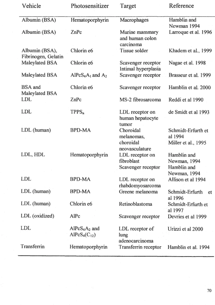
Table 6.1 Summary of photosensitizer serum-based vehicles and bioconjugatès
Vehicle
Photosensitizer
Target
Reference
Albumin (BSA) Hematoporphyrin Macrophages Hamblin and
Newman 1994
Albumin (BSA) ZnPc Murine mammary Larroque et al. 1996
and human colon carcinoma
Albumin (BSA), Chlorin e6 Tissue solder Khadem et al., 1999
Fibrinogen, Gelatin
Maleylated BSA Chlorin e6 Scavenger receptor Nagae et al. 1998
Intimal hyperplasia
Maleylated BSA AlPcS4A1 and A1 Scavengerreceptor Brasseur et al. 1999
BSAand Chlorin e6 Scavengerreceptor Hamblin et al. 2000
Maleylated BSA
LDL ZnPc MS-2 fibrosarcoma Reddi et al 1990
LDL TPPSn LDL receptor on de Smidt et al 1993
human hepatocyte tumor
LDL (human) BPD-MA Choroidal Schmidt-Erfurth et
melanomas, al 1994
choroidal Miller et al., 1995
neovasculature
LDL,HDL Hematoporphyrin LDL receptor on Hamblin and
fibroblast Newman, 1994
Scavenger receptor Hamblin and Newman, 1994
LDL BPD-MA LOL receptor on Allison et al 1994
rhabdomyosarcoma
LDL (human) BPD-MA Greene melanoma Schmidt-Erfurth et
al 1996
LDL (human) Chlorin e6 Retinoblastoma Schmidt-Erfurth et
al 1997
LDL ( oxidized) AlPc Scavenger receptor Devries et al l 999
LDL AlPcS4A2 and LOL receptor of Urizzi et al 2000
AlPcS4(C12) lung
adenocarcinoma
6.4 Antibody conjugates
A class of protein vehicles is the antibody vehicles. Photodynamic therapy using PS-Ab conjugates, termed photoimmunotherapy (PIT), has been developed over the past 15 years (Hasan et al., 1997; Klyashachitsky et al., 1994). PIT has evolved in parallel with the use of monoclonal antibodies as pharmaceuticals. PIT is an ever-expanding field of study as mAb generation techniques improve thus generating different antibodies to be coupled with various photosensitizers. Targeting of photosensitizers using antibodies and/or fragments has been extensively investigated. Table 6.2 is a summary of various studies.
6.4.1 Phthalocyanines
Phthalocyanines have likewise been targeted to various cell lines via antibody carriers. AlPcS0 was encapsulated in liposomes that were then linked to antibodies for
targeting purposes. It was clear from these early studies that the phototoxicity was directed by the mAb because free Pc or liposomal Pc were not phototoxic against the antigen-bearing cell lines under the same conditions (Morgan et al., 1989). Subsequently, this liposornal AlPcS0 formulation was used to target B-cells using an anti-B-cell Ab as
well as T-lymphocytes using anti-CD3 antibody. This could be applicable for ex vivo bone marrow purging (Morgan et al., 1992). A similar method has also been used to target hurnan bladder carcinorna (Morgan et al., 1994). An AlPcS4A1 (A1 being a 5 carbon spacer chain) has been used to label anti-carcinoembryonic antigen (CEA) monoclonal antibodies. This is the same Pc derivatives used to label proteins in the current study. The Pc-mAb conjugates had 5-16 moles of Pc per mol of antibody. The immunoreactivity of the bioconjugates was not altered due to the coupling with a
photosensitizer. ln nude mice, tumor to normal tissue ratios were similar for the Pc-mAb conjugates as compared to the free antibody. ln vitro, the bioconjugate was photocytotoxic against antigen positive cells. This, taken together with the in vivo tumor seeking capacity of the Pc-mAb, makes it a promising third generation photosensitizer (Carcenac et al., 1999). Conversely, an anti-CD3 antibody was covalently labeled with AlPcS4A1• Cell uptake of the bioconjugate was not impaired as measured by flow cytometry. However, the bioconjugate was not photoactive (Ménard, 1998). It was postulated that upon intemalization of the Pc-mAb, the photosensitizer was trapped in the endosome and following illumination, the singlet oxygen produced was quenched by the Pc-mAb microenvironment.
Table 6.2 Summary of photosensitizer-antibody conjugates
Antibody
Photosensitizer Target
Reference
Rabbit anti-Hu Hematoporphyrin Human perpherial blood Mew et al., 1985
CAMAL-1 leukocytes
Ll210 CAMAL antigen
Irrelevant MoAb
Anti-Leu/l Chlorin e6 T-leukemia cells Oseroff et al., 1986
Mab 3G2/C6 Chlorin e6 Bladder carcinoma Hasan et al., 1989
Mab 791T/36 Liposomal AlPcS Osteosarcoma, colorectal Morgan et al., 1989 carcmoma
Mab 309 Hematoporphyrin Gastrie cancer Xu et al., 1989
Mab 3Hl 1 derivative
Mab T48 BPD-MA Human chorionogonadotropic Jiang et al., 1990
(HCG) hormone
Rabbit IgG anti- Iodinated E. coli Devanathan et al.,
E.coli fluorescein 1990
Mab0Cl25 Chlorin e6 Ovarian carcinoma Goff et al., 1991,
1994, 1996
Hamblin et al., 1996
Mab HMFG-1 Hematoporphyrin Breast adenocarcinoma Prosser et al., 1991
MabE7 Liposomal AlPcS Urinary bladder carcinoma Morgan et al., 1994
Mab OC125 Chlorin e6 Human ovarian cancer Duska et al., 1997
Mab0KT3 AlPcS4A1 Leukemic T lymphocytes (CD3+) Menard, 1998
Mab U36 m-THPC Head and neck squamous cell Vrouenraets et al.,
carcmoma 1999
Mab 35A7 AlPcS4A1 Carcinoembryonic antigen (CEA) Carcanac et al., 1999
MabU36 TrisMPyP- Squamous cell carcinoma Vrouenraets et al.,
Mab425 <DC02H 2000
Murine Mab Chlorin e6 Human colorectal cancer antigen Del Governatore et
17.lA (EpCMA) al., 2000(a, b)
6.5 Epidermal growth factor
Epidermal growth factor (EGF) is a small protein that binds to a cell surface receptor. EGF is a potent mitogen found throughout the body and is classified as an angiogenesis stimulating factor. As seen with integrin receptor expression, the EGF receptor (EGF-R) is over expressed in a variety of tumor types, in particular ovarian cancers. 30% of ovarian cancers have increased EGF receptor numbers, making EGF a potential drug carrier (Moor et al., 2000). EGF has been coupled to various photosensitizers in the hopes of specifically targeting this receptor. The cytotoxic activities of an AlPc and a CoPc bound to EGF were determined against a human breast carcinoma cell line. The AlPc-EGF was seven times more photoactive as compared to the free AlPc. The CoPc-EGF conjugate was cytotoxic, requiring ascorbic acid for activation as opposed to light. In vivo studies using B 16 melanoma were subsequently carried out (Lutsenko et al., 1999).
More elaborate conjugates have been prepared using EGF and Sn(IV)chlorin e6 bound together via carriers such as dextran, polyvinyl alcohol and human serum albumin (HSA) (Gijsens and de Witte, 1998; Gijsens et al., 2000). The EGF-HSA-SnCe6 conjugate had the best receptor affinity displaying high phototoxicity against a cell line over-expressing the EGF-R. The effect was receptor related as native EGF could compete for binding sites and decreased photoactivity. The HSA conjugate also produced more intracellular ROS upon irradiation as compared to the dextran conjugate. In a previous study, the EGF-Dex-SnCe6 conjugate bound specifically to the EGF receptor on a squamous cell carcinoma line whereas the EGF-PV A-SnCe6 conjugate