P R O O F
Brain Behav Evol 429 DOI: 10.1159/0000XXXXXSpatial Distribution and Morphological
Characteristics of the Trunk Lateral Line
Neuromasts of the Sea Bass (
Dicentrarchus
labrax
, L.; Teleostei, Serranidae)
Karine Faucher
a, bAnne Aubert
aJean-Paul Lagardere
b aLaboratoire de Biologie et Environnement Marin, Université de La Rochelle, La Rochelle,bCentre de Recherche sur les Ecosystèmes Marins et Aquacoles, CNRS-IFREMER, L’Houmeau, France
Received: March 13, 2003 Returned for revision: May 8, 2003 Accepted after revision: July 8, 2003
Anne Aubert
ABC
© 2003 S. Karger AG, BaselKey Words
NeuromastW FishW TeleostW Sea bassW TrunkW Hair cellW Lateral line
Abstract
The morphology and spatial distribution of the different types of neuromasts encountered on the trunk lateral line of the sea bass (Dicentrarchus labrax) were exam-ined using scanning electron microscopy. The sea bass trunk lateral line exhibits a complete straight pattern. In their basic features, the two types of neuromasts present, canal and superficial, resemble what has been described in other fishes. They are similar in their gener-al cellular organization but differ in sizes, and shapes, as well as in the densities and lengths of their hair bundles. However, the sea bass trunk lateral line distinguishes itself in several ways. For instance, the pores of the canal segments are partially obstructed due to the overlap of scales throughout the trunk. Moreover, based on the density and length of the hair bundles, two distinct areas, central and peripheral, could be distinguished within the maculae of canal neuromasts. Their cupulae are also peculiar as they possess two wing-like extensions and that their central core appears to be organized in layers
instead of columns. In addition, the superficial neuro-masts, up to 6 per scale, are either round or elliptical and seem to be distributed serendipitously. Finally, within the maculae of both types of neuromasts, a significant number of hair bundles do not follow the two-directional polarity pattern usually described. Although some hy-potheses are proposed, the influence of these character-istics in terms of signal encoding and fish behavior is yet to be understood.
Copyright © 2003 S. Karger AG, Basel
Introduction
The mechanosensory lateral line system is found in all fishes and most amphibians [Dijkgraff, 1962; Blaxter, 1987; Lannoo, 1987; Coombs et al., 1989; Webb, 1989a; Northcutt, 1992]. In fishes this sensory system functions as a detector of low-frequency water displacements, rela-tive to the body of the animal [Denton and Gray, 1983; Bleckmann, 1993; Coombs and Montgomery, 1994; Coombs et al., 1996]. This capacity enables the fish to detect prey and/or predators [Hoekstra and Janssen, 1986; Montgomery, 1989; Bleckmann, 1993; Montgome-ry and Hamilton, 1997; Coombs, 1999; Janssen et al.,
ESTI:BBE:ZBRAI429XA.91 FF: ZUP9 E1:
P R O O F
1999], stationary objects [Dijkgraaf, 1962; Blaxter and Batty, 1985; Bleckmann, 1993] and congeners [Partridge and Pitcher, 1980; Janssen et al., 1995]. The lateral line also seems to be involved in rheotaxis, that is orientation and/or movement in regard to currents [Bleckmann, 1993; Pavlov and Tyuryukov, 1993; Montgomery et al., 1997; Northcutt, 1997; Baker and Montgomery, 1999a, b; Coombs et al., 2001].
The functional units of the lateral line system are the neuromasts [Coombs et al., 1989; Northcutt, 1992; Bleck-man, 1993]. These are present on the head, trunk and tail [Coombs et al., 1989; Northcutt, 1997]. Each neuromast is composed of sensory hair cells and support cells cov-ered by a cupula and surrounded by mantle cells [Münz, 1979]. In fishes, one distinguishes two major types of neu-romasts: canal neuromasts, contained in subdermal tube-like ducts; and superficial neuromasts, located on the epi-dermis [Coombs et al., 1989]. The overall morphology and spatial distribution of lateral line systems have been studied in many teleost [see Coombs et al., 1989 for review] and non-teleost [Maruska, 2001] fishes. The data indicate that there is a diversity of distribution patterns. For instance, regarding canal neuromasts, eight trunk canal patterns have been described [Webb, 1989b]. Like-wise, the network of ducts on the head exhibits diverse complexities according to species [Coombs et al., 1989; Webb, 1989b]. The variability is even more pronounced concerning superficial neuromasts as their number and spatial distribution vary considerably among species [Münz and Class, 1983; Song, 1984; Coombs et al., 1989; Northcutt, 1989].
Although similar in their basic structure, the two types of neuromasts differ in many ways. Canal neuromast are usually larger than superficial neuromasts [Münz, 1979; Webb, 1989c; Song and Northcutt, 1991; Maruska and Tricas, 1998; Northcutt et al., 2000] and possess more hair cells [Münz, 1979; Webb, 1989c; Song and North-cutt, 1991; Tsukamoto et al., 1995; Webb and NorthNorth-cutt, 1997]. Differences are also observed in their accessory structures such as the sizes and shapes of their cupulae and their respective afferent and efferent innervation [Flock and Wersäll, 1962; Münz, 1979, 1989; Münz and Class, 1983; Janssen et al., 1987; Coombs et al., 1989; Kroese and Schellart, 1992]. Taken together, these find-ings suggest that the contribution of the two types of neu-romasts during stimulus processing might be different. Indeed, it has been shown that superficial neuromasts mainly function as detectors of water velocity, whereas canal neuromasts appear to respond to water acceleration [Denton and Gray, 1983; Kroese and Schellart, 1992].
However, it is still unclear what are the respective biologi-cal functions of the two types of neuromasts in reference to the fish habitats or lifestyles [Dijjkgraaf, 1962; Janssen et al., 1987; Coombs et al., 1989, 2001; Münz, 1989; Maruska, 2001]. In addition, the great variety of structure and spatial organization of lateral lines observed among species suggest that they might fulfill different functions in terms of fish behavior.
Our present study describes the spatial distribution and morphological characteristics of the canal and super-ficial neuromasts observed on the trunk lateral line of the sea bass (Dicentrarchus labrax). We report the similarities and differences observed between those neuromasts. We also point out the peculiar aspects of the sea bass trunk lateral line as compared to that of other fishes. We do not intend, at this point, to establish any relationship between the morphological aspects and the biological functions of the different components of the lateral line. Rather, our study should be considered a basis for further experi-ments, which will examine the physiological responses of the lateral line systems of coastal fishes in regards to the constraints of their environmental conditions.
Materials and Methods
In order to identify and describe the different morphological types of lateral line neuromasts, thirty-seven specimens of sea bass (Dicentrarchus labrax, L.) were examined. The fish used in this study averaged 20 cm in length and 120 g in weight. They were obtained from a commercial source (Ferme des Baleines, Ile de Ré, France). All specimens were housed under natural photoperiod and constant temperature (16° C) in 240 liter tanks filled with filtered seawater.
The animals were fed with live molluscs and crustaceans.
After anesthesia with 75 mg/l MS222 (3-aminobenzoic acid ethyl, Sigma), the trunk lateral lines were isolated and transferred into an artificial solution (composition in mM: NaCl: 150; KCl: 5; CaCl2, 2 H2O: 3; MgCl2, 6 H2O: 1.5; HEPES: 10; adjusted to pH: 8). Sam-ples of 2 to 3 consecutive scales were obtained. When needed, the roof of the canal was carefully removed in order to expose the canal neuromast.
Each sample was fixed in 4% glutaraldehyde (Fisher Scientific Labosi) in sodium cacodylate buffer (0.4 M, pH 7.2), dehydrated in graded acetone and critical point-dried using liquid CO2 (BALTEC CPD 030). The samples were then mounted on brass supports and sputter coated with gold (Cressington Sputter Coat). Scanning elec-tron microscopic observations were performed with a JEOL JSM-5410LV microscope.
The quantitative measures reported herein were obtained from scanning electron microscopy recordings using the image analysis software Biocom Visiol b 200.
Quantitative data are expressed as the mean B SEM (standard error mean). Statistical analyses were performed using the non-para-metric test of Mann and Whitney.
P R O O F
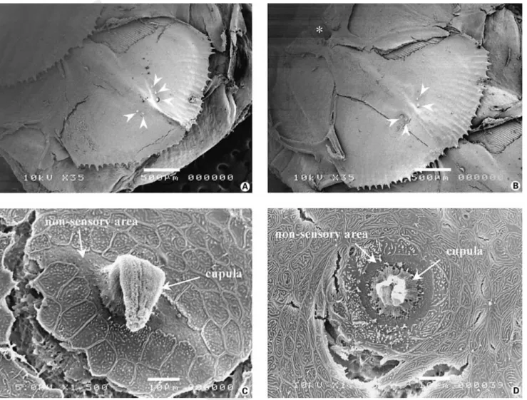
Fig. 1. Scanning electron micrographs of modified scales and superficial neuromasts. A and B Micrographs of modified scales showing that superficial neuromasts (arrowheads) are always observed lateral to the caudal end of the hump formed by the canal segment (scale bar = 500 Ìm). The asterisk on B indicates the suprascalar pore of the canal segment exposed after removal of the preceding scale. C and D Enlargements of the two shapes of superficial neuromasts observed: elliptical (C) and round (D) (scale bar = 10 Ìm). The sensory epithelia are still covered by an intact (C) or partially damaged cupula (D).
Results
The trunk lateral line of the sea bass consisted of a single row of modified scales which run within the mid-section of each flank from the operculum up to the tail. No canal nor superficial neuromast was observed outside that specific area. Each modified scale exhibited one subdermal tube, or canal segment, ending in two openings: a suprascalar pore on the rostral side of the scale and an infrascalar pore on the opposite end. No other opening was observed. Due to the overlap of the modified scales throughout the trunk, the suprascalar pore was located just below the opening of the infrascalar pore of the preceding scale.
According to their location on the scales, two different types of neuromasts were identified along the trunk lateral line. One could distinguish canal between neuromasts and superficial neuromasts.
Superficial Neuromasts
In order to study the presence of superficial neuro-masts, a total of 894 scales were examined. When present, superficial neuromasts were observed superficially on the epidermis of the modified scales. They were always local-ized on the sides of the caudal end of the canal segments (fig. 1A, B). The number of superficial neuromasts per scale varied from zero to six.
ESTI:BBE:ZBRAI429XA.91 FF: ZUP9 E1:
P R O O F
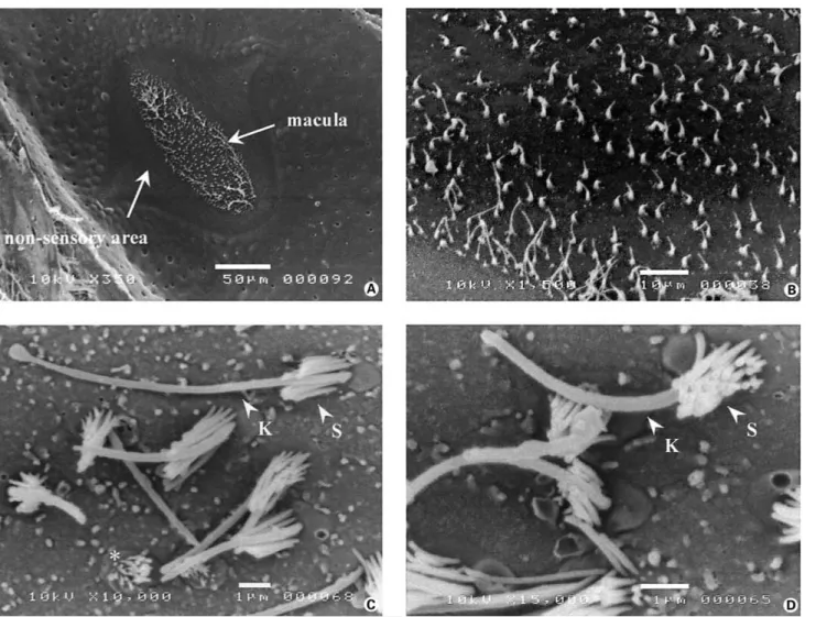
Fig. 2. Scanning electron micrographs of superficial neuromasts deprived of their cupula, following treatment with MS222. A and B Round-shaped maculae observed in both elliptical (A) and round (B) neuromasts (scale bar = 10 Ìm). C and D Details of the maculae which show the density, morphology (K: kinocilium; S: stereocilia) and respective orientation of the hair bundles (C: scale bar = 5 Ìm; D: scale bar = 1 Ìm).
Each superficial neuromast was composed of a sensory macula surrounded by non-sensory mantle cells (fig. 1C, D). The macula was often covered by a cupula as the latter could be partially or totally absent following the use of MS222. Two shapes of superficial neuromasts were ob-served: elliptical (fig. 1C) or round (fig. 1D). The former were, for the most part, oriented perpendicularly to the rostro-caudal axis of the scale. Measures indicated that elliptical neuromasts were 50.48 B 14.17 Ìm long (n = 134) and 28.15 B 9.34 Ìm (n = 134) wide for a mean surface area of 983.96 B 691.73 Ìm2 (n = 134). The
aver-age diameter for the round superficial neuromasts was 32.64 B 11.98 Ìm (n = 120) which corresponded to an
average surface area of 823.27 B 609.87 Ìm2 (n = 120).
When present the cupulae were usually cylindrical or ‘tongue-like’ (fig. 1C). Their mean surface area at the base
was 670.63 B 561.02 Ìm2 (n = 145). The cupulae
appeared to be organized in columnar compartments. In the absence of cupulae, one could observe the senso-ry maculae delimited by the hair bundles of the sensosenso-ry cells (fig. 2A, B). The sensory areas were always round whatever the overall shape of the neuromast. Their mean
surface area was 411.85 B 386.35 Ìm2 (n = 83).
Two types of hair bundles were observed: ‘normal’ ones with one kinocilium and about 20 stereocilia ar-ranged in 5 to 6 rows of increasing size (fig. 2C, D). Some ‘shorter’ hair bundles, deprived of kinocilium, with small-er stsmall-ereocilia wsmall-ere also obssmall-erved throughout the sensory
P R O O F
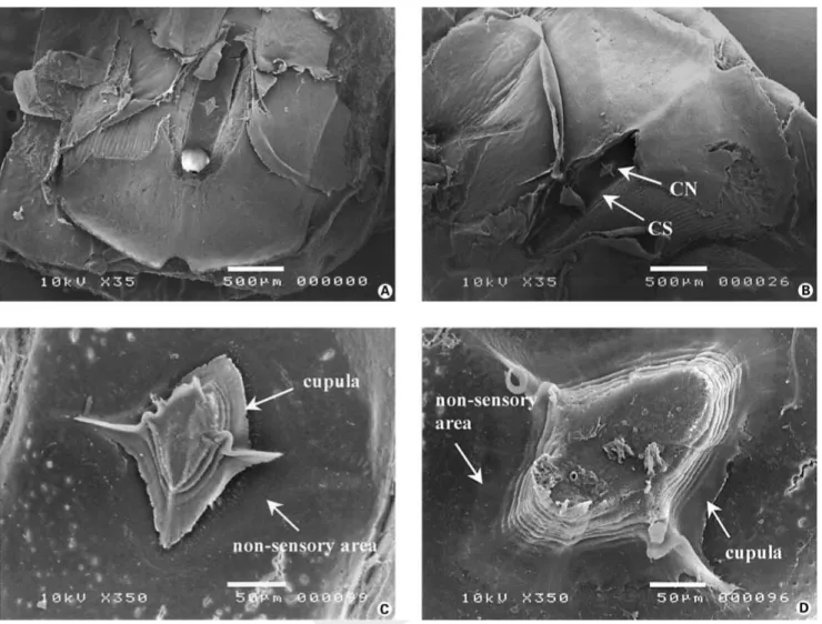
Fig. 3. Scanning electron micrographs of modified scales and canal neuromasts exposed after removal of the roof of the canal segment. A and B Two examples of canal neuromasts. There is only one canal neuromast per modified scale which is always located within the midsection of the canal segment (scale bar = 500 Ìm; CS: canal segment; CN: canal neuromast). C and D Enlargements of two canal neuromasts together with their cupula (scale bar = 50 Ìm). The main body of the latter runs parallel to the axis of the canal segment whereas the two wing-like extensions are perpendicularly oriented.
maculae. Most kinocilia exhibited a swollen tip. The aver-age number of hair bundles per macula, including the ‘shorter’ ones, was 43 B 15 (n = 16) which corresponded to an average density of 0.14 B 0.07 hair bundle/Ìm2.
When possible, the lengths of the kinocilia and stereocilia were measured. The kinocilia were 7.42 B 1.70 Ìm (n = 55) long on average whereas the lengths of the longest and shorter stereocilia were, respectively, 1.90 B 0.49 Ìm (n = 46) and 0.46 B 0.12 Ìm (n = 33).
The eccentric position of the kinocilium in regard to the stereocilia conferred a morphological orientation for each hair bundle. A closer look at the hair bundles within each macula did reveal a specific pattern of orientation.
However, pairs of hair bundles with the opposite morpho-logical orientation were also observed.
Canal Neuromasts
A total of 277 modified scales were examined in order to study the presence and morphological characteristics of canal neuromasts. These observations were made possible by removal of the roof of each canal segment. The total length of the segments was between 1.5 and 2.5 mm (n = 67). Their widths were larger in the middle than on the ends: 360 B 70 Ìm (n = 128) vs. 280 B 45 Ìm (n = 60) (U = 6418, p ! 0.005).
ESTI:BBE:ZBRAI429XA.91 FF: ZUP9 E1:
P R O O F
Fig. 4. Details of the sensory epithelia of canal neuromasts observed after removal of the cupulae following treatment with MS222. A Photomicrograph of a canal neuromast which shows the surface and shapes of the sensory and non-sensory epithelia. The macula is elliptical and oriented parallel to the axis of the canal segment (scale bar = 50 Ìm). B Enlargement of a macula which shows the different densities and sizes of the hair bundles observed between the edges and the center of the sensory epithelium (scale bar = 10 Ìm). C and D Morphological characteristics (K: kinocilium; S: stereocilia) and respective orientations of the hair bundles observed within the edges (C) and the center (D) of the macula (scale bar = 1 Ìm). The asterisk (C) indicates the presence of an atypical hair bundle (shorter stereocilia and absence of a kinocilium).
Only one canal neuromast could be observed on the floor of each segment (fig. 3A, B). It was positioned in the center of the canal segment. Although the structural orga-nization of the canal neuromasts was similar to that of the superficial neuromasts, differences were observed.
Canal neuromasts were always lozenge-shaped with a main axis oriented dorso-ventrally. The mean length and width of these neuromasts were 272.57 B 95.29 Ìm and 179.31 B 62.02 Ìm (n = 101), respectively, for a mean surface area of 0.0347 B 0.0249 mm² (n = 101).
The shape of the cupulae was elliptical with two wing-like extensions (fig. 3C, D). The ‘wings’ were always oriented perpendicularly to the canal’s main axis. In con-trast to superficial neuromasts, no columnar organization of the cupulae was observed. It seemed that the cupulae were composed of a succession of several layers of de-creasing sizes organized to form a pyramid-like structure. The mean surface area measured at the basis of the cupu-lae was 0.018 B 0.017 mm² (n = 79).
The maculae of canal neuromasts were elliptical with a rostro-caudal main axis (fig. 4A). Their mean surface area
P R O O F
was 0.016 B 0.013 mm² (n = 91). The average number ofhair bundles per maculae was 802 B 414 (n = 18), which corresponded to an average density of 0.052 B 0.027/ Ìm2. Statistical analyses indicated that the maculae of
canal neuromasts could be divided into two different areas. Indeed, the density of hair bundles in the periphery of the maculae was significantly higher than in the central part: 0.075 B 0.039 hair bundles/Ìm2 (n = 7) vs. 0.042 B
0.015 hair bundles/Ìm2 (n = 15; U = 81, p ! 0.045)
(fig. 4B). As for the superficial neuromasts, these numbers included hair bundles with smaller stereocilia and no kinocilium. Several kinocilia exhibited a bulbous ending (fig. 4C). In addition, significant differences in length for kinocilia and stereocilia were also observed (fig. 4C, D). The longest stereocilia located at the periphery of the maculae were on average shorter (2.25 B 0.69 Ìm, n = 34) than those present in the central part (2.98 B 0.80 Ìm, n = 13; U = 334, p ! 0.008). Peripheral kinocilia were 9.04 B 3.19 Ìm (n = 47) long on average whereas the central ones reached 5.18 B 1.41 Ìm (n = 24; U = 151, p ! 0.001).
As mentioned for superficial neuromasts, no preferen-tial orientation of the hair bundles within the maculae of canal neuromasts was clearly established, although some pairs of hair bundles with the opposite orientation were observed.
Discussion
The data presented herein are original observations of the spatial distribution and morphological characteristics of two types of neuromasts present on the trunk lateral line of the sea bass (Teleostei, Perciformes, Serranidae). These are preliminary results of a larger study, designed to address the possible involvement of the lateral line system in the physiological capacities of coastal fishes to adapt to the constraints of their environment.
The spatial distribution of the neuromasts throughout the body of the fish determines the amplitude of the receptive field of the lateral line sensory system [Denton and Gray, 1983]. Among teleosts, the Perciformes exhibit the whole spectrum of trunk patterns [Coombs et al., 1989; Webb, 1989b]. In the sea bass, the trunk lateral line showed the characteristics of a complete straight pattern according to the classification proposed by Webb [1989b]. Indeed, the presence of canal and superficial neuromasts was restricted to a single row of modified scales located within the midsection of both flanks. As observed in most teleosts [Coombs et al., 1989; Webb, 1989b], it was con-tinuous with the postotic canal of the head lateral line
sys-tem and runs from the slit of the operculum up to the cau-dal peduncle. It can be noted that this is the first time a straight pattern is described for a species of the Serranidae family.
All modified scales possessed a centered tube-like structure, or canal segment, oriented along a rostrocaudal axis. Each canal segment was spindle-shaped and was open by only two pores: one suprascalar pore on the ros-tral side of the segment and one infrascalar pore on the opposite side. Due to the partial overlap of the scales the infrascalar pore was positioned just above the suprascalar pore of the canal segment of the subsequent scale. This contrasts with several reports that indicate that in most teleosts the canal segment is generally directly connected to the external medium by one or two pores, or by tubules which allow water flow within the canal segment [Flock, 1965; Münz, 1979; Appelbaum and Schemmel, 1983; Coombs et al., 1989; Webb, 1990; Harvey et al., 1992; Kroese and Schellart, 1992]. This suggests that in the sea bass water entry within the canal segment might be greatly reduced.
According to their position, two types of neuromasts were identified along the trunk lateral line: superficial neuromasts, present on the upper surface of the modified scales and canal neuromasts, located on the floor of each canal segment. As expected, due to their common origin [Dijkgraaf, 1962; Coombs et al., 1989 for review; Webb, 1989c], the two types of neuromasts were similar in their general aspect. They were both composed of a central area, or macula, covered by a gelatinous cupula, which was surrounded by a differentiated epithelium. The use of scanning electron microscopy did not allow us to examine the ultrastructure of the neuromasts. However, the litera-ture indicates that the central area, or macula, is com-posed of sensory mechanoreceptors, or hair cells, inter-mingled with support cells, whereas the surrounding epi-thelium corresponds to non-sensory mantle cells [Flock, 1965; Yamada, 1973; Münz, 1979; Tatsuoka and Hama, 1987; Cernuda-Cernuda and Garcı´a-Ferna´ndez, 1996].
As it is usually observed in teleosts [Flock, 1965; Coombs and Montgomery, 1989; Münz, 1989; Webb, 1989a, b, 1990; Tsukamoto et al., 1995] there was only one canal neuromast per scale. It was positioned at the midpoint between the two pores where the canal segment was wider. In contrast to canal neuromasts, the number of superficial neuromasts per modified scale varied from 0 to 6. When present, they were positioned close to the cau-dal end of the canal segment. Consequently, they were never covered by the flap of the preceding scale. We were unable to establish a specific pattern of distribution for
ESTI:BBE:ZBRAI429XA.91 FF: ZUP9 E1:
P R O O F
these neuromasts along the trunk lateral line. For in-stance, they did not form distinctive lines or rows as observed in Cichlidae [Münz and Class, 1983; Webb, 1990], Congridae [Hama, 1978] or Notothenioidae [Coombs and Montgomery, 1994]. They appeared to be serendipitously distributed on the dorsal and/or ventral sides of the hump formed by the canal segment. Thus, according to their position on the scale, superficial neuro-masts might be potential targets for stimuli in the form of water disturbances coming from all directions.
Neuromast morphology defines which type of infor-mation is encoded [Kroese and Schellart, 1992]. Although similar in their general aspect, canal and superficial neu-romasts differed in many ways. For instance, as observed in studies performed on different fish species [Hama and Yamada, 1977; Münz, 1979; Best and Gray, 1982; Rouse and Pickles, 1991; Coombs and Montgomery, 1994; Tsu-kamoto et al., 1995; Maruska, 2001], canal neuromasts were always lozenge-shaped. Their maculae were invaria-bly elliptical with a longitudinal axis parallel to the canal axis. In contrast, superficial neuromasts were either round or elliptical. Elliptical superficial neuromasts were pre-viously observed on the trunk lateral lines of the Plaice [Harvey et al., 1992] and the channel catfish [Northcutt et al., 2000] whereas round ones were seen in the Herring [Blaxter et al., 1983] and the Florida gar [Song and North-cutt, 1991]. However, the concomitant presence of the two shapes of superficial neuromasts on the same scale, as reported herein, has only been described in cichlids [Webb, 1989c, 1990]. The author then suggested that elliptical neuromasts were precursors of canal neuro-masts. However, in the present study this appears to be unlikely as the maturation of canal neuromasts is already achieved. Finally it can be noted that the maculae of superficial neuromasts were always circular whatever the overall shape of the neuromast.
Other characteristics of hair cell densities and polariza-tions such as their size also need to be examined as those are directly related to the capacity of each neuromast to detect and encode stimuli. Among those parameters, the most striking difference is size. Although there is wide intra-type variability, canal neuromasts were 30 to 40 times bigger than superficial neuromasts. Similar size ratios were also observed for the maculae. These size dif-ferences are commonly observed in fishes [Münz, 1979; Webb, 1989c; Song and Northcutt, 1991; Maruska and Tricas, 1998; Northcutt et al., 2000]. However, one can observe that the actual sizes reported herein are lower than those measured in Siluridae [Northcutt et al., 2000] or Cichlidae [Münz, 1979] for example.
No hair cell per se was observed in this series of experi-ments. Their presence was implied by the observation of hair bundles. As previously reported [Münz, 1979; Webb, 1989c; Song and Northcutt, 1991; Tsukamoto et al., 1995; Webb and Northcutt, 1997], the number of hair bundles was higher in canal neuromasts than in superficial ones. However, an estimation of the hair bundle densities indi-cated that those were higher in superficial neuromasts. These data agree with those of Webb and Northcutt [1997] in non-teleost fishes. The hair bundles were homo-geneously distributed throughout the maculae of superfi-cial neuromasts. In contrast, there were clearly two differ-ent hair bundle regions within the maculae of canal neuro-masts. In the central part of the maculae the hair bundles were sparse, whereas in the vicinity of the periphery the density of hair bundles was significantly higher. To our knowledge, this is the first time that a different distribu-tion of mature hair bundles within the maculae of canal neuromasts is reported.
Each hair bundle was composed of about twenty ste-reocilia arranged in five to six rows of increasing size toward a kinocilium. The small number of stereocilia observed is consistent with the data reported by others [Flock, 1965; Hama, 1965; Yamada, 1973; Rouse and Pickles, 1991]. When possible the lengths of kinocilia and stereocilia were measured. The mean values obtained were similar to those reported in teleost [Flock, 1965; Blaxter et al., 1983; Webb, 1989c; Rouse and Pickles, 1991; Yamada and Hama, 1995] and non-teleost fishes [Yamada, 1973; Hama and Yamada, 1977; Webb and Northcutt, 1997; Peach and Rouse, 2000; Maruska, 2001]. Some studies have mentioned that the hair bundles were usually taller in superficial neuromasts than in canal neuromasts [Song and Northcutt, 1991; Webb and North-cutt, 1997]. This was indeed observed for the hair bundles present within the central area of canal neuromasts. How-ever, the hair bundles present in the peripheral area of canal neuromasts were taller than those of superficial neu-romasts.
A closer look at the maculae in both types of neuro-masts revealed the presence of smaller hair bundles in which the kinocilium was lacking and the stereocilia were shorter. These could correspond to damaged hair bundles, resulting from the treatment and handling of the tissue samples, or to immature, precursor, hair cells as seen by Rouse and Pickles [1991]. The general aspect of the hair bundles and the stereocilia seems to point toward the sec-ond hypothesis.
The eccentric position of the kinocilium in regards to the stereocilia confers a morphological orientation for
P R O O F
each hair bundle. This typical display is known to beresponsible for the directional sensitivity of each hair cell in response to stimulation [Flock and Wersäll, 1962; Flock, 1965; Wersäll et al., 1965; van Netten, 1997]. Therefore, an examination of the hair bundles orientation within the macula should indicate the axis of maximum sensitivity for a given neuromast in response to stimula-tion. Since Flock’s observation in 1965 on the burbot,
Lota vulgaris, it is generally accepted that within the
macula hair bundles are associated in pairs of opposing polarities [Best and Gray, 1982; Blaxter et al., 1983; Tat-suoka and Hama, 1987; Kroese and van Netten, 1989; Webb, 1989c; Song and Northcutt, 1991; Harvey et al., 1992; Tsukamoto et al., 1995; Webb and Northcutt, 1997]. However, in the present study this two-directional pattern could not be established in either canal or superfi-cial neuromasts. Paired hair bundles oriented along the main axis of the neuromasts were seen, but those were intermingled with hair bundles oriented in various direc-tions. Although puzzling these data are supported by the studies of Maruska [2001] and Harvey et al. [1992] which indicated the presence of hair bundles with different polarities throughout the maculae of the Atlantic Stingray and/or the Plaice and the Sole, respectively. In addition, Rouse and Pickles [1991] have shown in two species of teleosts that hair bundles develop in pairs but lose this characteristic as they mature. Therefore, although chal-lenging in terms of signal processing, it seems difficult to neglect the presence of hair bundles that do not fit the two-directional pattern. Only electrophysiological record-ings performed at the hair cell level and on the afferent fibers could determine the function of these sensory cells. The final significant difference between the two types of neuromasts relies on the shape and structure of their cupula. It is generally accepted that the presence of the cupulae is essential to the sensory role of the neuromasts. Its primary function is to transfer the motion of fluid
within the canal into displacement of the hair bundles [Denton and Gray, 1983, 1989; van Netten and Kroese, 1989]. The cupulae of superficial neuromasts were as expected: circular at the base and more or less cylindrical with a typical framework of vertically oriented columns [Munz and Claas, 1983; Kelly and van Netten, 1991]. Those of canal neuromasts were composed of two parts: a main body oriented along the main axis of the canal seg-ment and two lateral sail-like wings positioned perpendic-ular to the neuromast axis. The main body, which covers the entire macula, resembled a pyramid with a lozenge-shaped base. It does not appear to comply to the general framework of vertically oriented columns but rather seems to be constructed by a piling of different layers with decreasing sizes.
In conclusion, the gross morphology of the trunk later-al line of the sea bass resembles that of many teleosts. However, it presents some discrepancies, which might have implications for the biological functions of each type of neuromast, and for the lateral line as a whole. The phys-iological consequences of these differences and their im-plications in terms of behavior of the sea bass in response to stimulation are yet to be understood. In addition, these data need to be complemented by observations of the cephalic and tail components of this sensory system.
Acknowledgments
Supported by fundings from the Ministère de la Recherche et des Nouvelles Technologies and the Contrat de Plan Etat Région – IFREMER. Karine Faucher was a recipient of a doctoral fellowship from the Conseil Général de Charente-Maritime. Thanks to the Ferme des Baleines for providing the animals and the director and staff of the Aquarium of La Rochelle for housing and feeding them. We would also like to thank the Centre Commun d’Analyses (CCA), University of La Rochelle, for allowing the use of the Scanning Elec-tron Microscope.
References
Appelbaum A, Schemmel Ch (1983) Dermal sense organs and their significance in the feeding behaviour of the common sole Solea vulgaris. Mar Eco Prog Ser 13:29–36.
Baker CF, Montgomery JC (1999a) Lateral line mediated rheotaxis in the antarctic fish, Pago-thenia borchgrevinki. Polar Biol 21:305–309. Baker CF, Montgomery JC (1999b) The sensory
basis of rheotaxis in the blind mexican cave fish, Astyanax fasciatus. J Comp Physiol A 184:519–527.
Best ACG, Gray JAB (1982) Nerve fibre and recep-tor counts in the sprat utriculus and lateral line. J Mar Biol Assoc UK 62:201–213.
Blaxter JHS (1987) Structure and development of the lateral line. Biol Rev 62:471–514. Blaxter JHS, Batty RS (1985) Herring behaviour in
the dark: responses to stationary and contin-uously vibrating obstacles. J Mar Biol Assoc UK 65:1031–1049.
Blaxter JHS, Gray JAB, Best ACG (1983) Structure and development of the free neuromasts and lateral line system of the herring. J Mar Biol Assoc UK 63:247–260.
Bleckmann H (1993) Role of the lateral line in fish behaviour. In: Behaviour of Teleost Fishes (Pitcher TJ, ed) pp 201–246. London: Chap-man and Hall.
Cernuda-Cernuda R, Garcı´a-Ferna´ndez JM (1996) Structural diversity of the ordinary and special-ized lateral line organs. Microsc Res Tech 34: 302–312.
Coombs S (1999) Signal detection theory, lateral-line excitation patterns and prey capture be-haviour of mottled sculpin. Anim Behav 58: 421–430.
ESTI:BBE:ZBRAI429XA.91 FF: ZUP9 E1:
P R O O F
Coombs S, Montgomery JC (1989) The enigmatic lateral line system. In: The Mechanosensory Lateral Line. Neurobiology and Evolution (Coombs S, Görner P, Münz H, eds) pp 319– 362. New York: Springer-Verlag.
Coombs S, Montgomery J (1994) Function and evolution of superficial neuromasts in an an-tarctic notothenioid fish. Brain Behav Evol 44: 287–298.
Coombs S, Braun CB, Donovan B (2001) The orienting response of Lake Michigan mottled sculpin is mediated by canal neuromasts. J Exp Biol 204:337–348.
Coombs S, Hastings M, Finneran J (1996) Mod-eling and measuring lateral line excitation pat-terns to changing dipole source locations. J Comp Physiol A 178:359–371.
Coombs S, Janssen J, Webb JF (1989) Diversity of lateral line systems: evolutionary and function-al considerations. In: Sensory Biology of Aquatic Animals (Atema J, Fay RR, Popper AN, Tavolga WN, eds) pp 553–593. New York: Springer-Verlag.
Denton EJ, Gray JAB (1983) Mechanical factors in the excitation of clupeid lateral lines. Proc R Soc Lond B 218:1–26.
Denton EJ, Gray JAB (1989) Some observations on the forces acting on neuromasts in fish lateral line canals. In: The Mechanosensory Lateral Line. Neurobiology and Evolution (Coombs S, Görner P, Münz H, eds) pp 239–246. New York: Springer-Verlag.
Dijkgraaf S (1962) The functioning and signifi-cance of the lateral line organs. Biol Rev 38:51– 105.
Flock A (1965) Electron microscopic and electro-physiological studies on the lateral line canal organ. Acta Oto-Laryngol Suppl 199:1–90. Flock A, Wersäll J (1962) A study of the orientation
of the sensory hairs of the receptor cells in the lateral line organ of fish, with special reference to the function of the receptors. J Cell Biol 15: 19–27.
Hama K (1965) Some observations on the fine structure of the lateral line organ of the japa-nese sea eel, Lyncozymba nystromi. J Cell Biol 24:193–210.
Hama K (1978) A study of the fine structure of the pit organ of the common japanese sea eel Con-ger myriaster. Cell Tiss Res 189:375–388. Hama K, Yamada Y (1977) Fine structure of the
ordinary lateral line organ. II. The lateral line canal organ of the spotted shark, Mustelus ma-nazo. Cell Tissue Res 176:23–36.
Harvey R, Blaxter JHS, Hoyt RD (1992) Develop-ment of superficial and lateral line neuromasts in larvae and juveniles of plaice (Pleuronectes platessa) and sole (Solea solea). J Mar Biol Assoc UK 72:651–668.
Hoekstra D, Janssen J (1986) Lateral line receptivi-ty in the mottled sculpin (Cottus bairdi). Co-peia 1986:91–96.
Janssen J, Coombs S, Hoekstra D, Platt C (1987) Anatomy and differential growth of the lateral system of the mottled sculpin, Cottus bairdi (Scopaenaeformes: Cottidae). Brain Behav Evol 30:210–229.
Janssen J, Jones WR, Whang A, Oshel PE (1995) Use of the lateral line in particulate feeding in
the dark by juvenile alewife (Alosa pseudo-harengus). Can J Fish Aquat Sci 52:358–363. Janssen J, Sideleva V, Biga H (1999) Use of the
lat-eral line for feeding in two Lake Baikal scul-pins. J Fish Biol 54:404–416.
Kelly JP, van Netten SM (1991) Topography and mechanics of the cupula in the fish lateral line. I. Variation of cupular structure and composition in three dimensions. J Morphol 207:23–36. Kroese ABA, van Netten SM (1989) Sensory
trans-duction in lateral line hair cells. In: The Mecha-nosensory Lateral Line. Neurobiology and Evo-lution (Coombs S, Görner P, Münz H, eds) pp 266–284. New York: Springer-Verlag. Kroese ABA, Schellart NAM (1992) Velocity- and
acceleration-sensitive units in the trunk lateral line of the trout. J Neurophysiol 68:2212– 2221.
Lannoo MJ (1987) Neuromast topography in uro-dele amphibians. J Morphol 191:247–263. Maruska KP (2001) Morphology of the
mechano-sensory lateral system in elasmobranch fishes: Ecological and behavioral considerations. En-viron Biol Fish 60:47–75.
Maruska KP, Tricas TC (1998) Morphology of the mechanosensory lateral line system in the At-lantic stingray, Dasyatis sabina: the mechano-tactile hypothesis. J Morphol 238:1–22. Montgomery JC (1989) Lateral line detection of
planktonic prey. In: The Mechanosensory Lat-eral Line. Neurobiology and Evolution (Coombs S, Görner P, Münz H, eds) pp 561– 574. New York: Springer-Verlag.
Montgomery JC, Hamilton AR (1997) Sensory contribution to nocturnal prey capture in the dwarf scorpion fish (Scopaena papillosus). Mar Fresh Behav Physiol 30:209–223.
Montgomery JC, Baker CF, Carton AG (1997) The lateral line can mediate rheotaxis in fish. Na-ture 389:960–963.
Münz H (1979) Morphology and innervation of the lateral line system in Sarotherodon niloticus (L.) (Cichlidae, Teleostei). Zoomorphology 93: 73–86.
Münz H (1989) Functional organization of the lat-eral line periphery. In: The Mechanosensory Lateral Line. Neurobiology and Evolution (Coombs S, Görner P, Münz H, eds) pp 285– 297. New York: Springer-Verlag.
Münz H, Claas B (1983) The functional organiza-tion of neuromasts in the lateral-line system of a cichlid fish. In: Advances in Vertebrate Neu-roethology (Ewert JP, Capranica RR, Ingle DJ, eds) pp 301–307. New York: Plenum Press. Northcutt RG (1989) The phylogenetic
distribu-tion and innervadistribu-tion of craniate mechanore-ceptive lateral lines. In: The Mechanosensory Lateral Line. Neurobiology and Evolution (Coombs S, Görner P, Münz H, eds) pp 17–78. New York: Springer-Verlag.
Northcutt RG (1992) Distribution and innervation of lateral line organs in the axolotl. J Comp Neurol 325:95–123.
Northcutt RG (1997) Swimming against the cur-rent. Nature 389:915–916.
Northcutt RG, Holmes PH, Albert JS (2000) Dis-tribution and innervation of lateral line organs in the channel catfish. J Comp Neurol 421: 570–592.
Partridge BL, Pitcher TJ (1980) The sensory basis of fish schools: Relative roles of lateral line and vision. J Comp Physiol 135:315–325. Pavlov DS, Tyuryukov SN (1993) The role of
later-al-line organs and equilibrium in the behavior and orientation of the Dace, Leuciscus leucis-cus, in a turbulent flow. J Ichthyol 33:71–77. Peach MB, Rouse GW (2000) The morphology of
the pit organs and lateral line canal neuromasts of Mustelus antarcticus (Chondrichthyes: Tria-kidae). J Mar Biol Assoc UK 80:155–162. Rouse GW, Pickles JO (1991) Paired development
of hair cells in neuromasts of the teleost lateral line. Proc R Soc Lond B 246:123–128. Song J (1984) The distribution of the lateral line
receptors in garfish (Holostei). Am Zool 24: 134A.
Song J, Northcutt RG (1991) Morphology, distri-bution and innervation of the lateral-line recep-tors of the Florida gar, Lepisosteus platyrhin-cus. Brain Behav Evol 37:10–37.
Tatsuoka H, Hama K (1987) Freeze-fracture study of the lateral-line canal organ of the japanese sea eel, Lincozymba nystromi. Cell Tiss Res 249:523–531.
TsukamotoY, Tateyama H, Oohigashi S (1995) Ar-chitecture of the lateral line organ of the sea eel Conger myriaster. Okajimas Folia Anat Jpn 72: 51–58.
van Netten SM (1997) Hair cell mechano-transduc-tion: its influence on the gross mechanical char-acteristics of a hair cell sense organ. Biophys Chem 68:43–52.
van Netten SM, Kroese ABA (1989) Dynamic be-havior and micromechanical properties of the cupula. In: The Mechanosensory Lateral Line. Neurobiology and Evolution (Coombs S, Görn-er P, Münz H, eds) pp 247–263. New York: Springer-Verlag.
Webb JF (1989a) Developmental constraints and evolution of the lateral line system in teleost fishes. In: The Mechanosensory Lateral Line. Neurobiology and Evolution (Coombs S, Görn-er P, Münz H, eds) pp 79–97. New York: Springer-Verlag.
Webb JF (1989b) Gross morphology and evolution of the mechanoreceptive lateral-line system in teleost fishes. Brain Behav Evol 33:34–53. Webb JF (1989c) Neuromast morphology and
lat-eral line trunk canal ontogeny in two species of cichlids; an SEM study. J Morphol 202:53–68. Webb JF (1990) Ontogeny and phylogeny of the trunk lateral line system in cichlid fishes. J Zool 221:405–418.
Webb JF, Northcutt RG (1997) Morphology and distribution of pit organs and canal neuromasts in non-teleost bony fishes. Brain Behav Evol 50:139–151.
Wersäll J, Flock A, Lundquist PG (1965) Structural basis for directional sensitivity in cochlear and vestibular sensory receptors. Cold Spring Har-bor Symp Quant Biol 30:115–132.
Yamada Y (1973) Fine structure of the ordinary lateral line organ. I. The neuromast of lamprey, Entosphenus japonicus. J Ultrastruc Res 43:1– 17.
Yamada Y, Hama K (1995) Fine structure of the lateral-line organ of the common eel, Anguilla japonica. Z Zellforsch 124:454–464.