Author’s Accepted Manuscript
Fatty acid profile in cord blood of neonates born to
optimally controlled gestational diabetes mellitus
Pauline Léveillé, Jean-Luc Ardilouze, Jean-Charles
Pasquier, Charles Deacon, Kevin Whittingstall,
Mélanie Plourde
PII:
S0952-3278(16)30127-2
DOI:
http://dx.doi.org/10.1016/j.plefa.2016.10.006
Reference:
YPLEF1786
To appear in:
Prostaglandins Leukotrienes and Essential Fatty Acids
Received date: 16 August 2016
Revised date:
12 October 2016
Accepted date: 12 October 2016
Cite this article as: Pauline Léveillé, Jean-Luc Ardilouze, Jean-Charles Pasquier,
Charles Deacon, Kevin Whittingstall and Mélanie Plourde, Fatty acid profile in
cord blood of neonates born to optimally controlled gestational diabetes mellitus,
Prostaglandins
Leukotrienes
and
Essential
Fatty
Acids,
http://dx.doi.org/10.1016/j.plefa.2016.10.006
This is a PDF file of an unedited manuscript that has been accepted for
publication. As a service to our customers we are providing this early version of
the manuscript. The manuscript will undergo copyediting, typesetting, and
review of the resulting galley proof before it is published in its final citable form.
Please note that during the production process errors may be discovered which
could affect the content, and all legal disclaimers that apply to the journal pertain.
1 Fatty acid profile in cord blood of neonates born to optimally controlled gestational diabetes mellitus
Pauline Léveillé1,2, Jean-Luc Ardilouze2,3,4, Jean-Charles Pasquier3,4, Charles Deacon3, Kevin Whittingstall 3, Mélanie Plourde1,3*.
1
Research Center on Aging, Health and Social Services Centre – University Institute of Geriatrics of Sherbrooke, 1036 Belvédère Sud Sherbrooke (Québec), Canada, J1H 4C4.
2
Department of physiology, Université de Sherbrooke, 3001, 12e avenue Nord Sherbrooke (Québec), Canada, J1H 5N4.
3
Department of medecine, Université de Sherbrooke, 3001, 12e avenue Nord Sherbrooke (Québec), Canada, J1H 5N4.
4
Centre de recherche du CHUS, 3001, 12e avenue Nord Sherbrooke (Québec), Canada, J1H 5N4.
*Corresponding author: Mélanie Plourde, Ph.D. Research Center on Aging, 1036 Belvédère Sud
Sherbrooke, Canada, J1H 4C4, 819-780-2220 extension 45664. Melanie.Plourde2@usherbrooke.ca
Abstract
Objective
To evaluate the fatty acid profile of cord blood phospholipids (PL), cholesteryl esters (CE), triglycerides (TG) and non-esterified fatty acids (NEFA) in neonates born to mothers with gestational diabetes mellitus (GDM) compared to non-diabetic mothers.
Methods
The offspring of 30 pregnant women (15 non-diabetic controls, 15 with diet- or insulin-controlled GDM) were recruited before delivery. Cord blood was collected. After lipid extraction, PL, CE, TG and NEFA were separated by thin layer chromatography and analysed by gas chromatography.
2
In GDM vs. control mothers, maternal glycated haemoglobin (A1C, mean ± SD) was not different between
groups: 5.3 ± 0.5% vs. 5.3 ± 0.3% (p = 0.757), respectively. Cord plasma fatty acids were not different in TG, CE and NEFA between GDM and non-diabetic mothers. However, in PL, levels of palmitate, palmitoleate, oleate, vaccinate and di-homo-gamma-linolenate were significantly lower, with a trend for lower arachidonate (p = 0.078), in neonates born to GDM mothers compared to controls.
Conclusion
In contrast to other studies on cord blood docosahexaenoic acid (DHA) levels in GDM mothers, we did not found lower levels of DHA in cord PL, CE, TG or NEFA in neonates born to GDM compared to non-diabetic mothers.
Abbreviation
DHA, docosahexaenoic acid; GDM, gestational diabetes mellitus; PL, phospholipids; CE, cholesteryl esters, TG, triglycerides; NEFA, non-esterified fatty acids; A1C glycated haemoglobin; MUFA, monounsaturated fatty
acids.
Keywords: gestational diabetes, polyunsaturated fatty acids, docosahexaenoic acid
1. Introduction
Gestational diabetes mellitus (GDM) is a state of glucose intolerance diagnosed during pregnancy. GDM affects 5 to 20 % of pregnant women, depending on screening/diagnosis methods and ethnicity [1]. It is suggested that some children born to mothers with poor glucose control during pregnancy suffer lower behavioral and intellectual development compared to children born to non-diabetic women [2, 3]. A key fatty acid for healthy brain and nervous system development during pregnancy is docosahexaenoic acid (DHA). A DHA deficiency during this period might minimize neurodevelopment [4]. In humans, DHA is not efficiently synthesized by shorter-chain omega-3 fatty acids to fulfill the needs in DHA in the third trimester; so it has to be provided by dietary fat intake and transferred to the fetus via cord blood [5].
3
There is increasing evidence supporting that GDM modifies maternal fatty acid profile, which may in turn change the quantity/quality of lipids transferred to the fetus [6, 7]. To our knowledge, 9 case-control studies [8-16] evaluated DHA levels in cord blood of neonates born to GDM vs. non-diabetic mothers. Seven studies reported that DHA levels in cord blood were 12-36% lower in GDM vs. non-diabetic mothers. Most of these studies have evaluated DHA levels in phospholipids (PL), cholesteryl esters (CE) or in red blood cells although it is speculated that DHA in the non-esterified fatty acid (NEFA) pool is the preferred pool for reaching the brain for brain DHA accretion. We sought to evaluate the fatty acid profile and DHA in cord plasma PL, CE, triglycerides (TG) as well as in NEFA, in cord blood of neonates born to mothers with GDM compared to non-diabetic controls.
2. Material and Method
2.1. Population
Pregnant women (n = 30, aged 18 - 40 years, with singleton pregnancy) were recruited in the Centre Hospitalier Universitaire de Sherbrooke at 24 - 28 weeks of gestation between December 2013 and May 2014. Exclusion criteria were: smoking, illicit drug or omega-3/6 consumption, liver or renal disease, cancer, or any medical condition affecting glucose/lipid metabolism. GDM diagnosis and management were performed according to ADA criteria [17]. Individualized insulin therapy was initiated if dietary modification, physical activity, and self-monitoring of blood failed to control glycemic levels [17].
2.2. Sample size
The sample size (n = 12/group) was calculated considering that esterified DHA in cord plasma was 17% lower in GDM group vs. non-diabetic mothers [14], and anticipating a 20% withdrawal rate in both groups.
2.3. Ethic statement
The study was approved by the Human Ethics Research Committee of the Centre Hospitalier Universitaire de Sherbrooke; women gave informed written consent for themselves and their newborn.
4
Maternal weight was measured close to delivery and BMI was calculated. Blood samples for maternal A1C were drawn at hospital admission, before delivery. At delivery, 3 ml of cord blood was collected,
centrifuged, aliquoted, and stored at -80°C. Birth weight, length, head circumference, APGAR score and glycaemia were recorded.
2.5. Blood sample analyses
Maternal A1C, (normal range 3.6-5.8%) was measured using HPLC (Bio-Rad VARIANT, Hercules,
CA) in the Biochemistry lab of the Centre Hospitalier Universitaire de Sherbrooke. Total lipids from the cord plasma were extracted and separated into PL, TG, CE and NEFA by thin layer chromatography. NEFA were thereafter transmethylated using 14% methanol-boron trifluoride before being analysed by gas chromatography, as we previously described [18]. Cord plasma glucose, total cholesterol, total TG and total NEFA were measured using an automated analyzer (Dimension Xpand Plus; Siemens Healthcare Diagnostics, Deerfield, IL). Insulin was measured using an immunosorbent assay (Alpco, Salem, NH).
2.6. Statistical analysis
Normal distribution was evaluated using the Kolmogorov-Smirnov test. Chi-square tests were used to test the differences in the proportions for the categorical variables and t-tests or Mann or Whitney tests were used to compare means between the groups (SPSS software, version 12.0; SPSS Inc., Chicago, IL). Two-tailed p value < 0.05 was considered statistically significant.
3. Results
3.1. Mothers and newborns characteristics
Age, weight and BMI close to delivery were not different between groups (Table 1). A1C was identical
(p = 0.757) and in the normal range in both groups (Table 1). In newborns, clinical characteristics were not different between group except for gestational age (38.1 ± 1.4 vs. 39.6 ± 1.3; p = 0.007) (Table 1). In cord plasma, glucose, insulin, total cholesterol, TG, and total NEFA were not different between groups.
3.2 Fatty acid profile in the cord plasma PL, TG, CE and NEFA
GDM mothers had 15-27% lower C16:0, C16:1 n-7, C18:1 n-9, C18:1 n-7 and C20:3 n-6 in the PL compared to non-diabetic mothers (Table 2). We also found a trend for lower C20:4 n-6 in cord PL of neonates
5
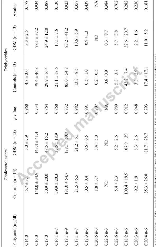
born to GDM vs. controls. The fatty acid profiles in cord NEFA, TG and CE were not different between groups (Table 2 and Table 3).
4. Discussion and conclusions
This study reports that newborns from mothers with optimally-controlled GDM received similar concentrations of esterified and non-esterified DHA. In GDM mothers, blood glucose over the last weeks of pregnancy was indeed well controlled in both groups as demonstrated by A1C levels that fall into the normal
range. This suggests that glucose control in the last months of gestation plays a role in DHA level transferred to the newborn during gestation.
The role of glucose control on the quantity/quality of most fatty acids transferred to the fetus is yet not clear. In our study, the fatty acid profile in PL was impaired in neonates born to GDM mothers: Monounsaturated fatty acids (MUFA) such as C16:1n-7, C18:1n-9, and C18:1n-7 were significantly lower than in controls. This result seems to be specific to the PL class since there was no difference in the other lipid classes neither in the total lipids (data not shown). This latter result is in line with other published studies where MUFA in total lipids of the cord blood in neonate born to well-controlled GDM vs. control were not different [8, 9]. In one study, the MUFA levels were 11-21 % lower in GDM mothers compared to non-diabetic mothers [19] while another study showed that the concentration of lysophospholipids, a class of phospholipids, were 20 % lower in GDM maternal serum compared to pregnant controls [15]. PL are structural lipids and can be used as biomarkers for dietary fatty acid intake. PL may although not be the best indicator of fatty acid that are readily available for placental transfer [20]. In neonates, palmitic, palmitoleic, and oleic acids are concentrated in brain membrane: their concentrations increase in the plasma PL and NEFA from birth to 6-8 hrs of age [21]. Moreover the cerebral white matter is particularly rich in n-9 MUFA [22].
We also report here that there was no difference in the esterified or non-esterified pool of DHA in neonates of women with well-controlled GDM vs. non-diabetic mothers. Our results are in line with two studies conducted in GDM [8, 23]. One of the paper reported that % DHA in the PL relative to other fatty acids was lower compared to control (2.9% vs 3.5%, P = 0.01) but this result became not significant after adjustment for confounding factors such as ethnicity, parity, pre-pregnancy BMI, smoking and alcohol use [9]. Ortega-Senovilla et al. studied the venous and arterial lipid profiles in cord blood of offspring from GDM. In arterial cord plasma of GDM, % DHA and ARA was 27% and 17% lower than in the control mothers but there was no difference in venous cord plasma [24]. There are two ways to interpret this finding: either the fetus
6
metabolizes more DHA, hence lowering the level of DHA in arterial cord blood, either DHA transfer to the fetus is disrupted. In line with this latter hypothesis, one study recently evaluated the transfer of DHA from the mothers’ blood to the fetus using uniformly labelled carbon 13 [13C]DHA in GDM vs. non-diabetic mothers [25]. This study supports the idea that placental DHA uptake is impaired in mothers with GDM whereas the transfer of the other fatty acids are less affected. Another study supporting this hypothesis evaluated the major facilitator super family domain containing 2a (Mfsd2a) transporter in the placenta of mothers with GDM compared to non-diabetic mothers. This transporter is described as being a lyso-phosphatidylcholine transporter required for brain DHA uptake [26]. It was shown that placenta Mfsd2a transporter levels was lower in GDM compared to non-diabetic mothers supporting that diabetic pregnancies is a medical condition that might influence the level of long chain fatty acid transporters in the placenta, hence disrupting DHA transport from the mother to the fetus.
The respective biological role of esterified and non-esterified DHA is currently debated. While some believe that DHA needs a specific transporter, others are reporting data on the importance of the non-esterified DHA pool as a very important pool for brain DHA accretion during the third trimester of gestation [4]. This hypothesis is supported by measurements of DHA in the NEFA in various designs: cell membranes, in vitro studies, study using intravenous non-esterified radiolabeled DHA bound to albumin, and studies of in situ cerebral perfusion with 14C-DHA (reviewed in [27]). In our study, we found that the concentration of non-esterified DHA in cord blood of neonates born to GDM vs. non-diabetic mothers varies between 1.3 and 2.9 mg/dL, with no difference between groups.
The selection criteria are one of the strengths of our study: mothers with confounding factors such as alcohol, tobacco consumption and omega-3 fatty acid supplementation were excluded. Moreover, we are the first to report the fatty acid profile in all lipid classes. The small size of our groups is the weakness of our study.
In conclusion, our results suggest that the control of blood glucose during pregnancy in women with GDM is potentially sufficient to provide equivalent esterified and non-esterified DHA levels in cord blood although some fatty acids are significantly lower in the phospholipids of GDM mothers compared to control.
Funding Source:
7 Financial Disclosure: None
Conflict of Interest:
The authors have no conflict of interest to disclose.
Contributor’s Statements:
Pauline Léveillé (post-doctoral fellow) collected the samples and data, performed the data analysis and the statistical analysis, interpreted the data and wrote the manuscript.
Jean-Luc Ardilouze (endocrinologist) was actively involved in recruiting volunteers and care of those with GDM; interpreted data; revised/edited the manuscript.
Jean-Charles Pasquier (obstetrician) and Charles Deacon (neurologist) participated in data analysis/interpretation and revised/edited the manuscript.
Kevin Whittingstall (physicist) analyzed and interpreted the EEG data and revised/edited the manuscript.
Mélanie Plourde was the PI of the project; she conceived the study design, participated in data analysis/interpretation and wrote the manuscript.
All authors approved the final manuscript as submitted and agree to be accountable for all aspects of the work.
Summary
Docosahexaenoic acid (DHA) is essential for neurodevelopment. We aimed to evaluate whether DHA status, in the esterified and non-esterified class of lipids in the cord blood, was modified in women with gestational diabetes mellitus (GDM) compared to non-diabetic women. Our results showed that GDM was optimally controlled, that the phospholipid profile was slightly modified in cord blood of neonates born to GDM, while DHA was not different between groups. Hence, in GDM women with optimal control of blood glucose during pregnancy, DHA levels were not different than control women.
8 Acknowledgements
We thank Maude Gérard, RN, and Julie Ménard, PhD, research assistant to JLA, for assistance in recruiting women and study management.
Pauline Léveillé is holding a fellowship from Department of Medicine of Université de Sherbrooke and a Fonds de la Recherche du Québec – Santé (FRQ-S) fellowship, Jean-Luc Ardilouze, Jean-Charles Pasquier and Mélanie Plourde are FRQ-S Junior 2 research scholars, Kevin Whittingstall holds a Canada Research Chair in neurovascular coupling, Mélanie Plourde is holding a salary award from the Canadian Institutes of health Research and holds a Chair on lipid metabolism during aging donated by the Medical Research Center of the University of Sherbrooke.
Table 1: Characteristics of mothers and newborns
n Controls (n = 15) n GDM (n = 15) p value Maternal characteristics Age (years) 15 27.9 ± 4.3 15 30.9 ± 5.2 0.102
Weight close to delivery (kg) 15 84.5 ± 18.0 15 90.4 ± 18.0 0.378 Weight gain (kg) 15 14.8 ± 8.3 15 15.3 ± 5.3 0.835
9
BMI close to delivery (kg/m2) 15 31.1 ± 7.1 15 33.3 ± 6.1 0.381 A1C at delivery (%) 13 5.3 ± 0.3 15 5.3 ± 0.5 0.757
Caesarean section, n (%) 15 2 (7) 15 5 (17) 0.390
Newborns characteristics
Sex ratio (boy/girl), n 15 9/6 15 8/7 0.999
Gestational age (weeks) 15 39.6 ± 1.3 15 38.1 ± 1.4 0.007
Birth weight (g) 15 3442 ± 346 15 3213 ± 479 0.145
Length (cm) 15 51.5 ± 1.7 15 50.0 ± 3.0 0.106
Head circumference (cm) 15 34.5 ± 1.3 15 34.1 ± 1.4 0.372 APGAR score (1 min) 15 8.0 ± 1.5 15 7.7 ± 2.1 0.695 APGAR score (5 min) 15 9.3 ± 0.7 15 9.0 ± 1.0 0.405
Cord plasma measurements
Glucose (mM) 13 5.3 ± 0.9 15 5.4 ± 1.3 0.684
Insulin (pmol/L) 12 26.5 ± 24.4 14 36.6 ± 29.3 0.198 Total cholesterol (mM) 13 1.7 ± 0.3 13 1.6 ± 0.4 0.789 Triglycerides (mM) 13 0.6 ± 0.4 15 0.4 ± 0.2 0.151 Non-esterified fatty acids (mM) 14 0.4 ± 0.3 15 0.4 ± 0.2 0.410
Abbreviation: GDM, gestational diabetes mellitus; Chi-square, t-tests were used to test statistical differences between the groups. p value in bold is significant. Data presented are mean ± standard error.
10
Table 2 : Cord plasma fatty acid concentration in phospholipids and non-esterified fatty acids from newborns of mothers with GDM vs. controls.
Abbreviation: GDM, gestational diabetes mellitus; NA, not available; ND, Not detected. Data presented are mean ± standard error; p values in bold are significant. Data presented are mean ± standard error.
Phospholipids Non-esterified fatty acids Fatty acid
(mg/dl)
Controls (n = 13) GDM (n = 13) p value Controls (n = 15) GDM (n = 9) p value
C14:0 3.0 ± 1.6 2.3 ± 1.0 0.182 3.1 ± 2.4 2.1 ± 1.8 0.308 C16:0 273.1 ± 73.8 229.2 ± 37.0 0.050 64.2 ± 44.6 38.4 ± 20.3 0.098 C18:0 134.4 ± 18.7 124.7 ± 19.0 0.197 33 ± 19.4 25.6 ± 9.9 0.272 C16:1 n-7 8.1 ± 3.7 5.9 ± 2.0 0.050 8.7 ± 10.2 3.6 ± 5.0 0.153 C18:1 n-9 75.7 ± 19.5 61.7 ± 13.0 0.030 46.5 ± 57.1 25.1 ± 31.4 0.294 C18:1 n-7 24.0 ± 6.2 19.6 ± 3.9 0.030 5.8 ± 6.6 2.8 ± 3.7 0.210 C18:3 n-3 ND ND NA 0.5 ± 0.5 0.3 ± 0.3 0.260 C20:5 n-3 2.7 ± 1.0 2.6 ± 1.2 0.802 0.8 ± 2.3 1.0 ± 3.2 0.936 C22:5 n-3 4.0 ± 1.4 3.2 ± 2.0 0.300 0.4 ± 0.8 ND NA C22:6 n-3 50.5 ± 16.6 43.0 ± 12.1 0.178 2.9 ± 4.1 1.3 ± 1.9 0.255 C18:2 n-6 69.7 ± 19.3 61.4 ± 15.1 0.216 22.7 ± 21.8 10.6 ± 11.6 0.124 C20:3 n-6 49.0 ± 10.7 40.4 ± 8.3 0.023 1.3 ± 1.8 0.9 ± 1.3 0.571 C20:4 n-6 180.2 ± 57.2 150.4 ± 29.1 0.078 6.5 ± 8.5 3.1 ± 4.8 0.266
1 2 Ta ble 3 : C or d plasma f at ty a cid conc entra ti on in c holester ol ester s and t rig ly ce ride s f rom ne wbo rns of mot he rs w it h GD M vs . c ontrol Abbr eviation: GD M, g es tational diabe tes me ll it us, ND , not dete cted. Da ta pr ese nted are me an ± st anda rd e rror . C holester ol ester s Tr ig ly ce ride s F att y a cid (m g /dl ) C ontrols (n = 13) GD M (n = 15) p va lue C ontrols (n = 13) GD M (n = 13) p va lue C 14:0 5.7 ± 3.1 5.0 ± 2.9 0.960 4.8 ± 3.0 3.3 ± 2.5 0.178 C 16:0 148.0 ± 34.9 143.4 ± 41.4 0.734 79.4 ± 46.8 78.1 ± 37.2 0.934 C 18:0 50.9 ± 20.0 48.5 ± 15.2 0.864 29.9 ± 16.4 24.9 ± 12.8 0.388 C 16:1 n -7 39.9 ± 20.3 37.5 ± 18.9 0.860 21.1 ± 17.6 13.1 ± 7.6 0.130 C 18:1 n -9 181.0 ± 54.7 178.1 ± 50.7 0.832 85.0 ± 54.8 83.2 ± 41.2 0.923 C 18:1 n -7 21.5 ± 5.5 21.2 ± 6.1 0.982 13.3 ± 8.5 10.6 ± 5.9 0.357 C 18:3 n -3 0.5 ± 0.4 0.6 ± 0.5 0.991 0.5 ± 1.0 0.9 ±1.3 0.439 C 20:5 n -3 1.8 ± 3.7 3.4 ± 5.0 0.907 0.2 ± 0.5 N D N A C 22:5 n -3 N D N D N A 0.6 ±0.9 0.3 ± 0.7 0.384 C 22:6 n -3 5.4 ± 2.3 5.2 ± 2.6 0.989 5.3 ± 3.7 5.7 ± 3.8 0.762 C 18:2 n -6 109.4 ± 19.9 107.9 ± 39 0.912 43 2 ± 7.4 32.6 ± 20.7 0.282 C 20:3 n -6 9.2 ± 1.9 8.3 ± 2.6 0.948 3.3 ± 2.9 2.2 ± 1.6 0.230 C 20:4 n -6 85.3 ± 26.8 81.7 ± 28.7 0.793 17.4 ± 17.1 11.0 ± 5.2 0.181
13 References
[1] Standards of Medical Care in Diabetes-2016: Summary of Revisions, Diabetes care, 39 Suppl 1 (2016) S4-5. [2] J.R. Castro Conde, N.L. Gonzalez Gonzalez, D. Gonzalez Barrios, C. Gonzalez Campo, Y. Suarez Hernandez, E. Sosa Comino, Video-EEG recordings in full-term neonates of diabetic mothers: observational study, Archives of disease in childhood. Fetal and neonatal edition, 98 (2013) F493-498.
[3] G.L. Nielsen, C. Dethlefsen, H.T. Sorensen, J.F. Pedersen, L. Molsted-Pedersen, Cognitive function and army rejection rate in young adult male offspring of women with diabetes: a Danish population-based cohort study, Diabetes care, 30 (2007) 2827-2831.
[4] S.M. Innis, J. Gilley, J. Werker, Are human milk long-chain polyunsaturated fatty acids related to visual and neural development in breast-fed term infants?, The Journal of pediatrics, 139 (2001) 532-538.
[5] M. Plourde, S.C. Cunnane, Extremely limited synthesis of long chain polyunsaturates in adults: implications for their dietary essentiality and use as supplements, Applied physiology, nutrition, and metabolism =
Physiologie appliquee, nutrition et metabolisme, 32 (2007) 619-634.
[6] Y. Min, O. Djahanbakhch, J. Hutchinson, S. Eram, A.S. Bhullar, I. Namugere, K. Ghebremeskel, Efficacy of docosahexaenoic acid-enriched formula to enhance maternal and fetal blood docosahexaenoic acid levels: Randomized double-blinded placebo-controlled trial of pregnant women with gestational diabetes mellitus, Clin Nutr, 35 (2016) 608-614.
[7] E. Larque, S. Krauss-Etschmann, C. Campoy, D. Hartl, J. Linde, M. Klingler, H. Demmelmair, A. Cano, A. Gil, B. Bondy, B. Koletzko, Docosahexaenoic acid supply in pregnancy affects placental expression of fatty acid
transport proteins, The American journal of clinical nutrition, 84 (2006) 853-861.
[8] J.K. Yee, C.S. Mao, M.G. Ross, W.N. Lee, M. Desai, A. Toda, S.L. Kjos, R.A. Hicks, M.E. Patterson, High oleic/stearic fatty-acid desaturation index in cord plasma from infants of mothers with gestational diabetes, Journal of perinatology : official journal of the California Perinatal Association, 34 (2014) 357-363.
[9] J.P. Zhao, E. Levy, W.D. Fraser, P. Julien, E. Delvin, A. Montoudis, S. Spahis, C. Garofalo, A.M. Nuyt, Z.C. Luo, Circulating docosahexaenoic acid levels are associated with fetal insulin sensitivity, PloS one, 9 (2014) e85054. [10] V. Wijendran, R.B. Bendel, S.C. Couch, E.H. Philipson, S. Cheruku, C.J. Lammi-Keefe, Fetal erythrocyte phospholipid polyunsaturated fatty acids are altered in pregnancy complicated with gestational diabetes mellitus, Lipids, 35 (2000) 927-931.
[11] B.A. Thomas, K. Ghebremeskel, C. Lowy, B. Offley-Shore, M.A. Crawford, Plasma fatty acids of neonates born to mothers with and without gestational diabetes, Prostaglandins, leukotrienes, and essential fatty acids, 72 (2005) 335-341.
[12] M. Zornoza-Moreno, S. Fuentes-Hernandez, V. Carrion, M.V. Alcantara-Lopez, J.A. Madrid, C. Lopez-Soler, M. Sanchez-Solis, E. Larque, Is low docosahexaenoic acid associated with disturbed rhythms and
neurodevelopment in offsprings of diabetic mothers?, European journal of clinical nutrition, 68 (2014) 931-937. [13] D.A. Dijck-Brouwer, M. Hadders-Algra, H. Bouwstra, T. Decsi, G. Boehm, I.A. Martini, E. Rudy Boersma, F.A. Muskiet, Impaired maternal glucose homeostasis during pregnancy is associated with low status of long-chain polyunsaturated fatty acids (LCP) and essential fatty acids (EFA) in the fetus, Prostaglandins, leukotrienes, and essential fatty acids, 73 (2005) 85-87.
[14] Y. Min, C. Lowy, K. Ghebremeskel, B. Thomas, D. Bitsanis, M.A. Crawford, Fetal erythrocyte membrane lipids modification: preliminary observation of an early sign of compromised insulin sensitivity in offspring of gestational diabetic women, Diabetic medicine : a journal of the British Diabetic Association, 22 (2005) 914-920.
[15] M.T. Prieto-Sanchez, M. Ruiz-Palacios, J.E. Blanco-Carnero, A. Pagan, C. Hellmuth, O. Uhl, W. Peissner, A.J. Ruiz-Alcaraz, J.J. Parrilla, B. Koletzko, E. Larque, Placental MFSD2a transporter is related to decreased DHA in cord blood of women with treated gestational diabetes, Clin Nutr, (2016).
[16] H. Ortega-Senovilla, U. Schaefer-Graf, K. Meitzner, M. Abou-Dakn, K. Graf, U. Kintscher, E. Herrera, Gestational diabetes mellitus causes changes in the concentrations of adipocyte fatty acid-binding protein and other adipocytokines in cord blood, Diabetes care, 34 (2011) 2061-2066.
14 [17] Diagnosis and classification of diabetes mellitus, Diabetes care, 33 Suppl 1 (2010) S62-69.
[18] M. Plourde, R. Chouinard-Watkins, M. Vandal, Y. Zhang, P. Lawrence, J.T. Brenna, S.C. Cunnane, Plasma incorporation, apparent retroconversion and beta-oxidation of 13C-docosahexaenoic acid in the elderly, Nutrition & metabolism, 8 (2011) 5.
[19] V. Wijendran, R.B. Bendel, S.C. Couch, E.H. Philipson, K. Thomsen, X. Zhang, C.J. Lammi-Keefe, Maternal plasma phospholipid polyunsaturated fatty acids in pregnancy with and without gestational diabetes mellitus: relations with maternal factors, The American journal of clinical nutrition, 70 (1999) 53-61.
[20] K. Ghebremeskel, M.A. Crawford, C. Lowy, Y. Min, B. Thomas, I. Golfetto, D. Bitsanis, K. Costeloe, Arachidonic and docosahexaenoic acids are strongly associated in maternal and neonatal blood, European journal of clinical nutrition, 54 (2000) 50-56.
[21] C. De Lucchi, M.L. Pita, M.J. Faus, J.L. Periago, A. Gil, Changes in the fatty acid composition of plasma and red blood cell membrane during the first hours of life in human neonates, Early human development, 15 (1987) 85-93.
[22] Y. Kishimoto, B.W. Agranoff, N.S. Radin, R.M. Burton, Comparison of the fatty acids of lipids of subcellular brain fractions, Journal of neurochemistry, 16 (1969) 397-404.
[23] J.P. Zhao, E. Levy, B. Shatenstein, W.D. Fraser, P. Julien, A. Montoudis, S. Spahis, L. Xiao, A.M. Nuyt, Z.C. Luo, Longitudinal circulating concentrations of long-chain polyunsaturated fatty acids in the third trimester of pregnancy in gestational diabetes, Diabetic medicine : a journal of the British Diabetic Association, (2015). [24] H. Ortega-Senovilla, G. Alvino, E. Taricco, I. Cetin, E. Herrera, Gestational diabetes mellitus upsets the proportion of fatty acids in umbilical arterial but not venous plasma, Diabetes care, 32 (2009) 120-122. [25] A. Pagan, M.T. Prieto-Sanchez, J.E. Blanco-Carnero, A. Gil-Sanchez, J.J. Parrilla, H. Demmelmair, B. Koletzko, E. Larque, Materno-fetal transfer of docosahexaenoic acid is impaired by gestational diabetes mellitus, American journal of physiology. Endocrinology and metabolism, 305 (2013) E826-833.
[26] L.N. Nguyen, D. Ma, G. Shui, P. Wong, A. Cazenave-Gassiot, X. Zhang, M.R. Wenk, E.L. Goh, D.L. Silver, Mfsd2a is a transporter for the essential omega-3 fatty acid docosahexaenoic acid, Nature, 509 (2014) 503-506. [27] P. Schonfeld, G. Reiser, Why does brain metabolism not favor burning of fatty acids to provide energy? Reflections on disadvantages of the use of free fatty acids as fuel for brain, Journal of cerebral blood flow and metabolism : official journal of the International Society of Cerebral Blood Flow and Metabolism, 33 (2013) 1493-1499.