Fatty Acid Esters of Azaspiracids Identified in Mussels (Mytilus edulis) using Liquid Chromatography-High Resolution Mass Spectrometry
Elizabeth M. Mudge, Christopher O. Miles, William R. Hardstaff, Pearse McCarron*
Biotoxin Metrology, National Research Council Canada, 1411 Oxford St., Halifax, Nova Scotia, B3H 3Z1, Canada
*Corresponding Author:
Phone: +1 (902) 426-6182
Email: [email protected]
Supplementary Data
Table S1. Inclusion list for DIA acquisition with the mass centers for each of the 15 acquisition windows with a mass width of m/z 39, and the stepped collision energy (NCE).
Mass (m/z) Polarity NCE
668.0000 Positive 35, 65
705.0000 Positive 35, 65
742.0000 Positive 35, 65
778.0000 Positive 35, 65
815.0000 Positive 35, 65
852.0000 Positive 35, 65
888.0000 Positive 35, 65
925.0000 Positive 35, 65
962.0000 Positive 35, 65
998.0000 Positive 35, 65
1035.0000 Positive 35, 65
1072.0000 Positive 35, 65
1108.0000 Positive 35, 65
1145.0000 Positive 35, 65
1182.0000 Positive 35, 65
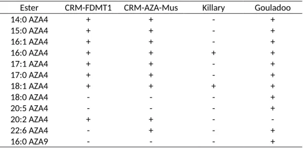
Table S2. Summary of 3-O-acyl AZA esters detected in whole mussel tissues. (+) detected, (-) not detected.
Ester CRM-FDMT1 CRM-AZA-Mus Killary Gouladoo
14:0 AZA4 + + - +
15:0 AZA4 + + - +
16:1 AZA4 + + - +
16:0 AZA4 + + + +
17:1 AZA4 + + - +
17:0 AZA4 + + - +
18:1 AZA4 + + + +
18:0 AZA4 - - - +
20:5 AZA4 - - - +
20:2 AZA4 + + - -
22:6 AZA4 - + - +
16:0 AZA9 - - - +
Figure S1. (A) Extracted ion chromatogram of AZA4, the product at m/z 826.4743 and 3-O-palmitoyl AZA4 from semi-synthesis of 3-O-palmitoyl (16:0) AZA-4. (B) Full scan MS spectrum of the product with [M+H]+ of m/z 826.4743, suspected to be due to 2,3-dehydroAZA4 (C46H68O12N+, Δ 0.8 ppm) based on periodate cleavage of the 20,21-diol (data not shown).
Figure S2. Extracted ion chromatogram of m/z 1082.7138 (±5 ppm) of the Bruckless HP tissue (black) and the semi-synthesized 3-O-palmitoylAZA4 (red).
Figure S3. Product ion spectra of 3-O-pamitoylAZA7 co-eluting with 3-O-margaroylAZA4 in the concentrated 7:3 EtOAc:MeOH silica gel fraction of the hepatopancreas tissue. The vertical scale from
Figure S4. Product ion spectrum of semi-synthetic 3-O-palmitoylAZA9, which was confirmed to be present in the HP tissue.
Figure S5. Product ion spectrum of the semi-synthetic 3-O-palmitoylAZA7.
Figure S6. (A) Extracted ion chromatogram of AZA1 (m/z 842.5049) and of the semi-synthetic 16:0 ester of AZA1 (presumed to be 20-O-palmitoylAZA1, m/z 1080.7345) (B) Product ion spectrum of the semi- synthesized 20-O-palmitoylAZA1), which was not detected in the HP tissue.
Figure S7. Extracted ion chromatogram of the product ions at m/z 362.2690 and 168.1381 from the mass range of m/z 650 to 1200 using DIA acquisition in the HP tissue in a control extract (black) and 2 h after treatment with sodium periodate.