Copyright © 2002, American Society for Microbiology. All Rights Reserved.
Biochemistry and Comparative Genomics of SxxK Superfamily
Acyltransferases Offer a Clue to the Mycobacterial Paradox:
Presence of Penicillin-Susceptible Target Proteins versus
Lack of Efficiency of Penicillin as Therapeutic Agent
Colette Goffin and Jean-Marie Ghuysen*
Center for Protein Engineering, Institut de Chimie, University of Lie`ge, B-4000 Sart Tilman, Lie`ge, Belgium
INTRODUCTION ...702
PREDICTIVE STUDIES...705
LIPID II PRECURSORS: PEPTIDOGLYCAN ASSEMBLY...706
SxxK ACYLTRANSFERASE SUPERFAMILY ...707
STRUCTURE-ACTIVITY RELATIONSHIPS OF !-LACTAM ANTIBIOTICS ...709
GROUP I SxxK ACYLTRANSFERASES: IMPLICATED IN (433) PEPTIDOGLYCAN BIOCHEMISTRY ...712
Class A PBP Fusions: Nascent (433) Peptidoglycan ...712
Class B PBP Fusions: Morphogenetic Apparatus...715
Free-Standing PBPs: Auxiliary Cell Cycle Proteins...717
E. coli PBP Fusions as Targets for !-Lactam Antibiotics ...719
Mosaic PBP Fusions...720
GROUP II SxxK ACYLTRANSFERASES...720
GROUP III SxxK ACYLTRANSFERASES: PENICILLIN-RESISTANT PROTEIN FUSIONS ...722
Class B PenrProtein Fusions of Gram-Positive Bacteria...723
Class A PenrProtein Fusions of Gram-Negative Bacteria ...725
SxxK ACYLTRANSFERASES IN SPECIFIC ORGANISMS ...728
Class A PBP Fusions of M. tuberculosis, M. leprae, M. smegmatis, and C. diphtheriae ...728
Class A PenrProtein Fusions of M. tuberculosis, M. leprae, M. smegmatis, and C. diphtheriae...729
Subclass B3 and B-Like I PBP Fusions of M. tuberculosis, M. leprae, and C. diphtheriae ...729
Class B-Like II PenrProtein Fusions of M. tuberculosis, M. leprae, and C. diphtheriae...730
Free-Standing PBPs, !-Lactamases, and Related Polypeptides of M. tuberculosis and M. leprae...730
PBP Fusions and PenrProtein Fusions of S. coelicolor...730
Detected versus Predicted PBPs of M. tuberculosis H37Ra...731
WHERE NEXT?...731
ACKNOWLEDGMENTS ...733
REFERENCES ...733 INTRODUCTION
The bacterial wall peptidoglycans are covalently closed, net-like polymers (77, 203). The glycan chains are made of alter-nating "-1,4-linked N-acetylglucosamine and N-acetylmuramic acid residues. TheD-lactyl groups of the muramic acid residues are amidated with L-alanyl-#-D-glutamyl-(L)-diaminoacyl-D-alanine stem tetrapeptides and L-alanyl-#-D-glutamyl-L-diami-noacid stem tripeptides. The stem peptides can be branched, in which case the $ amino group of the diaminoacid residue is substituted by an additional amino acid residue or a short peptide. Unbranched or branched stem peptides belonging to adjacent glycan strands are covalently linked, resulting in poly-meric peptidoglycan.
All peptidoglycan-containing bacteria in the exponential phase of growth manufacture a (433) peptidoglycan (Fig. 1) in
a penicillin-susceptible manner. Peptidoglycan crosslinking ex-tends from the carboxy-terminalD-alanine residue at position 4 of a stem tetrapeptide to the lateral amino group at position 3 of another, unbranched or branched, stem peptide. The (433) interpeptide linkages or cross-bridges are made by spe-cialized acyltransferases which are immobilized by penicillin in the form of stable, enzymatically inactive penicillin-binding proteins (PBPs) (214). Escherichia coli, the mycobacteria, and leprosy-derived corynebacteria (111) produce unbranched (433) peptidoglycans with meso-diaminopimelic acid at posi-tion 3 of the stem peptides. Peptidoglycan crosslinking is me-diated by direct (D)-alanyl-(D)-meso-diaminopimelic acid inter-peptide linkages (Fig. 1). In mycobacteria, however, muramic acid is either acylated or glycolylated (12). The %-carboxylate ofD-glutamic acid can be amidated. A glycine residue substi-tutes for theL-alanine residue at the amino end of the stem peptides in Mycobacterium leprae.
Bacteria also exist which have the dual ability to manufac-ture a peptidoglycan of the (433) type and another pepti-doglycan of the (333) type (Fig. 2). The (333) interpeptide
* Corresponding author. Mailing address: Center for Protein Engi-neering, Institut de Chimie, University of Lie`ge, B6, B-4000 Sart Til-man, Lie`ge, Belgium. Phone: 32 4 366.33.96/95. Fax: 32 4 366.33.64. E-mail: jmghuysen@ulg.ac.be.
linkages or cross-bridges extend from the %-carbonyl of the diaminoacid residue (L-center) at position 3 of a stem peptide to the lateral amino group at position 3 of another, unbranched or branched, stem peptide.
The identification in the early 1970s of the occurrence of (333) peptidoglycans in Streptomyces albus G, Clostridium
per-fringens (139), Mycobacterium smegmatis ATCC 21732, and Mycobacterium tuberculosis BCG Pasteur strain (244) came as
an exclamation point. Mycobacteria have a highly crosslinked peptidoglycan. About 70% to 80% of the total meso-diamin-opimelic acid residues are involved in interpeptide linkages. In
Mycobacterium smegmatis grown in Sauton’s medium in Roux
bottles for 9 days at 37°C, the (433) and (333) peptidoglycans
occur in the proportion of about 2 to 1. The pattern of distri-bution of the two peptidoglycans is not known, but (333)-linked peptide trimers occur in M. smegmatis and M.
tubercu-losis BCG.
Subsequently, it was found that Escherichia coli manufac-tures a (333) peptidoglycan (Fig. 2) in increasing proportions of total peptidoglycan and becomes increasingly more resistant to "-lactam antibiotics as the generation time increases (227). Penicillin-induced lysis also causes an increased proportion of (333) peptidoglycan (127). The contribution of (333) peptidoglycan in Aeromonas spp., Acinetobacter
acetoaceti-cus, Agrobacterium tumefaciens, Enterobacter cloacae, Proteus morganii, Pseudomonas aeruginosa, Pseudomonas putida, Sal-FIG. 1. (433) peptidoglycans.D-Alanyl-diaminoacyl interpeptide linkages and cross-bridges (boxed) betweenL-Ala-#-D-Glu-(L )-diaminoacyl-D-alanine stem peptides. The diamino acid residues are meso-diaminopimelic acid (Dpm),L,L-diaminopimelic acid, orL-lysine. The stem peptides are unsubstituted at position 3 in Mycobacterium, Corynebacterium, and Bacillus spp. and E. coli. They are substituted at position 3 by one or several additional amino acid residues (%%) in the other organisms shown. G, N-acetylglucosamine; M, N-acetylmuramic acid (see Fig. 4); COX & COOH or CONH2. In S. pneumoniae, the cross-bridges are Nε-(L-alanyl-L-alanyl)- or (L-alanyl-L-seryl)-L-lysine. In S. aureus, the cross-bridges comprise five glycine residues or three glycine and twoL-serine residues.
monella enterica serovar Typhimurium, Vibrio parahaemolyti-cus, Yersinia enterocolitica, and E. coli grown to late
exponen-tial phase varies from 1% to 45% of total peptidoglycan (183, 184). Finally, Enterococcus faecium is also a (333) peptido-glycan manufacturer (Fig. 2). A laboratory mutant has been isolated which resists penicillin in the exponential phase of growth, conditions under which it manufactures a wall pepti-doglycan of the (333) type exclusively (145, 205).
From the foregoing, it follows that, in all likelihood, (333) peptidoglycan crosslinking is carried out by acyltransferases that escape penicillin action and that, in particular genetic backgrounds or under specific growth conditions, the penicil-lin-resistant (333) peptidoglycan assembly molecular machine can substitute for the penicillin-susceptible (433) peptidogly-can assembly molecular machine. This conclusion raises ques-tions of fundamental and practical importance, related, in par-ticular, to the mycobacterial pathogens.
E. coli has a 4.60-Mb genome (24). M. tuberculosis H37Rv
has a 4.41-Mb genome (38). Mycobacterium leprae has a 3.27-Mb genome (39) that shows extensive decay and down-sizing. M. tuberculosis and M. leprae share about 1,500 genes. As stated above, E. coli and Mycobacterium spp. manufacture
similar, unbranched, meso-diaminopimelic acid-containing peptidoglycans of the (433) and (333) types. The nonpepti-doglycan wall polymers, however, are different. In E. coli, sev-eral lipoproteins are linked to the peptidoglycan. The 56-ami-no-acid residue Braun’s lipoprotein (29) contains one fatty acid bound as an amide to the amino group of a cysteine residue and two fatty acids bound as esters to the hydroxyl groups of S-glyceryl cysteine (Fig. 3). The fatty acids are em-bedded in the outer membrane. Lipoprotein molecules occur both in a free form and in covalent linkage with the underlying peptidoglycan. The linkage is an amide bond between the ε-amino group of theL-lysine residue at the carboxy end of the lipoprotein and the carbonyl group at theL-center of the meso-diaminopimelic acid residue at position 3 of a stem tripeptide of the peptidoglycan.
In mycobacteria, mycolic acid, arabinogalactan, and pepti-doglycan form a covalent complex (30). Mycolic acids are 2-alkyl 3-hydroxy branched-chain fatty acids. They are ester linked to arabinogalactan, which itself is linked to carbon C-6 of muramic acid via phosphodiester bonds. One may also note that a large portion of the coding ability to M. tuberculosis H37Rv is involved in lipid and polyketide metabolism (38) and
FIG. 2. (333) peptidoglycans. Diaminoacyl-diaminoacyl interpeptide linkages and cross-bridges (boxed) betweenL-Ala-#-D-Glu-(L
)-diami-noacyl-D-alanine stem peptides. The stem peptides are unsubstituted at position 3 in Mycobacterium spp. and E. coli. They are substituted
8% of the genome is devoted to the production of two families of glycine-rich proteins of repetitive structure (20, 186).
M. tuberculosis and M. leprae have different life styles. M. tu-berculosis enters host macrophages at cholesterol-rich domains
of the plasma membrane (75). It survives in the macrophage by preventing fusion of the mycobacterial phagosomes with lyso-somes (50, 201, 218, 236). It may persist inside macrophages lodged in calcified structures of the lungs and be reactivated decades after initial infection (153). It grows in synthetic media with a generation time of 12 to 24 h. Tuberculosis, once almost vanquished, resurged in the late 1980s because of the emer-gence of multidrug-resistant strains (25, 250). Often acting in deadly combination with AIDS, it kills more than 2 million people each year. Mycobacterium leprae enters the body via the mucosal linings of the nose or through open wounds. It shows a marked tropism for myelin-producing Schwann cells, which insulate nerves (187, 188). It may be dormant for years until the body’s immune system attacks the infected cells, destroying the nerves and degrading soft tissues and even bones. It is unculturable but can be grown in the nine-banded armadillo and the footpad of mice with an estimated generation time of about 2 weeks. Leprosy still flourishes in developing countries, where more than 750,000 people contract the disease each year.
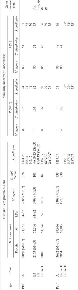
Consistent with its ability to manufacture a (433) pepti-doglycan, M. tuberculosis H37Ra (ATCC 25177, an attenuated laboratory strain) produces four major PBPs of 94, 82, 52, and 37 kDa (35). The inactivation of the 94-, 82-, and 52-kDa PBPs in cells grown to mid-exponential phase is associated with antibacterial activity. "-Lactamase production (240), more than the relatively low permeability of the cell envelope, is the major determinant of resistance (35, 63, 112, 185). Conse-quently, the MICs of ampicillin associated with the "-lacta-mase inactivator sulbactam are!0.1 'g ml(1for M. tubercu-losis H37Ra and!2 'g ml(1for M. tuberculosis H37Rv (35). The drug combination is bactericidal to M. tuberculosis in ex-ponential-phase cultures. It is bactericidal to M. tuberculosis multiplying in macrophages (35) and to M. leprae multiplying in mouse footpads (180, 189).
Paradoxically in view of the above data, the "-lactam
anti-biotics are not effective therapeutic agents for the treatment of tuberculosis and leprosy (101). This lack of efficiency could be due to a shift of peptidoglycan synthesis from the (433) type to the (333) type. Information that can help apprehend the problem was sought from comparative genomics of bacterial species (E. coli, Enterobacter faecium, Mycobacterium spp., and others) which have the ability to assemble, presumably from the same pool of precursors, peptidoglycans of the (433) and (333) types. Searches were also made for the positions of genes on the chromosomes because operons are common in prokaryotes and genes that are neighbors tend to be function-ally linked.
PREDICTIVE STUDIES
Data concerning unfinished genome sequences were from the Institute for Genomic Research (TIGR) website at http: //www.tigr.org. They were produced at TIGR with the support of the National Institute of Allergy and Infectious Diseases for
Mycobacterium avium 104, at the Sanger Center/Institut
Pas-teur/VLA Weibridge with the support of MAFF/Beowulf Genomics for Mycobacterium bovis AF2122/97, at the Sanger Center/World Health Organization/Public Health Laboratory with the support of Beowulf Genomics for Corynebacterium
diphtheriae NCTC 13129, and at the Sanger Center/John Innes
Center with the support of BBSRC/Beowulf Genomics for
Streptomyces coelicolor A3 (2).
Amino acid sequence similarities were searched with the Basic Local Alignment Search Tool Blast programs of the National Center for Biotechnology Information (NCBI) from their website at http://www.ncbi.nlm.nih.gov/Blast (4). Program BlastP compares a query amino acid sequence against a pro-tein database. Program tBlastN compares a query amino acid sequence against a nucleotide sequence database dy-namically translated in all six reading frames. E. coli, M.
tu-berculosis, M. leprae, C. diphtheriae, and S. coelicolor proteins
were searched in the nonredundant protein database with BlastP and in the finished and unfinished genome database with tBlastN. The Expect value E of the NCBI’s programs allows estimation of how far from the background noise is the
similarity between aligned sequences (3). For values of !0.01,
E becomes equivalent to the probability P that the sequences
align by chance (118). P values smaller than 10(3 to 10(4 (depending on the sizes of the databanks) indicate statistically significant similarity.
The P values depend on the number and size of the gaps introduced to optimize the alignments. When the P value is small, equal or close to 0.0, the percentage of identical amino acid residues allows the sequences to be hierarchically classi-fied. Combining the P values, the lengths of the overlapping regions, and the percentages of identities (I) present in the aligned regions gives an estimate of the extent of similarity between the sequences under comparison. Proteins encoded by genes having a common ancestor are homologues (193). Proteins encoded by genes related by duplication within a genome followed by diversification normally evolve new func-tions (103); they are paralogues. Homologous proteins en-coded by genes in different organisms separated by speciation are orthologues (51, 103).
Protein orthologues that are essential and related by small
P values and large I values normally retain similar biological
functions. This conclusion is especially pertinent when low
P values and large I values apply, over the entire sequences, to
protein fusions for which the constitutive modules evolved from different protein ancestors. In contrast, little information is obtained when the similarities are restricted to peptide stretches or amino acid groupings. Conserved motifs defining the boundary of a catalytic center give valuable information on the catalytic mechanism, not on the specificity and fate of the reactions catalyzed.
LIPID II PRECURSORS: PEPTIDOGLYCAN ASSEMBLY Lipid II molecules are the immediate biosynthetic precur-sors of the wall peptidoglycans (102). A disaccharide peptide exposed on the outer face of the plasma membrane is linked to a C55H89undecaprenyl carrier via a pyrophosphate bridge in-volving carbon C-1 of N-acetylmuramic acid. The stem peptide borne by N-acetylmuramic acid is a pentapeptide terminating in D-alanyl-D-alanine. It can be unsubstituted or branched at position 3.
In E. coli, the synthesis of lipid II (Fig. 4) involves an inter-change of carriers that are compatible with the environments of the cell (231). UDP-N-acetylglucosamine is converted into UDP-N-acetylglucosamine-enolpyruvate by MurA and from this into UDP-N-acetylmuramic acid by MurB. The Ddl (ATP,ADP ! Pi) ligase catalyzes the formation of a D-alanyl-D-alanine dipeptide, and the MurC, MurD, MurE, and MurF (ATP,ADP ! Pi) ligases catalyze the formation of UDP-N-acetylmuramoyl pentapeptide by the sequential additions to UDP-N-acetylmuramic acid of L-alanine, D-glutamic acid,
meso-diaminopimelic acid, and the preformed D-alanyl-D-ala-nine dipeptide. Then, MraY transfers the phospho-N-acetyl-muramoyl pentapeptide from its uridylic carrier to a mem-brane-bound C55-isoprenoid alcohol phosphate, giving rise to lipid I. MurG transfers the N-acetylglucosamine from its uridylic carrier to carbon atom C-4 of N-acetylmuramic acid, giving rise to lipid II. Somehow, lipid II flips over the mem-brane bilayer, and the disaccharide pentapeptide moiety is exposed on the outer face of the membrane (Fig. 4). The genes
ddl, murC, murD, murE, murF, murG, and mraY reside in the dcw (or mra) cluster at the 2-min region of the chromosome
(Fig. 5).
The stem pentapeptides can be modified at different stages of the biosynthesis of lipid II (234). Amidation of the %-car-boxylate of D-glutamic acid (88) and the carboxylate at the D-center of meso-diaminopimelic acid probably takes place at the level of the UDP-N-acetylmuramoyl pentapeptide precur-sors. In many gram-positive bacteria, the diaminoacid residue at position 3 of the stem pentapeptide isL-lysine. Addition of one or several amino acid residues to the ε-amino group of the L-lysine residue, resulting in the formation of branched-stem pentapeptides, probably occurs at the level of lipid II while it is still oriented toward the cytosol.
In Staphylococcus aureus, there are four glycyl tRNAs. One is involved in protein synthesis. The three others seem to be used exclusively for peptidoglycan synthesis. Lipid II precursor molecules with one, three, and five glycine residues attached to the ε-amino group of the L-lysine residue are formed by the sequential actions of the glycine-adding enzymes FemX (also called FemhB), FemA, and FemB (128, 197). Loss of FemX is lethal. In some strains, the FemAB-like Lif and Epr incorpo-rate anL-serine residue instead of a glycine residue within the branches at positions 3 and 5 (60). In Streptococcus
pneu-moniae, the Nε-(L-alanyl-L-alanyl)-L-lysine and Nε -(L-alanyl-L-seryl)-L-lysine branches are synthesized by MurM and MurN, respectively (64, 242), with MurM being responsible for the addition of the first amino acid residue to the ε-amino group of L-lysine.
Vancomycin-resistant enterococci are programmed to pro-duce lipid II precursor molecules which terminate in D-alanyl-D-lactate (8, 136). Five enzymes are necessary and sufficient. The Zn2!-dependent VanX dipeptidase hydrolyzes the D-ala-nyl-D-alanine dipeptide produced by the Ddl ligase. VanH and VanA act sequentially to synthesize the depsipeptide D-alanyl-D-lactate, which is incorporated in lipid II rather than the dipeptideD-alanyl-D-alanine. The production of VanHAX is controlled by a two-component regulatory system that involves the transmembrane sensor kinase VanS and the transcription factor VanR, which becomes active when phosphorylated by VanS (8).
The conversion of the disaccharide peptide (depsipeptide) moiety of lipid II into polymeric (433) peptidoglycan requires the sequential actions of two transferases (Fig. 4). A glycosyl-transferase catalyzes attack of carbon C-1 of N-acetylmuramic acid of a lipid II molecule by the acceptor nucleophile OH of carbon C-4 of N-acetylglucosamine of another lipid II mole-cule (or at the nonreducing end of a growing glycan chain; not shown). At each transfer, the delivery of a disaccharide peptide unit generates an undecaprenyl pyrophosphate which is de-phosphorylated. The C55-isoprenoid alcohol phosphate turns over the membrane bilayer so that the phosphate group faces the cytosol, allowing a new cycle to start.
In turn, the requiredD,D-acyltransferase (Fig. 4 and reaction I in Fig. 6) catalyzes attack of the carbonyl of the D-alanine residue at position 4 of a pentapeptide (depsipeptide) borne by a glycan chain, by the lateral amino group at position 3 of a peptide borne by another glycan chain. The reaction proceeds via the transitory formation of an N-acyl-D-alanyl-enzyme in-termediate in which theD-alanine residue is linked as an ester
to a serine residue at the catalytic center (69, 70, 78). The carbonyl donor involved in enzyme acylation is invariably a D-alanyl-D-alanine(-D-lactic acid) sequence. In contrast, the ac-ceptor involved in enzyme deacylation can be the amino group of a glycine residue, the ε-amino group of anL-lysine residue, or an amino group borne by anL- or aD-configured carbon atom (Fig. 1).
Water can also serve as an acceptor. Stem pentapeptides are hydrolyzed into stem tetrapeptides, preventing further (433) peptidoglycan crosslinking. The "-lactam antibiotics act as sui-cide carbonyl donors of the D,D-acyltransferases (224). Rup-ture of the "-lactam amide bond produces a serine ester-linked acyl (penicilloyl, cephalosporoyl, . . .) enzyme, which is a dead end because the bulky acyl moiety is a steric hindrance to the approach of any attacking nucleophile. The catalytic center turns over very slowly, once or less per hour (71, 72), and the inactivatedD,D-acyltransferases are detectable as PBPs (214). No one knows exactly how the (333) peptidoglycans are made in a penicillin-resistant manner. The (333) interpeptide linkages or cross-bridges could be made through the action of
anL,D-acyltransferase that cleaves the bond between position 3 and position 4 of pentapeptide carbonyl donors, with release of the dipeptideD-alanyl-D-alanine (reaction II in Fig. 6). Alter-natively, they could be made through the sequential actions of aD,D-carboxypeptidase catalyzing reaction IIIa (Fig. 6) and an L,D-acyltransferase that cleaves the bond between position 3 and position 4 of tetrapeptide carbonyl donors (reaction III in Fig. 6). Reactions IIIa and III each cause the release of a single D-alanine residue.
SxxK ACYLTRANSFERASE SUPERFAMILY The serine acyltransferases implicated in wall peptidoglycan assembly are members of a superfamily of SxxK serine en-zymes. The term superfamily is used to include proteins with no statistically significant sequence similarities but with similar structures in the classical sequence-based families (169). With x denoting a variable amino acid residue, the acyltransferases of this superfamily have a specific bar code in the form of three motifs, SxxK, SxN (or analogue), and KTG (or analogue). The
FIG. 4. Lipid II precursor (bottom) and polymeric (433) peptidoglycan (top) of E. coli. Reactions catalyzed by the glycosyl- and acyltrans-ferases. For details, see the legend to Fig. 1.
motifs occur at equivalent places and with roughly the same spacing along the polypeptide chains. In the three-dimensional structures, they are brought close to each other at the imme-diate boundary of the catalytic center between an all-% domain and an %/" domain, the five-stranded "-sheet of which is cov-ered by %-helices (79, 123, 124).
The serine residue of the invariant motif SxxK occupies a central position in the catalytic center and is central to the
enzyme acylation and deacylation steps, which, mechanisti-cally, are similar to the proteolytic reactions of the trypsin protein family. The mature SxxK acyltransferase of
Streptomy-ces sp. strain K15 is 262 amino acid residues long. It is one of
the smallest members of the superfamily (Fig. 7) (68). Diverg-ing evolution gave rise to a constellation of SxxK acyltrans-ferases with various amino acid sequences. Fusion to other polypeptide chains resulted in a combinatorial system of
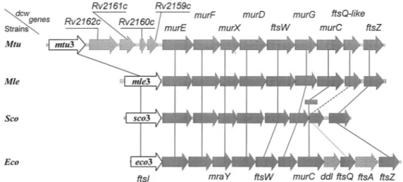
struc-FIG. 5. Organization of the division cell wall (dcw) gene clusters of M. tuberculosis (Mtu), M. leprae (Mle), Streptomyces coelicolor (Sco), and
E. coli (Eco). All the coding sequences are on the same DNA strands. Open arrows, mtu3, mle3, sco3, and eco3 (synonymous to ftsI), coding for
cell septation SxxK PBP fusions. Shaded arrows, with fts standing for the filamentous phenotype of thermosensitive E. coli mutants, ftsW, ftsQ, ftsA, and ftsZ code for cell septation-related, non-penicillin-binding proteins. Genes murE, murF, murX (synonymous to mraY), murD, murG, and murC code for enzymes of the lipid II-synthesizing pathway. In S. coelicolor, murC (shaded rectangle) is not in the dcw cluster. Stippled grey arrows, genes of a dcw cluster having no homologues in the other clusters. In M. tuberculosis, the genes inserted between mtu3 and murE probably code for a glycine-rich protein of repetitive structure (Rv2162c), a protein of the lincomycin-synthesizing pathway (Rv2161c), and proteins of unknown function (Rv2160c and Rv2159c). A gene homologous to E. coli ddl (coding for the ligase which catalyzes the formation ofD-alanyl-D-alanine) is
located elsewhere in the chromosomes of M. tuberculosis, M. leprae, and S. coelicolor. E. coli ftsA codes for a cell division, actin-like ATPase. Solid, dashed, and dotted lines connecting the dcw genes indicate the extents, in decreasing order, of similarity between homologous genes. The E. coli cell septation genes mraZ, mraW, and ftsL (not shown) are upstream from eco3.
FIG. 6. Acyl transfer reactions. Formation of (433) peptidoglycan interpeptide linkages by penicillin-susceptibleD,D-N-acyl-D-alanyl-D-alanine
transpeptidases (reaction I). Formation of (333) peptidoglycan interpeptide linkages by penicillin-resistantL,D-N-acyl-L-diaminoacyl-D-dipeptidyl
transpeptidases (reaction II) andL,D-N-acyl-L-diaminoacyl-D-alanine transpeptidases (reaction III). Hydrolysis of stem pentapeptides into stem
tural modules that contributed to a massive increase in func-tional diversity.
The acyltransferases of the SxxK superfamily are a paradigm of catalytic versatility. They fall into three main groups. The D,D-acyltransferases–PBPs, implicated in (433) peptidoglycan synthesis, are of group I. Acyltransferases endowed with di-verse functions unrelated to peptidoglycan biochemistry are of group II. Penicillin-resistant acyltransferases that could act as L,D-peptidases in (333) peptidoglycan synthesis are of group III. The ensuing sections provide critical coverage of mature topics and speculations on emerging topics. The identifiers of the SxxK acyltransferases are shown in Table 1. They combine the name of the producing bacterial species as a three-letter
prefix flanked by a number or additional letters that specify the protein under consideration (e.g., Eco1a, protein 1a of E. coli).
STRUCTURE-ACTIVITY RELATIONSHIPS OF !-LACTAM ANTIBIOTICS
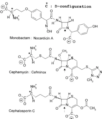
The SxxKD,D-acyltransferases of group I are the target pro-teins of the "-lactam antibiotics. These compounds are a large group of molecules of which the common structural feature is the presence of a "-lactam (azetidinone) ring (Fig. 8). Cur-rently, they are regarded as a molecular mimic of N-acyl-D-alanyl-D-alanine (224), explaining why they are suicide
car-FIG. 7. Basic polypeptide fold of the acyltransferases of the SxxK superfamily and spatial disposition of the three catalytic center-defining amino acid groupings. The structure shown is that of the 262-amino-acid DD-transpeptidase-PBP of Streptomyces sp. strain K15. The SxxK
acyltransferases comprise an all-% domain (left side) and an %/" domain (right side). The invariant motif 1, S*xxK, where S* is the essential serine nucleophile, forms the amino-terminal turn of helix %2 of the all-% domain. Motif 2, most often SxN or SxD (here SxC), is on a loop connecting two helices of the all-% domain. Motif 3, most often KTG or KSG, is on strand "3 of the %/" domain. The structure was built with Molscript (130) and Raster3D (155). Illustration courtesy of Paulette Charlier, Eveline Fonze´, Michae¨l Delmarcelle, and Andre´ Piette, Center for Protein Engineering.
TABLE 1. Prefixes identifying bacterial species
Prefix Species Prefix Species
Aae... Aquifex aeolicus
Ana... Anabaena sp. strain PCC7120 Bbu... Borrelia burgdorferi
Bsu... Bacillus subtilis
Cdi... Corynebacterium diphtheriae Eco ... Escherichia coli
Efam... Enterococcus faecium Efas ... Enterococcus faecalis Ehi... Enterococcus hirae Hin ... Haemophilus influenzae Hpy... Helicobacter pylori Mle ... Mycobacterium leprae Msm ... Mycobacterium smegmatis
Mtu ... Mycobacterium tuberculosis Nme ... Neisseria meningitidis Pae ... Pseudomonas aeruginosa Sau ... Staphylococcus aureus Scl... Streptomyces clavuligerus Sco... Streptomyces coelicolor Sgr ... Streptomyces griseus Sor... Streptococcus oralis Spn ... Streptococcus pneumoniae Ssc ... Staphylococcus sciuri Spy... Streptococcus pyogenes Sth ... Streptococcus thermophilus Syn... Synechocystis sp. strain PCC6203
bonyl donors of the D,D-acyltransferases–PBPs implicated in (433) peptidoglycan synthesis (Fig. 4 and reaction I in Fig. 6). The penams, 3-cephems, and N-acyl-D-alanyl-D-alanine-ter-minated peptides (in the extended conformation) have a com-mon backbone, CON-C2-CON-C3-COO((Fig. 9, upper part). Carbon atoms C-2 and C-3 (marked by asterisks) have a D-configuration. They are the pivots that connect the central CON amide plane at position I to the CON amide plane at position II (through the rotation angles )2 and *2) and the carboxylate at position III (through the rotation angles *3and )3). The scissile CON bonds at position I, however, are far from being isosteric. The nitrogen atom is planar in the
pep-tides. It is pyramidal in the azetidinones. The bond angle % is 117° in the peptides and 90° in the azetidinones. The rotation angle $ is 180° in the peptides, 155° in the 3-cephems, and 135° in the penams.
Rather than being related to similarities between linked atoms, structural analogy between the most stable D-alanyl-D-alanine-terminated peptide, penam, and 3-cephem conformers relies on the spatial disposition of the electrostatic negative wells created by the carbonyl CO at position I, the carbonyl CO at position II, and the carboxylate COO(at position III (80, 131, 132). These three groupings are almost coplanar, and the spanning distances between the oxygen atoms at position I and
FIG. 8. "-Lactam antibiotic family. Only 6-epoxypenicillin S is active. *,D-configured carbon atom. (“Mecillinam” is another name for
position II (!5 A˚) and between the oxygen atom at position I and the carbon atom of the carboxylate at position III (3.5 to 4.5 A˚ ) are roughly similar. Figure 9 (lower part) shows the three-dimensional electrostatic potentials of the optimized benzylpenicillin and extended bisacetyl-L-lysyl-D-alanyl-D-ala-nine molecules. The two additional negative wells seen in the peptide are due to the acetyl groupings that substitute the %-and ε-amino groups of theL-lysine residue.
As a result of the coplanarity of the negative wells at
posi-tions I, II, and III, the %-face of the azetidinone ring of the "-lactam antibiotics is well exposed, facilitating the attack of the electrophilic center at position I by the serine nucleophile of the SxxK D,D-acyltransferases. The backbone CON-C2 -CON-C3-COO(is modified into CON-C2-CON-SO3(in the monobactam aztreonam (Fig. 8). It is modified into C&N-C2 -CON-C3-COO(in the penam amdinocillin. The carbon atom C-2 of the carbapenem imipenem has anL-configuration. In spite of these structural variations, the disposition of the
sulfa-FIG. 9. Common backbone of extended N-acyl-D-alanyl-D-alanine peptides and penams (top) and ab initio electrostatic potentials of bisacetyl-L-lysyl-D-alanyl-D-alanine and benzylpenicillin (bottom) optimized at level AM1 (the terminal carboxylates are protonated). The electrostatic
negative wells I, II, and II are shown at levels of (40 kcal (solid contours) and (30 kcal (dotted contours). They are coplanar. The negative wells generated by the acetyl substituents of the %- and ε-amino groups of theL-lysine residue of bisacetyl-L-lysyl-D-alanyl-D-alanine are above and below
the plane, respectively. The negative well of small amplitude seen between well I and well III of benzylpenicillin is due to the pair of free electrons of the nitrogen atom of the azetidinone ring. Illustration courtesy of Georges Dive, Center for Protein Engineering.
mate in aztreonam is roughly comparable to that of the car-boxylate at position III in the bicyclic penams and 3-cephems. The + environment of the CH&N amidino bond of amdino-cillin generates a negative well at position II, albeit of reduced amplitude. Because the rotation angle , (Fig. 9, upper part) is less open in the carbapenems than in the penams and 3-cephems, the carboxylate at position III of imipenem is moved upward, ensuring coplanarity with the oxygen atom at position I and the alcohol oxygen atom at position II. Of the two 6-epoxypenicillin isomers (Fig. 8), the side chain of which, at position II, has a frozen conformation due to the epoxy cycle, the S isomer is active, but the R isomer is not.
Depending on the (noncyclic, monocyclic, or bicyclic) frame-work and conformation of the backbone and the presence of bulky, ionized, and electron-withdrawing side chains, the trostatic negative wells at positions II and III around the elec-trophilic center at position I of the carbonyl donors can vary in shape and strength, be displaced along the reference plane, be better expressed in other sections of the molecules, be fused, or be masked. In turn, the SxxK acyltransferases are large bodies studded with positively and negatively charged magnets. Upon binding of a carbonyl donor to the catalytic center, a dense hydrogen bonding network is formed, and a multimem-bered ring that is both enzyme and ligand specific is created, in which the scissile CO-N amide bond at position I of the car-bonyl donor is connected to the catalytic serine #OH of motif SxxK of the acyltransferase by one or several water molecules and the side chains of several amino acid residues (53, 54, 80). This multimembered ring is utilized as a motorway, allowing the proton of the serine #OH to be transferred via a transition state to the nitrogen atom of the scissile bond, resulting in enzyme acylation. For the reaction to reach completion, the serine ester-linked acyl enzyme must adopt a conformation that allows entry of an amino group or a water molecule and formation of a new multimembered ring that performs enzyme deacylation.
Potential energy hypersurfaces best portray the geometric rearrangements, electronic redistributions, and free energy barriers that occur along the reaction pathways (80). Depend-ing on the freedom of the water molecules, the ease with which the ligands undergo deformation, and the ease with which the enzyme backbone undergoes relaxation, numerous possible routes for proton transfer exist. The energetically most favor-able route dictates the specificity of the SxxK acyltransferases and the completeness (protein binding, protein acylation, pro-tein acylation, and deacylation) and productiveness of the cat-alyzed reactions.
TheD,D-acyltransferases–PBPs of group I perform multiple functions related to (433) peptidoglycan assembly (see be-low). Changes in protein structure, with conservation of the overall fold, could go as far as a change fromD,Dspecificity to L,D specificity (see the chapter on SxxK acyltransferases of group III).
GROUP I SxxK ACYLTRANSFERASES: IMPLICATED IN (433) PEPTIDOGLYCAN BIOCHEMISTRY The SxxK D,D-acyltransferases–PBPs of group I occur as independent entities referred to as free-standing PBPs. They also occur as modules of PBP fusions. A D,D-acyltransferase
module of class A or class B is fused, in a class-dependent manner, to polypeptides having their own bar codes and three-dimensional structures (78, 84). Traces of similarity between the free-standing PBPs and the acyltransferase modules of class A or B are generally limited to the three active-site defining motifs. Similarity between the acyltransferase mod-ules of class A and class B is marginal (P " 1 - 10(5).
The PBPs are bound to the plasma membrane, with the bulk of the polypeptide chains exposed on the outer face. The PBP fusions are synthesized with an amino-terminal hydrophobic segment that functions as both a signal sequence for secretion and a stop transfer signal that serves as a membrane anchor. Despite a great divergence in the amino acid sequences, the bar codes SxxK, SxN, and KTG of the free-standing PBPs and the acyltransferase modules of the PBP fusions are conserved except that, occasionally, SxD substitutes for SxN and KSG substitutes for KTG.
The core of an SxxK acyltransferase is defined as the se-quence starting about 70 amino acid residues upstream from the SxxK motif and terminating about 70 amino acid residues downstream from the KT(S)G motif. Adducts may occur as inserts and/or as carboxy-terminal extensions. The free-stand-ing PBP5 of E. coli (45) and the class B PBP fusion Spn2x of
Streptococcus pneumoniae (86, 173) are of known structure.
They each have carboxy-terminal extensions. That of E. coli PBP5 is made of "-structures that form a loose "-barrel; that of Snp2x is made of two %/"/"/" domains.
Class A PBP Fusions: Nascent (433) Peptidoglycan The class A PBP fusions consist of a penicillin-binding acyl-transferase module of class A linked to the carboxy end of a glycosyltransferase module, itself linked to the carboxy end of the membrane anchor (Fig. 10). The full bar code comprises motifs 1 to 5 of the glycosyltransferase module, motif 6 of the intermodule junction, and motifs 7 to 9 of the acyltransferase module (84). By combining a glycosyltransferase and an acyl-transferase in a single polypeptide chain, class A PBP fusions catalyze the polymerization of the disaccharide pentapeptide units borne by lipid II precursor molecules into nascent (433) peptidoglycans (Fig. 4) (162, 222, 232, 235).
The structure of the glycan backbones of the wall peptido-glycans is invariant. Consistently, the glycosyltransferase mod-ules of the class A PBP fusions form a continuum of sequences (Fig. 10), indicating steady divergence and functional conser-vation. In contrast, the structures of the peptidoglycan inter-peptide linkages and cross-bridges are variable (Fig. 1 and 2). Consistently, the acyltransferase modules of class A PBP fu-sions cluster into several subclasses (Fig. 10), indicating func-tional diversification. Modules of the same subclass are related by P values smaller than !1 - 10(30. Subclasses A1 and A2 are typical of gram-negative bacteria. Subclasses A3, A4, and A5 are typical of gram-positive bacteria.
E. coli produces two class A PBP fusions, Eco1a and Eco1b
(Fig. 11). The Eco1a-encoding gene, at the 75-min region of the chromosome, and the Eco1b-encoding gene, at the 4-min region, are not linked to particular operons. An insert occurs downstream from the membrane anchor in Eco1b. Inserts oc-cur downstream from the intermodule junction and between motifs 8 and 9 in Eco1a. Eco1b occurs in three forms, each of
which is functional and encoded by the same gene (219). The % form, shown in Fig. 11, has a cytosolic amino-terminal tail which is 70 amino acid residues long. The glycosyltransferase modules of Eco1a and Eco1b are related by a P value of 4 -10(33 (identity [I], 35%). The acyltransferase modules of Eco1a and Eco1b are distantly related by a P value of 2 -10(15 (identity, 27%) for overlaps 190 amino acid residues long. They belong to distinct subclasses. Eco1a (subclass A1)
and Eco1b (subclass B2) are produced in large quantities, amounting together to several thousand molecules per cell. In in vitro assays in the absence of preformed peptidoglycan primer, they each catalyze the conversion of the disaccharide peptide moiety of lipid II into polymeric (433) peptidoglycan. Lysozyme cleaves glycosidic bonds with net retention of con-figuration of the anomeric center via a covalent enzyme intermediate that involves Asp52 (239). The
glycosyl-FIG. 10. Hierarchical distribution of the SxxK PBP fusions and Penrprotein fusions of classes A and B. The occurrence of class-specific motifs 1 to 9 (class A) and 1 to 7 (class B) along the polypeptide chains is shown. Adapted from reference 84. Scores (vertical axis of the dendrograms) are the standard deviation values above that expected from a run of 100 randomized pairs of sequences with the same amino acid composition as the two sequences under comparison. The protein identifiers (bottom of the dendrograms) are defined in Table 1. The clusters are labeled by two circles, one of which defines a particular subclass (A1 to A5 and B1 to B5) and the other the prototypic protein. Solid arrowheads help identify proteins that are discussed in the text. Solid stars help identify the mycobacterial proteins. The Penracyltransferase modules are underlined.
FIG. 11. Class A PBP fusions. Bar code, motifs 1 to 9 and inserts (underlined). For protein identi fiers, see Table 1. mTgase, free-standing transglycosylase of E. coli .
transferase of class A PBP fusions make new glycosidic bonds with inversion of configuration at carbon C-1, from the %-con-figuration in lipid II to the "-con%-con-figuration in the peptidoglycan (Fig. 4). The essential Glu233 of Eco1b, at the amino end of motif 1 of the glycosyltransferase module (Fig. 11), is involved in proton donation to the oxygen atom of the scissile phos-phoester bond, resulting in the formation of a muramic oxo-carbonium intermediate (222). Asp234 of motif 1 and Glu290 of motif 3 could be responsible for the activation of the 4-OH of the nucleophile N-acetylglucosamine and the attachment on the "-face of N-acetylmuramic acid.
Crosslinking between peptide-substituted glycan chains is an acylation-deacylation reaction that is strictly dependent on the serine residue of motif SxxK of the acyltransferase module. It is aided by amino acid residues of the other catalytic center-defining motifs and one or several catalytic water molecules. In in vitro assays with lipid II, Eco1b transfers the carbonyl of the D-alanine residue at position 4 of stem pentapeptides to water (reaction products, tetrapeptide monomers) and to the D-amino group of the meso-diaminopimelic acid residue at position 3 of pentapeptide and tetrapeptide monomers [re-action products: (433)-linked tetrapeptide-pentapeptide and tetrapeptide-tetrapeptide dimers]. Peptide oligomers larger than dimers are not produced in detectable amounts, indicat-ing that, in vitro, the stem pentapeptide of the tetrapeptide-pentapeptide dimers is not used as a carbonyl donor for further crosslinking.
The acyltransferase of Eco1b is inert on exogenous N-acyl-D-alanyl-D-alanine-terminated peptides (222). However, it cat-alyzes hydrolysis and aminolysis of ester and thiolester ana-logues, indicating that theD-alanyl-D-alanine-cleaving activity is glycosyltransferase dependent. Conversely, Eco1b, the acyl-transferase module of which is inactivated by penicillin, cata-lyzes glycan chain elongation, indicating that the glycosyltrans-ferase module is acyltransglycosyltrans-ferase independent.
Loss of Eco1a and Eco1b is fatal, but loss of either Eco1a or Eco1b is tolerated, indicating that Eco1a and Eco1b can substitute for each other (119, 220, 252). PBP fusions of sub-class A2 are dispensable in some gram-negative bacteria. The coccus-shaped Neisseria meningitidis, the genome sequence of which is known (223), produces a single class A PBP fu-sion of subclass A1 (199). Consistently, the cluster formed by the acyltransferase modules of subclass A1 (to which Eco1a belongs) is more populated, i.e., comprises more sequences, than the cluster formed by the acyltransferase modules of subclass A2 (to which Eco1b belongs) (Fig. 10). It remains possible that Eco1a and Eco1b cause distinct, subtle traits in peptidoglycan crosslinking in E. coli that are difficult to detect (34).
Class B PBP Fusions: Morphogenetic Apparatus The class B PBP fusions are components of the morphoge-netic apparatus that are dynamic in abundance and composi-tion. They control wall expansion, ensure cell shape mainte-nance, and direct and carry out septum formation and cell division (55, 146, 163). The class B PBP fusions consist of a penicillin-binding acyltransferase module of class B linked to the carboxy end of a module which does not have the bar code of a transglycosylase and is itself linked to the carboxy end of
a membrane anchor (Fig. 10). The non-penicillin-binding mod-ule mediates protein-protein interactions critical for pepti-doglycan assembly in a cell cycle-dependent manner. For this reason, it is referred to as a linker (or protein recognition) module. The full bar code of the class B PBP fusions comprises motifs 1 to 3 of the linker module, motif 4 of the intermodule junction, and motifs 5 to 7 of the acyltransferase module.
The acyltransferase and linker modules diverged in concert (Fig. 10). In gram-negative bacteria, an acyltransferase module of subclass B2 or B3 is fused to a linker module of subclass B2 or B3, respectively. In gram-positive bacteria, an acyltrans-ferase module of subclass B4 or B5 is fused to a linker module of subclass B4 or B5, respectively. E. coli produces two class B PBP fusions, Eco2 of subclass B2 and Eco3 of subclass B3 (Fig. 10 and 12). The 588-amino-acid Eco3 is processed into M1-V577 Eco3 (161).
The cell shape apparatus of E. coli comprises at least six proteins encoded by genes located at the 14-min and the 71-min regions of the chromosome. Genes of the 14-71-min cluster code for the PBP fusion Eco2, RodA, and the free-standing PBP Eco5. RodA is an integral membrane protein (108). It belongs to a large protein family called SEDS for shape, elon-gation, division, and sporulation (100) or SFR for SpoVE, FtsW, and RodA (32). Eco2, RodA, and ribosomal activities are coordinated by a chain of relaying elements, one of which is regulated by the alarmone ppGpp (115, 191). Genes of the 71-min cluster code for MreB, MreC, and MreD (241). MreB and actin, a central component of the eukaryotic cytoskeleton, are very similar in three dimensions (229). MreB-like proteins are widely distributed among rod-shaped, filamentous and he-lical bacteria, suggesting that an MreB cytoskeleton is impor-tant to generate a nonspherical shape.
The cell septation apparatus of E. coli, known as the divi-some, is a Lego-like interlocking of a set of at least 10 proteins that assemble into a septal ring at midcell. With Fts standing for temperature-sensitive filamentous phenotype, the PBP fu-sion Eco3 (or FtsI), FtsW, FtsQ, FtsA, FtsZ are encoded by genes of the 2-min dcw cluster, which also contains several lipid II synthetase-encoding genes (Fig. 5) (162, 237). FtsK, ZipA, FtsN, and YgbQ are encoded by genes occurring at different places on the chromosome. Like Eco3, FtsL, YgbQ, FtsQ, and FtsN are bitopic membrane proteins with a short cytoplasmic amino tail and a relatively large periplasmic domain (31, 36). FtsL and YgbQ have a leucine-like zipper motif in the periplasmic domain. They belong to a family of proteins that exhibit a great propensity to form coiled-coil structures (31). FtsW is a homologue of RodA (28). By analogy with FtsW of
S. pneumoniae (76), the E. coli FtsW probably comprises 10
transmembrane segments, with a large extracytoplasmic loop extending between segments 7 and 8. FtsA and the eukaryotic actin are members of the same ATPase domain protein super-family (to which Hsp70 also belongs) (237). FtsZ is a GTPase homologue of tubulin, the building block of the eukaryotic cell division microtubules (97, 142, 143).
FtsZ arrives first at mid-cell; it serves as scaffold holding the other proteins of the divisome together, and it provides the driving force for cytokinesis (2). FtsA and ZipA localize to the septum independently but in an FtsZ-dependent manner. The other proteins then assemble in a sequential dependency order as follows: FtsK, FtsQ, FtsL, YgbQ, FtsW, Eco3, and FtsN. It
FIG. 12. Class B PBP fusions. Bar code, motifs 1 to 7. For protein identi fiers, see Table 1.
is likely that class A PBP fusions are also components of the divisome (233). The class A PBP fusion Bsu1 (subclass A3) of
Bacillus subtilis is localized at the division septum in vegetative
cells (175).
The cell shape PBP fusion Eco2 of subclass B2 and the cell septation PBP fusion Eco3 of subclass B3 are produced in small amounts: a few tens and about 200 molecules per cell, re-spectively. Their acyltransferase modules are distantly related by a P value of 6 - 10(15(identity, 29%). Loss of Eco2 results in a block of cell division, transformation of the E. coli cells into spherical bodies, and cell death. Loss of Eco3 causes a block of cell septation, formation of multigenomic filaments, and cell death. Loss of either Eco2 or Eco3 is fatal (211). PBP fusions of subclass B2, however, are dispensable in some gram-negative bacteria. Neisseria meningitidis possesses a single PBP fusion of class B that belongs to subclass B3 (255). This PBP fusion may fulfil functions comparable to those of Eco2 and Eco3 in E. coli (14). Consistently, the clusters formed by the linker and acyl-transferase modules of the PBP fusions of subclass B3 are more populated than the clusters formed by the corresponding modules of the PBP fusions of subclass B2 (Fig. 10).
Both Eco2 and Eco3 are implicated in the exponential phase of growth of E. coli cells (19), conditions under which the peptidoglycan precursors are incorporated all over the lateral wall in a diffuse way (49). Newly formed (433) peptidoglycan is mixed with existing (433) peptidoglycan except at the time of cell septation. At this stage, peptidoglycan synthesis is strict-ly localized to the septum in an Eco3-dependent manner (27). In the course of the hierarchical assembly of the divisome, FtsZ, FtsQ, FtsL, and YgbQ are required for the septal local-ization of FtsW, and FtsW is essential for the subsequent recruitment of Eco3 (154).
Consistently, the linker module of Eco3 appears to be de-signed in such a way that the acyltransferase module is posi-tioned, in an active form, within the divisome where it needs to be (148) (Fig. 13). The membrane anchor (243) and the seg-ment upstream from motif 1 of the linker module (148) have the information ensuring that Eco3 localizes at the cell septa-tion site. Motifs 1, 2, and 3 and other peptide segments which form the core of the linker module have the information en-suring that Eco3 folds correctly and that the acyltransferase catalytic center adopts the active conformation. The Glu206-Val217 peptide segment at the surface of the linker module has the information ensuring that Eco3 fulfils its cell septation activity within the fully complemented divisome by interacting with other components of the morphogenetic apparatus.
In in vitro assays, Eco3, which lacks glycosyltransferase ac-tivity, is inert on lipid II. As observed with the PBP fusion Eco1b of class A, Eco3 is inert on N-acyl-D-alanyl-D-alanine-terminated peptides, but it catalyzes the hydrolysis and amin-olysis of thiolester analogues of the peptides (1), indicating that in vivo, Eco3 identifies N-acyl-D-alanyl-D-alanine se-quences as carbonyl donors. As derived from studies carried out both in vivo and with ether-permeabilized cells (179), the acceptor of the Eco3-catalyzed transfer reaction could be the lateral amino group at position 3 of tripeptide-derived precur-sors, resulting in the formation of (433)-linked tetrapeptide-tripeptide dimers. The required stem tetrapeptide-tripeptides could be brought into the periplasm in the form of incomplete
(disac-charide tripeptide) lipid II molecules lacking the D-alanyl-D-alanine dipeptide normally incorporated by the ligase MurF.
E. coli produces, in the cytosol, penicillin-resistant
pepti-dases that hydrolyze the bond between meso-diaminopimelic acid (L-center) andD-alanine at positions 3 and 4 of the stem peptides. AnL,D-endopeptidase (85) and an L,D-carboxypep-tidase I (156) act on the nucleotides UDP-N-acetylmuramoyl-(D-alanyl-D-alanine-terminated) pentapeptide and UDP-N-acet-ylmuramoyl-(D-alanine-terminated) tetrapeptide, respectively. The L,D carboxypeptidase LdcA (221, 228) is made without signal peptide. Loss of LdcA causes cell lysis, and the effect is restricted to the onset of the stationary phase. LdcA does not belong to the SxxK peptidase family. The required stem trip-eptides can also be produced, in the periplasm, by the action of theL,D-carboxypeptidase II (17). LdcII acts on stem tetrapep-tides of nascent peptidoglycan at the time of cell division. It may be present in nondividing cells, but then its activity is masked. LdcII has not been characterized biochemically.
Eco2 is essential to multiplying rod-shaped E. coli cells. It may be also important in the stationary phase (23). In rounded
E. coli cells, in which Eco2 is impaired, the incorporation of the
peptidoglycan precursors is a zonal process (49). There is no mixing of new peptidoglycan and old peptidoglycan, and the wall polymer cannot undergo remodeling. In vitro assays that would help identify the reaction that the acyltransferase mod-ule of Eco2 performs in vivo have not been developed. Eco2 binds benzylpenicillin and ampicillin with high affinity, suggest-ing that the acyltransferase module is targeted to N-acyl-D-alanyl-D-alanine sequences. However, Eco2 is also very suscep-tible to the nonclassical penam amdinocillin, the carbapenem thienamycin, and the oxapenem clavulanic acid (213) (Fig. 8). The linker modules of Eco2 and Eco3 have the same con-served motifs 1, 2, and 3. They probably adopt the same fold. As a corollary, peptide segments other than the conserved motifs must serve as recognition sites specifying the morpho-genetic apparatus to which Eco2 and Eco3 attach. Cell division and viability in the absence of Eco2 but in the presence of Eco3 are restored by increasing the pool of ppGpp or the level of FtsQAZ (115, 238). Hence Eco2, but not Eco3, is dispensable in particular genetic backgrounds. It is likely that the morpho-genetic apparatus is not composed of fixed structures and pro-teins can belong to several morphogenetic apparatuses simul-taneously or at different times.
Free-Standing PBPs: Auxiliary Cell Cycle Proteins Free-standing PBPs are autonomous folding and catalysis entities. They utilize water as the acceptor nucleophile for the deacylation of the serine ester-linked N-acyl-D-alanyl enzyme intermediate. They are implicated, one way or another, in cell morphogenesis. E. coli produces multiple free-standing PBPs (Fig. 14). Eco5, Eco6, and Eco6b (13) have carboxy-terminal membrane-anchoring domains, and the bulks of the polypep-tide chains are exposed in the periplasm. These D-alanyl-D-alanine-cleaving carboxypeptidases, together with other fac-tors, control the balance between different peptidoglycan precursors and help determine whether E. coli cells will elon-gate or divide (19). The similarities between the pair Eco5-Eco6 (P, 10(141; identity, 60%) and the pair Eco5-Eco6b (P, 10(104; identity, 47%) are great, indicating that Eco5, Eco6,
and Eco6b arose by gene duplication with a high level of sequence conservation. Eco5 and Eco6, however, are not func-tionally redundant. Loss of Eco5 but not of Eco6 suppresses the block in cell division caused by a single base change in the gene encoding FtsK, a protein that performs a septation func-tion and a chromosome partifunc-tion funcfunc-tion (18). Overproduc-tion of Eco5 (147) but not of Eco6 (230) results in growth of wild-type E. coli as spherical cells.
The free-standing PBPs Eco4 (129) and Eco7 (198) hydro-lyze D-alanyl-(D)-meso-diaminopimelic acid interpeptide link-ages made by transpeptidation. Eco7 is peculiar. It is released from whole cells by osmotic shock. It hydrolyzes the pepti-doglycan sacculus but is inert on isolated disaccharide peptide dimers. It is inactivated by low concentrations of penems with an L,D-configured scissile bond (Fig. 8), and its inactivation causes lysis of nonmultiplying cells. Eco7, Eco5, Eco6, and Eco6b belong to the S11 family in the database MEROPS (190). Eco4 belongs to the S13 family. One may note here that
E. coli also produces a penicillin-resistant D-alanyl-(D)-meso-diaminopimelic acid-cleaving peptidase, MepA (120). MepA is secreted in the periplasm. Surprisingly, a fivefold superproduc-tion does not modify the overall extent of peptidoglycan cross-linking. MepA does not belong to the SxxK peptidase family.
E. coli produces two additional chromosome-encoded
free-standing SxxK acyltransferases, AmpC and AmpH (99). They are similar in sequence. AmpC is a "-lactamase (see Group II SxxK Acyltransferases below) or a PBP, depending on the structure of the "-lactam antibiotic with which it reacts (74). AmpH is a high-affinity PBP for benzylpenicillin and cephalo-sporin C (99).
The free-standing PBPs are nonessential auxiliary cell cycle proteins. A triple deletion of Eco4, Eco5, and Eco6 has no obvious effects apart from a slightly longer generation time and slightly altered cell morphology (59). The genes encoding Eco4, Eco5, Eco6, Eco6b, Eco7, AmpC, and AmpH have been deleted in every possible combination; all the deletions give rise to viable E. coli cells. Some of them, however, cause significant morphological aberrations (48, 164, 251).
E. coli PBP Fusions as Targets for !-Lactam Antibiotics To kill bacteria, a "-lactam antibiotic must inactivate the acyltransferase module of one of several PBP fusions at ther-apeutically achievable concentrations in time periods shorter than the generation time of the bacterium. Currently, 50% and 90% inhibitory concentration (IC50and IC90) values, i.e., the concentrations (in micrograms of antibiotic per milliliter) at which a "-lactam compound inhibits the PBPs by 50% or 90%, respectively, after a given time of incubation with isolated membranes, are used to estimate the inactivating efficiency of the drug. IC values are not intrinsic parameters of the
inter-action. With K (molarity) denoting the dissociation constant of the protein–"-lactam Michaelis complex and k!2(per second) denoting the first-order rate constant of protein acylation, the second-order rate constant k!2/K expresses the inactivating efficiency of a "-lactam antibiotic if the value of the rate of breakdown of the acyl enzyme is small.
Under the experimental conditions defined in reference 82,
k!2/K is equal to (lnX/(["-lactam] ! t), where X is the per-centage of the target protein left intact, ["-lactam] is the molar drug concentration, and t is the incubation time in seconds. Benzylpenicillin, at 1 'g ml(1(i.e., 3.0 - 10(6M) concentra-tion, inactivates a target protein by 50% in 600 s if the k!2/K value is!400 M(1s(1. At the same concentration and in the same time period, benzylpenicillin inactivates another target protein by 95% if the k!2/K value is 1,600 M(1s(1.
Table 2 gives the k!2/K values of the interactions between "-lactam antibiotics and the isolated free-standing PBP Eco4* (194), the His-tagged (Met46-Asn844) PBP fusion Eco1b* (222), the OmpA signal peptide-transported (Gly57-Val577) PBP fusion Eco3* (1), and the membrane-bound PBPs of E.
coli, as calculated from published IC50values (41). Binding of a "-lactam antibiotic to isolated membranes is a competition between multiple target proteins with various affinities for the drug. Traces of a "-lactamase can interfere with the assays. Hence, the k!2/K values of the isolated Eco4*, Eco1b*, and Eco3* are considerably larger than those derived from the corresponding IC50values, yet conversion of the IC50values into k!2/K values allows the "-lactams to be roughly classified in the order of their acylating potencies.
E. coli is killed via cell spheroplasting and lysis as a result of
the selective inactivation of both Eco1a and Eco1b by cepha-loridine and cefsulodin; via transformation of the cells into round bodies as a result of the selective inactivation of Eco2 by amdinocillin; via cell filamentation as a result of the selective inactivation of Eco3 by mezlocillin, cefaperazone, cefotaxime, cefuroxime, cephalothin, and aztreonam (data not shown); or via different combinations of these morphological alterations by ampicillin, benzylpenicillin, carbenicillin, and cefoxitin.
The membrane-bound free-standing PBPs Eco4, Eco5, and Eco6 are, in general, less susceptible to the "-lactam antibiotics than the acyltransferase modules of the membrane-bound PBP fusions. However, Eco4 and the PBP fusions Eco1a, Eco1b, Eco2, and Eco3 have comparable susceptibilities to ampicillin, benzylpenicillin, and carbenicillin. Eco5, Eco6, and the PBP fusions Eco1a and Eco1b have comparable susceptibilities to cefoxitin.
From the foregoing, it follows that the essential class A and B PBP fusions present in a bacterial cell each has its own specificity profile for "-lactam antibiotics. Most often, they perform different functions and belong to different subclasses. As shown in the ensuing section, variants of a PBP fusion can
FIG. 13. Schematic structure of the membrane-bound SxxK subclass B3 PBP fusion protein Eco3 of E. coli. Spatial disposition along the polypeptide chain of the essential S*307 of the SxxK motif of the acyltransferase module (see Fig. 7) and of motifs 1, 2, and 3 (identified by the residue at the amino side of the sequences) and segment E206 to V217 of the linker module. The acyltransferase module is in yellow, with S*307 of the SxxK motif in red. The linker module is in black with motif 1 (R71 to G79) in green, motif 2 (R167 to G172) in dark blue, motif 3 (G188 to D197) in orange, and segment E206 to V217 in light blue. Motifs 1, 2, and 3 and other peptide segments form the core of the linker module in interaction with a noncatalytic groove of the acyltransferase module. The peptide segment M1 to R71 is of unknown structure. Adapted from reference 148. Illustration courtesy of Robert Brasseur, Faculte´ Universitaire des Sciences Agronomiques, Gembloux.
arise. The variants belong to the same subclass and presumably perform the same function as the wild-type PBP fusion, but their susceptibilities to the drugs are changed.
Mosaic PBP Fusions
Streptococcus pneumoniae is of known genome sequence
(105). It is a paradigm of genetic plasticity and adaptation to the environment via natural transformation (37). DNA se-quences that are likely to be functional in a bacterium donor are introduced by homology-dependent recombination in a bacterium acceptor. Factors regulating genetic competence are involved in this process.
S. pneumoniae strains are normally very susceptible to
ben-zylpenicillin (MIC, 0.02 'g of antibiotic ml(1). Determinants conferring reduced susceptibility to "-lactam antibiotics have evolved in commensal Streptococcus spp., presumably by the accumulation of point mutations in genes that code for PBP fusions of classes A and B. The shuffling and capture of DNA sequences from strains that have a low susceptibility to the drugs give rise to S. pneumoniae isolates that carry genes which code for mosaic PBP fusion variants of classes A and/or B having a decreased affinity for one or several "-lactam antibi-otics (56, 89, 90, 257). In the laboratory, several independent mutations are necessary for a penicillin-susceptible PBP fusion to be converted into a mosaic PBP fusion.
In nature, Streptococcus oralis and Streptococcus mitis are likely to act as DNA donors (6). As the result of one transfor-mation event with chromosomal DNA from an S. mitis strain having a low susceptibility to penicillin, S. pneumoniae pro-duces an assortment of mosaic PBP fusions that substitute for the wild-type PBP fusions Spn1a, Spn2a, and Spn1b of sub-classes A3, A4, and A5 and the PBP fusions Spn2x and Spn2b of subclasses B4 and B5 (91). Mosaic and wild-type PBP fu-sions with different susceptibilities to "-lactam antibiotics but of the same subclass differ in amino acid residues by up to 15%.
S. pneumoniae strains producing mosaic PBP fusions often
manufacture a peptidoglycan which is enriched in interpeptide cross-bridges (64, 65). The inactivation of murMN in S.
pneu-moniae Pen6 (MIC, 6 'g of benzylpenicillin ml(1), with loss of the enzymes of the pathway to branched peptidoglycan pre-cursors (see the section on lipid II prepre-cursors), restores sus-ceptibility to the drug (MIC, 0.03 'g of benzylpenicillin ml(1). N. gonorrhoeae strains that have decreased susceptibility to
penicillin have also emerged (212). The mosaic PBP fusions that they produce arise by amino acid substitution, insertion, and exchange of regions of the penicillin-suceptible acyltrans-ferase modules with the homologous regions of resistant PBPs of closely related species.
GROUP II SxxK ACYLTRANSFERASES
The SxxK acyltransferases of group II are indirectly or not implicated in wall peptidoglycan synthesis and metabolism. Many of them help, in one way or another, the SxxK PBP fusions of group I escape penicillin action. Others have very diverse functions. The SxxK acyltransferases of group II occur as free-standing polypeptides and as modules of protein fu-sions. They illustrate exemplarily the concept of protein super-family: polypeptide chains adopting the same fold can perform
extremely various functions. Often, the SxN motif and/or the KTG motif is modified. The SxxK motif, however, is strictly conserved (Fig. 15).
Streptomyces sp. strain K15 produces a free-standing PBP
which acts as aD,D-transpeptidase. SxC substitutes for the SxN motif (171). The mature enzyme is membrane associated but is secreted when overproduced. In aqueous media which contain 55.5 M H2O, proper amino compounds, at millimolar concen-trations, compete successfully with water as acceptors of the transfer reaction. With the carbonyl donor-amino acceptor
FIG. 14. Occurrence of catalytic center-defining motifs along the sequences of free-standing SxxK polypeptides of E. coli, M.
tuberculo-sis, and M. leprae. Polypeptides of group 1 bear the bar codes SxxK,
SxN, and KTG. They are auxiliary cell cycle PBPs in E. coli. Inserts are underlined. The M. tuberculosis polypeptides of groups 2 and 3 have no equivalent in E. coli. Their bar codes are modified or incomplete. It is likely that they are not implicated in wall peptidoglycan metabolism. The M. tuberculosis "-lactamase of class A (group 4) has a class-specific Ex2LN motif. E. coli can produce two "-lactamases, one of which (plasmid coded) is of class A (Fig. 15).
system bisacetyl-L-lysyl-D-alanyl-D-alanine/glycylglycine, the product of the reaction is almost exclusively bisacetyl-L-lysyl-D-alanyl-glycyl-glycine (140, 165). Streptomyces sp. strain R61 and Actinomadura sp. strain R39 secrete free-standing PBPs which act mainly as D,D-carboxypeptidases. In the R61 PBP, YxN substitutes for the SxN motif and HTG substitutes for the KTG motif (57). In the R39 PBP, motifs SxxK, SxN, and KTG are conserved, but a large insert occurs between the SxxK motif and the SxN motif (87).
The primary response of Streptomyces spp. and other soil bacteria to exposure to "-lactam antibiotics might have been to develop a protective mechanism through the secretion of free-standing PBPs with increasing affinities for the drugs (122). Thus, the k!2/K values of the interactions with benzylpenicillin are 135 M(1s(1for the K15 enzyme, 18,000 M(1s(1for the R61 enzyme, and 300,000 M(1s(1for the R39 enzyme.
The conversion of free-standing PBPs into SxxK "-lacta-mases gave rise to a defense mechanism of great efficiency (125, 126). The SxxK "-lactamases have a very low activity on
N-acyl-D-alanyl-D-alanine peptides because the enzyme acyla-tion step is severely rate limiting (195). They react with and rupture the "-lactam amide bond and produce serine ester-linked acyl (penicilloyl, cephalosporoyl, . . .) enzyme interme-diates that are hydrolytically labile. On good "-lactam sub-strates, the catalytic centers of the serine "-lactamases can turn over 1,000 times or more per second. In "-lactamases of class A, the SxxK, SxN, and KTG motifs are conserved, but an additional motif, ExxLN, occurs in the catalytic center. In "-lactamases of classes C and D, YxN and SxV substitute for the SxN motif, respectively.
Penicillin sensory transducers are components of regulatory pathways leading to the inducible synthesis of penicillin resis-tance determinants (96, 254). BlaR proteins are implicated in "-lactamase synthesis in Bacillus licheniformis and
Staphylococ-cus aureus. The MecR protein is implicated in synthesis of the
penicillin-resistant SxxK protein fusion Sau2a in S. aureus (see next section). The tripartite BlaR and MecR proteins comprise a signal receptor which is at the carboxy end of the polypeptide
chains and is exposed on the outer face of the plasma mbrane; a four %-helix bundle signal transmitter which is em-bedded in the membrane; and a cytosolic signal emitter which possesses the HisGluLeuTyrHis consensus of a Zn2! -depen-dent peptidase.
The Met346-Arg601 penicillin receptor of the B.
licheniformis BlaR is related to the Oxa2 "lactamase by a P value of 6
-10(36 (identity, 36%). As an independent entity, the BlaR receptor lacks detectable peptidase and "-lactamase activities and behaves as a high-affinity PBP (96), yet signal reception by the full-size BlaR leading to transcription of the "-lactamase-encoding gene does not involve penicilloylation of the serine residue of the SxxK motif of the receptor. In S. aureus, un-blocking transcription of the Sau2a-encoding gene is the result of site-specific proteolytic cleavage of the zinc peptidase of the transmitter of MecR, which is activated, and of the repressor, which is inactivated (254).
There are SxxK acyltransferases which are not targeted to peptide bonds extending between twoD-alanine residues in an %-position to a carboxylate. Bacillus cereus secretes a peptidase ADP which hydrolyzes the D-phenylalanyl-D-phenylalanyl bond in an endoposition (10). The peptidase has some "-lac-tamase activity. Its bar code is similar to that of the
Streptomy-ces sp. strain R61D,D-carboxypeptidase-PBP. The two proteins are related by a P value of 10(48(identity, 36%). Ochrobac-trum anthropi produces intracellularly a D-aminopeptidase, DAP, which acts on D-alanine amide and peptides with a D-alanine residue at the amino end (11, 26). The carbonyl side only of the scissile bond is borne by a D-configured carbon atom. "-Lactam compounds and the tripeptide bisacetyl-L-lysyl-D-alanyl-D-alanine behave as competitive inhibitors.
The catalytic module of the peptidase DAP and the free-standingD,D-carboxypeptidase-PBP of Streptomyces sp. strain R61 are distantly related by a P value of 3 - 10(18(identity, 26%),yet the two polypeptides adopt the same fold (26). The noncatalytic carboxy-terminal module of the peptidase DAP comprises two eight-stranded "-barrels. A loop protruding from the last "-barrel interacts with the catalytic module. This
TABLE 2. Second-order rate constant k!2/K values of acylation by "-lactam antibiotics of the membrane-bound PBPs of E. coli and
M. tuberculosis RvH37Ra, the water-soluble Met46-Asn844 Eco1b* and Gly57-Val577 Eco3*, and purified Eco4*
"-Lactam
k!2/K (M(1s(1)
E. coli PBPs M. tuberculosis PBPs
1a 1b 1b* 2 3 3* 4 4* 5 6 94 kDa 82 kDa 52 kDa 37 kDa
Cephaloridine 2,000 200 350 10 65 30 .2 .2 600 200 400 .5 Cefsulodin 1,300 130 .2 .2 .2 .2 .2 Mecillinam .2 .2 .2,000 .2 .2 .2 .2 Mezlocillin 400 80 700 27,000 290,000 .30 .30 .30 Cefoperazone 1,500 500 1,800 830 15,000 80,000 .15 .15 .15 30 20 200 .5 Cefotaxime 10,500 800 2,600 100 .10,500 80,000 20 .10 .10 Cefuroxime 4,000 300 700 40 6,000 66,000 3 .2 3 Cephalothin /1,800 30 10 440 7 4 4 80 30 300 .5 Ampicillin 300 100 130 500 450 5,000 200 3 40 2,000 300 250 .5 Benzylpenicillin 800 100 600 480 430 4,000 385 /100,000 16 20 !600 !600 !600 !30 Carbenicillin 200 100 120 100 200 1,500 120 3 3 Cefoxitin 5,000 150 900 .2 90 1,000 75 800 550 1,200 600 100 800 Imipenem 6,000 2,000 400 250 Clavulanate 230 90 10 10 Sulbactam 330 150 .3 .3