IDENTIFICATION ET CARACTÉRISATION DE
PROTÉINES ANTIGÉNIQUES DE BORRELIA
BURGDORFERI SENSU LATO
Thèse présentée à la Faculté des Sciences
Institut de Biologie
Université de Neuchâtel
Pour l’obtention du grade de docteur ès sciences
Par
Jean-Christophe Wyss
Acceptée sur proposition du jury :
Dr Olivier Peter ……… Co-directeur de thèse
Prof. Bruno Betschart …………... Co-directeur de thèse
Dr Lise Gern ………... Rapporteur
Dr Guy Baranton ……… … Rapporteur
Sountenue le 24 mai 2012
Université de Neuchâtel
2012
•
U1
1
UNIVERSITÉ DE
NEUCHÂTEL
FAClJL TE DES SCIENCES Secrétariat·Décanat de la faculté
Rue Emlle-A.rgand 11
CH-2000 Neuchâtel
IMPRIMATUR POUR
LA
THESE
Identification et caractérisation de protéines
antigéniques
·
de Borrelia B
.
urgdorleri
sensu
lato
Jean~Christophe
WYSS
UNIVERSITE DE NEUCHATEL
FACULTE DES SCIENCES
La Faculté des sciences de l'Université de Neuchâtel, sur le rapport des membres du jury
Prof. Bruno Betsch~t (co-directeur de thèse), Neuchâtel
Dr Olivier Péter (co-directeur de thèse),
Institut Central des Hôpitaux Valaisans, Sion
Prof. ass. Lise Gern, Neuchâtel
Prof. Guy s·aranton, Institut Pasteur, Paris
autorise l'impression de la présente thèse.
î?
L
lA_.f
Neuchâtel, le 18 juin 2012 Le doyen :
TABLE DES MATIERES
1 Résumé / Abstract 3
2 Introduction 5
2.1 Borrelia burgdorferi 5
2.1.1 Classification 5
2.1.2 Structure et exigences de croissance in vitro 5
2.1.3 Génome 5
2.1.4 Les protéines de surface externe 6
2.2 Borréliose de Lyme 6
2.2.1 Epidémiologie 6
2.2.2 Pathologie 7
2.2.3 Réponse immunitaire à B. burgdorferi 8
2.2.4 Manifestations cliniques 8
2.2.5 Diagnostic et traitement 9
2.3 Problématique et objectifs 10
3 Articles 13
3.1 Comparison of antigens of Borrelia afzelii, B. burgdorferi and B. garinii isolated with immuno-affinity columns on two-dimensional
electrophoresis maps 13
3.1.1 Introduction 14
3.1.2 Material and methods 16
3.1.3 Results 18
3.1.4 Figures and tables 19
3.1.5 Discussion 23
3.1.6 Bibliography 25
3.2 Analysis of putative extracellular proteins of Borrelia afzelii
(strain PKo), B. burgdorferi (strain B31) and B. garinii (strain PBi) 29
3.2.1 Introduction 30
3.2.2 Material and methods 32
3.2.3 Results 33
3.2.4 Figures and tables 34
3.2.5 Discussion 37
3.2.6 Bibliography 38
3.3 Characterization of the protein recognized by the monoclonal
antibody D6 specific for Borrelia garinii isolates 43
3.3.1 Abstract 44
3.3.2 Introduction 45
3.3.3 Material and methods 46
3.3.4 Results 49
3.3.5 Figures and tables 51
3.3.6 Discussion 57 3.3.7 Bibliography 59 4 Discussion 63 5 Bibliographie 69 6 Remerciements 73 7 Annexes 75 7.1 Annexe_I : BB0112 76 7.2 Annexe_II : 2D immunoblots 81
Mots clefs
Algorithme; Alignement de séquence; Amérique du nord; Analyse de motif; Analyse de séquence; Anticorps; Antigène; Bactérie gramme-négative; Bactérie; Biologie informatique; Biologie moléculaire; Borrelia ; Borrelia burgdorferi ; Borrelia garinii ; Borrelia azelii; Chromosome; Clonage; Coloration; Conformation de protéine; Détergent; Diagnostique; Electrophorèse; Electrophorèse à deux dimensions; ELISA; Épidémiologie; Epitope; Etiologie; Europe; Expression de gène; Focalisation isoélectrique; Génétique; Génome; Génomique; Génotype; Humain; Immunoblotting; Immunoglobuline; Immunologie; Infection; Informatique; Interaction hôte-pathogène; Internet; Isolement & purification; Lipoprotéine; Logiciel; Maladie de Lyme; Microbiologie; Monoclonal; Motif d'acide aminé; Pathogenicité; PCR; Phénotype; Phylogénie; Plasmide; Pliage de protéine; Protéine membranaire; Protéine recombinante; Proteome; Proteomique; Sécrétion; Sensibilité et spécificité; Séquence d'acide aminé; Sera; Serotypage; Signal de protéine; Solubilité; Spécificité de l'espèce; Spectrométrie de masse; Spirochète; Structure de protéine; Syndrome; Test sérologique; Tique; Traitement ; Vaccin;
Keywords
Algorithm; Amino acid motif; Amino acid sequence; Antibody; Antigen; Bacterial; Borrelia ; Borrelia afzelii; Borrelia burgdorferi; Borrelia garinii; Chromosome; Cloning; Cluster analysis; Computational biology; Detergent; Diagnostic; Electrophoresis; Enzyme-linked immunosorbent assay; Epidemiology; Epitope; Etiology; Europe; Gene expression ; Genetic; Genome; Genomic; Genotype; Gram-negative bacteria; Host-pathogen interaction; Human; Immunoblotting; Immunoglobulin; Immunology; Infection; Informatic; Internet; Isoelectric focusing; Isolation & purification; Lipoprotein; Lyme disease; Mass spectrometry; Membrane protein; Microbiology; Molecular biology; Monoclonal; North America; Pathogenicity; Phenotype; Phylogeny; Plasmid; Polymerase chain reaction; Protein conformation; Protein folding; Protein sorting signal; Protein structure; Proteome; Proteomic; Recombinant protein; Secretion; Sensitivity and specificity; Sequence alignment; Sequence analysis; Sera; Serologic test; Serotyping; Software; Solubility; Species specificity; Spirochaeta; Staining and labelling; Syndrome; Tick; Treatment ; Two-dimensional electrophoresis; Vaccine;
1 Résumé
Borrelia afzelii, B. garinii, et B. burgdorferi sont trois des cinq espèces de Borrelia définitivement reconnues comme responsables de la borréliose de Lyme en Europe. Cette maladie infectieuse est transmise par les tiques et se caractérise par des symptômes multiples en plusieurs étapes touchant le derme, les articulations, le système neurologique et cardiaque. Le but de cette étude est de révéler de nouvelles protéines antigéniques spécifiques de B. burgdorferi sensu lato et de les caractériser. L’'intérêt serait d’améliorer la détermination sérologique, la PCR de routine, et le diagnostic médical (symptômes / antigènes particuliers). Dans cette étude, deux approches globales ont été utilisées pour étudier les protéines antigéniques d'intérêts: une démarche protéomique et une démarche génomique.
La partie protéomique consiste à étudier l’immunoprotéome de chaque espèce pathogène. Les fractions antigéniques des lysats protéiques totaux de B. burgdorferi sensu stricto VS215, B.garinii VS102 et B. afzelii VS461 ont été préparées en utilisant différentes colonnes d’immuno-affinités ayant une réactivité sérologique spécifique. Les protéines ont ensuite été séparées par électrophorèse bidimensionnelle pour obtenir des cartes de références spécifiques à chacune des espèces. 4 spots spécifiques ont été observés pour B. afzelii et 2 pour B. burgdorferi.
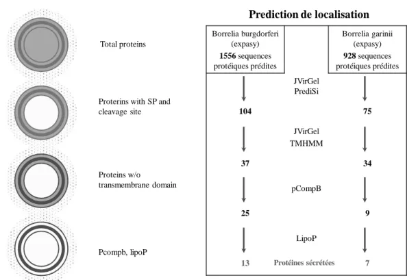
La partie génomique consiste à étudier le protéome prédit de chaque espèce. Des algorithmes bioinformatiques ont été utilisés et une méthode a été décrite pour sélectionner les protéines potentiellement sécrétées par B. afzelii, B. burgdorferi et B.garinii (méthode rapide, facile et librement disponible à partir d’internet). Cette sélection a mis en évidence 3 candidats pour B. afzelii, 7 pour B. burgdorferi et 2 pour B.garinii.
Ces deux approches donnet une vue de l’ensemble des antigènes de B. burgdorferi s.l.
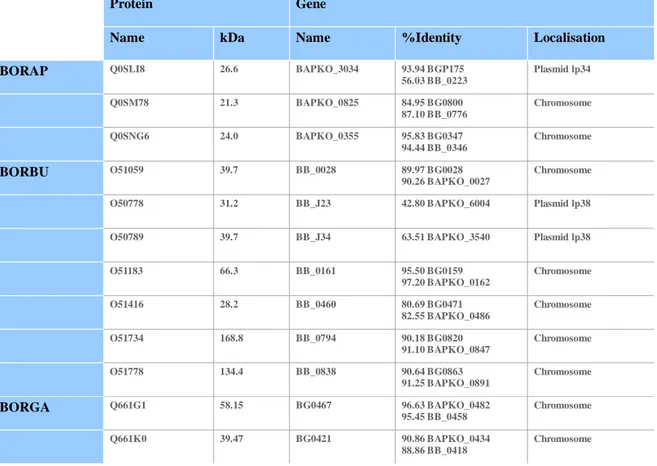
Finalement un exemple d’identification et de caractérisation de candidat a été faite. L'objectif de cette étude était d'identifier une protéine de 12 kDa de B. garinii réagissant avec l’anticorps monoclonal D6. La protéine a été extraite et soumise à une analyse de séquence LC-MS/MS. Cette analyse a révélé trois séquences polypeptidiques analogues à BB0477 (30S ribosomique S10), à BB0061 (thiorédoxine A), et à BB0390 (50 S ribosomique L7/L12).
Les analyses génétiques ont été réalisées et deux polypeptides de la thiorédoxine A et un de la protéine ribosomique 50S ont ainsi été identifiés comme des épitoques potentiels.
L’expression dans le vecteur PQR9 de E. coli suivie d’immunoblots a permit de montrer que les résidus 7-12 de la thiorédoxine A. sont reconnu par le D6. Cela a été confirmé par des expériences de compétition avec un peptide synthétique.
Abstract
Borrelia afzelii, B. garinii, and B. burgdorferi are three of the five Borrelia species definitely recognized as responsible for Lyme borreliosis in Europe. This infectious disease is transmitted by ticks and is characterized by multistage skin, joint, neurological and cardiac manifestations. The aim of the present study is to reveal new species specific proteins of B. burgdorferi sensu lato and to characterize them. The points of interest consist in serologic determination, routine PCR, chips technology and in medical diagnostic (symptoms / particular antigen).
In this study, two global approaches were used to study antigenic proteins of interest: the proteomic way and the genomic way.
The proteomic part consists to study the immune-proteome of each pathogenic genospecies. Antigenic fractions of total bacterial protein lysate of B. burgdorferi sensu stricto VS215, B.garinii VS102 and B. afzelii VS461 were prepared using different immune-affinity columns with specific serological reactivity. Proteins were then separated by two-dimensional electrophoresis to obtain specific reference map for each species. 4 specific spots were observed for B. afzelii and 2 for B. burgdorferi.
The genomic part consists to study the predicted proteome of each genospecies. Bioinformatic algorithms were used and a method was described to select potential secreted proteins from B. afzelii, B. burgdorferi and B.garinii proteome in a simple way (fast, easy and freely available from internet). At the end of selection, 3 candidates were found for B. afzelii, 7 for B. burgdorferi and 2 for B.garinii.
These two approaches resume the antigenic state of B. burgdorferi s.l.
Finally an example of identification and characterisation of such candidates was made. The objective of this study was to identify a12 kDa protein from B. garinii reacting with the D6 monclonal antibody. Protein was extracted and submitted to LC-MS/MS sequence analysis. This analysis revelead three polypeptide sequences analogous to BB0477 (30S ribosomal protein S10), BB0061 (thioredoxine A), and BB0390 (50 S ribosomal protein L7/L12).
Genetic analyses were performed and two polypeptides from thioredoxin A and one from 50S ribosomal protein were thus identified as potential epitopes. Expression in E.coli PQR9 followed by by immunoblotting identified residues 7-12 of thioredoxin A as the D6 mab epitope. This was confirmed by competition experiments with a synthetic peptide.
2 Introduction
2.1 Borrelia burgdorferi 2.1.1 Classification
Borrelia burgdorferi sensu lato appartient à l'ordre des Spirochaetales, comme les genres Leptospira et Treponema. Le genre Borrelia comprend une trentaine d’espèces, dont Borrelia burgdorferi sensu lato qui regroupe 19 espèces et 5 sont des espèces pathogènes majeures, B. burgdorferi sensu stricto, B. garinii, B. afzelii, B.bavaiensis et B. spielmani responsable de la borréliose de Lyme (BL) (Baranton et al. 1992; Canica et al. 1993). Il contient aussi au moins huit espèces étroitement apparentées qui ne provoquent que très rarement des infections humaines.
Borrelia burgdorferi sensu lato est abrégé dans cette thèse comme Bb ou B. burgdorferi, Borrelia burgdorferi sensu stricto comme Bb s.s., Borrelia garinii comme Bg et Borrelia afzelii comme Ba.
2.1.2 Structure et exigences de croissance in vitro
Bb est une bactérie Gram-négative hélicoïdale qui mesure 15-25 µm de long sur 0,2-0,5 µm de large, qui est dotée de 7-11 flagelles périplasmiques (Barbour, Hayes 1986), qui permettent la motilité et lui confèrent sa forme. D'une manière analogue aux autres bactéries Gram-négatives, Bb a une membrane externe qui entoure l'espace periplasmique et une membrane cytoplasmique interne qui protège le cytoplasme (Barbour, Hayes 1986). Exceptionnellement, aucun lipopolysaccharide (LPS) n'est présent dans la membrane externe de Bb. Bb peut être visualisé sans coloration par champ noir ou par microscopie à contraste de phase (Preac-Mursic et al. 1993). L'organisme peut être détecté par microscopie après coloration de Wright, Giemsa ou argent, ainsi que par microscopie à fluorescence suite à un marquage immunocytochimique.
Bb peut être cultivé in vitro dans des conditions microaérophiliques à 33 °C dans un milieu liquide appelé Barbour-Stoenner-Kelly (BSK II) (Barbour 1984). Lors de conditions défavorables, tel que pH bas ou sous l'influence d'antibiotiques, le spirochete développe des blebs de membrane (Preac-Mursic et al. 1986).
2.1.3 Génome
Bb a un chromosome linéaire d'une taille approximative de 1Mb (Fraser et al. 1997), et il contient généralement au moins quatre plasmides linéaires et plusieurs plasmides circulaires. La plupart du génome de la souche B31 de Bb s.s. a été publié en 1997 (Fraser et al. 1997) et
le génome complet a été publié en 2000 (Casjens et al. 2000). Le génome contient un grand nombre de gènes qui codent pour des lipoprotéines qui incluent des protéines de surface externes (Osps) de A à F (Fraser et al. 1997). Par contre, le génome de Bb ne code que pour peu de protéines impliquées dans la biosynthèse, signifiant que le spirochète est dépendant de l'hôte pour les exigences alimentaires.
2.1.4 Les protéines de surfaces externes
Les protéines de la surface externe OspA et OspB sont deux lipoprotéines majeures de surface de la membrane externe de Bb. Dans le génome de la souche B31 de Bb s.s, les gènes codant OspA et OspB sont localisés sur le plasmide linéaire 54 (lp54). Les deux gènes partagent un promoteur et sont transcrits de manière coordonnée (Howe et al. 1986; Bergström et al. 1989). Bb est transmis aux êtres humains par les tiques et les études ont montré que Bb régule l'expression de beaucoup de protéines de surface lors de la phase de transmission. OspA et OspB sont exprimés par Bb dans l'intestin moyen de la tique. Peu après l'entrée dans l'hôte vertébré, l'expression est diminuée, et l'expression d'OspC est augmentée (Schwan et al. 1995; Schwan, Piesman 2000). Des anticorps dirigés contre OspA et OspB sont détectables chez quelques malades lors de la phase précoce de la maladie, et lors de l'étape tardive pendant l’arthrite (Kalish et al. 1995; Chen et al. 1999), ainsi que lors de traitement-réfractaire à l'arthrite de Lyme (Lengl-Janssen et al. 1994; Chen et al. 1999) suggérant qu'OspA et OspB sont aussi exprimés lors de certaines étapes de l'infection persistante. De plus, des antigènes OspA (Coyle et al. 1993) ainsi que ces anticorps (Schutzer et al. 1997) ont été identifiés dans le fluide cérébrospinal de malades atteints de neuroborréliose. Dans une étude publiée par Batsfor, approximativement 80% de sera de malades ayant une arthrite ou une acrodermatite chronique atrophiante (ACA) et 23% des sera de malades ayant un érythème migrant (EM) reconnaissent les antigènes OspA ou OspB (Batsford et al. 1998).
2.2 Borréliose de Lyme 2.2.1 Epidémiologie
Les spirochètes Bb vivent dans un cycle enzootique impliquant des tiques et une grande gamme d'animaux comprenant des mammifères, en particulier des souris, des oiseaux et même des reptiles (Anderson, Magnarelli 1984; Gern et al. 1998; Xu et al. 2007). La borréliose de Lyme apparaît comme l’affection vectorielle la plus fréquente aux USA et probablement en Europe. Les vecteurs sont de la famille des Ixodidae. Selon le lieu géographique l’espèce diffère : Ixodes ricinus en Europe occidentale, I. persulcatus en Europe
de l’Est et Asie, I. ovatus au Japon, et I. scapularis et I. pacificus pour l’Amérique du Nord, respectivement à l’est et à l’ouest. Le risque de contamination dépend directement de la densité en tiques et de leurs pourcentages d’infestations, ainsi que des facteurs géographiques et climatiques (Magnarelli, Anderson 1988; Wittenbrink et al. 1994).
Le cycle d’Ixodes comprend trois stades : larve, nymphe et adulte. Le passage d’un stade au suivant nécessite un repas sanguin. La larve se fixe généralement sur des insectivores ou petits rongeurs, la nymphe sur des vertébrés de taille moyenne ou des oiseaux et finalement l’adulte sur des mammifères de plus grande taille. Aux trois stades, l‘homme est un hôte accidentel, mais Bb est habituellement transmis par les nymphes et les tiques adultes (femelles).
Les études sur les animaux de laboratoire ont montré que la transmission de la maladie exige généralement un attachement de la tique d'une durée de 48-72 heures (selon l’espèce), signifiant que si la tique a été attachée durant moins de 24 heures, le risque d'infection peut être considéré comme bas (Piesman et al. 1987; Piesman 1993; Des Vignes et al. 2001).
2.2.2 Pathologie
Pour maintenir son cycle enzootique, Bb doit s'adapter aux environnements des différents hôtes. À l'intérieur de la tique, Bb exprime la protéine de surface externe A (OspA) qui reste attaché au récepteur OspA de la tique (TROSPA) au niveau de l'intestin moyen (Pal et al. 2004). Lors du repas sanguin de la tique, Bb change l'expression de plusieurs gènes, y compris la régulation du gène qui code pour l’OspA. Simultanément, l'expression de l’OspC est augmentée, ce qui est nécessaire pour infecter les mammifères (Steere et al. 2004; Hu et al. 1996).
Il a été démontré que l’OspC lie la protéine salivaire Salp15 de la tique, Salp15 ayant des propriétés immunosuppressives (Hovius et al. 2008).
Plusieurs jours à semaines après la transmission au niveau de la peau du mammifère, Bb peut migrer par voie hématogène vers plusieurs organes. Pour faciliter cette dissémination, Bb adhère aux intégrines, protéoglycanes, ou glycoprotéines des cellules hôtes ainsi qu'aux matrices extracellulaires (Steere et al. 2004).
Bb peut se lier à des plasminogènes et des urokinases activatrices de plasminogènes, pour mieux pénétrer à travers les couches cellulaires endothéliales. D’autres protéines importantes de Borrelia interviennent, tel BBK32 (47 kDa), qui a été démontrée comme se liant aux fibronectines; p66 (66 kDa), une protéine de surface externe qui se lie aux récepteurs du fibrinogène et au récepteur du vitronectine; p26 (26 kDa), qui se lie aux glycosaminoglycanes
des cellules endothéliales et neuronales. De plus, les protéines dbp A et B lient un collagène associé aux décorines du protéoglycane (Steere et al. 2004).
2.2.3 Réponse immunitaire à B. burgdorferi
Une fois que Bb est dans la peau, les premiers facteurs de l'hôte que rencontrent les bactéries sont des composants du système du complément et les cellules du système immunitaire inné. Selon l'espèce de Bb, la lyse médiée par le complément contre les spirochètes peut être le premier mécanisme de défense de l'hôte (Breitner-Ruddock et al. 1997). Le premier symptôme clinique, la lésion EM, est la réponse des lymphocytes, cellules dendritiques (DCs), macrophages et un petit nombre de cellules du plasma (Müllegger et al. 2000). Cependant, seulement très peu de neutrophiles sont présents, ce qui est atypique lors d’une infection bactérienne (Steere et al. 1983a). Les cellules inflammatoires dans l'EM produisent des cytokines pro-inflammatoires, les plus abondantes étant TNFα et INFγ. Une réponse Th1 optimale dans la phase précoce de la maladie a été associée avec de bons résultats (Sjöwall et al. 2005). L'infection peut être limitée à la peau et peut se résorber même sans traitement, mais pour des raisons encore inconnues, dans quelques cas la bactérie envahit la circulation sanguine et se dissémine vers plusieurs organes. Les symptômes peuvent paraître même des années après l'infection primaire. Il semblerait que dans la phase précoce de la maladie, la réponse Th1 serait importante dans la défense contre les bactéries, alors que la réponse Th2 serait importante dans la phase plus tardive de la borréliose de Lyme (Oksi et al. 1996; Sjöwall et al. 2005).
2.2.4 Manifestations cliniques
La borréliose de Lyme est caractérisée par un polymorphisme clinique, auquel il faut de plus distinguer la forme européenne de l’affection américaine (Stanek, Strle 2003; Nadelman, Wormser 1998).
Le développement de la maladie peut être divisé en infection locale précoce, disséminée et chronique tardive (Steere et al. 2004). Cependant, les trois étapes se chevauchent et leurs présence n'est pas claire dans tous les cas de BL. Le premier symptôme dermatologique de l'infection, l'érythème migrant (EM), a lieu habituellement 3-32 jours après l'infection. Cette phase primaire peut s’accompagner de symptômes apparentés à la grippe, tel que malaise, fatigue, maux de tête, arthralgies, myalgies et fièvre (Steere et al. 1983b). Cet EM est parfois suivi d’une phase dite secondaire, le plus souvent neurologique, tel que des névropathies crâniennes et périphériques (un exemple typique étant une paralysie du nerf facial),
accompagné de méningite lymphocytaire, plus précisément de meningoradiculite lymphocytaire.
Les manifestations typiques au niveau de la peau de la BL disséminée sont des lésions EM multiples, des lymphocytomes cutanés et des Acrodermatites chroniques atrophiantes (ACA) (Hansen, Asbrink 1989) qui sont des manifestations tardives de la BL. L'arthrite de Lyme est généralement une manifestation tardive de la BL mais elle peut se produire quelquefois plus tôt lors de la maladie. L’arthrite est caractérisée typiquement par des attaques périodiques au niveau des (grosses) articulations. Les manifestations neurologiques tardives de la BL incluent méningites, et radiculites.
Il y a un grand nombre d’autres manifestations disséminées et tardives engendrées par la BL : au niveau du cœur il peut y avoir des cardites, plus précisément des endomyocardites ou péricardites (Steere 1989) ; au niveau des yeux on trouve des conjonctivites, des kératites et uvéites (Lesser 1995); au niveau de foie des hépatites (Kazakoff et al. 1993), des splénomégalies au niveau de la rate (Cimmino et al. 1989) ; ou encore des orchites et des hématuries microscopiques (Steere 1989). L'infection Bb a une forte tendance à devenir chronique (Berger et al. 1983; Steere et al. 1983b; Steere et al. 1983a).
Il existe des différences cliniques et épidémiologiques entre l’expression de la BL entre l’Europe et les USA. Aux USA le pathogène n’est représenté que par Borrelia burgdorferi sensu stricto alors que les trois espèces pathogènes sont représentées en Europe. Les différences cliniques s’expliquent par l’organotropisme existant entre les différentes espèces de Borrelia. Ainsi les complications rhumatologiques sont dans la plupart des cas causées par Bb s.s., alors que Bg est responsable des symptômes de type neurologiques, et que les manifestations dermatologiques tardives sont souvent associées avec les infections de Ba (Assous et al. 1993; Péter et al. 1997; Wang et al. 1999).
2.2.5 Diagnostic et traitement
Dans la phase précoce de la BL, le diagnostic est basé habituellement sur des conclusions cliniques typiques, la plus évidente et spécifique étant la rougeur de l'EM, et dans ce cas le diagnostic de laboratoire n'est pas recommandé. Si une rougeur EM est suspecte, le malade devrait être traité aux antibiotiques. L’agent causal étant de la famille des Spirochaetacea, les Beta-lactamines et les tétracyclines sont les antibiotiques de choix et les nouveaux macrolides peuvent être utilisés en deuxième intention.
Au stade tardif de la maladie, le diagnostic sérologique est recommandé en aide au diagnostic clinique. La production d'anticorps IgM a lieu durant les deux premières semaines suite à
l'infection et atteint un maximum après deux mois. Progressivement, la production d'anticorps IgG commence en même temps que le déclin des IgM. Une approche en deux étapes est généralement utilisée dans la sérologie de la BL (Wilske 2003). La première étape est un test ELISA sensible qui est suivi par un test immunoblot lors d'un positif ou cas positif limite. Bb peut être mis en culture dans un milieu liquide BSK II (Barbour 1984). Le temps de génération de Bb étant long, les cultures nécessitent d’être incubées quelques semaines. Une méthode moléculaire consiste à détecter l'ADN de Bb dans des échantillons de biopsie ou fluides corporels (par exemple fluide synovial, fluide cérébrospinal, sang) en utilisant la technique de la « polymerase chain reaction » (PCR) (Stanek, Strle 2003). En général, les sensibilités des cultures de Bb et des PCR ne sont pas des méthodes de choix dans le diagnostic de laboratoire de la BL, et sont donc négligeables en dehors d’études cliniques.
2.3 Problématique et objectifs
BL est l’une des infections bactériennes persistantes les plus communes en Europe qui n'offrent pas encore de sérologie adéquate. Ceci paraît être lié aux réponses immunes uniques résultant des stratégies de fuite de la bactérie par rapport au système immunitaire de l’hôte. Cette thèse a pour but d’identifier de nouveaux antigènes pertinents pour le diagnostic.
Le diagnostic de l’infection de Borrelia burgdorferi sensu lato, agent étiologique de la BL, est basé sur les manifestations cliniques et est confirmé par tests sérologiques. Ces tests ont néanmoins pour défauts leurs manques de sensibilité et spécificité. L'usage de nouveau marqueurs spécifiques et fortement antigéniques pourraient améliorer ces limitations.
• La première partie de cette thèse a pour but d’identifier de nouveaux antigènes de B. burgdorferi en employant une approche protéomique.
• La deuxième partie de cette thèse a pour but d’identifier de nouveaux antigènes de B. burgdorferi en employant une approche génomique.
• La troisième partie est un exemple d’identification et de caractérisation d’une protéine antigénique.
DNA mRNA protein TRANSCRIPTION TRADUCTION genome proteome P R O T E O M IQ U E GE N O M IQ U E
Résumé des deux démarches : L’étude protéomique se faisant à partir des protéines (approche pratique : détection physique grâce aux anticorps) pour aboutir aux gènes, alors que l’étude génomique (approche informatique : prédiction grâce à des algorithmes) est à l’inverse : on part du gène pour aboutir aux protéines. Ceci permet donc d’avoir une vision globale et complète des antigènes potentiels de Borrelia.
3 Articles
3.1 Comparison of antigens of Borrelia afzelii, B. burgdorferi and B. garinii isolated with immuno-affinity columns on two-dimensional electrophoresis maps
WYSS Jean-Christophe1,2; BETSCHART Bruno2; PETER Olivier1§.
1 Institut Central des Hôpitaux Valaisans (ICHV), Microbiologie, Av. du Grand-Champsec 86
1951 Sion, Switzerland.
2
Institut de biologie, Université de Neuchâtel, Rue Emile-Argand 11, 2000 Neuchâtel, Switzerland.
§Reprints or correspondence: PETER Olivier, Institut Central des Hôpitaux Valaisans (ICHV), Microbiologie,
Av. du Grand-Champsec 86, 1951 Sion, Switzerland, telephone : +41 27 6034862 Fax : +41 27 6036650, e-mail : olivier.peter@hopitalvs.ch
Email addresses:
WYSS Jean-Christophe (jean-christophe.wyss@unine.ch) BETSCHART Bruno (bruno.betschart@unine.ch) PETER Olivier (olivier.peter@hopitalvs.ch)
3.1.1 Introduction
The bacterium B. burgdorferi sensu lato (s.l.), the causative agent of Lyme borreliosis (LB), belongs to Gram-negative, spirochetal bacteria (Paster et al. 1991) characterised by a spiral morphology and flagella that function as motility organs.
B. burgdorferi s.l. is divided into different genospecies of which three have been identified as major human pathogens: B. burgdorferi sensu stricto (s.s.), B. garinii and B. afzelii (Baranton et al. 1992; Marconi, Garon 1992; Canica et al. 1993). All three species can be found in Europe and Asia whereas in the USA only B. burgdorferi s.s. occurs.
The genome of B. burgdorferi s.l. consists of a linear chromosome and numbers of linear and circular plasmids (Barbour 1988; Casjens et al. 2000). The large number of plasmids may enable an extensive antigen variation to adapt to the different environments the bacterium may encounter. Outer surface proteins exposed on the cell surface interact with the host and contribute to the pathogenesis of LB.
Lyme borreliosis, the most prevalent and widespread vector-borne infectious disease in the northern hemisphere, is a multisystem disease involving many organs, mainly the skin, nervous system, joints and heart (Steere 1989; Pfister et al. 1994; Stanek, Strle 2003).
Different clinical forms are found and are directly correlated to phenotype and genotype heterogeneity of the pathogen (Pachner et al. 2004). Arthritis and carditis are preferentially associated with B. burgdorferi sensu stricto, the degenerative skin disorder acrodermatitis chronica atrophicans (ACA) with B. afzelii and neuroborreliosis with B. garinii (Demaerschalck et al. 1995; Balmelli, Piffaretti 1995).
If there are no pathognomonic symptoms such as a typical erythema migrans, clinical diagnosis of Lyme borreliosis usually requires confirmation by means of a laboratory-diagnostic assay. Antibody detection methods mainly are used for this purpose, whereas detection of the causative agent by culture isolation and nucleic acid techniques is confined to special situations.
Antibody detection is made by serological tests, currently using a two step approach: a screening assay, mostly ELISA, confirmed by immunoblot analysis (Wilske et al. 2007). Recently, immunoblots have been improved by addition of recombinant antigens (i.e. VlsE and DbpA) (Goettner et al. 2005; Schulte-Spechtel et al. 2003). Despite recent improvements, the limitations regarding sensitivity and specificity of the tests still restrict their use as routine diagnostic tools (Aguero-Rosenfeld et al. 2005). To date, the cross-reactivity of borrelia antigens, the delayed appearance or even lack of measurable immune responses in the early stage of LB and the absence of a marker for persistent or active infections are the main challenges for serodiagnosis (Brouqui et al. 2004). Identifying and characterizing antigenic components that are involved in the pathogenesis of B. burgdorferi s.l. will permit a better understanding of the disease and lead to more effective diagnosis and treatment.
A way to detect and identify specific antigens is to work with complete immunoproteomes of each pathogenic genospecies. The method consists of two-dimensional electrophoresis (adequate resolution for microorganisms proteome) followed by blotting on membranes and overlaying the protein pattern with patient’s sera (Krah, Jungblut 2004).
Generally, prior to electrophoresis, proteins are enriched in proteins of interest to increase the resolution of the separation.
For Borrelia various techniques have been attempted (Bledsoe et al. 1994): surface labelling using I-125 or biotin (Luft et al. 1989), antibody labelling (Barbour et al. 1984), accessibility to proteases (Norris et al. 1992) , and differential solubilisation with detergents (Sambri, Cevenini 1991; CUNNINGHAM et al. 1988).
However, the techniques are subject to artefacts, and consider only a fraction of protein of interest for diagnosis purpose.
Here we isolated first the antigenic proteins to obtain directly immunoproteome by 2d-electrophoresis. Antigenic fractions of the total bacterial protein lysate of B. burgdorferi s.s. VS215, B. garinii VS102 and B. afzelii VS461 were prepared using different immune-affinity columns with specific serological reactivity. The following immunoblot step is used to validate the results.
The study allowed identifying new species-specific antigens of B. burgdorferi s.l., and obtaining antigenic maps for B. burgdorferi s.s., B. garinii and B. afzelii. Such studies were already made by American groups, but focalised only on B.b.ss(Nowalk et al. 2006a). Maps of all 3 genospecies were elaborated by Jungblut (Jungblut et al. 1999), but the antigenic characteristics were focused on B. garinii exclusively.
3.1.2 Material and methods Bacteria cultures
Low passage Borrelia strains, B. burgdorferi s.s. VS215, B. garinii VS102 and B. afzelii VS461 were used for antigen preparation (Péter et al. 1997). All strains were isolated from ticks (I. ricinus) (Péter, Bretz 1992). Spirochetes were cultured in BSK II medium. During the late logarithmic phase of growth, the culture was centrifuged at 14,000g for 15 min and washed twice in phosphate-buffered saline (pH 7.2) to which MgCl2 (0.05 M) was added. After sonication, the protein concentrations of the suspensions were determined by the Biuret method and adjusted to 1 mg/ml in distilled water.
Sera
A minimum of three sera were mixed to constitute a pool. Selection consisted of sera from patients with characterized late LB symptoms, i.e. with neuroborreliosis, acrodermatitis or arthritis (stage III). Each serum was highly reactive in ELISA and presented strong species specific IgG reactivities in immunoblots (Ryffel et al. 1999). They were pooled according to their reactivities.
Affinity chromatography
Hitrap protein G columns (Amersham bioscience) were equilibrated with 10 volume of binding buffer (B-buffer; 20mM Sodium Phosphate, pH7.0). Each pool of sera was diluted 1:1 with B-buffer, filtered (0.45µm) and titration of IgG was carried out by nephelometry. At least 25mg IgG of each elaborated pool of sera were incubated for 30 min in the corresponding column. The Ab-bead conjugates were washed with eight volumes of B-buffer and with eight volumes of washing buffer (W2-buffer; 20mM Sodium Phosphate buffer pH8.2). The antibodies were then covalently linked for one hour to the protein G beads using dimethylpimeliminidate (DMP) solution (0.2M Triethanolamine pH8.2 + DMP (40mg/10ml)). After an eight volume wash with 0.2M Triethanolamine pH8.2, remaining protein G sites were blocked with fifteen volumes of saturation buffer (0.1M Ethanolamine buffer pH8.2). Columns are ready for use after a pre-elution with four volumes of elution buffer (E-buffer; 0.1M Glycine-HCl pH2.8) and equilibration with ten volumes of W2-buffer. For long term storage, H2O with 10% ethanol was used.
Immunoprecipitation
Columns were washed with lysis buffer (L-buffer; 150mM NaCl, 1%NP-40 (v/v), 0.1% SDS (w/v), 50mM Tris-HCl pH8.0) containing protease inhibitors. Two mg of each Borrelia antigen were resuspended in a final volume of 10ml L-buffer, sonicated, incubated 15 min on a rotating wheel, and centrifuged at 18000g. Supernatants were incubated in the corresponding columns during 30 min with continuous running. Columns were then washed with eight volumes of L-buffer and followed by elution with eight volumes of E-buffer. Elutions were stabilized with 175µl 2.0M Tris-HCl pH 8.0 for a final volume of 1.5 ml. Columns were regenerated with ten volumes W2-buffer and finally stored in 10% ethanol.
Two-dimensional Gel Electrophoresis
Volumes of 100µl (for gel staining) or 50µl (for immunoblots) of eluted material were concentrated and precipitated with trichloroacetic acid (20%). Individual 7cm ReadyStrips IPG strips (Bio-Rad), pH 5–8 were rehydrated with 125 µl of urea buffer (7 M urea, 2 M thiourea, 4% (w/v) CHAPS, 1% (v/v) Np40, 0.2% (v/v) Bio-Lytes, 1% (w/v) dithiothreitol, 0.002% (v/v) bromophenol blue) containing the sample. Isoelectric focusing was carried out using a Protean IEF Cell according to the instructions from the manufacturer (Bio-Rad): 14
hour 50V active rehydration; 20 min 250V linear slope; 120 min 4000V linear slope; 4000V 12000VH rapid slope. Prior to the second dimension, the IPG gel strips were equilibrated twice in 2.5 ml of equilibration solution (50 mM Tris-HCl buffer, pH 8.8, containing 6 M urea, 30% (v/v) glycerol, 1% (w/v) SDS) for 20 min, first in the presence of dithiothreitol (65 mM) and then in the presence of iodoacetamide (87 mM). Second dimension gels consisted in a 12% tris-glycine SDS-PAGE. Gels were run using the Protean II xi Cell (Bio-Rad), at a constant current of 15 mA/gels. The analytical two-dimensional gels were stained with zinc (Hardy, Castellanos-Serra 2004), with silver (Shevchenko et al. 1996; Yan et al. 2000) or were transferred for immunoblots and scanned with G-700 from Bio-rad.
Immunoblots
After electrophoresis the proteins were transferred to a polyvinylidenedifluoride membrane with constant voltage (120V) during one hour. Before further use, the membranes were blocked for 1 h at 37°C with Tris-buffered saline (TBS; pH 7.2) with 5% gelatine and were washed three times for 5 min each time with washing buffer (W-buffer; TBS with 0.1% gelatine and 0.05% Tween 20) at room temperature. The following steps were also performed at room temperature. Each membranes were incubated for 2 h with pools of human sera diluted 1/200 in D-buffer (TBS with 1% gelatine and 0.05% Tween 20). For monoclonal antibodies dilutions were 1:1500 for LA114 ZS7 (93 kDa protein), 1: 100 for LA 18 (66 kDa protein), 1:1000 for H9724 (41 kDa protein; flagellin), LA112 ZS7 (39 kDa protein), LA222B8 (OspA) and LA31 (OspC) (kindly provided by A. G. Barbour, University of California, Irvine; R. Wallich, Ruprecht-Karls-Universität, Heidelberg, Germany; and B. Wilske, Max von Pettenkofer Institut, Munich, Germany) (Ryffel et al. 1999). The membranes were washed three times for 5 min with W-buffer. After the washing, rabbit anti-human IgG (Sigma, St-Louis, USA) conjugated to alkaline phosphatase diluted 1/1000 in D-buffer was added for human sera. The secondary antibodies for monoclonal antibodies were goat anti-mouse polyglobulin (Sigma, St-Louis, USA) conjugated to alkaline phosphatase diluted 1/1000. At an incubation of 2 hours, two washes were done with W-buffer and one was done with TBS. The bound conjugate was visualized by addition of the chromogenic substrate 5-bromo-4-chloro-3-indolylphosphate-Nitro Blue Tetrazolium (Kirkegaard & Perry Laboratories). The reaction was stopped 30 min later by two rinses in distilled water.
Image analysis
Image analyses were performed with Flicker version 0.83.6 (Lemkin, Thornwall 1999), a program belonging to the Open2Dprot Software, with ImageMaster 2D Platinum 6.0 (GE Healthcare) and TopSpot (Pleissner et al. 2002).
3.1.3 Results
On every gel (Figure 1), two horizontal diffuse streaks (~50kD and ~25kD) are observed that represent a small amount of eluted IgG from the column (heavy and light chains). These streaks were used to align gels together.
After two-dimensional electrophoresis, different staining techniques (zinc and silver) were used to visualize proteins on gels. The general pattern (Figure 1) between species is very similar. Staining differs in sensitivity, in the increasing order from silver to zinc. Less spots are present with silver staining. This gives a semi-quantitative aspect of the visualized proteins.
With all the landmarks and in comparison with the literature it was possible to identify the main spots (listed on Table 1). For B. burgdorferi s.s., 90 spots were detected with zinc staining, 22 were well identified and 2 were described as species specific: spot n°23 and n°51. For B. afzelii, on a total of 78 spots, 16 were identified and 8 were species specific: spot n°13, 56, 65, 22, 26, 35, 52 & 75. No species specific spots were observed for B. garinii among the 71 spots (19 with identity).
More spots seem to be seen for zinc maps, but they appear more distinct with silver staining. With identical parameters (area; separation), more spots are detected on silver maps: 98 for B. burgdorferi (36 identified); 102 for B. afzelii (23 identified) and 76 for B. garinii (17 identified). Species specific spots are similar to spots observed on zinc maps, just one supplementary isoform for B. afzelii: spot n°84 is dectectable. Every data are visualized on Figure 1 and data are referenced on Table 1.
The proteins p83/100, Oms66, ErpB, OppA, FlaB, BmpA, OspC and LA7 are detected in all species. Some spots are only present in one species like Hsp90, ErpA and OspB for B. burgdorferi or GADPH for B. garinii. Some proteins are represented by one spot like p83/100, whereas others with spot string like Oms66 or FlaB (Table 1). Differences could also be observed between staining methods. Hsp 90; OspB or RevA are only detected on zinc maps of B. burgdorferi s.s. On the contrary, OspA and OspB are well represented on silver maps.
Most of identified spots are concentrated in acidic range of the gel. Some proteins like p83/100, Oms66, ErpB and FlaB are similar in MW and PI between species. Other proteins differ in PI like OspC or in MW and PI like OspA (not present for B. garinii).
Finally some proteins are only represented in one species: spots 23, 51 for B. burgdorferi, (13, 56, 65), (22, 26, 35), 52, 75 for B. afzelii. Corresponding respectively to 14, 46 for B burgdorferi s.s silver map and (67; 75; 83; 84); (15; 21; 28); 66; 97 for B. afzelii. No species specific spots were detected for B. garinii. To confirm the antigenic properties of the proteins, immunoblots were performed with monoclonal antibodies (p100, p66, p41, p39, OspA, OspC) in order to localize the major proteins, to scale gels and to align them together (data not shown). This standardisation was made with B. burgdorferi s.s only. Recognition of the corresponding proteins for the other species was deduced since these known common proteins have identical patterns and molecular mass, excepted OspA. All proteins detected on the 2D gels were also present on immunoblots (data not shown)
Z in c st a in in g S il v er s ta in in g B. afz eli i < ---SD S-P AG E IE F - --> A c id ic b as ic IE F - --> A c id ic b as ic M r[ k D A ] _ 5 0 _ 2 5 B. bu rg do rfe ri < ---SD S-P AG E IE F - --> A c id ic b as ic IE F - --> A c id ic b as ic _ 5 0 _ 2 5
B. ga rin ii < ---SD S-P AG E IE F - --> A c id ic b as ic IE F - --> A c id ic b as ic _ 5 0 _ 2 5 F ig . 1 : 2 D r ef er en ce m ap s o f B . a fz el ii , B . g a ri n ii a n d B . b u rg d o rf er i. E x tr ac te d p ro te in s w e re s ep ar a te d b y I E F i n t h e p H r an g e 5 t o 8 , fo ll o w ed b y S D S -P A G E g el ( 1 2 % ). S p o t n u m b er s in d ic at e id en ti fi ed p ro te in s b y T o p S o p t p ro g ra m .
T a b le 1 : L is t o f sp o ts o f in te re st f o r z in c an d s il v e r re fe re n ce m ap s --Z IN C S T A IN IN G ---S IL V E R S T A IN IN G --B . a fz el ii B . b u rg d o rf er i B .g a ri n ii B . a fz el ii B . b u rg d o rf er i B .g a ri n ii S h o rt -N a m e R ef sp o t n ° sp o t n ° sp o t n ° sp o t n ° sp o t n ° sp o t n ° p 8 3 /1 0 0 m o n o A B ; (N o w a lk e t al . 2 0 0 6 b ) 2 6 4 4 2 3 H sp 9 0 (N o w a lk e t al . 2 0 0 6 a ) 4 3 O m s6 6 (J u n g b lu t et a l. 1 9 9 9 ) 1 4 ; 1 9 ; 3 2 3 5 ; 4 4 ; 5 0 1 1 ; 1 5 ; 1 9 1 1 ; 1 7 ; 2 7 2 7 ; 3 5 ; 4 4 7 ; 1 1 ; 1 4 ; 2 1 E rp B m o n o A B ; (N o w a lk e t al . 2 0 0 6 a) 6 ; 1 ; 3 9 ; 1 4 ; 7 6 6 7 ; 9 ; 6 6 7 ; 1 ; 2 5 ; 7 ; 1 7 8 ; 1 3 ; 2 0 O p p A (N o w a lk e t al . 2 0 0 6 a ) 2 3 ; 2 7 ; 3 6 7 3 ; 3 7 ; 3 2 1 ; 2 4 ; 2 9 2 0 ; 2 6 ; 3 5 2 9 ; 3 ; 4 2 2 5 ; 3 0 ; 3 4 F la B m o n o A B ; (N o w a lk e t al . 2 0 0 6 a) 2 0 ; 2 9 ; 4 1 3 3 ; 4 1 ; 5 2 2 0 ; 2 6 ; 3 5 1 8 ; 2 5 ; 4 4 2 2 ; 3 1 ; 4 8 1 6 ; 2 6 ; 3 5 B m p A m o n o A B ; (N o w a lk e t al . 2 0 0 6 a) 5 ; 1 1 2 ; 7 ; 1 6 5 ; 8 5 4 ; 8 9 G A P D H (J u n g b lu t et a l. 1 9 9 9 ) 6 2 7 0 O sp B (N o w a lk e t al . 2 0 0 6 b ) 7 6 O sp A m o n o A B ; 3 6 1 3 ; 2 3 ; 3 6 ; 5 3 O sp D (N o w a lk e t al . 2 0 0 6 b ) 2 4 2 3 3 3 O sp C m o n o A B ; 7 2 4 1 ; 4 2 ; 5 5 ; 3 ; 5 9 ; 6 3 4 3 ; 4 7 ; 5 4 ; 6 4 , 6 7 ; 7 0 ; 7 3 ; 7 1 ; 7 6 ; 7 8 ; 8 0 ; 8 2 ; 8 4 ; 8 6 2 7 L A 7 (N o w a lk e t al . 2 0 0 6 a ) 1 7 2 6 ; 1 5 1 0 1 9 1 5 E rp A (N o w a lk e t al . 2 0 0 6 a ) 2 0 1 2 R ev A (N o w a lk e t al . 2 0 0 6 a ) 3 6 7 0 u n k n o w n s p s p o t 1 3 ; 5 6 ; 6 5 2 3 6 7 ; 7 5 ; 8 3 ; 8 4 1 4 2 2 ; 2 6 ; 3 5 5 1 1 5 ; 2 1 ; 2 8 4 6 5 2 6 6 7 5 9 7 S ta ti st ic n b s p o t n b s p o t n b s p o t n b s p o t n b s p o t n b s p o t n b t o ta l o f sp o t 7 8 9 0 7 1 1 0 2 9 8 7 6 n b u n k n o w n s p o t 6 2 6 8 5 2 7 9 6 2 5 9 k n o w n s p o t 1 6 2 2 1 9 2 3 3 6 1 7 k n o w n p ro t 7 1 2 1 0 1 0 1 1 9
T a b le 2 : C h ar ac te ri st ic s o f p ro te in s id en ti fi ed a n d c it ed i n t h e li te ra tu re P ro te in M W ( k D a ) K n o w n f u n ct io n o r lo ca li sa ti o n Im p o rt a n ce i n d ia g n o si s p 8 3 /1 0 0 8 0 /1 0 0 P ro to p la sm ic c y li n d er o r fl ag el lu m -a ss o ci at ed ? + + + H sp 9 0 h ea t sh o ck p ro te in s ro le i n i ro n m et ab o li sm O m s6 6 6 6 M em b ra n e-as so ci at ed p o ri n + E rp B 6 0 n o O p p A 5 9 O li g o p ep ti d e p er m ea se h o m o lo g A F la B 4 1 fl ag el li n + B m p A 3 9 M em b ra n e-as so ci at ed + + + G A P D H 3 7 G ly ce ra ld eh y d e 3 -p h o sp h at e d eh y d ro g en as e n o O sp B 3 4 -3 6 O u te r m em b ra n e p ro te in n o O sp A 3 1 -3 3 O u te r m em b ra n e p ro te in + + + O sp D 2 8 O u te r m em b ra n e p ro te in n o O sp C 2 1 -2 4 O u te r m em b ra n e p ro te in + + + L A 7 2 2 L ip o p ro te in n o E rp A 1 9 n o R ev A 1 7 O u te r m em b ra n e p ro te in
3.1.5 Discussion
By definition, the proteome is the totality of all proteins in a genome, or more precisely in two gel analysis the total of all proteins at a particular place under certain environmental conditions (Wasinger et al. 1995). The method of choice for the study of the proteome is two-dimensional polyacrylamide gel electrophoresis (2-DE) (Rabilloud 2002), (Klose 1975; O'Farrell 1975). Complex mixtures of proteins are separated by isoelectric point (pI) and molecular weight (MW). The result is a pattern of dots (spots), representing single protein (Jungblut et al. 1996).
The 2-DE is in their high-resolution version capable of separating (identifying) approximately 5000 different proteins (spots) (Jungblut et al. 1996). This is largely sufficient for prokaryotes (Jungblut 2001), B. burgdorferi s.s. having for example 1556 predicted proteins (Casjens et al. 2000).
To visualise only antigenic proteins, two-dimensional blots were revealed with antibodies from patient sera. The resulting spots permit to identify specific antigens (vaccine candidates), their pathogenicity and association with disease (diagnostic markers).
Immunoblots have with regard to the antigen-recognition a limitation: through the separation of proteins using 2-DE using high concentrations of urea, the proteins partially unfold. This destroys the native three-dimensional structure irrevocably for many proteins.
Antibodies are directed not only against sequential but also against conformational epitopes, so not all epitopes can be detected with immunoblots. Here we isolated antigens first under semi-native conditions with an immunoprecipitation (IP) step. The IP is based on the relationship of antibodies with sepharose beads coupled with protein G, which have a high affinity for the Fc fragment of immunoglobulins. An additional advantage of IP is that no restriction occurs in terms of pI and MW precipitable proteins. A series of studies already used IPs successfully in connection with 2-DE (Chang et al. 2001; Houry et al. 1999; Imam-Sghiouar et al. 2002; Stancato, Petricoin 2001).
Another advantage of enrichment with IP is that Borrelia burgdorferi s.l.produces excessive amounts of membrane lipoproteins such as OspA. So, when grown in vitro, many low or moderately abundant proteins are underrepresented when cell lysates are examined by 2-DE (Schulte-Spechtel et al. 2003). Enrichment with IP advantages soluble proteins and minimizes strikes on gel due to high level of lipoproteins.
To validate the method, immunoblots were performed using the same sera as used to conjugate to the sepharose beads. All spots present on gels (silver staining) were identified on immunoblots, meaning that all proteins have antigenic properties. Separation of spots was better on gel maps, where fewer strikes were present. Coloration of proteins is more homogeneous than immunologic staining of immunoblots, resulting in more exact and proper reference maps.
IP allowed us to have nice reference results, but we have to keep in mind that collected proteins depend on solubilisation and elution buffer. The method has to be strong enough to avoid to have proteins from secondary interaction (solubilisation) and not to strong, but enough to elute highly antigenic proteins (elution).
Many studies were done to investigate proteomes of Borrelia using 2-DE (Nowalk et al. 2006a; Norris et al. 1992; Norris 2006), chromatography (Jacobs et al. 2005; Angel et al. 2010), or proteome array (Barbour et al. 2008), but only with B. burgdorferi s. s. Maps of all 3 genospecies were elaborated by Jungblut (Jungblut et al. 1999), but the antigenic characteristics were focused on B. garinii exclusively.
The general profile between the three species seems similar. Most of the identified spots are present on each map. A few spots are present in one species only, explaining the low numbers of unknown specific spots.
Proteins between 60 and 41 kDa are normaly conserved between species (Wilske et al. 1988), but here 3 specific spots are found for B. afzelii. For lower weight it is known that profiles are variables between species, that explain the two species specific spots found for B. burgdorferi and the two smaller proteins of B. afzelii.
If we consider the genome of the three species, it is known that B. afzelii appears to be the more divergent compare to B. garinii and B. burgdorferi s.s. (Wang et al. 1999). It could explain why we found more species specific spots for B. afzelii.
This study serves as antigenic reference maps for each pathogenic species of Borrelia. Next step could be to present a universal map of antigenic protein of Borrelia or maps of specific antigens for each genospecies. In the first case, 2DE has to be made with the elution of antigens from columns built with a pool made with each species specific human hyperimmune serum. In the second case, the idea is to pass the elutions of each species through the two other columns to eliminate cross-antigenicities. Proteins of the last method could be directly studied using chromatographic separations coupled with tandem mass spectrometry (Angel et al. 2010).
These references maps were made for the three human pathogenic genospecies of B. burgdorferi s.l. and not only with B. burgdorferi s.s.. Another point of interest is to work directly with antigens and to obtain antigenic maps in a one step procedure.
3.1.6 Bibliography
Aguero-Rosenfeld, M. E.; Wang, G.; Schwartz, I.; Wormser, G. P. (2005): Diagnosis of lyme borreliosis. In Clin. Microbiol. Rev 18 (3), pp. 484–509.
Angel, T. E.; Luft, B. J.; Yang, X.; Nicora, C. D.; Camp, D. G.; Jacobs, J. M.; Smith, R. D. (2010): Proteome analysis of Borrelia burgdorferi response to environmental change. In PLoS ONE 5 (11), pp. e13800.
Balmelli, T.; Piffaretti, J. C. (1995): Association between different clinical manifestations of Lyme disease and different species of Borrelia burgdorferi sensu lato. In Res. Microbiol 146 (4), pp. 329–340.
Baranton, G.; Postic, D.; Saint Girons, I.; Boerlin, P.; Piffaretti, J. C.; Assous, M.; Grimont, P. A. (1992): Delineation of Borrelia burgdorferi sensu stricto, Borrelia garinii sp. nov., and group VS461 associated with Lyme borreliosis. In Int. J. Syst. Bacteriol 42 (3), pp. 378–383. Barbour, A. G. (1988): Plasmid analysis of Borrelia burgdorferi, the Lyme disease agent. In J. Clin. Microbiol 26 (3), pp. 475–478.
Barbour, A. G.; Jasinskas, A.; Kayala, M. A.; Davies, D. Huw; Steere, A. C.; Baldi, P.; Felgner, P. L. (2008): A genome-wide proteome array reveals a limited set of immunogens in natural infections of humans and white-footed mice with Borrelia burgdorferi. In Infect. Immun 76 (8), pp. 3374–3389.
Barbour, A. G.; Tessier, S. L.; Hayes, S. F. (1984): Variation in a major surface protein of Lyme disease spirochetes. In Infect. Immun 45 (1), pp. 94–100.
Bledsoe, H. A.; Carroll, J. A.; Whelchel, T. R.; Farmer, M. A.; Dorward, D. W.; Gherardini, F. C. (1994): Isolation and partial characterization of Borrelia burgdorferi inner and outer membranes by using isopycnic centrifugation. In J. Bacteriol 176 (24), pp. 7447–7455.
Brouqui, P.; Bacellar, F.; Baranton, G.; Birtles, R. J.; Bjoërsdorff, A.; Blanco, J. R. et al. (2004): Guidelines for the diagnosis of tick-borne bacterial diseases in Europe. In Clin. Microbiol. Infect 10 (12), pp. 1108–1132.
Canica, M. M.; Nato, F.; Du Merle, L.; Mazie, J. C.; Baranton, G.; Postic, D. (1993): Monoclonal antibodies for identification of Borrelia afzelii sp. nov. associated with late cutaneous manifestations of Lyme borreliosis. In Scand. J. Infect. Dis 25 (4), pp. 441–448. Casjens, S.; Palmer, N.; van Vugt, R.; Huang, W. M.; Stevenson, B.; Rosa, P. et al. (2000): A bacterial genome in flux: the twelve linear and nine circular extrachromosomal DNAs in an infectious isolate of the Lyme disease spirochete Borrelia burgdorferi. In Mol. Microbiol 35 (3), pp. 490–516.
Chang, J. W.; Young, D. A.; Coleman, P. D.; O'Banion, M. K. (2001): Two-dimensional gel analysis of secreted proteins induced by interleukin-1 beta in rat astrocytes. In Neurochem. Int 39 (5-6), pp. 349–359.
Cunningham, T. M.; Thomas, D. D.; Thompson, S. D.; Miller, J. N.; Lovett, M. A. (1988): Identification of Borrelia burgdorferi surface components by Triton X-114 phase partitioning. In Annals of the New York Academy of Sciences 539 (1), pp. 376‐378.
Demaerschalck, I.; Ben Messaoud, A.; Kesel, M. de; Hoyois, B.; Lobet, Y.; Hoet, P. et al. (1995): Simultaneous presence of different Borrelia burgdorferi genospecies in biological fluids of Lyme disease patients. In J. Clin. Microbiol 33 (3), pp. 602–608.
Goettner, G.; Schulte-Spechtel, U.; Hillermann, R.; Liegl, G.; Wilske, B.; Fingerle, V. (2005): Improvement of Lyme borreliosis serodiagnosis by a newly developed recombinant immunoglobulin G (IgG) and IgM line immunoblot assay and addition of VlsE and DbpA homologues. In J. Clin. Microbiol 43 (8), pp. 3602–3609.
Hardy, Eugenio; Castellanos-Serra, Lila R. (2004): "Reverse-staining" of biomolecules in electrophoresis gels: analytical and micropreparative applications. In Anal. Biochem 328 (1), pp. 1–13.
Houry, W. A.; Frishman, D.; Eckerskorn, C.; Lottspeich, F.; Hartl, F. U. (1999): Identification of in vivo substrates of the chaperonin GroEL. In Nature 402 (6758), pp. 147–154.
Imam-Sghiouar, Naïma; Laude-Lemaire, Isabelle; Labas, Valérie; Pflieger, Delphine; Le Caër, Jean-Pierre; Caron, Michel et al. (2002): Subproteomics analysis of phosphorylated proteins: application to the study of B-lymphoblasts from a patient with Scott syndrome. In Proteomics 2 (7), pp. 828–838.
Jacobs, Jon M.; Yang, Xiaohua; Luft, Benjamin J.; Dunn, John J.; Camp, David G.; Smith, Richard D. (2005): Proteomic analysis of Lyme disease: global protein comparison of three strains of Borrelia burgdorferi. In Proteomics 5 (5), pp. 1446–1453.
Jungblut, P. R. (2001): Proteome analysis of bacterial pathogens. In Microbes Infect 3 (10), pp. 831–840.
Jungblut, P. R.; Grabher, G.; Stöffler, G. (1999): Comprehensive detection of immunorelevant Borrelia garinii antigens by two-dimensional electrophoresis. In Electrophoresis 20 (18), pp. 3611–3622.
Jungblut, P.; Thiede, B.; Zimny-Arndt, U.; Müller, E. C.; Scheler, C.; Wittmann-Liebold, B.; Otto, A. (1996): Resolution power of two-dimensional electrophoresis and identification of proteins from gels. In Electrophoresis 17 (5), pp. 839–847.
Klose, J. (1975): Protein mapping by combined isoelectric focusing and electrophoresis of mouse tissues. A novel approach to testing for induced point mutations in mammals. In Humangenetik 26 (3), pp. 231–243.
Krah, Alexander; Jungblut, Peter R. (2004): Immunoproteomics. In Methods Mol. Med 94, pp. 19–32.
Lemkin, P. F.; Thornwall, G. (1999): Flicker image comparison of 2-D gel images for putative protein identification using the 2DWG meta-database. In Mol. Biotechnol 12 (2), pp. 159–172.
Luft, B. J.; Jiang, W.; Munoz, P.; Dattwyler, R. J.; Gorevic, P. D. (1989): Biochemical and immunological characterization of the surface proteins of Borrelia burgdorferi. In Infect. Immun 57 (11), pp. 3637–3645.
Marconi, R. T.; Garon, C. F. (1992): Phylogenetic analysis of the genus Borrelia: a comparison of North American and European isolates of Borrelia burgdorferi. In J. Bacteriol 174 (1), pp. 241–244.
Norris, S. J.; Carter, C. J.; Howell, J. K.; Barbour, A. G. (1992): Low-passage-associated proteins of Borrelia burgdorferi B31: characterization and molecular cloning of OspD, a surface-exposed, plasmid-encoded lipoprotein. In Infect. Immun 60 (11), pp. 4662–4672. Norris, Steven J. (2006): The dynamic proteome of Lyme disease Borrelia. In Genome Biol 7 (3), p. 209.
Nowalk, A. J.; Gilmore, R. D.; Carroll, J. A. (2006a): Serologic proteome analysis of Borrelia burgdorferi membrane-associated proteins. In Infect. Immun 74 (7), pp. 3864–3873.
Nowalk, Andrew J.; Nolder, Christi; Clifton, Dawn R.; Carroll, James A. (2006b): Comparative proteome analysis of subcellular fractions from Borrelia burgdorferi by NEPHGE and IPG. In Proteomics 6 (7), pp. 2121–2134.
O'Farrell, P. H. (1975): High resolution two-dimensional electrophoresis of proteins. In J. Biol. Chem 250 (10), pp. 4007–4021.
Pachner, Andrew R.; Dail, Donna; Bai, Yunhong; Sondey, Marie; Pak, Lena; Narayan, Kavitha; Cadavid, Diego (2004): Genotype determines phenotype in experimental Lyme borreliosis. In Ann. Neurol 56 (3), pp. 361–370.
Paster, B. J.; Dewhirst, F. E.; Weisburg, W. G.; Tordoff, L. A.; Fraser, G. J.; Hespell, R. B. et al. (1991): Phylogenetic analysis of the spirochetes. In J. Bacteriol 173 (19), pp. 6101–6109. Péter, O.; Bretz, A. G. (1992): Polymorphism of outer surface proteins of Borrelia burgdorferi as a tool for classification. In Zentralbl. Bakteriol 277 (1), pp. 28–33.
Péter, O.; Bretz, AG; Postic, D.; Dayer, E. (1997): Association of distinct species of Borrelia burgdorferi sensu lato with neuroborreliosis in Switzerland. In Clin. Microbiol. Infect 3 (4), pp. 423–431.
Pfister, H. W.; Wilske, B.; Weber, K. (1994): Lyme borreliosis: basic science and clinical aspects. In Lancet 343 (8904), pp. 1013–1016.
Pleissner, Klaus-Peter; Eifert, Till; Jungblut, Peter R. (2002): A European pathogenic microorganism proteome database: construction and maintenance. In Comp. Funct. Genomics 3 (2), pp. 97–100.
Rabilloud, T. (2002): Two-dimensional gel electrophoresis in proteomics: old, old fashioned, but it still climbs up the mountains. In Proteomics 2 (1), pp. 3–10.
Ryffel, K.; Péter, O.; Rutti, B.; Suard, A.; Dayer, E. (1999): Scored antibody reactivity determined by immunoblotting shows an association between clinical manifestations and presence of Borrelia burgdorferi sensu stricto, B. garinii, B. afzelii, and B. Valaisiana in humans. In J. Clin. Microbiol 37 (12), pp. 4086–4092.
Sambri, V.; Cevenini, R. (1991): Comparative ability of various detergents to extract proteins of Borrelia burgdorferi and Borrelia hermsii. In Microbiologica 14 (4), pp. 307–313.
Schulte-Spechtel, U.; Lehnert, G.; Liegl, G.; Fingerle, V.; Heimerl, C.; Johnson, B. J. B.; Wilske, B. (2003): Significant improvement of the recombinant Borrelia-specific immunoglobulin G immunoblot test by addition of VlsE and a DbpA homologue derived from Borrelia garinii for diagnosis of early neuroborreliosis. In J. Clin. Microbiol 41 (3), pp. 1299–1303.
Shevchenko, A.; Wilm, M.; Vorm, O.; Mann, M. (1996): Mass spectrometric sequencing of proteins silver-stained polyacrylamide gels. In Anal. Chem 68 (5), pp. 850–858.
Stancato, L. F.; Petricoin, E. F. (2001): Fingerprinting of signal transduction pathways using a combination of anti-phosphotyrosine immunoprecipitations and two-dimensional polyacrylamide gel electrophoresis. In Electrophoresis 22 (10), pp. 2120–2124.
Stanek, G.; Strle, F. (2003): Lyme borreliosis. In Lancet 362 (9396), pp. 1639–1647. Steere, A. C. (1989): Lyme disease. In N. Engl. J. Med 321 (9), pp. 586–596.
Wang, G.; van Dam, A. P.; Schwartz, I.; Dankert, J. (1999): Molecular typing of Borrelia burgdorferi sensu lato: taxonomic, epidemiological, and clinical implications. In Clin. Microbiol. Rev. 12 (4), pp. 633–653.
Wasinger, V. C.; Cordwell, S. J.; Cerpa-Poljak, A.; Yan, J. X.; Gooley, A. A.; Wilkins, M. R. et al. (1995): Progress with gene-product mapping of the Mollicutes: Mycoplasma genitalium. In Electrophoresis 16 (7), pp. 1090–1094.
Wilske, B.; Fingerle, V.; Schulte-Spechtel, U. (2007): Microbiological and serological diagnosis of Lyme borreliosis. In FEMS Immunol. Med. Microbiol 49 (1), pp. 13–21.
Wilske, B.; Preac-Mursic, V.; Schierz, G.; Kühbeck, R.; Barbour, A. G.; Kramer, M. (1988): Antigenic variability of Borrelia burgdorferi. In Ann. N. Y. Acad. Sci. 539, pp. 126–143. Yan, J. X.; Wait, R.; Berkelman, T.; Harry, R. A.; Westbrook, J. A.; Wheeler, C. H.; Dunn, M. J. (2000): A modified silver staining protocol for visualization of proteins compatible with matrix-assisted laser desorption/ionization and electrospray ionization-mass spectrometry. In Electrophoresis 21 (17), pp. 3666–3672.
3.2 Analysis of putative extracellular proteins of Borrelia afzelii (strain PKo), B. burgdorferi (strain B31) and B. garinii (strain PBi)
WYSS Jean-Christophe1,2; BETSCHART Bruno2; PETER Olivier1§.
1 Institut Central des Hôpitaux Valaisans (ICHV), Microbiologie, Av. du Grand-Champsec 86
1951 Sion, Switzerland.
2
Institut de biologie, Université de Neuchâtel, Rue Emile-Argand 11, 2000 Neuchâtel, Switzerland.
§Reprints or correspondance: PETER Olivier, Institut Central des Hôpitaux Valaisans (ICHV), Microbiologie,
Av. du Grand-Champsec 86, 1951 Sion, Switzerland, telephone : +41 27 6034700 Fax : +41 27 6034701, e-mail : olivier.peter@ichv.ch
Email addresses:
WYSS Jean-Christophe (jean-christophe.wyss@unine.ch) BETSCHART Bruno (bruno.betschart@unine.ch) PETER Olivier (olivier.peter@hopitalvs.ch)