HAL Id: hal-02140370
https://hal.archives-ouvertes.fr/hal-02140370
Submitted on 27 May 2019HAL is a multi-disciplinary open access archive for the deposit and dissemination of sci-entific research documents, whether they are pub-lished or not. The documents may come from teaching and research institutions in France or abroad, or from public or private research centers.
L’archive ouverte pluridisciplinaire HAL, est destinée au dépôt et à la diffusion de documents scientifiques de niveau recherche, publiés ou non, émanant des établissements d’enseignement et de recherche français ou étrangers, des laboratoires publics ou privés.
Towards Light Activated Ruthenium-Arene
(RAPTA-type) Prodrug Candidates
Anna Renfrew, Johannes Karges, Rosario Scopelliti, Felix Bobbink, Patrycja
Nowak-Sliwinska, Gilles Gasser, Paul Dyson
To cite this version:
Anna Renfrew, Johannes Karges, Rosario Scopelliti, Felix Bobbink, Patrycja Nowak-Sliwinska, et al.. Towards Light Activated Ruthenium-Arene (RAPTA-type) Prodrug Candidates. ChemBioChem, Wiley-VCH Verlag, In press, �10.1002/cbic.201900236�. �hal-02140370�
1
Towards Light Activated Ruthenium-Arene (RAPTA-type) Prodrug
Candidates
Anna K. Renfrew,a, # Johannes Karges,b, # Rosario Scopelliti,a Felix D. Bobbink,a Patrycja Nowak-Sliwinska,c Gilles Gasser,b,* and Paul J. Dysona,*
a
Institut des Sciences et Ingénierie Chimiques, Ecole Polytechnique Fédérale de Lausanne
(EPFL), CH-1015 Lausanne, Switzerland.
b
Chimie ParisTech, PSL University, CNRS, Institute of Chemistry for Life and Health
Sciences, Laboratory for Inorganic Chemical Biology, F-75005 Paris, France.
c
School of Pharmaceutical Sciences, University of Geneva and University of Lausanne, 1211 Geneva4, Switzerland.
# These authors have contributed equally to the work.
* Email: [email protected]; Tel. +33 1 44 27 56 02, [email protected]; Tel. +41 693 98 53 ORCID-ID: Johannes Karges: 0000-0001-5258-0260 Rosario Scopelliti: 0000-0001-8161-8715 Patrycja Nowak-Sliwinska: 0000-0002-8299-0444 Gilles Gasser: 0000-0002-4244-5097 Paul J. Dyson: 0000-0003-3117-3249 Keywords
Bioinorganic Chemistry, Metal-based drugs, Anti-cancer compounds, Photoactivated chemotherapy (PACT).
2 Abstract
Cancer is currently one of the deadliest diseases worldwide. Based on the high incidence of this disease, the side-effects associated with current chemotherapies and the appearance of drug resistance, considerable efforts have been directed towards the development of new anticancer drugs with new modes of action during. Metal-based compounds were shown to be particularly attractive candidates due to their metabolic mechanisms which differ substantially to organic drugs. Of special interest in this context are organometallic ruthenium(II) complexes of the type [Ru(η6-arene)(pta)Cl2] (arene = p-cymene, toluene, benzene, etc.; pta =
1,3,5-triaza-7-phosphaadamantane), abbreviated RAPTA. Complementary to chemotherapy, photoactivated chemotherapy (PACT) is a technique that has received increasing attention towards the development of treatment of numerous kinds of cancer. With this in mind, we have designed a photoactive RAPTA-type complex bearing azide ligands. The diazide complex, [Ru(η6-p-cymene)pta(N3)2], is inert in water but slowly releases the azide ligand on
exposure to light. Consequently, the in vitro cytotoxicity of the complex in the dark as well as upon light exposure at 450 nm in human cervical carcinoma (HeLa) and non-cancerous retinal pigment epithelium (RPE-1) cells was investigated. While the cytotoxicity of the complex was found to be modest in the dark, an increase in toxicity upon light exposure was observed.
3 Graphical abstract
The diazide complex, [Ru(η6-p-cymene)pta(N3)2], is inert in water but slowly releases the
azide ligand on exposure to light. For this reason, the in vitro cytotoxicity of the complex in the dark as well as upon light exposure at 450 nm in human cervical carcinoma (HeLa) and non-cancerous retinal pigment epithelium (RPE-1) cells was investigated.
4 Introduction
Cancer is one of the deadliest diseases worldwide,[1] and patients who are diagnosed with cancer are commonly treated via chemotherapy. Since cisplatin entered the clinic it rapidly became the most used anticancer drug worldwide. Despite its undoubted success, cisplatin is associated with severe side effects, e.g. nerve and kidney damage, nausea, vomiting and bone marrow suppression, and both intrinsic and acquired resistance. To overcome these issues, considerable efforts have been made in the development of new metal-based drug candidates.[2]
Among others, organometallic ruthenium(II) complexes of the type [Ru(η6-arene)(pta)Cl2]
(arene = p-cymene, toluene, benzene, etc.; pta = 1,3,5-triaza-7-phosphaadamantane), abbreviated RAPTA, were shown to exhibit relevant and unique in vivo properties in the absence of a strong cytotoxic effect. The prototype complex RAPTA-C 1 (Figure 1) damages DNA at the pH typically found in hypoxic tumour cells whereas little or no damage was found at the pH typically in healthy cells.[3] Although 1 reacts with isolated DNA, it preferentially binds to histone proteins in chromatin, which appears to be a major target.[4] In addition, studies have highlighted the promising in vivo properties of RAPTA compounds, i.e. their ability to inhibit the growth of primary tumors[5], to reduce the number and weight of metastases,[6] and also to exhibit an antiangiogenic effect.[7] Importantly, very low doses of 1 can be applied in the absence of observable side effects when used in combination with targeted drugs.[8]
Figure 1. Structure of [Ru(η6-p-cymene)(pta)Cl2] (pta = 1,3,5-triaza-7-phosphaadamantane),
known as RAPTA-C 1.
Complementary to chemotherapy, photoactivated chemotherapy (PACT) has expanded the techniques used for the treatment of patients with cancer.[9] During PACT, a (preferably) nontoxic compound is injected and, upon light activation, triggered to form different kinds to chemical modifications on the injected compound, which result in the generation of toxic
5
species. Importantly, PACT does not rely on the presence of oxygen, allowing hypoxic tumours to be treated, contrary to type II photodynamic therapy (PDT), which is the most common mechanism in PDT.[2h, 10]
To date, metal-based compounds developed for application with PACT has focused on Ru, Rh and Pt complexes.[11] Notably, upon irradiation at 365 nm, the dinuclear complex [((η6 -indane)RuCl)2(µ-2,3-dpp)](PF6)2 2 (2,3-dpp = 2,3-bis(2-pyridyl)pyrazine, Figure 2) releases
the indane ligand to generate a reactive Ru species which forms mono- and bifunctional adducts with DNA.[12] It has also been shown that the cytotoxicity of Ru(II)-arene complexes may be slightly increased upon light irradiation thanks to the decomposition of the complexes.[13] In addition, Ru(II) polyazaaromatic complexes with π-acceptor ligands, e.g. 1,4,5,8-tetraazaphenanthrene (TAP) 3 or 1,4,5,8,9,12- hexaazatriphenylene (HAT) 4, are able to selectively form adducts with amino acids or DNA bases upon excitation of their MLCT band.[14]
A different concept has been applied to a series of platinum(IV) complexes containing azide ligands that act as PACT agents. Upon activation by light, the azide ligands undergo photodissociation and/or photodecomposition, releasing nitrogen and the Pt(IV) core is simultaneously reduced to Pt(II). Importantly, the compounds can also be activated at longer wavelengths where no absorption is observed in the UV/VIS spectra, since σ-antibonding orbitals are populated which are promoting the release of the azide ligand. In vitro phototoxicity assays on trans, trans, trans-[Pt(N3)2(OH)2(NH3)(py)] 5 confirmed the complex
to be non-toxic in the dark, but highly cytotoxic when cells treated with the complex were irradiated with UV light.[15] Worthy of note, arene ruthenium(II) azido compounds of the type [(η6-p-cymene)Ru(N⋂O)N
3] 6 have already been prepared and used in 1,3-dipolar additions
with substituted alkynes to yield [(η6-p-cymene)Ru(N⋂O)(N
6
Figure 2. Structures of [((η6-indane)RuCl)2(µ-2,3-dpp)]2+ 2,
[Ru(1,4,5,8-tetraazaphenanthrene)] 2+ 3, [Ru(1,4,5,8,9,12- hexaazatriphenylene)] 2+ 4, trans, trans, trans-[Pt(N3)2(OH)2(NH3)(py)] 5 and [(η6-p-cymene)Ru(N⋂O)N3] 6.
7
Inspired by these studies, we designed a RAPTA-type complex as a prodrug for PACT. For this purpose, we modified the prototype compound, 1, with azide ligands replacing the chlorides. Since 1 binds to its targets by the loss of the chloride ligand(s), photoactivatable azides should allow targeting by PACT. Herein, we disclose the synthesis and characterization of the diazide complex (8), and show that it has an improved cytotoxicity upon irradiation at 450 nm in comparison to the cancer cells treated in the dark.
Results and discussion
The synthesis of the RAPTA-azide complex was attempted in two steps; first, preparation of the azo-bridged-Ru-cymene dimer and, second, reaction of the dimer with two equivalents of pta. The method described by Bates et al.[17] for the synthesis of [Ru(η6 -p-cymene)(PPh3)Cl(N3)] from the azo-bridged dimer [Ru(η6-p-cymene)Cl(N3)]2 was followed,
in which the dimer is prepared in quantitative yield by addition of excess trimethylsilylazide to the chloro-bridged dimer [Ru(η6-p-cymene)Cl2]2 in dichloromethane (Scheme 1). As
reported by the authors, only partial substitution of the chloride ligands occurs, to give the bridging azide complex, [Ru(η6-p-cymene)Cl(N3)]2.
In the preparation of [Ru(η6-p-cymene)(PPh3)Cl(N3)], the authors reported a single product
following addition of PPh3 to the dimer. However, reaction with pta under the same
conditions resulted in a mixture of [Ru(η6-p-cymene)(pta)Cl2] 1, [Ru(η6-p-cymene)pta(N3)Cl]
7 and [Ru(η6-p-cymene)pta(N3)2] 8. An alternative strategy, in which an excess of
trimethylsilylazide was added directly to 1, the same mixture of products was obtained. Slow diffusion of hexane into a dichloromethane solution of the mixture afforded single crystals suitable for X-ray diffraction with both 1 and 7 present in the unit cell in a 1:1 ratio (see below).
As separation of the three complexes is problematic, a method described by Wehlan et al. for the preparation the fully substituted precursor [Ru(η6-p-cymene)(N3)2]2 was undertaken.[18]
The one pot reaction involves preparation of the azido dimer in situ from [Ru(η6 -p-cymene)Cl2]2 with a large excess of sodium azide in water, followed by direct addition of two
equivalents of pta. Complex 8 was obtained in good yield as the only product (see Figures S6-S7). Slow diffusion of hexane into a dichloromethane solution of 8 yielded single crystals suitable for X-ray diffraction.
8
Scheme 1. Synthetic routes to RAPTA-type compounds with azide ligands, 7 and 8.
The structures of 7 and 8 are shown in Figure 3 and a comparison of selected bond parameters is provided in Table 1 along with RAPTA-C 1. The unit cell of 7 is highly disordered with 1 and 7 occupying the same positions and consequently only the structure of 7 is described in Table 1.
7 8
Figure 3. ORTEP representation of one of the two independent molecules of 7 and 8. Ellipsoids are drawn at the 50% probability level.
9
Table 1. Selected bond lengths (Å) and angles (°) of 1, 7 and 8. 1a, b 7b 8 Ru-(Ar)centroid 1.692(4) 1.701(4) 1.697(2) 1.697(2) 1.708(1) Ru-P 2.296(2) 2.298(3) 2.2939(11) 2.2910(11) 2.2991(7) Ru-Cl 2.412(3) 2.429(3) 2.425(3) 2.425(3) 2.4407(13) 2.4256(12) - Ru-N - 2.06(2) 2.22(3) 2.111(2) 2.112(2) Cl-Ru-P 87.09(9) 83.42(8) 85.29(10) 82.78(9) 81.94(4) 83.56(4) - N-Ru-P - 90.3(5) 89.9(8) 84.87(7) 84.46(8) Cl-Ru-Cl 91.8(3) 88.97(9) - - Cl-Ru-N - 82.7(5) 84.5(8) - N-Ru-N - - 84.93(10) a
Taken from [3a].
b
Structure contains two crystallographically independent molecules within the unit cell.
Both 7 and 8 exhibit the piano-stool structure typical of Ru(η6-arene) compounds and the ruthenium-cymene and ruthenium-pta bond lengths are similar to those observed for RAPTA-C and other RAPTA complexes.[3a, 19] The ruthenium-azide bond lengths are markedly shorter than the ruthenium-chloride bonds (ca. 0.3 Å), but are comparable to other Ru(η6-arene)-azide complexes.[20] The structure of 7 contains two crystallographically independent molecules in the unit cell, where each molecule has a slightly different geometry, with the principal difference being the Cl-Ru-N and Cl-Ru-P bond angles. In the case of 8, one molecule of
10
water is also present in the unit cell and hydrogen bonding interactions are observed with terminal azide nitrogen atoms, N6 and N9 (d(H-N6) = 1.98(2) Å, d(H-N9) = 1.93(2) Å) (Figure 4).
Figure 4. Hydrogen bonding between terminal azide nitrogens of 8 and water forming an infinite chain in the crystal. Ellipsoids are drawn at the 50% probability level.
Stability studies
Hydrolysis inside a cell is thought to activate cisplatin and is also proposed to be important to mono and dichloro ruthenium(II)-arene based drug candidates, which undergo rapid hydrolysis in water containing 5 mM NaCl (the approximate salt concentration inside a cell). The process is usually reversed in the presence of 100 mM NaCl, the approximate salt concentration in the blood. Consequently, the stability of 8 in an aqueous solution was investigated. For this purpose, 8 was monitored at regular time intervals over 24 h using UV-vis, 1H and 31P NMR spectroscopy. The 31P NMR spectrum of a freshly prepared D2O
solution of 8 shows a single peak at -29.2 ppm which has been assigned to the intact (unhydrolysed) complex. No evidence of decomposition was observed in either the NMR or UV-vis spectra (see Figure S8). The experiment was repeated with an additional 2 equivalents of glutathione over the same time period, and again decomposition was not observed (note that 1 reacts rapidly with glutathione).[21]
Next, the stability of 8 was repeated in a 100 mM NaCl solution, corresponding to the concentration of free chloride ions in the blood stream since it is important that the azide ligands are not replaced by chloride before reaching the tumor environment. After 2 h in solution, a new peak is observed in the 31P NMR spectrum at -32.1 ppm, which has been assigned to [Ru(η6-p-cymene)pta(N3)Cl], as it corresponds to a freshly prepared solution of 7
11
in D2O (see Figure S9). Equilibrium of 7:8 is reached after 4 h with a ratio of 1:2, following
which no further change is observed. In the UV-vis spectra of 8 in 100 mM NaCl (Figure S2), only a small increase in absorbance at 335 nm and decrease at 400 nm is observed. Despite the slow conversion of 8 to 7, the compound should display sufficient stability for application as a prodrug for PACT.
Stability upon exposure to light
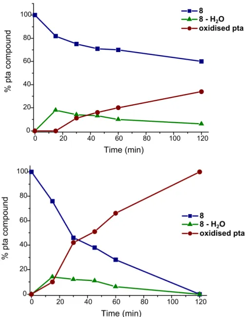
A solution of 8 in D2O was irradiated with UV light (λ = 366 nm) and aliquots were removed
at intervals of 15 min and analysed by 1H and 31P NMR and UV-vis spectroscopy. After the first 15 min of irradiation, a new species is observed in the 31P NMR spectrum of 8 at -33.0 ppm, tentatively assigned to the aqua complex [Ru(η6-p-cymene)pta(N3)(H2O)]+ as it is also
formed from 7 in water. On further irradiation, a second new peak is observed in the 31P NMR spectrum at -2.5 ppm, corresponding to oxidised pta (Figure 5 and Figure S10). The concurrent appearance of free p-cymene in the 1H NMR spectrum suggests that the resulting ruthenium complex extensive ligand loss. Note that the release of a the arene ligand upon light exposure has been already observed for other Ru(II)-arene complexes.[12, 22]
Decreasing the wavelength of the light source to 254 nm accelerates the photolytic decomposition (Figure 5). After 30 min, the intensity of the signal corresponding to the original complex in the 31P NMR spectrum has decreased by a factor of 2, and is replaced by the signal corresponding to oxidised pta. Following 2 h of irradiation, the original complex is no longer present, being replaced by the signal of free oxidised pta at -2.5 ppm. Emergence of a new species is also evident in the UV-vis spectra between 45 min and 2 h with an isosbestic point at 382 nm (Figure S3). Between 0 and 45 min the only change in the spectra corresponds to an increase in absorption at 344 and 400 nm.
12
Figure 5. Graphs showing the percentage of pta compound present in an aqueous solution of 8 following UV irradiation as a function of time. Top: irradiation at 366 nm. Bottom: irradiation at 254 nm. Percentages were determined by integration of the signals in the 31P NMR spectra.
(Photo-)cytotoxicity studies
Having assessed the stability 8 in aqueous media and confirmed that the majority of 8 in its diazide form would accumulate in cells, the influence of 8 on cell viability in cancerous human cervical carcinoma (HeLa) and non-cancerous retinal pigment epithelium (RPE-1) cells was investigated to assess its suitability as a prodrug in PACT. For this purpose, 8 was incubated for 1 h, the time in which the majority of the structurally related compound RAPTA-C enters cells, and for 4 h, the time for equilibrium between 7 and 8 to be reached in
13
a 100 mM NaCl solution. After incubation for the appropriate time (in the dark) the cells were incubated additional 44 h and the cell viability determined using a fluorometric resazurin assay (Table 2). Comparison of the data shows that at the longer incubation time the cytotoxicity is slightly higher. Notably, 8 is about 1.2 - 1.4 times more toxic in the cancerous cell line HeLa in comparison to the non-cancerous cell line RPE-1. Having assessed the cytotoxicity, the effect of light irradiation (450 nm, 10 min, light dose 4.7 J/cm2) was investigated. As anticipated, the cytotoxicity of 8 increases upon light exposure. The phototoxic index (PI), defined as the ratio between the IC50 value in the dark and the IC50
value upon light exposure, was calculated for 8 and is similar in both cell lines with values of 1.5-1.6 at 1 h incubation and 1.2 at 4 h incubation. This decrease of the PI with incubation time is probably due to the modest instability of 8 with time. As recently demonstrated, similar PI values were observed with other Ru(II)-arene complexes bearing 3,5-cycloheptadienyldiphenylphosphine or cycloheptyldiphenylphosphine and ethylbenzoate or p-cymene ligands.[13]
Table 2. IC50 values in the dark and following irradiation of 8 incubated in human cervical
carcinoma (HeLa) and non-cancerous retinal pigment epithelium (RPE-1) cells. Average of three independent measurements.
Incubation time HeLa RPE-1 dark 450 nm (10 min, 4.7 J/cm2) PI dark 450 nm (10 min, 4.7 J/cm2) PI 1 h 275 ± 14 176 ± 9 1.6 332 ± 18 225 ± 9 1.5 4 h 214 ± 16 181 ± 5 1.2 290 ± 6 249 ± 13 1.2 Conclusions
An azide-containing analogue of the drug candidate RAPTA-C, 1, was prepared and evaluated as potential prodrug for PACT. The diazide complex, 8, is remarkably stable in water and in the presence of glutathione, whereas 1 reacts rapidly. However, at chloride concentrations equivalent to that of the blood stream, 8 undergoes a slow ligand exchange. However, during exposure to UV light (λ = 366 nm or 254 nm), the compound undergoes more rapid and extensive ligand loss. As expected from the stability studies, the cytotoxicity in the dark was modest and increases upon light exposure, confirming a photoactivated effect. In this context, it should be noted that in vitro cytotoxicities of this class of compounds are
14
generally low and do not closely reflect their activity in vivo. Since RAPTA compounds are highly active in vivo against primary and metastatic tumours, it is conceivable that 8 could combine the advantages of a RAPTA chemotherapy and PACT.
Experimental
Materials
All chemicals were obtained from commercial sources and used without further purification. Dulbecco’s Modified Eagles Medium (DMEM), Dulbecco’s Modified Eagles Medium supplemented with nutrient mixture F-12 (DMEM/F-12), Fetal Bovine Serum (FBS), Gibco Penicillin-Streptomycin-Glutamine (Penstrep), Dulbecco’s Phosphate-Buffered Saline (PBS) were purchased from Fisher Scientific and Resazurin from ACROS Organics.
Instrumentation and Methods
All manipulations were carried out using standard Schlenk techniques with solvents degassed prior to use. [Ru(
η
6-p-cymene)Cl2]2 and pta were synthesised according to literatureprocedures.[23]1H and 31P NMR spectra were recorded on a Bruker Avance DPX spectrometer at room temperature. Ho and Hm describe aromatic protons H-ortho and H-para respectively.
UV-vis spectra were recorded on a JASCO V-550 UV-vis spectrophotometer at room temperature. Microanaylsis was performed at the EPFL.
Synthesis of [Ru(
η
6-p-cymene)(pta)Cl(N3)](7)1) To a solution of [Ru(η6-p-cymene)Cl2]2 (200 mg, 0.33 mmol) in dry dichloromethane (10
mL),N3SiMe3 (0.2 mL, 2 mmol) was added dropwise at room temperature and the mixture
stirred for 30 min. Hexane (5 mL) was added and the solvent volume reduced to give 7as a red powder. The product was dissolved in dichloromethane (20 mL) and pta (111 mg, 0.7 mmol) added and the solution stirred for 4 h. Hexane (10 mL) was added and the solvent volume reduced to give the product as orange powder. Recrystallisation from methanol afforded an orange, semi-crystalline solid. Yield: 69 mg (21%).
2) To a solution of 1(100 mg, 0.21 mmol) in dry dichloromethane (10 mL), N3SiMe3 (0.46
mL, 4.6 mmol) was added dropwise at room temperature and the mixture stirred for 30 min. Hexane (5 mL) was added and the solvent volume reduced to give 7 as an orange powder. Slow diffusion of hexane into a dichloromethane solution afforded the product as orange crystals suitable for X-ray diffraction (see Figures S4-S5) Yield: 34 mg (35%).
15
1
H NMR (CDCl3): δ 5.58 (d, 3JHH = 5.6 Hz, 2H, Hm), 5.55 (d, 3JHH = 5.6 Hz, 2H, Ho), 4.66 (d, 2
JHH = 12.9 Hz, 3H, pta), 4.60 (d, 2JHH = 13.3 Hz, 3H, pta), 4.41 (s, 6H, pta), 2.74-2.91 (m,
1H, CH(CH3)2), 2.11 (s, 3H, arene-CH3), 1.24 (d, 3JHH = 6.8 Hz, 6H, CH(CH3)2). 31P NMR
(CDCl3):δ -33.5 (s, pta).
Synthesis of [Ru(
η
6-p-cymene)pta(N3)2](8)To a solution of [Ru(η6-p-cymene)Cl2]2 (250 mg, 0.41 mmol) in water (50 mL), NaN3 (500
mg 7.7 mmol) was added and the resulting solution stirred for 30 min at room temperature. Next, pta (141 mg, 0.89 mmol) was added and the solution stirred for a further 12 h. The product was extracted with dichloromethane (3 x 50 mL) and the combined organic phases dried over sodium sulphate. The solution was concentrated in vaccuo and hexane (10 mL) added to precipitate 8 as a red powder. Yield: 344 mg (88%).
1
H NMR (CDCl3): δ 5.51 (d, 3JHH = 5.8 Hz, 2H, Hm), 5.44 (d, 3JHH = 5.6 Hz, 2H, Ho), 4.6 (s,
6H, pta), 4.29 (s, 6H, pta), 2.70-2.86 (m, 1H, CH(CH3)2), 2.21 (s, 3H, arene-CH3), 1.29 (d,
3
JHH = 6.4 Hz, 6H, CH(CH3)2). 31P NMR (CDCl3):δ -30.6 (s, pta). ESI-MS (MeOH) m/z =
912.0 [(Ru(η6-p-cymene)pta(N3))2(
µ
-N3)]+ (60%), m/z = 478.1 [Ru(η6-p-cymene)(pta-H)(N3)2]+ (10%), m/z = 407.0 [Ru2(η6-p-cymene)2(pta)(pta-H)(N3)2(
µ
-N3)]2+ (100%). UV/Vis(H2O): λ (ε) = 205 (18800), 254 (6100), 366 (1200), 450 (670 M-1.cm-1). Anal: Calculated for
C16H28N9OPRu ([Ru(η6-p-cymene)pta(N3)2]⋅H2O C, 38.86; H, 5.71; N, 25.49. Found C,
38.57; H, 5.65; N, 25.63.
X-ray structure determination
Data for 7 and 8 were collected on a KUMA CCD diffractometer system using graphite-monochromated Mo Kα radiation. Data reduction was performed with CrysAlisPro.[24] The structures were solved by SHELXT[25] and refined by least squares fit on F2 using SHELX.[26] The absorption correction was applied using the multi-scan procedure SCALE3 ABSPACK.[24] All non-hydrogen atoms were refined anisotropically with hydrogen atoms placed in geometrically calculated positions and refined using a riding model. In 7, positional disorder of the chloride and azide group was observed and resolved by splitting the atoms over two positions and then fixing the occupancy factors to 0.5. For structure 7, a number of disordered dichloromethane molecules were present, which were removed using the SQUEEZE algorithm of Platon.[27] For structure 8, hydrogen atoms which belong to the water molecule were located on the electron density difference map and the H-O bond lengths and
16
the H-O-H bond angles constrained to reasonable values. ORTEP3[28] was used to produce the graphical representation of 7 and 8. Relevant data is given in Table 3.
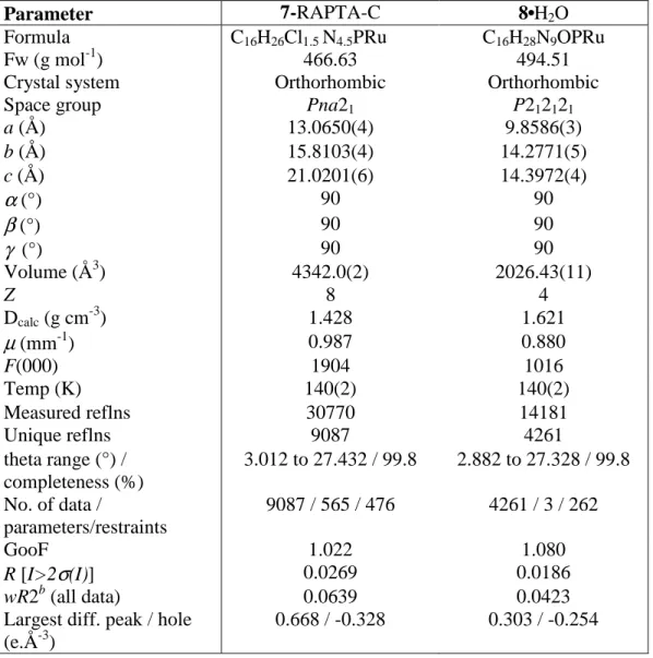
Table 3. Selected crystallographic data for 7 and 8.
Parameter 7-RAPTA-C 8•H2O
Formula C16H26Cl1.5 N4.5PRu C16H28N9OPRu
Fw (g mol-1) 466.63 494.51
Crystal system Orthorhombic Orthorhombic
Space group Pna21 P212121
a (Å) 13.0650(4) 9.8586(3) b (Å) 15.8103(4) 14.2771(5) c (Å) 21.0201(6) 14.3972(4)
α
(°) 90 90β
(°) 90 90γ
(°) 90 90 Volume (Å3) 4342.0(2) 2026.43(11) Z 8 4 Dcalc (g cm-3) 1.428 1.621µ
(mm-1) 0.987 0.880 F(000) 1904 1016 Temp (K) 140(2) 140(2) Measured reflns 30770 14181 Unique reflns 9087 4261 theta range (°) / completeness (%) 3.012 to 27.432 / 99.8 2.882 to 27.328 / 99.8 No. of data / parameters/restraints 9087 / 565 / 476 4261 / 3 / 262 GooF 1.022 1.080 R [I>2σ
(I)] 0.0269 0.0186 wR2b (all data) 0.0639 0.0423Largest diff. peak / hole (e.Å-3)
0.668 / -0.328 0.303 / -0.254
Stability studies in aqueous solution
A freshly prepared 10 mM solution of 8 in D2O or (0.1 M NaCl D2O, equivalent to ca. pH =
7) or upon addition of 2 equivalents of glutathione was monitored by 1H and 31P NMR and UV-vis spectroscopy over a period of 24 h.
Stability upon light exposure
A freshly prepared 10 mM solution of 8 in D2O was irradiated with UV light at 366 or 254 nm
17
the lamp, with a power of ca. 8 Wcm-2. Aliquots were taken after 15, 30, 45, 60 and 120 min and analysed by 1H NMR, 31P NMR and UV-vis spectroscopy.
Cell culture
Human cervical carcinoma (HeLa) cells were cultured using DMEM media and retinal pigment epithelium (RPE-1) cells using DMEM/F-12 with addition of 10% FBS and 1% penstrep. The cells were cultivated and maintained at 37 °C in a cell culture incubator at 37 °C with 5% CO2 atmosphere. Before use the cells were passaged three times.
(Photo)cytotoxicity studies
Cytotoxicity was assessed by measuring cell viability using the fluorometric resazurin assay. The cultivated cells were seeded in triplicates in 96 well plates with a density of 4000 cells per well in 100 µL of media. After 24 h the medium was removed and the cells were treated with increasing concentrations of the appropriate compound diluted in cell media achieving a total volume of 200 µL. The cells were incubated with the compound for 1 or 4 h. After this time, the media was removed and replaced with 200 µL of fresh medium. For the phototoxicity studies, the cells were exposed to light with an Atlas Photonics LUMOS BIO irradiator. Each well was constantly illuminated with 450 nm (10 min, 4.7 J.cm2) irradiation. During this time, the temperature was maintained constantly at 37 °C. The cells were grown in the incubator for additional 44 h. For the determination of the dark cytotoxicity, the cells were not irradiated and after the medium exchange directly incubated for 44 h. After this time, the medium was replaced with fresh medium containing resazurin with a final concentration of 0.2 mg/mL. After 4 h incubation, the amount of the fluorescent product resorufin was determined upon excitation at 540 nm and measurement its emission at 590 nm using a SpectraMax M2 Microplate Reader (Molecular Devices). The obtained data was analyzed with the GraphPad Prism software.
Acknowledgement
This work was financially supported by the Swiss National Science Foundation to P.J.D., an ERC Consolidator Grant PhotoMedMet to G.G. (GA 681679) and has received support under the program “Investissements d’ Avenir” launched by the French Government and implemented by the ANR with the reference ANR-10-IDEX-0001-02 PSL (G.G.).
18 References
[1] A. Jemal, F. Bray, M. M. Center, J. Ferlay, E. Ward, D. Forman, CA Cancer J. Clin. 2011, 61, 69-90.
[2] a) A. Urruticoechea, R. Alemany, J. Balart, A. Villanueva, F. Vinals, G. Capella,
Curr. Pharm. Des. 2010, 16, 3-10; b) F. E. Poynton, S. A. Bright, S. Blasco, D. C.
Williams, J. M. Kelly, T. Gunnlaugsson, Chem. Soc. Rev. 2017, 46, 7706-7756; c) J. Shum, P. K.-K. Leung, K. K.-W. Lo, Inorg. Chem. 2019, 58, 2231-2247; d) L. Zeng, P. Gupta, Y. Chen, E. Wang, L. Ji, H. Chao, Z.-S. Chen, Chem. Soc. Rev. 2017, 46, 5771-5804; e) C. S. Allardyce, A. Dorcier, C. Scolaro, P. J. Dyson, Appl. Organomet.
Chem. 2005, 19, 1-10; f) T. C. Johnstone, K. Suntharalingam, S. J. Lippard, Chem. Rev. 2016, 116, 3436-3486; g) A. Notaro, G. Gasser, Chem. Soc. Rev. 2017, 46,
7317-7337; h) F. Heinemann, J. Karges, G. Gasser, Acc. Chem. Res. 2017, 50, 2727-2736; i) M. Jakubaszek, B. Goud, S. Ferrari, G. Gasser, Chem. Commun. 2018, 54, 13040-13059; jJ. Karges, P. Goldner, G. Gasser, Inorganics 2019, 7, 4.
[3] a) C. S. Allardyce, P. J. Dyson, D. J. Ellis, S. L. Heath, Chem. Commun. 2001, 1396-1397; b) A. Dorcier, C. G. Hartinger, R. Scopelliti, R. H. Fish, B. K. Keppler, P. J. Dyson, J. Inorg. Biochem. 2008, 102, 1066-1076; c) P. J. Dyson, G. Sava, Dalton
Transactions 2006, 1929-1933; d) R. Lee, S. Escrig, C. Maclachlan, G. Knott, A.
Meibom, G. Sava, P. Dyson, Int. J. Mol. Sci. 2017, 18, 1869.
[4] Z. Adhireksan, G. E. Davey, P. Campomanes, M. Groessl, C. M. Clavel, H. Yu, A. A. Nazarov, C. H. F. Yeo, W. H. Ang, P. Dröge, U. Rothlisberger, P. J. Dyson, C. A. Davey, Nature Communications 2014, 5, 3462.
[5] A. Weiss, R. H. Berndsen, M. Dubois, C. Müller, R. Schibli, A. W. Griffioen, P. J. Dyson, P. Nowak-Sliwinska, Chemical Science 2014, 5, 4742-4748.
[6] a) C. Scolaro, A. Bergamo, L. Brescacin, R. Delfino, M. Cocchietto, G. Laurenczy, T. J. Geldbach, G. Sava, P. J. Dyson, J. Med. Chem. 2005, 48, 4161-4171; b) A.
Bergamo, A. Masi, P. J. Dyson, G. Sava, Int. J. Oncol. 2008, 33, 1281-1289.
[7] P. Nowak-Sliwinska, J. R. van Beijnum, A. Casini, A. A. Nazarov, G. Wagnières, H. van den Bergh, P. J. Dyson, A. W. Griffioen, J. Med. Chem. 2011, 54, 3895-3902. [8] a) A. Weiss, X. Ding, J. R. van Beijnum, I. Wong, T. J. Wong, R. H. Berndsen, O.
Dormond, M. Dallinga, L. Shen, R. O. Schlingemann, R. Pili, C.-M. Ho, P. J. Dyson, H. van den Bergh, A. W. Griffioen, P. Nowak-Sliwinska, Angiogenesis 2015, 18, 233-244; b) R. H. Berndsen, A. Weiss, U. K. Abdul, T. J. Wong, P. Meraldi, A. W.
Griffioen, P. J. Dyson, P. Nowak-Sliwinska, Sci. Rep. 2017, 7, 43005.
[9] a) S. Bonnet, Dalton Transactions 2018, 47, 10330-10343; b) J. D. Knoll, C. Turro,
Coord. Chem. Rev. 2015, 282-283, 110-126.
[10] a) L. N. Lameijer, D. Ernst, S. L. Hopkins, M. S. Meijer, S. H. C. Askes, S. E. Le Dévédec, S. Bonnet, Angew. Chem. Int. Ed. 2017, 56, 11549-11553; b) D. E. Dolmans, D. Fukumura, R. K. Jain, Nat. Rev. Cancer 2003, 3, 380-387.
[11] a) N. J. Farrer, L. Salassa, P. J. Sadler, Dalton Transactions 2009, 10690-10701; b) N. A. Smith, P. J. Sadler, The Royal Society Publishing, 2013; c) U. Schatzschneider,
Eur. J. Inorg. Chem. 2010, 2010, 1451-1467; d) D. Kessel, J. Reiners Jr, Isr. J. Chem.
2012, 52, 674-680; e) S. Bonnet, Comments Inorg. Chem. 2015, 35, 179-213; f) C. Mari, V. Pierroz, S. Ferrari, G. Gasser, Chem. Sci. 2015, 6, 2660-2686; g) B. S. Howerton, D. K. Heidary, E. C. Glazer, J. Am. Chem. Soc. 2012, 134, 8324-8327; hR. Sharma, J. D. Knoll, P. D. Martin, I. Podgorski, C. Turro, J. J. Kodanko, Inorg. Chem. 2014, 53, 3272-3274.
[12] S. W. Magennis, A. Habtemariam, O. Novakova, J. B. Henry, S. Meier, S. Parsons, I. D. Oswald, V. Brabec, P. J. Sadler, Inorg. Chem. 2007, 46, 5059-5068.
19
[13] U. Basu, J. Karges, F. Chotard, C. Balan, P. Le Gendre, G. Gasser, E. Bodio, R. Malacea Kabbara, Polyhedron 2019.
[14] a) B. Elias, A. Kirsch-De Mesmaeker, Coord. Chem. Rev. 2006, 250, 1627-1641; b) L. Herman, S. Ghosh, E. Defrancq, A. K. D. Mesmaekera, J. Phys. Org. Chem. 2008, 21, 670-681.
[15] a) F. S. Mackay, N. J. Farrer, L. Salassa, H.-C. Tai, R. J. Deeth, S. A. Moggach, P. A. Wood, S. Parsons, P. J. Sadler, Dalton Transactions 2009, 2315-2325; b) F. S.
Mackay, S. A. Moggach, A. Collins, S. Parsons, P. J. Sadler, Inorg. Chim. Acta 2009,
362, 811-819; c) F. S. Mackay, J. A. Woods, P. Heringová, J. Kašpárková, A. M.
Pizarro, S. A. Moggach, S. Parsons, V. Brabec, P. J. Sadler, Proceedings of the
National Academy of Sciences 2007, 104, 20743-20748; d) L. Salassa, H. I. A.
Phillips, P. J. Sadler, PCCP 2009, 11, 10311-10316; eL. Ronconi, P. J. Sadler, Chem.
Commun. 2008, 235-237.
[16] K. S. Singh, W. Kaminsky, Polyhedron 2014, 68, 279-286.
[17] R. S. Bates, M. J. Begley, A. H. Wright, Polyhedron 1990, 9, 1113-1118.
[18] M. Wehlan, R. Thiel, J. Fuchs, W. Beck, W. P. Fehlhammer, J. Organomet. Chem. 2000, 613, 159-169.
[19] a) A. Dorcier, W. H. Ang, S. Bolano, L. Gonsalvi, L. Juillerat-Jeannerat, G.
Laurenczy, M. Peruzzini, A. D. Phillips, F. Zanobini, P. J. Dyson, Organometallics 2006, 25, 4090-4096; b) A. Casini, C. Gabbiani, F. Sorrentino, M. P. Rigobello, A. Bindoli, T. J. Geldbach, A. Marrone, N. Re, C. G. Hartinger, P. J. Dyson, J. Med.
Chem. 2008, 51, 6773-6781.
[20] a) K. S. Singh, V. Svitlyk, P. Devi, Y. Mozharivskyj, Inorg. Chim. Acta 2009, 362, 5252-5258; b) Y.-L. Liu, F.-H. Wu, T.-K. Duan, Q.-F. Zhang, Chinese J. Struct.
Chem. 2009, 28, 995-997.
[21] C. G. Hartinger, A. Casini, C. Duhot, Y. O. Tsybin, L. Messori, P. J. Dyson, J. Inorg.
Biochem. 2008, 102, 2136-2141.
[22] a) F. Chotard, R. Malacea-Kabbara, C. d. Balan, E. Bodio, M. Picquet, P. Richard, M. Ponce-Vargas, P. Fleurat-Lessard, P. Le Gendre, Organometallics 2018, 37, 812-820; b) L. Delaude, A. Demonceau, A. F. Noels, Chem. Commun. 2001, 986-987; cA. Fürstner, M. Liebl, C. W. Lehmann, M. Picquet, R. Kunz, C. Bruneau, D. Touchard, P. H. Dixneuf, Chemistry–A European Journal 2000, 6, 1847-1857.
[23] a) M. A. Bennett, A. K. Smith, J. Chem. Soc., Dalton Trans. 1974, 233-241; b) D. J. Daigle, T. J. Decuir, J. B. Robertson, D. J. Darensbourg, Inorg. Synth. 1998, 32, 40-45.
[24] CrysAlisPro, Rigaku Oxford Diffraction, release 1.171.39.46, 2018.
[25] SHELXT - Integrated space-group and crystal-structure determination¸ G. M.
Sheldrick, Acta Crystallogr., Sect. A 2015, 71, 3-8.
[26] SHELXL - Crystal structure refinement, G. M. Sheldrick, Acta Crystallogr., Sect. C
2015, 71, 3-8.
[27] PLATON, A.L.Spek, Acta Crystallogr., Sect. D 2009, 65, 148-155.
![Figure 2. Structures of [((η 6 -indane)RuCl) 2 (µ-2,3-dpp)] 2+ 2, [Ru(1,4,5,8- [Ru(1,4,5,8-tetraazaphenanthrene)] 2+ 3, [Ru(1,4,5,8,9,12- hexaazatriphenylene)] 2+ 4, trans, trans, trans-[Pt(N 3 ) 2 (OH) 2 (NH 3 )(py)] 5 and [(η 6 -p-cymene](https://thumb-eu.123doks.com/thumbv2/123doknet/7767388.256176/7.892.107.765.109.1082/figure-structures-indane-rucl-tetraazaphenanthrene-hexaazatriphenylene-trans-cymene.webp)