HAL Id: hal-02105865
https://hal.archives-ouvertes.fr/hal-02105865
Submitted on 22 Apr 2019
HAL is a multi-disciplinary open access
archive for the deposit and dissemination of
sci-entific research documents, whether they are
pub-lished or not. The documents may come from
teaching and research institutions in France or
abroad, or from public or private research centers.
L’archive ouverte pluridisciplinaire HAL, est
destinée au dépôt et à la diffusion de documents
scientifiques de niveau recherche, publiés ou non,
émanant des établissements d’enseignement et de
recherche français ou étrangers, des laboratoires
publics ou privés.
para-sulphonato-calix[8]arene and phenanthroline in the
solid state
Barbara Leśniewska, Anthony Coleman, Yannick Tauran, Florent Perret,
Kinga Suwińska
To cite this version:
Barbara Leśniewska, Anthony Coleman, Yannick Tauran, Florent Perret, Kinga Suwińska.
Pseu-dopolymorphs – a variety of self-organization of para-sulphonato-calix[8]arene and phenanthroline
in the solid state.
CrystEngComm, Royal Society of Chemistry, 2016, 18 (46), pp.8858-8870.
�10.1039/C6CE01753C�. �hal-02105865�
This is an Accepted Manuscript, which has been through the Royal Society of Chemistry peer review process and has been accepted for publication.
Accepted Manuscripts are published online shortly after
acceptance, before technical editing, formatting and proof reading. Using this free service, authors can make their results available to the community, in citable form, before we publish the edited article. We will replace this Accepted Manuscript with the edited and formatted Advance Article as soon as it is available.
You can find more information about Accepted Manuscripts in the author guidelines.
Please note that technical editing may introduce minor changes to the text and/or graphics, which may alter content. The journal’s standard Terms & Conditions and the ethical guidelines, outlined in our author and reviewer resource centre, still apply. In no event shall the Royal Society of Chemistry be held responsible for any errors or omissions in this Accepted Manuscript or any consequences arising from the use of any information it contains.
Accepted Manuscript
rsc.li/crystengcomm
www.rsc.org/crystengcomm
CrystEngComm
HIGHLIGHT Tiddo J. Mooibroek, Antonio Frontera et al. Towards design strategies for anion–π interactions in crystal engineering
Volume 18 Number 1 7 January 2016 Pages 1–184
CrystEngComm
This article can be cited before page numbers have been issued, to do this please use: B. Lesniewska, A. Coleman, Y. Tauran, F. PERRET and K. Suwinska, CrystEngComm, 2016, DOI: 10.1039/C6CE01753C.Pseudopolymorphs
–
a
variety
of
self-organization
of
para-sulphonato-calix[8]arene and phenanthroline in the solid state
Barbara Leśniewska
1*, Anthony W. Coleman
2, Yannick Tauran
2, Florent Perret
3and Kinga Suwińska
4,5* 1Institute of Physical Chemistry, Polish Academy of Sciences, Kasprzaka 44/52, 01-224 Warsaw, Poland
2
LMI, Université Lyon 1, CNRS UMR 5615, 43 bvd 11 novembre, 69622 Villeurbanne, France
3
Institut de Chimie et Biochimie Moléculaires et Supramoléculaires, Université Lyon 1, 43 bvd 11 novembre, 69622 Villeur-banne, France
4
Faculty of Mathematics and Natural Sciences, Cardinal Stefan Wyszynski University, Wóycickiego 1/3, PL-01 938 Warsaw
5
A. M. Butlerov Institute of Chemistry, Kazan Federal University, Kremlevskaya 18, 420008 Kazan, Russia
The paper presents results concerning construction of inclu-sion complexes of para-sulphonato-calix[8]arene with 1,10-phenanthroline. Structures of crystalline host-guest complexes were determined by means of X-ray diffraction. Intermolecu-lar interactions were analyzed and the organization of ions and molecules in the crystal lattice was described.
INTRODUCTION
Supramolecular chemistry is an interdisciplinary field of sci-ence that deals with the design, formation and exploration of complex chemical systems formed from smaller building blocks joined together by non-covalent intermolecular interac-tions.1 However, small here can be a relative term as
supramo-lecular systems can vary in size from clathrate hydrates,2 and Werner clathrates,3 through organic systems,4 metal organic
frameworks,5 to micellar and liposomal structures,6 to pro-teins,7 or DNA8 or even virus assemblies.9 Yet strangely the
simple assembly of water can generate extremely large and complex supramolecular assemblies.10
One major task is the search for new molecular receptors, and to acquire knowledge about molecular recognition involving selective binding between a receptor and a guest or ligand, and resulting in host-guest complex formation. Molecules will recognize each other when there are efficient intermolecular interactions between them, such as preferred spatial relation-ships or the complementarity of the electrostatic fields of their molecules.
Suitably modified receptors may exhibit supramolecular reac-tivity and the spontaneous formation of complex well-defined structures such as, for example capsules, pseudo-capsules, columns and chains, that is, to self-assemble, into auto-associated complex supramolecular systems.11
In terms of organic systems, artificial molecular receptors include the cyclodextrins,12 crown ethers, cryptands and
spherands,13 cyclic polyamines,14 and cucurbiturils15 and the
class of systems based on the calix[n]arenes.16,17
One of the most important tasks of supramolecular chemistry is to attempt to understand the construction and functioning of self-assembled structures systems in nature. This may be achieved through their modeling by means of simpler biomi-metic systems, for example enzyme models,18 or artificial
membranes.19
The panoply of processes of molecular recognition and com-plex formation occurring in nature include, enzymatic reac-tions in living organisms,20 distinguishing fragrance by olfac-tory receptors and synthetic analogues,21 active transport of
compounds through the membrane of the cell walls,22 (indeed
back transport gives rise to multi-drug resistance23) the for-mation of the DNA double helix,24 and virus assembly.9
Recent work by ourselves in the area of the biochemistry of the para-sulphonato-calix[n]arenes,25,26 has shown that there
are considerable differences between the four, six and eight membered macrocycles. In particular para-sulphonato-calix[8]arene has been shown to be a more effective endonu-clease inhibitor than the smaller analogs27 and interestingly shows the ability to transport across the CaCo-2 cell model of the human gut.28 Thus structural information on the binding of para-sulphonato-calix[8]arene to substrates will have
consid-erable relevance in the biological and pharmacological fields, but also for the construction of novel solid-state systems. The aim of this work was to obtain self-organizing systems of
para-sulphonato-calix[8]arene (C8S) with 1,10-phenanthroline (Phen) and to undertake structural analysis of the obtained solid-state supramolecular assemblies by X-ray diffraction on single crystals.
The obtained structural data were the basis for a detailed anal-ysis of the conformation of the para-sulphonato-calix[8]arene receptor and for analyzing interactions between the host and guest species. Also the analysis of the organization of ions and molecules in the crystal lattices has been carried out, allowing us to observe pseudo-polymorphism in two of the systems,
where the core structure varied in form, as a result of the differences in solvation. Such behavior is common in large
bio-molecular structures, but less well known in molecular supramolecular complexes. The ligand, 1,10-Phenanthroline is a flat and rigid molecule which is not subject to conformation-al changes, but which can influence conformationconformation-al changes
CrystEngComm
Accepted
Manuscript
Published on 31 October 2016. Downloaded by Tokyo Daigaku on 01/11/2016 10:54:54.
for other flexible molecules which co-create the supramolecu-lar complex. Previously we have studied solid state complexes of 1,10-phenanthroline with para-sulphonato-calix[4]arene29
and also para-sulphonato-calix[8]arene with flexible guest molecules 1,2-bis(4-pyridyl)-ethane and 1,3-bis(4-pyridyl)-propane.30
EXPERIMENTAL SECTION Crystal growth
Synthesis of para-sulphonato-calix[8]arene
para-Sulphonato-calix[8]arene was syntheised by the method
previously used for para-sulphonato-calix[4]arene by Cole-man.31 The physical properties, in particular, the ES/MS
ac-cord with the structure, it should be noted that this method generates calix[8]arene-para-sulphonic acid, however the pKa of the sulphonic acids is around 0 so the compound is fully deprotonated.
Crystal growth
Suitable crystals for X-ray diffraction were grown by slow diffusion of a solution of the Phen in ethanol and methanol at 4 equiv. into 1 mL of a 100 mg mL-1 solution of para-sulphonatocalix[8]arene in water. The crystallization experi-ments were carried out at 20 °C in sealed tubes. The solvent
ratios were varied from 0.5 Alcohol:1 Water to 2 Alcohol:1 Water to screen crystallization conditions. The crystals are colourless.
X-ray structure determination
The X-ray data were collected at 100 K on a Nonius Kap-paCCD diffractometer using MoKα radiation for complex 1 and on an Agilent SuperNova diffractometer equipped with a CuKα micro-focus X-ray source for complex 2 and 3. Struc-tures were solved by direct methods with SHELXS-9732 and
refined against F2 with full-matrix least-squares using SHELXL-9732. Hydrogen atoms were calculated at their
ideal-ized positions and were refined as riding atoms with isotropic thermal parameters based upon the corresponding bonding carbon atom (Uiso = 1.2Ueq, Uiso = 1.5Ueq for CH3 and OH
hydrogens). Hydrogen atoms of methyl and hydroxyl groups were refined in geometric positions for which the calculated sum of the electron density is the highest (rotating group re-finement). Where possible hydrogen atoms of amino groups and water molecules were located on Fourier difference maps and refined with positional parameters. Because of the poor diffracting quality and disorder, the R and wR of structures 1 and 2 are high. Crystal structures are deposited with the Cambridge Crystallographic Data Centre as CCDC 1437840-1437842. Crystal data for 1, 2 and 3 are shown in Table 1.
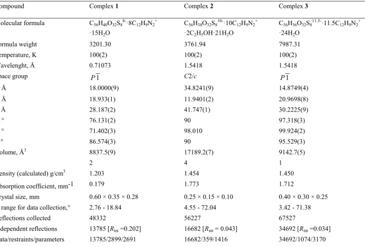
Table 1. Crystal data for 1, 2, 3.
Compound Complex 1 Complex 2 Complex 3
Molecular formula C56H40O32S8 8-·8C12H9N2 + ·15H2O C56H38O32S8 10-·10C12H9N2 + ·2C2H5OH·21H2O C56H36O32S8 11.5-·11.5C12H9N2 + ·24H2O Formula weight 3201.30 3761.94 7987.31 Temperature, K 100(2) 100(2) 100(2) Wavelenght, Å 0.71073 1.5418 1.5418 Space group P1 C2/c P1 a, Å 18.0000(9) 34.8241(9) 14.8749(4) b, Å 18.933(1) 11.9401(2) 20.9698(8) c, Å 28.187(2) 41.747(1) 30.2225(9) α, ° 76.131(2) 90 97.318(3) β, ° 71.402(3) 98.010 99.924(2) γ, ° 86.574(3) 90 95.529(3) Volume, Å3 8837.5(9) 17189.2(7) 9142.7(5) Z 2 4 1 Density (calculated) g/cm3 1.203 1.454 1.450 Absorption coefficient, mm-1 0.179 1.773 1.712 Crystal size, mm 0.60 × 0.35 × 0.28 0.25 × 0.15 × 0.10 0.40 × 0.30 × 0.25
Ɵ range for data collection,° 2.76 - 18.84 4.55 - 72.04 3.42 - 71.38 Reflections collected 48332 56227 67527
Independent reflections 13785 [Rint =0.202] 16682 [Rint = 0.043] 34692 [Rint =0.034]
Data/restraints/parameters 13785/2899/2691 16682/359/1416 34692/1074/3170
CrystEngComm
Accepted
Manuscript
Published on 31 October 2016. Downloaded by Tokyo Daigaku on 01/11/2016 10:54:54.
Goodness-of-fit on F2 1.68 1.20 0.95
Final R indices [I > 2σ(I)] R = 0.220, wR = 0.485 R = 0.137, wR = 0.149 R = 0.078, wR = 0.246 R indices (all data) R = 0.321, wR = 0.524 R = 0.371, wR = 0.375 R = 0.098, wR = 0.261
RESULTS AND DISCUSSION
Complex 1
Crystals of the complex 1 were obtained from a methanol-water crystallization medium. The asymmetric part of the unit cell contains one C8S octaanion, eight monoprotonated phe-nanthroline cations and 15 water molecules (Figure 1). The exact charge on the para-sulphonato-calix[8]arene anion is subject to debate, as one or more of the phenolic residues may be deprotonated,33 and as with cationic complexes of DNA,34,35 at least some of the water molecules may be protonated.
Figure 1. The asymmetric unit of the complex 1.
The structure is characterized by a high degree of disorder. Six
Phen ions are disordered, four of which are in two orientations
Y/O, V/M, T/L, K/Q, with site occupation factors, (s.o.f.) = 0.5 each, and two other Phen R and J cations are located on the center of symmetry, each of the two symmetrically-dependent orientation of these two cations is half-occupied. Additionally five of the eight calixarene sulphonate groups and almost all water molecules are disordered. The octaanion of para-sulphonato-calix[8]arene adopts a new, previously undescribed conformation (Figure 2).
(a)
(b)
Figure 2. The unusual conformation of the para-sulphonato-calix[8]arene in the complex 1; (a) view from above showing intermolecular O‒H···O hydrogen bonding, (b) side view.
This conformation is different from the pleated loop and dou-ble cone which have been highlighted as two extreme confor-mations for eight-membered calixarenes.36 One of the phenolic
rings H of C8S lies flat in the plane of the macrocyclic ring and its sulphonate group pointing toward the inside of the macrocycle. The two neighboring to it phenolic rings A and G are oriented anti-parallel one to another and are perpendicular to the plane of the macrocycle, the remaining five phenolic rings are inclined to the plane of macrocycle with sulphonate groups facing outwards and forming a folded loop. para-Sulphonato-calix[8]arene in this untypical conformation has the cavity inside of macrocycle blocked by self-inclusion of the sulphonate group of the phenolic ring H (Figure 3). Angles between planes of phenolic rings to the reference plane of calixarene (the plane defined by eight carbon atoms of
meth-ylene groups of C8S) are presented in Table 2.
CrystEngComm
Accepted
Manuscript
Published on 31 October 2016. Downloaded by Tokyo Daigaku on 01/11/2016 10:54:54.
4
Figure 3. Self-Inclusion of one of the sulphonate group of C8S in complex 1.
Table 2. Dihedral angles between the planes of the phenolic rings of C8S and the reference plane (the plane defined by the carbon atoms of methylene groups of C8S) in the crys-tal of complex 1.
Ring Angle (°) Ring Angle (°)
A 73.92(5) E 57.05(5)
B 26.90(5) F 10.89(5)
C 52.79(5) G 87.27(5)
D 52.92(4) H 13.07(5)
Conformation of the C8S anion is stabilized by two hydrogen bonds O‒H···O [2.86(2) and 2.89(2) Å] between the oxygen atoms of the phenolic hydroxyl groups and the one O‒H···O bonding [2.71(2) Å], in which the donor is the oxygen atom of the hydroxyl group and the acceptor is an oxygen atom of sulphonate group of C8S. The C‒H···O, C‒H···π and π-π weak interactions present between the calixarene anion and
Phen cations have an effect on the conformation adopted by C8S (Figure 4).
Figure 4. Cations of Phen (X – blue, Y – red, J – green, V – pink, T – purple and K – yellow) located in the immediate vicinity of the C8S anion and their influence on the calixarene conformation.
Conformation of the calixarene anion in the crystal of complex
1 prevents the formation of an inclusion compound. Phen
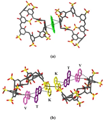
cations J are π-π stacked [3.52(2)–4.81(2) Å] between aro-matic rings of two adjacent calixarenes and additionally this interaction is stabilized by the C‒H···π [3.73(2) Å] interaction between carbon atoms of the methylene group of C8S and the central ring of Phen (Figure 5a and Figure 1). Selected inter-actions present in the structure of complex 1 are presented in Table S1 (supplementary information).
(a)
(b)
Figure 5. Phen cations intercalated between two adjacent C8S anions; (a) intercalation of a single cation Phen J (b) intercalation of Phen6 hexamer (Phen cations are disordered, only one
orienta-tion of caorienta-tions V/M, T/L and K/Q is shown for clarity).
Phen cations K, T and V form a centrosymmetric π-stacked Phen6 hexamer V-T-K-K-V-T where the interplanar distance
for K···K is 3.72(4) Å and estimated distances for K···T and T···V interactions are 3.4 and 3.2 Å, respectively (Figure 5b). The hexamer is located between two anions of C8S. External
Phen ion V interacts with calixarene by C‒H···π [3.61(2) Å]
interaction present between the carbon atom of the methylene group of C8S and the central ring of Phen V as well as by C‒ H···O [3.06(2) Å] hydrogen bonding present between the carbon atom of the nonprotonated ring of Phen and oxygen of sulphonate group of calixarene, and also forms π-π interaction (with distances between centroids: 4.48(2) and 4.62(2) Å) between the aromatic ring of C8S and central and protonated rings of Phen cation V. The Phen cation T interacts with the calixarene anion by C‒H···O [3.21(3) Å] interaction between the carbon atom of no protonated Phen ring and the oxygen atom of a sulphonate group of C8S, and through C‒H···O 3.24(4) Å interaction between the carbon atom of the central ring of Phen and the oxygen atom of the hydroxyl group of
C8S. Ion Phen K does not interact directly with calixarene.
Other phenantroline cations marked with symbols Y, X, Z, W, R create infinite stack (Figure 6) stabilized by π-π interactions, the estimated distance between Phen cations in the stack is 3.4 Å. Phenanthroline cations forming the stack interact with
CrystEngComm
Accepted
Manuscript
Published on 31 October 2016. Downloaded by Tokyo Daigaku on 01/11/2016 10:54:54.
5
calixarenes via C‒H···O and C‒H···π hydrogen bonds and π-π interactions see Table S1). A similar stacked self-organization of phenanthroline cations was previously observed in crystal structures of complexes of smaller para-sulphonato-calix[4]arene with phenanthroline.29,37
Figure 6. The infinite stack formed by Phen cations in structure of complex 1.
Each anion of C8S interacts directly with two neighboring calixarenes (Figure 7) through π‒π interactions between two adjacent aromatic rings of C8S with distances between cen-troids: 3.69(2) and 3.98(2) Å and by O‒H···O hydrogen bond-ing [2.59(3), 2.82(3), 2.74(2) Å] in which H-donors are oxy-gen atoms of hydroxyl groups of one C8S and acceptors are oxygen atoms of sulphonate groups of adjacent C8S anions, as well as by C‒H···O interaction [3.23(3) Å] between the car-bon atom of methylene group of one calixarene and the oxy-gen atom of the sulphonate group of adjacent calixarene. C8S anions are organized in double tapes shown in (Figure 7). Between the adjacent tapes of calixarenes no interactions are observed.
Figure 7. Adjacent anions of C8S interacting via π-π, O‒H···O and C‒H···O interactions ‒ construction of a double tape.
The crystal packing of ions and molecules in the complex 1 is close to layered. Stacks and hexamers of phenanthroline are separated from each other by C8S anions (Figure 8). The structure is quite highly hydrated but the conventional hydro-philic or hydrophobic layers cannot be unambiguously distin-guished. Similar crystal packing was observed in the already mentioned structures of complexes formed by para-sulphonato-calix[4]arene with phenanthroline29, but the current
structure of complex 1 is more “dense” due to the presence of stronger interactions.
(a)
(b)
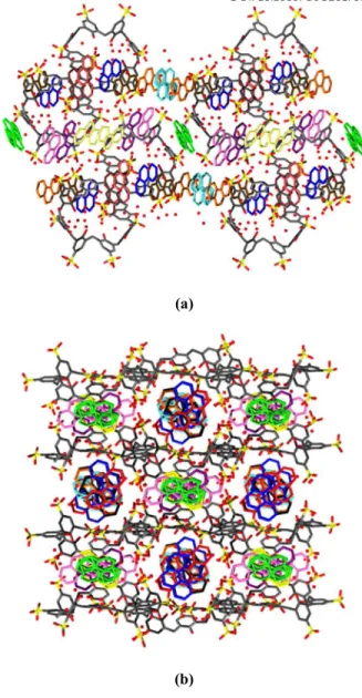
Figure 8. Packing of ions and molecules in the crystal of complex
1; (a) view along [010] direction; (b) view along [11-1] direction.
The self-assembly of C8S anions and Phen cations, in the crystal lattice of complex 1 leads to the formation of cages with approx. dimensions of 16 × 6 × 6 Å. The solvent accessi-ble volume is 733 Å3, which is 8.3% of the volume of the unit cell (Figure 9).
CrystEngComm
Accepted
Manuscript
Published on 31 October 2016. Downloaded by Tokyo Daigaku on 01/11/2016 10:54:54.
6
Figure 9. Solvent accessible cages in the crystal lattice of complex
1; view along the [100] direction. Water molecules are omitted for
clarity.
The cages are filled with water molecules. Three walls of cage are formed by hydrophobic parts of Phen cations, one wall is formed by the sulphonate groups of C8S and protonated rings of Phen cations.
Complex 2
Crystals of complex 2 were obtained from an ethanol-water crystallization medium. The asymmetric part of the unit cell (Figure 10) contains half of the C8S anion in which three sulphonate groups are disordered, five monoprotonated cations of phenanthroline, one molecule of ethanol, and 10.5 water molecules. In the structure one cation Phen Y/W is disordered with site occupation factors (s.o.f.) equal to 0.79 and 0.21 for orientations Y and W, respectively; 4.5 out of 10.5 water molecules are also disordered.
Figure 10. The asymmetric unit of the complex 2. Part of mole-cule which is marked in fuchsia is symmetry related.
The presence of water molecules and an oxygen atom of sul-phonate group of C8S near the nitrogen atoms of Phen at distances typical to hydrogen bonds indicates that all phenan-throline molecules are monoprotonated and bearing a total charge +5. Consequently, the charge balance in the unit cell indicates deprotonation of two hydroxyl groups of calixarene in addition to deprotonation of eight sulphonate groups. It is evidenced by the very short distances between oxygen atoms of the phenolic groups O4C and O4D [2.477 (5) Å] which indicates the presence of a hydrogen bond supported by elec-tric charge. C8S anion is located on a crystallographic center of inversion and adopts centrosymmetric inverted double partial cone conformation (Figure 11) similar, but not identi-cal, to these observed for the structures [LUKBUP]38 and
[QULJUD]39 [QULJUD01]39, [QULKAK]39 and [VERBEA]36.
Superposition of conformations of
para-sulphonato-calix[8]arene in complex 2 and VERBEA is shown on Figure 12.
(a)
(b)
Figure 11. Conformation of the para-sulphonato-calix[8]arene in the complex 2; (a) top view showing intramolecular O‒H···O hydrogen bonding, (b) side view.
(a)
CrystEngComm
Accepted
Manuscript
Published on 31 October 2016. Downloaded by Tokyo Daigaku on 01/11/2016 10:54:54.
7
(b)
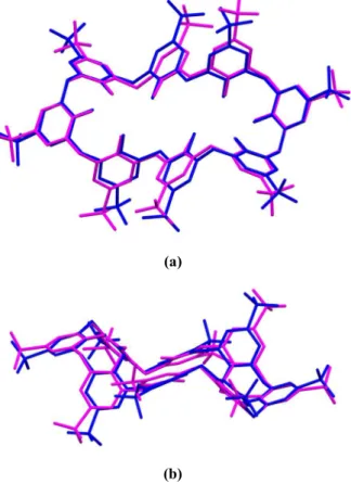
Figure 12. The superposition of the inverted double partial cone conformations of C8S in complex 2 (red) and in [VERBEA] (blue); (a) top view, (b) side view.
The two partial cones of calix[8]arene anion are oriented anti-parallel to each other (Figure 11b) and each partial cone is formed by phenolic rings A, C, and D, while phenolic ring B is inverted with an angle of -36.73 (2)° to the reference plane of calixarene (the plane defined by the carbon atoms of meth-ylene groups of C8S). Phenolic rings located between the two partial cones are at a considerable distance from each other and thus a cavity inside the macrocycle is formed. As a conse-quence, the conformation of para-sulphonato-calix[8]arene in complex 2 can be also considered as a distorted pleated loop and compared to this conformation36 (Figure 13).
(a)
(b)
Figure13.The superposition ofpleatedloopconformation of
C8S in complex 2(red) and [VERBAW]36(gray); (a) top view, (b) side view.
Dihedral angles between the planes of the phenolic rings of
C8S and the reference plane of calixarene are presented in
Table 3.
Table 3. Dihedral angles between the planes of the phenolic rings of C8S and the reference plane of macrocycle (the plane defined by the carbon atoms of methylene groups of C8S) in the crystal of complex 2.
Ring Angle (°)
A 77.65(2)
B -36.73(2)
C 33.59(2)
D 37.87(2)
Conformation of C8S ion in the structure of complex 2 is stabilized by intramolecular O‒H···O hydrogen bonds [2.628(8), 2.685(7) and 2.477(8) Å] between oxygen atoms of the phenolic hydroxyl groups and by aromatic, electrostatic and hydrophobic ions between C8S anion and the surrounding
Phen cations (Figure 14). Such conformation of C8S anion as
described above results in a presence of two cone-like cavities and additional creation of four "folds" formed between twisted phenolic groups. Cations Phen X and U are located in these "folds", while cations Phen Z are located within the partial cone cavities of C8S (Figure 14).
Figure 14. Host-guest interactions in complex 2: two cations Phen Z (orange) located in partial cone cavities (the second Phen Z cation is located in the second cavity of C8S and is invisible in this orientation) and cations Phen X (blue) and U (violet) located in "folds" of the calixarene anion.
Anion of C8S, four cations of Phen (two Phen Z and two
Phen X), and two molecules of ethanol form an
inclusion-additive complex (Figure 15).
Figure 15. Inclusion-additive complex formed by the anion of
C8S, four phenanthroline cations and two molecules of ethanol
(green).
The depth of inclusion of cation Phen Z within the partial cone of C8S measured as the distance between the centroid of
CrystEngComm
Accepted
Manuscript
Published on 31 October 2016. Downloaded by Tokyo Daigaku on 01/11/2016 10:54:54.
8
included phenanthroline protonated ring and the reference plane of C8S is 3.782(6) Å, the dihedral angle between the plane of Phen Z and the reference plane of C8S is 43.89(5)°. Cation Phen Z is not located parallel to the axis of the partial cone, as it was observed in the case of para-sulphonato-calix[4]arene29, but is tilted within the cavity to allow π-π
interaction [estimated plane-to plane distance of 3.4 Å] be-tween the phenanthroline protonated ring and ring C of C8S (similarly to the complexes of para-sulphonato-calix[6]arene with Phen).40 Inclusion of cations Phen Z in complex 2 is additionally stabilized by C‒H···π interaction [3.643(9) Å] between the carbon atom of the methylene group of C8S and the central ring of guest cation, C‒H···π interactions [3.526(8) and 3.453(8) Å] between carbon atoms belonging to the heter-ocyclic protonated ring and/or central ring of Phen and aro-matic rings of C8S, and also by one C‒H···O interaction [3.24(1) Å] in which the H-donor atom is the carbon atom belonging to the protonated ring of Phen and the H-acceptor atom is an oxygen atom of the sulphonate group of the host anion. Cations Phen X which are located in the "folds" of the host anion interact with the C8S by means of π‒π interactions [3.497(4) and 4.763(5) Å] between the aromatic ring C of C8S and heterocyclic protonated and central rings of Phen X, C‒ H···π interaction [3.656(8) Å] between the carbon atom of the methylene group of C8S and the central ring of phenanthroline cation and C‒H···π interaction [3.816(9) Å] between the car-bon atom belonging to the heterocyclic protonated ring of
Phen and the aromatic ring B of C8S. The distance between
the centroid of protonated ring of Phen X and the reference plane of C8S is 2.931(4) Å, the angle between the plane of the cation Phen X and the reference plane is 38.80(5)°. Cation
Phen U located in the second "fold" of calixarene interacts
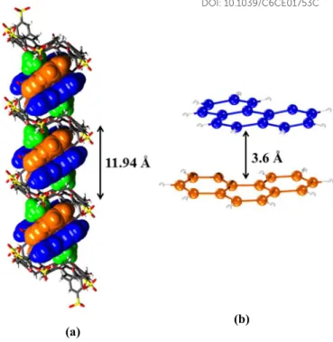
with the host anion only by two C‒H···O hydrogen bonds [3.43(2) and 3.27(2) Å] between carbon atoms belonging to a heterocyclic protonated ring of Phen U and oxygen atoms of the sulphonate and hydroxyl groups of C8S. The ethanol mol-ecule which is the part of inclusion-additive complex interacts with calixarene by C‒H···O hydrogen bond [3.37(1) Å] occur-ring between the carbon atom of the methylene group of C8S and the oxygen atom of ethanol. Selected interactions present in the structure of complex 2 are listed in the Table S2 (sup-plementary information). Cations Phen X and Z belonging to the neighboring supramolecular complexes form dimers (Fig-ure 16b) arranged along the [010] crystallographic axis in a polymeric system of pseudo-capsules formed between adja-cent calixarenes and accommodating two dimers of Phen and two molecules of ethanol (Figure 16a). Phen X and Z in the dimer are rotated relative to each other by 43°. Each dimer is stabilized by π-π interaction [estimated distance of 3.6 Å] between the two of Phen ions.
(a)
(b)
Figure 16. (a) Columns of inclusion-additive complexes compris-ing two dimers of Phen and two molecules of ethanol formcompris-ing an inclusion-additive polymer; (b) π-π interaction between cations
Phen X and Phen Z in the dimer.
The height of the repeating unit in such an inclusion-additive polymer is 11.94 Å Similar structural motifs have been ob-served in crystals of para-sulphonato-calix[6]arene with
Phen40,41 but in this case the pseudo-capsules are smaller and are able to accommodate in a single dimer of Phen only. The three other cations Phen U, V and Y/W present in the crystal of complex 2 form infinite stacks stabilized by π-π interactions along the [001] crystallographic axis. The estimated distances between planes of Phen molecules are the same and all equal to 3.4 Å. The stacks of Phen and form together with water molecules present in the crystal separate layers in the structure (Figure 17). Phen ions belonging to the infinite stacks interact with calixarenes through the C‒H···O (Phen U) and π-π (Phen Y/W) interactions. A similar stacking self-organization of phenantroline cations takes place in the structures of para-sulphonato-calix[4]arene with Phen29,37 and in the complex 1.
In the crystal of complex 2, no direct interactions between
C8S anions are observed.
CrystEngComm
Accepted
Manuscript
Published on 31 October 2016. Downloaded by Tokyo Daigaku on 01/11/2016 10:54:54.
9
(a)
(b)
Figure 17. The crystal packing of ions and molecules in the crys-tal of complex 2; (a) view along the [010] cryscrys-tallographic axis; (b) view along the [001] crystallographic axis. Phen U cations are marked in purple, V in cyan Y/W in fuchsia.
The structure of complex 2 show a typical bilayer packing. One layer consists of the columns of C8S anions, cations
Phen X and Z and ethanol molecules involved in
inclusion-addition complex formation; the second layer consists of the remaining three Phen cations forming the infinite stacks and water molecules. In this structure the distinct hydrophilic and hydrophobic layers are not observed. Most of the water mole-cules present in the crystal, are located near the sulphonate groups of C8S and all of them are inside the elongated centro-symmetric S-shaped cages of approx. dimensions 6 × 8 × 22 Å and running parallel to Phen stacks (Figure 17b).
Complex 3
Further evaporation of solvent from the solution containing crystals of complex 2 led to crystallization of new crystalline phase named complex 3. The asymmetric part of the unit cell consists one C8S anion in which four sulphonate groups are disordered, 11.5 phenanthroline cations and 24 water mole-cules of which 7 are disordered (Figure 18). Also three Phen cations are disordered; the one marked as Z/L was found in two orientations with s.o.f. = 0.69 and 0.31, respectively for Z and L, the second one occurs in three orientations V/M/K rotated relative to each other in the plane of the molecule and with the site occupancy factors of 0.34; 0.33 and 0.33, respec-tively for V, M and K, and the third disordered Phen cation denoted as N is located on a crystallographic center of inver-sion and is half-occupied (Figure 19).
Figure 18. The asymmetric unit of the crystal 3.
(a) (b)
(c)
Figure 19. Disorder of phenantroline cations in complex 3; (a) cation Z/L (Z – black and L – red); (b) cation V/M/K (V – black, M – yellow and K – green); (c) cation N.
The presence of water molecules next to the 10 nitrogen atoms of Phen cations at distances typical for hydrogen bonds sug-gests that the molecules of phenanthroline are monoprotonat-ed. The structural model assumes protonation of these nitrogen atoms. In order to balance the electric charges, it was assumed that half of the calixarenes in the unit cell have three deproto-nated hydroxyl groups and the other half have the four depro-tonated hydroxyl groups. Deprotonation of OH groups of calixarene may also be evidenced by a short distances between the phenolic oxygen atoms: O4A and O4B [2.436(4) Å]; O4E and O4F [2.448(4) Å]; O4F and O4G [2.547(4) Å]; O4C and O4B [2.558(4) Å]. It was not possible to locate all of the hy-drogen atoms on Fourier difference maps, due to a large num-ber of atoms in the independent part of the unit cell and the high degree of disorder in the structure. C8S anion adopts pseudo-centrosymmetric conformation similar to the inverted double partial cone and similar to that observed in the crystal structure of complex 2 (Figure 20).
CrystEngComm
Accepted
Manuscript
Published on 31 October 2016. Downloaded by Tokyo Daigaku on 01/11/2016 10:54:54.
10
(a)
(b)
Figure 20. Superposition of pseudo-centrosymmetric C8S anion in complex 3 (magenta) and centrosymmetric C8S anion found complex 2 (blue); (a) top view, (b) side view.
Partial cones of C8S are formed by three phenolic rings A, B, C, and E, F, G, while the phenolic groups of rings D and H between the two partial cones are directed outside of the mac-rocyclic ring. The dihedral angles between the planes of phe-nolic rings to the reference plane of C8S are listed in Table 4.
Table 4. Dihedral angles between the planes of the phenolic rings of C8S and the reference plane of the macrocycle (the plane defined by the carbon atoms of methylene groups of C8S) in the crystal of complex 3.
Ring Angle (°) Ring Angle (°)
A 35.91(4) E 33.51(4)
B 28.70(4) F 31.96(4)
C 72.44(4) G 67.26(4)
D -36.31(4) H -38.68(4)
In each partial cone of C8S one cation of Phen is included – X in one and Y in the second partial cone (Figure 21).
Figure 21. An inclusion host-guest complex formed by the C8S anion and two cations X and Y of phenanthroline.
The depth of inclusion of Phen cations measured as the dis-tance between centroids of included heterocyclic monoproto-nated rings of phenantroline and reference plane of C8S is 3.926(5) and 3.979(5) Å for Phen X and Y, respectively, and the dihedral angles between the reference plane of calixarene and the planes of Phen X and Y are 40.87(3) and 46.82(3)°, respectively. Similarly to the complex 2, Phen cations are not included vertically in the calixarene cavities but are tilted within the cavity to allow π-π interaction between Phen X and ring E of C8S [estimated plane to plane distance of 3.4 Å], and between Phen Y and ring A of C8S [estimated plane to plane distance of 3.3 Å]. However, in contrary to complex 2 where the protonated rings of included Phen cations was found to be inserted within the cavities of C8S, in complex 3 the nonpro-tonated heterocyclic rings of Phen are located within the par-tial cones. Such type of complex formation was found in two crystal structures of para-sulphonato-calix[4]arene with
Phen.29 Additionally, the inclusion complex is stabilized by
C‒H···π interactions [3.342(2), 3.689(3) and 3.475(2) Å] between carbon atoms belonging to the heterocyclic nonproto-nated rings of Phen and aromatic rings of C8S, and C‒H···O hydrogen bonds [3.180(5), 3.205(8), 3.180(7), 3.45(1), 3.34(1) Å] in which the proton donors are atoms belonging to the protonated rings of Phen cations and acceptors are the sulpho-nate oxygen atoms of the host molecule. Protosulpho-nated nitrogen atoms of included Phen cations are also involved in N‒H···O hydrogen bonds [2.901(4) and 2.760(5) Å] to water molecules.
Selected interactions present in crystals of complex 3 are listed in the Table S3 (supplementary information). Each of the included Phen cations form a π-stacking dimers (X···X) and (Y···Y) to the related by crystallographic centers of symmetry inclusion complexes (Figure 22b and c), and thereby forming one-dimensional inclusion polymer (Figure 22a) containing two types of pseudo-capsules of estimated height 9.7 and 10.1 Å for capsules containing dimers (X···X) and (Y···Y), respec-tively.
CrystEngComm
Accepted
Manuscript
Published on 31 October 2016. Downloaded by Tokyo Daigaku on 01/11/2016 10:54:54.
11
(a)
(b)
(c)
Figure 22. (a) Inclusion polymer formed by C8S, Phen X and
Phen Y in the crystal of complex 3; (b) a dimer formed by cations Phen X; (c) dimer formed by cations Phen Y.
Chains of polymeric capsules in complex 3 are arranged paral-lel to the [011] crystallographic direction (Figure 23).
Figure 23. Crystal packing of ions in complex 3, the view along the [011] crystallographic direction. Water molecules are omitted for clarity.
Between phenolic groups of C8S anions and sulphonate groups of the calixarenes from adjacent chains propagated in [100] direction weak C‒H···O interactions occur [3.375(2) and 3.454(1) Å] thus forming a dense layer. The spaces be-tween layers are filled with the remaining cations of Phen and water molecules. The O and P cations of Phen, which are not involved in the inclusion complex formation, form (O···P) dimers which are intercalated between six surrounding ca-lixarene anions (Figure 24a).
(a)
(b)
Figure 24. (a) Phen (O···P) dimer intercalated between six C8S anions in complex 3; (b) π-stacking between cations Phen O and P forming a dimer.
Figure 25. The stack composed of cations Phen W, U, T, R, Q, Z, V, N in complex 3; W – blue, U – white T – green, R – blue, Q – pink, Z – yellow, V – magenta, N – purple.
The dimer Phen (O···P) shown on (Figure 24b) consists of two crystallographically independent, almost parallel cations of phenantroline with the estimated distance between the planes of Phen of 3.4 Å. Each cation of Phen participates in one C‒H···π interaction [3.360(2) and 3.284(3) Å for O and P, respectively] between the carbon atom of the heterocyclic protonated ring of Phen and an aromatic ring of one of the surrounding calixarene. Intercalation of dimer Phen (O···P) is also stabilized by several C‒H···O interactions [range 3.03(2)–3.34(2) Å] to sulphonate groups of C8S. The
remain-CrystEngComm
Accepted
Manuscript
Published on 31 October 2016. Downloaded by Tokyo Daigaku on 01/11/2016 10:54:54.
12
ing 7.5 crystallographically independent cations of Phen W, U, T, R, Q, Z, V, N form infinite stacks along the [11-2] crys-tallographic direction (Figure 25 and Figure 26). The estimat-ed distances between Phen cations in the stack are in the nar-row range of 3.3–3.5 Å.
Figure 26. The crystal packing of ions in the crystal of complex 3, view along the [11-2] crystallographic direction. Phen W, U, T, R, Q, Z, V, N cations are marked in purple. Water molecules are omitted for clarity.
Phenantroline cations forming the stack are located closed to calixarene anions and they interact with them by numerous N– H···O, C–H···O, C–H···π and π–π interactions. A similar self-organization of Phen cations in stacks was observed in the structures of complexes of para-sulphonato-calix[4]arene with
Phen29,37and complexes 1 and 2 described above. The struc-ture of complex 3 represents the layered-columnar type of packing (Figure 27).
(a)
(b)
Figure 27. The crystal packing of ions in the crystal of complex 3: (a) view along the [100] crystallographic axis; (b) view along the [010] crystallographic axis. Water molecules have been omitted for clarity.
The layers formed by the inclusion polymers C8S/Phen (X, Y) are arranged alternately with layers constructed of other
Phen cations. The entire structure is strongly hydrated,
there-fore, the conventional hydrophilic and hydrophobic layers cannot be distinguished.
CONCLUSIONS
As a result of co-crystallization of
para-sulphonato-calix[8]arene with phenanthroline from an alcohol/water sol-vent mixtures crystals of two pseudopolymorphs were ob-tained. The term pseudopolymorph is emphasized by the var-iation in the core helical structural generated by a small differ-ence in the solvation, such behavior is more common for large biomolecules than the smaller molecules typical of supramo-lecular chemistry. All the described complexes are organic salts. Structural diversity of obtained crystals is caused by the wealth and competitiveness of mutual interactions involved in the complex formation. In all three structures calixarene ani-ons interact with Phen catiani-ons mainly through π-π and C‒ H···π interactions. Complex 1 does not form inclusion com-plex, while in complexes 2 and 3 inclusion in the partial cone cavities of the C8S anion takes place. In each of the presented crystal structures occurs an intercalation of Phen cations be-tween C8S anions: in complex 1 bebe-tween neighboring C8S anions single cations and hexamers of Phen are located. In complex 3 dimers are present. Furthermore infinite stacks of
Phen exist in all the three complexes. On the basis of the
obtained structural data it can be stated that para-sulphonato-calix[8]arene/1,10-phenanthroline systems are able to sponta-neous generation of well-defined, structured supramolecular architectures. The obtained results show structural diversity of supramolecular assemblies which can be obtained using these compounds, a richness of their supramolecular chemistry, and demonstrate that they are able to construct supramolecular architectures of high complexity. The information gained about the crystal and molecular structures of para-sulphonato-calix[n]arenes complexes with aromatic amines can contribute significantly to the development of supramolecular chemistry and crystal engineering – and more importanatly to our under-standing of how para-sulphonato-calix[8]arene interacts with biopolymers including endonucleases, that is to lead to a better understanding of an analogy between para-sulphonato-calix[8]arene and DNA. Current work is underway to study how far this analogy extends.
AUTHOR INFORMATION E-mail: [email protected] E-mail: [email protected] E-mail: [email protected] E-mail: [email protected] E-mail : [email protected] ACKNOWLEDGMENTS
CrystEngComm
Accepted
Manuscript
Published on 31 October 2016. Downloaded by Tokyo Daigaku on 01/11/2016 10:54:54.
13
The scientific work was funded by the National Science Cen-tre, number of decision-DEC-2013/11/N/ST5/01920.
REFERENCES
1
J. W. Steed, J. L. Atwood, Supramolecular Chemistry, 2nd ed., John Wiley & Sons, Ltd, Chichester, 2009.
2 E. D. Sloan, Jr., Clathrate hydrates of natural gases, 2nd ed.,
Marcel Dekker Inc.: New York, 1998.
3
E. Jóna, R. Boča, J Incl Phenom Macrocycl Chem, 1992, 14, 65–71.
4
S. Sadjadi, Organic Nanoreactors: From Molecular to
Supra-molecular Organic Compounds, 1st ed., Academic Press, 2016.
5
L. R. MacGillivray Metal-Organic Frameworks: Design and
Application, 1st ed., Wiley, 2010.
6
G. G. M. D'Souza, Liposomes: Methods and Protocols (Methods
in Molecular Biology), Humana Press; 2nd ed. 2017, 2016.
7
A. L. Cortajarena, T. Grove, Protein-based Engineered
Nanostructures, 1st ed., Springer, 2016.
8
N. C. Seeman, Structural DNA Nanotchnology, Cambridge University Press, Cambridge, UK, 2015.
9 M. G. Mateu, Structure and Physics of Viruses: An Integrated
Textbook (Subcellular Biochemistry), Springer; Softcover
re-print of the original 1st ed. 2013, 2016.
10
F. Franks, Water a matrix of life, 2nd ed., RSC, 2000.
11M. Albrecht, F. E. Hahn, Chemistry of Nanocontainers (Topics
in Current Chemistry 319), Springer, 2016.
12
Z.-Y. Jin, Cyclodextrin Chemistry: Preparation and
Applica-tion, 1st ed., World Scientific Publishing Company, 2013.
13
M. Hiraoka, Crown Ethers and Analogous Compounds (Studies
in Organic Chemistry 45), Elsevier Science, 1992.
14E. K. Barefield, Coord. Chem. Rev., 2010, 254, 1607–1627.
15
K. Kim, Y. H. Ko, N. Selvapalam, Cucurbiturils: Chemistry,
Supramolecular Chemistry and Applications, Imperial
Col-lege Press, 2011.
16
C. D. Gutsche, Calixarenes : An Introduction : Edition 2 RSC, Cambridge, 2008.
17
T. Boinski, A. Cieszkowski, B. Rosa, B. Leśniewska and A. Szumna, New J. Chem., 2016, DOI: 10.1039/c6nj01736c.
18
A. J. Kirby, F. Hollfelder, From Enzyme Models to Model
Enzymes, RSC 1st ed., Cambridge, 2009.
19A. Catalá, Membrane Organization and Lipid Rafts in the Cell
and Artificial Membranes, Nova Science Publishers, Inc., 2016.
20
B. H. A. Rehm, Bionanotechnology: Biological Self-assembly
and its Applications, Caister Academic Press, 2013.
21
A. K. Kaushik, C. K. Dixit, Nanobiotechnology for Sensing
Applications: From Lab to Field, Apple Academic Press,
2016.
22
R. Krämer, C. Ziegler, Membrane Transport Mechanism: 3D
Structure and Beyond, Springer, 2014.
23
A. Boumendjel, J. Boutonnat, J. Robert, ABC Transporters and
Multidrug Resistance, 1st ed., Wiley, 2009.
24
J. D. Watson, A. Gann, J. Witkowski, The Annotated and
Illus-trated Double Helix, 1st ed., Simon & Schuster, 2012.
25
F. Perret and A. W. Coleman, Chem. Commun., 2011, 47, 7303–7319.
26
Y. Tauran, A. W. Coleman, F. PerretandB. Kim, Current Org
Chem, 2015, 19, 2250-2270.
27
Y. Tauran, C. Anjard, B. Kim, M. Rhimi and A. W. Coleman,
Chem. Commun., 2014, 50, 11404–11406.
28
E. Roka, M. Vecsernyes, I. Bacskay, C. Félix, M. Rhimi, A. W. Coleman, F. Perret, Chem. Commun., 2015, 51, 9374-9376.
29
B. Leśniewska, O. Danylyuk, K. Suwińska, T. Wojciechowski, A. W. Coleman, CrystEngComm. 2011, 13, 3265-3272.
30
B. Lesniewska, F. Perret, K. Suwinska, A. W. Coleman,
CrystEngComm. 2014, 16, 4399-4405.
31
A. W. Coleman, S. Jebors, S. Cecillon, P. Perret,D. Garin,D. Marti-Battle and M. Moulin, New J Chem, 2008, 32, 780-782.
32
G. M. Sheldrick, SHELX97: Program for Crystal Structure
Analysis, University of Göttingen, Germany, 1997.
33
O. Danylyuk, F. Perret, A. W. Coleman and K. Suwinska, Open
Crystallogr. J., 2008, 1, 18-23.
34
B. Bagchi, Water in Biological and Chemical processes, Cam-bridge University Press, 2013.
35
M. Soler-López, L. Malinina, and J. A. Subirana, J. Biol.
Chem., 2000, 275, 23034-23044.
CrystEngComm
Accepted
Manuscript
Published on 31 October 2016. Downloaded by Tokyo Daigaku on 01/11/2016 10:54:54.
14
36F. Perret, V. Bonnard, O. Danylyuk, K. Suwinska, A. W. Coleman, New J. Chem. 2006, 30, 987-990.
37
Y. Liu, D.-S. Guo, H.-Y. Zhang, Y.-H. Ma, E.-C. Yang, J.
Phys. Chem. B, 2006, 110, 3428-3434.
38
W. He, Y. Bi, W. Liao, D. Li, J. Mol. Struct. 2009, 937, 95-99.
39
Y. Liu, W. Liao, Y. Bi, M. Wang, Z. Wu, X. Wang, Z. Su, H. Zhang, CrystEngComm., 2009, 11, 1803-1806.
40
B. Lesniewska, K. Suwinska, A. W. Coleman, Unpublished results.
41Y. Liu, Q. Li, D.-S. Guo, K. Chen, Cryst. Growth Des. 2007, 7,
1672-1675.
CrystEngComm
Accepted
Manuscript
Published on 31 October 2016. Downloaded by Tokyo Daigaku on 01/11/2016 10:54:54.
![Figure 2. The unusual conformation of the para-sulphonato- para-sulphonato-calix[8]arene in the complex 1; (a) view from above showing intermolecular O‒H···O hydrogen bonding, (b) side view](https://thumb-eu.123doks.com/thumbv2/123doknet/14640461.549250/5.918.81.433.386.684/figure-unusual-conformation-sulphonato-sulphonato-complex-intermolecular-hydrogen.webp)
![Figure 9. Solvent accessible cages in the crystal lattice of complex 1; view along the [100] direction](https://thumb-eu.123doks.com/thumbv2/123doknet/14640461.549250/8.918.483.829.59.563/figure-solvent-accessible-cages-crystal-lattice-complex-direction.webp)
![Figure 13. The superposition of pleated loop conformation of C8S in complex 2 (red) and [ VERBAW] 36 (gray ); (a) top view, (b) side view](https://thumb-eu.123doks.com/thumbv2/123doknet/14640461.549250/9.918.109.411.336.784/figure-superposition-pleated-loop-conformation-complex-verbaw-gray.webp)
![Figure 17. The crystal packing of ions and molecules in the crys- crys-tal of complex 2; (a) view along the [010] crystallographic axis;](https://thumb-eu.123doks.com/thumbv2/123doknet/14640461.549250/11.918.518.793.56.323/figure-crystal-packing-ions-molecules-crys-complex-crystallographic.webp)
![Figure 23. Crystal packing of ions in complex 3, the view along the [011] crystallographic direction](https://thumb-eu.123doks.com/thumbv2/123doknet/14640461.549250/13.918.505.802.216.622/figure-crystal-packing-ions-complex-view-crystallographic-direction.webp)
