Université de Montréal
Regulation of gene expression in the dinoflagellate Lingulodinium
polyedrum
par Sougata Roy
Départment de Sciences Biologiques Faculté des Arts and Sciences
Thèse présentée à la Faculté Faculté des etudes supérieures en vue de l’obtention du grade de Ph.D
en Sciences Biologiques
July, 2013
Université de Montréal
Faculté des études supérieuresCette thèse intitulée:
Regulation of gene expression in the dinoflagellate Lingulodinium polyedrum
Présentée par: Sougata Roy
a été évaluée par un jury compose des personnes suivantes
………Annie Angers……… président rapporteur ………David Morse……… directeur de recherche ……..………Armand Séguin……… membre du jury
….………..Frances M. Van Dolah……….. examinateur externe
……… Annie Angers ……… représentant du doyen de la FES
Résumé
Les dinoflagellés sont des eucaryotes unicellulaires que l’on retrouve autant en eau douce qu’en milieu marin. Ils sont particulièrement connus pour causer des fleurs d’algues toxiques nommées ‘marée-rouge’, ainsi que pour leur symbiose avec les coraux et pour leur importante contribution à la fixation du carbone dans les océans. Au point de vue moléculaire, ils sont aussi connus pour leur caractéristiques nucléaires uniques, car on retrouve généralement une quantité immense d’ADN dans leurs chromosomes et ceux-ci sont empaquetés et condensés sous une forme cristalline liquide au lieu de nucléosomes. Les gènes encodés par le noyau sont souvent présents en multiples copies et arrangés en tandem et aucun élément de régulation transcriptionnelle, y compris la boite TATA, n’a encore été observé. L’organisation unique de la chromatine des dinoflagellés suggère que différentes stratégies sont nécessaires pour contrôler l’expression des gènes de ces organismes. Dans cette étude, j’ai abordé ce problème en utilisant le dinoflagellé photosynthétique Lingulodinium polyedrum comme modèle. L. polyedrum est d’un intérêt particulier, car il a plusieurs rythmes circadiens (journalier). À ce jour, toutes les études sur l’expression des gènes lors des changements circadiens ont démontrées une régulation à un niveau traductionnel. Pour mes recherches, j’ai utilisé les approches transcriptomique, protéomique et phosphoprotéomique ainsi que des études biochimiques pour donner un aperçu de la mécanique de la régulation des gènes des dinoflagellés, ceci en mettant l’accent sur l’importance de la phosphorylation du système circadien de L. polyedrum.
L’absence des protéines histones et des nucléosomes est une particularité des dinoflagellés. En utilisant la technologie RNA-Seq, j’ai trouvé des séquences complètes encodant des histones et des enzymes modifiant les histones. L polyedrum exprime donc des séquences conservées codantes pour les histones, mais le niveau d’expression protéique est plus faible que les limites de détection par immunodétection de type Western.
Les données de séquençage RNA-Seq ont également été utilisées pour générer un transcriptome, qui est une liste des gènes exprimés par L. polyedrum. Une recherche par
homologie de séquences a d’abord été effectuée pour classifier les transcrits en diverses catégories (Gene Ontology; GO). Cette analyse a révélé une faible abondance des facteurs de transcription et une surprenante prédominance, parmi ceux-ci, des séquences à domaine Cold Shock. Chez L. polyedrum, plusieurs gènes sont répétés en tandem. Un alignement des séquences obtenues par RNA-Seq avec les copies génomiques de gènes organisés en tandem a été réalisé pour examiner la présence de transcrits polycistroniques, une hypothèse formulée pour expliquer le manque d’élément promoteur dans la région intergénique de la séquence de ces gènes. Cette analyse a également démontré une très haute conservation des séquences codantes des gènes organisés en tandem.
Le transcriptome a également été utilisé pour aider à l’identification de protéines après leur séquençage par spectrométrie de masse, et une fraction enrichie en phosphoprotéines a été déterminée comme particulièrement bien adapté aux approches d’analyse à haut débit. La comparaison des phosphoprotéomes provenant de deux périodes différentes de la journée a révélée qu’une grande partie des protéines pour lesquelles l’état de phosphorylation varie avec le temps est reliées aux catégories de liaison à l’ARN et de la traduction. Le transcriptome a aussi été utilisé pour définir le spectre des kinases présentes chez L. polyedrum, qui a ensuite été utilisé pour classifier les différents peptides phosphorylés qui sont potentiellement les cibles de ces kinases. Plusieurs peptides identifiés comme étant phosphorylés par la Casein Kinase 2 (CK2), une kinase connue pour être impliquée dans l’horloge circadienne des eucaryotes, proviennent de diverses protéines de liaison à l’ARN.
Pour évaluer la possibilité que quelques-unes des multiples protéines à domaine Cold Shock identifiées dans le transcriptome puissent moduler l’expression des gènes de L. polyedrum, tel qu’observé chez plusieurs autres systèmes procaryotiques et eucaryotiques, la réponse des cellules à des températures froides a été examinée. Les températures froides ont permis d’induire rapidement un enkystement, condition dans laquelle ces cellules deviennent métaboliquement inactives afin de résister aux conditions environnementales défavorables. Les changements dans le profil des phosphoprotéines seraient le facteur majeur causant la formation de kystes. Les phosphosites prédits pour être phosphorylés par la CK2
sont la classe la plus fortement réduite dans les kystes, une découverte intéressante, car le rythme de la bioluminescence confirme que l’horloge a été arrêtée dans le kyste.
Mots-clés: dinoflagellé, Lingulodinium, expression de gène, RNA-Seq, transcriptome, transcription, traduction, horloge circadienne, histones, kystes, modification post-traductionnelle, kinase, CK2, phosphoprotéomique
Abstract
Dinoflagellates are unicellular eukaryotes found in both marine and freshwater environments. They are best known for causing toxic blooms called ‘red-tides’, for their symbiosis with corals, and for their important contribution to carbon fixation in the ocean. On a more molecular level, they are also known for their unique nuclear characteristics, as they generally have huge amount of DNA found in chromosomes that are permanently condensed and packaged into liquid crystalline forms instead of nucleosomes. Nuclear-encoded genes are often present in multiple copies and arranged in tandem, and no putative promoter elements including the conserved TATA box, have yet been observed. The unique organization of dinoflagellate chromatin suggests different strategies may be required to regulate gene expression in these organisms. In this study, I have started to address this problem using the photosynthetic dinoflagellate Lingulodinium polyedrum as a model. L. polyedrum is of particular interest because it shows a number of circadian (daily) rhythms. To date, all circadian changes in gene expression studied are regulated at a translational level. I have used transcriptomic, proteomic and phosphoproteomic approaches along with biochemical studies to provide insight into the gene regulatory mechanisms in dinoflagellates, with particular emphasis on the importance of phosphorylation in the L. polyedrum circadian system.
The absence of histone proteins and nucleosomes is a hallmark of the dinoflagellates. Using high throughput RNA-seq technology, I found complete set of sequences encoding the core histones as well as sequences encoding histone-modifying enzymes in L. polyedrum. Thus L. polyedrum expresses conserved histone transcripts, although levels of proteins are still below what can be detected using immunoblotting studies.
Using the de novo assembly algorithm the RNA-seq data was used to generate a transcriptome. This transcriptome, a list of genes expressed by L. polyedrum, has been extensively characterized. First, homology based sequence searches were used to classify the transcripts in gene ontology (GO) categories, and this analysis revealed a reduced number of
transcription factor types and a surprising predominance of sequences containing a cold shock domain. Alignments of reads from the RNA–seq to genomic copies of L. polyedrum tandem repeat sequences was performed to assess the possibility of polycistronic transcripts, a hypothesis proposed to explain the lack of promoter elements in the intergenic region of the tandem repeat gene sequences. This analysis also showed a surprisingly high conservation of tandemly repeated gene sequences.
The transcriptome database was also used to fuel gene identification after protein sequencing by mass spectrometry, and a purified phosphoproteome fraction was found to be particularly amenable to high throughput approaches. A comparison of the phosphoproteome at two different times of day revealed that a major class of proteins whose phosphorylation state varied over time belonged to the RNA binding and translation GO category. The transcriptome was also used to define the spectrum of kinases present in L. polyedrum, which in turn was used to classify the different phosphorylated peptides as potential kinase targets. Predicted peptides of casein kinase 2 (CK2), a kinase known to be involved in the circadian clocks of other eukaryotes, were found to include many RNA binding proteins.
To assess the possibility that some of the many cold shock domain proteins identified in the transcriptome might modulate gene expression in L. polyedrum, as has been observed in many other eukaryotic and prokaryotic systems, the cellular response to cold temperatures was examined. Cold temperatures were found to induce rapid encystment, a metabolically inactive cell type whose role is to combat unfavourable environmental conditions. Changes in phosphoproteome profile were found to be the major molecular correlates to cyst formation. Predicted CK2 phosphosites are the most highly reduced class of kinase targets, a finding of interest as measurements of the bioluminescence rhythm confirmed that the clock is stopped in cysts.
Keywords: dinoflagellate, Lingulodinium, gene expression, RNA‐seq, transcriptome, transcription, translation, circadian clock, histones, cysts, posttranslational
Table des matières
Résumé ...iv
Abstract... vii
Table des matières ...ix
Liste des tableux ...xiv
Liste des figures...xv
List of abbreviations ... xvii
Dedication...xx
Remerciements ...xxi
My Project ... xxiii
CHAPTER 1-INTRODUCTION ...1
1.1. The Dinoflagellate ...2
1.1.1. Lingulodinium polyedrum (previously Gonyaulax polyedra) ...5
1.1.2. Circadian clocks and L. polyedrum ...6
1.1.3. Dinoflagellate nuclear genome...9
1.1.4. Nucleosomes and Chromatin...9
1.1.5. The Dinoflagellate Cysts ...11
1.2. Transcription and Maturation of mRNA in Dinoflagellates...15
1.2.1. Abstract...16
1.2.3. Transcription and its regulation...18
1.2.3.1. Cis-acting sequences and RNA polymerase components ...18
1.2.3.2. Basal/General Transcription factors...21
1.2.3.3. DNA binding proteins...22
1.2.3.4. Transcriptional regulation ...25
1.2.4. Splicing and the spliceosome ...31
1.2.5. RNA transport and mRNA surveillance pathways...34
1.2.6. Conclusions and perspectives...36
1.3. Translation in Dinoflagellates ...51 1.3.1. General Translation ...51 1.3.2. Translation factors ...52 1.3.3. Aminoacyl-tRNA synthetases ...54 1.3.4. Translational regulation...54 1.3.4.1. By protein factors ...54 1.3.4.2. By small RNAs ...59
1.3.5. Posttranslational regulation of gene expression ...61
CHAPTER 2 – PUBLICATION # 1 ...69
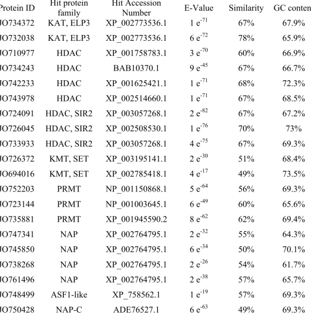
A full suite of histone and histone modifying genes are transcribed in the dinoflagellate Lingulodinium...70
2.1. Abstract...71
2.2. Introduction ...72
2.3. Materials and Methods ...74
2.3.1. Cell Culture ...74
2.3.2. Acid Extraction of proteins...74
2.3.3. SDS-PAGE and Immunoblotting ...75
2.3.4. Mass Spectrometric analysis ...76
2.3.5. Bioinformatic and Phylogenetic Analysis...77
2.4. Results ...78
2.4.1. All core histone and many histone modifying enzyme sequences are present in the Lingulodinium transcriptome ...78
2.4.2. Phylogenetic grouping identifies at least two major variants of all histone sequences within Lingulodinium ...78
2.4.3. Histone mRNAs abundance levels are uniform throughout ...79
2.4.4. Histone protein accumulation is below current detection limits...79
2.5. Discussion...81
CHAPTER 3 – PUBLICATION # 2 ...112
Transcripts from dinoflagellate tandem array genes are highly conserved and are not polycistronic ...113
3.1. Abstract...114
3.2. Introduction ...115
3.3. Materials and Methods ...117
3.5.1. Cell culture ...117
3.5.2. RNA purification and sequencing...117
3.5.3. Sequence assembly and analysis ...117
3.4. Results ...120
3.3.1. The de novo assembly is an authentic portrait of the transcriptome...120
3.3.2. Tandem gene array sequences are highly conserved in the transcriptome...122
3.3.3. Sequences of potential bacterial origin have the same GC-content as the host123 3.3.4. Assessing the potential for polycistronic transcripts...125
3.5. Discussion...127
3.6. Acknowledgements ...156
CHAPTER 4 – PUBLICATION # 3 ...157
Predicted Casein Kinase 2 sites in RNA binding proteins of Lingulodinium show daily variations in phosphorylation state ...158
4.1. Abstract...159
4.2. Introduction ...160
4.3. Materials and methods...163
4.5.1. Cell Culture ...163
4.5.2. Phosphoprotein purification and gel electrophoresis ...163
4.5.3. Mass Spectrometric analysis ...164
4.5.4. Bioinformatic Analysis ...166
4.3.1 Phosphoprotein purification yields more peptides than phosphopeptide
enrichment ...168
4.3.2. The phosphoproteome fraction is enriched in proteins involved in translation and RNA binding ...169
4.3.3. Phosphopeptide intensity comparisons between ZT2 and ZT14 reveal many RNA binding proteins ...170
4.3.4. Orthologs of kinases involved in circadian regulation in other eukaryotes may regulate translation in Lingulodinium...171
4.5. Discussion...173
4.6. Acknowledgements ...211
CHAPTER 5 – PUBLICATION # 4 ...212
Cold-induced cysts of the dinoflagellate Lingulodinium have low levels of protein phosphorylation and lack a normal circadian bioluminescence rhythm...213
5.1. Abstract...214
5.2. Introduction ...215
5.3. Materials and Methods ...218
5.3.1. Cell Culture ...218
5.3.2. Cyst formation and purification ...218
5.3.3. RNA Extraction and sequencing...219
5.3.4. Sequence Analysis...219
5.3.5. Northern hybridization ...219
5.3.6. Microscopy ...220
5.3.7. Protein and phosphoprotein extraction...221
5.3.8. 2-D gel electrophoresis ...222
5.3.9. SDS-PAGE and Western blotting ...223
5.3.10. Mass spectrometry analysis...223
5.3.11. Bioinformatic analysis...225
5.3.12. Bioluminescence assay ...225
5.4. Results ...226
5.4.2. Protein phosphorylation is reduced in cysts...226
5.4.3. Cysts have an arrested clock and show a decreased level of Casein Kinase 2 phosphosites...228
5.4.4. Plastid-encoded RNAs have decreased levels in cysts ...229
5.5. Discussion...231
5.6. Acknowledgements ...265
CHAPTER 6 – GENERAL DISCUSSION...266
6.1. General Discussion ...267
6.2. Future perspectives ...271
Liste des tableaux
Table 1.2.1. mRNA transport components ...38
Table 1.2.2. mRNA surveillance components...39
Table 1.3.1. Translational factors in dinoflagellates ...64
Table 1.3.2. Aminoacyl-tRNA synthetases in dinoflagellates...66
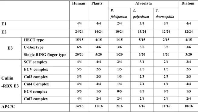
Table 1.3.3. Ubiquitin mediated proteolysis...68
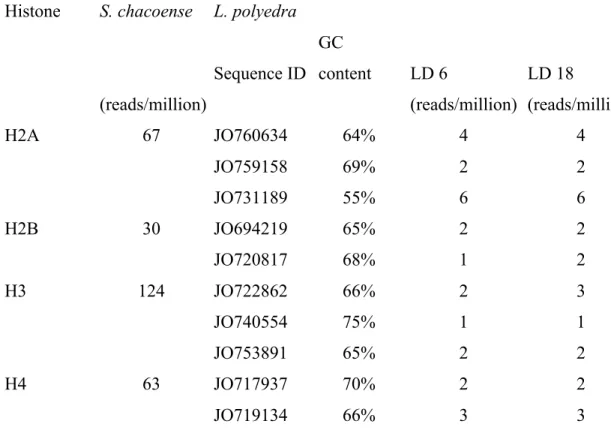
Table 2.1. Description of histone sequences and their relative abundance in Lingulodinium ..85
Table 2.2. Description of histone modifying enzymes and histone chaperones...86
Table2.3. Proteins found by LC-MS/MS sequencing of total acid soluble proteins from Lingulodinium and yeast...87
Table 2.4. mRNA abundance of expressed proteins detected by LC-MS/MS in an acid-extracted protein fraction...88
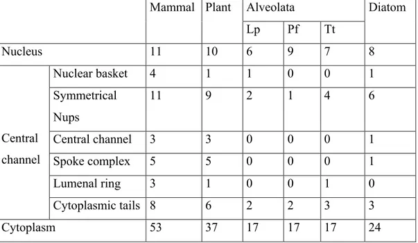
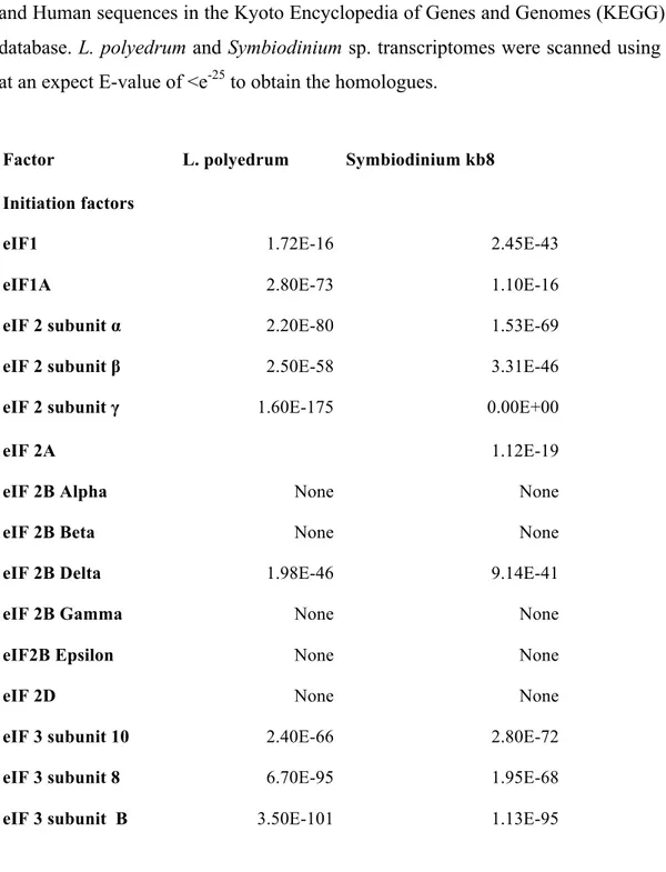
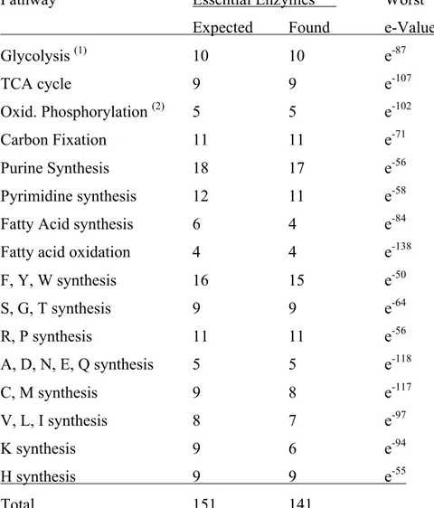
Table 3.ST1. Number of KEGG genes found for a variety of pathways ...151
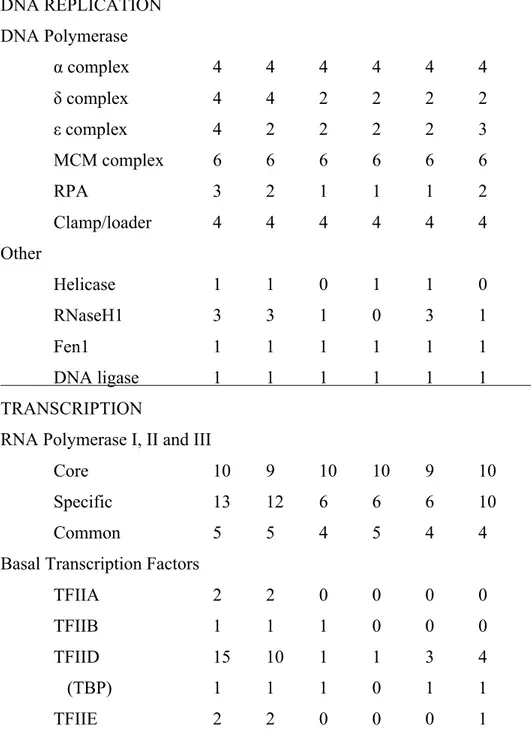
Table 3.ST2. Number of KEGG pathway sequences found in mammals, plants, apicomplexans, dinoflagellates, ciliates and diatoms for replication, transcription, splicing and translation...152
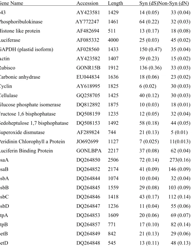
Table 3.ST3. Nuclear- and plastid-encoded reference sequences from GenBank used for comparison of synonymous and non-synonymous mutations...155
Table 4.1. Comparison of phosphopeptide and phosphoprotein enrichment protocols ...178
Table 4.2. Numberof hyperphosphorylated RNA-binding proteins at either ZT2 or ZT14....179
Table 4.ST1. Lingulodinium kinases ...194
Table 4.ST2. The identification of proteins containing the 527 phosphopeptides in ZT2 and ZT14 extracts of Lingulodinium ...195
Table 5.ST1. Cyst hyperphosphorylated peptides ...252
Table 5.ST2. Cyst hypophosphorylated peptides ...256
Liste des figures
Figure 1.1.1. The scanning electron microscopy photograph of a single cell of the dinoflagellate Lingulodinium polyedrum taken with a FEI Quanta 200 3D (Dualbeam)
microscope...13
Figure 1.2.1. Superphylum Alveolata...40
Figure 1.2.2. Lingulodinium polyedrum nuclear morphology ...42
Figure 1.2.3. RNA polymerase components in Lingulodinium ...44
Figure 1.2.4. TBP phylogenetic classification...46
Figure 1.2.5. General transcription factors in dinoflagellates ...48
Figure 2.1. Two variants of Histone H2A in Lingulodinium...89
Figure 2.2. The acid soluble protein profiles of Lingulodinium and Yeast differ ...91
Figure 2.3. Histone H3 protein levels in Lingulodinium are below current immunodetection limits ...93
Figure 2.S1.Cladogram of histone H2B ...95
Figure 2.S2. Cladogram of histone H3 ...97
Figure 2.S3. Cladogram of histone H4 ...99
Figure 2.S4. Histone H2B protein is not detected in Lingulodinium ...101
Figure 2.S5. Alignment of H2AX sequences. ...103
Figure 2.S6. Alignment of H2B sequences ...105
Figure 2.S7. Alignment of H3 sequences ...107
Figure 2.S8. Alignment of H4 sequences ...109
Figure 3.1.Global analysis of the Lingulodinium assembly ...131
Figure 3.2. Sequence variation among transcripts...133
Figure 3.3. Bacteria-like sequences in the transcriptomes of different dinoflagellates have GC-contents commensurate with the host ...135
Figure 3.4. RNA-Seq does not support a polycistronic transcription mechanism ...137
Figure 3.S1. Size distribution of sequences in the transcriptome and in the mRNA ...139
Figure 3.S3. Characterization of sequence common to Lingulodinium, Alexandrium and
Karenia ...143
Figure 3.S4. Frequency spectrum of sequence variation in PCP and Luc TAG transcripts....145
Figure 3.S5. Characteristics of the bacterial-like sequence in the transcriptome...147
Figure 3.S6. Detection of reads aligning to the Luciferase intergenic spacer ...149
Figure 4.1. Efficient enrichment of Lingulodinium phosphophoproteins by affinity chromatography ...180
Figure 4.2. RNA rather than DNA related processes are preferred in the total enriched protein pool...182
Figure 4.3. Phosphopeptide Intensity at ZT2 is much pronounced than at ZT14 ...184
Figure 4.4. Many RBP are differentially phosphorylated at ZT2 and ZT14 ...186
Figure 4.5. Daily variation of kinase activity and their efficiency in Lingulodinium ...188
Figure 4.6. Many RNA binding proteins are among the predicted CK2 targets ...190
Figure 4.S1. Comparison of kinases...192
Figure 5.1. Cyst morphology differs from that of motile cells...236
Figure 5.2. Phosphoprotein profiles of cyst and motile cell extracts differ...238
Figure 5.3. Cyst phosphopeptides are generally hypophosphorylated and fall into categories regulating the amount and activity of proteins ...240
Figure 5.4. Casein Kinase 2 phosphosites are the most hypophosphorylated class ...242
Figure 5.5. RNA-Seq of cyst extracts reveals most RNAs with altered levels have decreased abundance ...244
Figure 5.S1. Western blot analysis of three nuclear encoded proteins show no significant decrease in cyst extracts. ...246
Figure 5.S2. Comparison of phosphoprotein enrichment fraction from cyst and motile cells248 Figure 5.S3. Northern blot analyses of two plastid-encoded and two nuclear-encoded RNAs confirms a decrease in plastid RNAs...250
List of abbreviations
2D-PAGE: 2-dimensional polyacrylamide gel electrophoresis AMPK: 5' adenosine monophosphate-activated protein kinase ATP: adenosine 5′-triphosphate
bp: base pair BSA: bovine albumin cDNA: complementary DNA
CDPK: calcium-dependent protein kinase
CHAPS: 3-[(3-Cholamidopropyl) dimethylammonio]-1-propanesulfonate hydrate CK: casein Kinase
CSD: cold shock domain CT: circadian time
DBP: DNA binding protein
Dinap1: dinoflagellate nuclear associated protein 1 Dip: Dinap interacting protein
DNA: deoxyribonucleic acid DTT: dithiothreitol
EDTA: ethylenediaminetetraacetic acid
GAPDH: glyceraldehyde-3-phosphate dehydrogenase GDP: guanosine 5′-diphosphate
GSK: glycogen synthase kinase GTP: guanosine 5′-triphosphate
IEF: isoelectric focussing Kb: kilobase
kDa: kilodalton
KH: K-homology
LB: luria-Bertoni
LBP: luciferin binding protein LCF: luciferase
LD: Light/Dark
LDS: lithium dodecyl sulfate
mg: miligram
miRNA: microRNA mL: mililiter mM: millimolar
MMG: monomethylguanosine
MOPS: 3-(N-Morpholino propanesulfonic acid mRNA: messenger RNA
MS: mass spectrometry
NADP: nicotinamide adenine dinucleotide phosphate nm: nanomolar
OD: optical density ORF: open reading frame
PCP: peridinin Chlorophyll-a binding protein PCR: polymerase chain reaction
PEG: polyethylene glycol PFAM: protein families
PMSF: phenylmethanesulfonyl fluoride PPR: pentatricopeptide
PSI: photosystem I PSII: photosystem II
PTM: posttranslational modification RBPs: RNA binding proteins
RNA: ribonucleic acid RNAi: RNA interference RRM: RNA Recognition motif rRNA: ribosomal RNA
SDS: sodium dodecyl sulfate SEM: scanning electron microscopy SL: splice leader
TBP: TATA-box binding-protein TLF: TATA-box like factor TMG: trimethylguanosine
Tris: 2-amino-2-hydroxyméthyl-1,3-propanediol tRNA: transfer RNA
TRP: TATA-box related proteins
TTFL: transcription translation feedback loop UTR: Untranslated region
UV: ultra violet ZT: Zeitgeber time
Dedication
I want to dedicate this thesis to my (Mother)
Ma- my epitome, with her loving and caring
attitude has been always by my side and my
(Father) Baba, my philosopher and guide has
been a source of inspiration to me.
Remerciements
I would like to convey my deep respect and heartfelt gratitude to Prof. David Morse, my research director, whose constructive criticism and methodical problem solving approach helped me to develop my scientific outlook. I admire his ability as a teacher to captivate the audience with his enthusiastic, energetic and informative explanation. I am thankful to Prof. Morse for the liberty and encouragement he has offered me to pursue several projects. It has been an extraordinary experience to work with Prof. Morse and I am grateful to him for giving me this opportunity. He is one of the most fun-loving and knowledgeable people I have known.
I am grateful to my PhD committee, Prof. Daniel P. Matton and Dr. Jean Rivoal for their helpful suggestions and insightful discussion. I would like to take this opportunity to thank Prof. Mario Cappadocia for his support and guidance during the sabbatical period of Prof Morse. I would like to extend my gratitude to all the faculty members, students and administrative staff of IRBV for their help.
I consider myself lucky to be able to work with very intelligent, passionate and helpful co-researchers Steve and Mathieu. Working with them has been an excellent and pleasant experience, which cannot be explained by mere words. I would also like to thank Philippe for his help during my early days of PhD. Thank you all for making my stay in the Morse Lab memorable. Thanks to Jonathan for the elaborate and interesting discussions we had on different scientific as well as extracurricular topics.
For the complete duration of my PhD, I have always felt the warmth of a family although I was far away from my home. Thanks to Leo and Lise and their sons Etienne and Julien, with whom I have been staying for the last five years. Thank you all for including me as one of the members of your lovely family.
Some debts we can never think of repaying and indebtness to family comes among the foremost ones. I have no words of appreciation to describe the contribution of my family. Without the continuous and unconditional love, encouragement and guidance of my mother
and father, I would not have come so far in my career. I would like to thank my elder brother, who has always remained by my side inspiring and motivating me. I appreciate the calm presence of my sister-in-law and my special thanks to the newcomer in my family, my niece, Srita.
Coming to Canada and completing my studies would not have been possible without the financial help through grants and scholarships from the IRBV, FESP, the Department of Biological Sciences and the NSERC grant funding our laboratory. Thanks to all the organizations for their generous support.
My Project
My curiosity about Lingulodinium was impelled by the observation that gene expression can occur rhythmically, under the control of an internal daily (circadian) clock, even though this species has the permanently condensed chromatin characteristic of the dinoflagellates. My particular interest was to try to understand expression of genes present in multiple copies and arranged in tandem arrays, and I began these studies by analysis of a transcriptome prepared from high throughput sequencing (RNA-Seq) data. During these studies I noticed that the transcriptome contained all core histone genes, as well as many histone modifying enzyme genes. The fact that these sequences were conserved and expressed thus argues against the dogma that histones have been lost in dinoflagellates. However, I was unable to show the presence of histones immunologically, indicating that protein levels are still below the limits of what can be detected experimentally. I also became interested in the study of gene expression at the post-transcriptional level. In particular, I was interested in using the transcriptome database to allow a mass spectrometry protein sequencing approach. I found that an enriched phosphoprotein fraction could be directly analyzed. Phosphoproteomics is fast emerging as an important field to study the role of gene expression at a posttranslational level. The Lingulodinium phosphoproteome shows important differences between day and night suggesting that protein phosphorylation may be a mechanism whereby the Lingulodinium circadian system influences cell behavior. Lastly, the phosphoproteome is markedly different in cold-induced cysts, which together with the observation that the circadian clock has stopped in cysts, may help to identify components of the clock machinery.
1.1. The Dinoflagellate
Dinoflagellates are unicellular eukaryotic protists found widely in marine as well as freshwater environments. The fossil record reveals traces of dinoflagellates 245 million years ago, confirming their presence during the Mesozoic and Cenozoic era. However, molecular phylogenetic studies coupled with anatomical comparison suggests that origin of the modern dinoflagellates may actually be found in the Precambrian, more than 570 million years ago [1]. Sequence analyses and the construction of phylogenetic trees place dinoflagellates within the kingdom Alveolata, which also contains the parasitic group of apicomplexans and ciliates [2-4], which are unified by the presence of large flattened cortical vesicles termed alveoli. Dinoflagellates themselves are further sub-divided into oxyrrhinales, syndinales and the core dinoflagellates groups [5]. Dinoflagellates are generally microscopic, ranging from 15-50 µM in size, with the largest reported dinoflagellate, Noctiluca, up to 2 mm in diameter [6-8]. Some dinoflagellates, termed armored, contain a series of cellulosic thecal plates inside the alveoli [9], although unarmored species lacking the thecal plates also exists [10]. The dinoflagellates demonstrate extensive diversity with respect to morphology and food habits with photosynthetic [11], heterotrophic [12], symbiotic [13], parasitic [14] or mixotrophic [15] behavior all found in the group. A large number of the dinoflagellates are photosynthetic, and most contain distinctive plastids surrounded by three membranes, thought to result from a secondary endosymbiosis, as well as containing the unique xanthophyll peridinin, a carotenoid responsible for the characteristic red color in these species [16, 17]. The peridinin-containing dinoflagellates have the smallest chloroplast genome of any functioning plastid because of frequent gene transfer events from the chloroplast to the nucleus [18]. However, other types of plastid are occasionally found in some dinoflagellate species such as the fucoxanthin containing dinoflagellates Karenia and Karlodinium. These organisms are thought to have acquired their chloroplasts during a tertiary endosymbiosis, where the peridinin-containing plastids have been replaced by a fucoxanthin-containing plastid [19, 20]. Dinoflagellates also have a characteristic helical swimming behavior due to its two flagella, one running like a belt in a transverse groove around the cell, which acts to spin the cell around its axis, the other lying in a longitudinal groove toward the base of the cell which pushes the cell forward [21, 22]. Dinoflagellates
can decide the direction of their movement by sensing chemicals, light as well as gravity [21].
Another interesting feature of dinoflagellates is its capability to form symbioses with different marine protists and invertebrates such as foraminifera, radiolarians, flatworms, anemones, jellyfish, and mollusks [23-25]. In particular, the association of Symbiodinium with the reef-forming corals has been of great interest because of the immense importance reefs play in marine ecology [25, 26]. In this mutual relationship, which benefits both the organisms, the photosynthetic dinoflagellates fix atmospheric CO2 and transfer a
considerable part of it as food for the corals [27] in exchange of shelter.
Toxic dinoflagellates can have a huge negative impact on different forms of marine life [27-30] and on human [31] through the formation of harmful algal blooms (HABs). HAB outbreaks are becoming more frequent in the coastal areas around the world, and at least in part, are due to the increase of nitrogen and phosphate (fertilizer) runoff in the coastal waters [32]. HAB-causing dinoflagellates can release different types of toxins, the most studied of which are neurotoxins, resulting in various syndromes and can even be fatal to humans. The most common toxin, saxitoxin, is a sodium channel blocker that causes paralytic shellfish poisoning (PSP), and is produced mainly by Alexandrium species and to some extent by Gymnodinium catenatum [33, 34] and Pyrodinium bahamense var compressum [35]. Brevetoxins, released by Karenia brevis [36], also cause the neurotoxic shellfish poisoning (NSP) and marine mammals have been found to be highly susceptible to ingestion or even inhalation of this toxin. Gambierdiscus toxicus produces the polyether-based ciguatoxins [37] which cause ciguatera fish poisoning (CFP), while diarrhetic shellfish poisoning (DSP) and azaspiracid shellfish poisoning (AZP) are caused by toxins from Dinophysis or Prorocentrum species and Protoperidinium crassipes [38], respectively.
Horizontal gene transfer (HGT), also known as lateral gene transfer, is the exchange of genetic material between two organisms. HGT can either occur between prokaryotes and eukaryotes or within prokaryotes or eukaryotes themselves [39]. HGT has been reported in many eukaryotes and the genome-wide investigations are now being used to identify the
novel genes introduced into genomes from different sources [40-42]. The use of phylogenomics approachs in particular [43, 44] are extremely useful in revealing the extent of gene transfer from bacteria to protists [45], with up to 7.5% of the genes in diatoms derived from bacterial sources by HGT [45]. In dinoflagellates, many genes such as the histone-like proteins (HLPs), the form II Rubisco, aroB and O-methyltransferase have been acquired from bacteria through HGT [46]. Proteorhodopsins of marine proteobacteria have been horizontally transferred to other prokaryotic classes, and recently have also been found in two different dinoflagellates, a result of independent HGT events [47]. The full extent of HGT events has not yet been unveiled for dinoflagellates.
Bioluminescence is a fascinating process found in at least 30 different systems, belonging to phylogenetic groups as diverse as bacteria, fungi, dinoflagellates or insects. The general mechanism of light production involves the oxidation of a substrate (luciferin) by molecular oxygen and is catalyzed by a Luciferase enzyme specific to each system. The marine environment seems to be preferred habitat for bioluminescent species [48]. The dinoflagellates constitute one of the most common sources of bioluminescence in marine waters [49] and at least 18 genera of dinoflagellates are known to produce bioluminescence [50]. However it is curious that among many marine bioluminescent organisms only dinoflagellates can perform photosynthesis [51]. Particularly, the dinoflagellate Lingulodinium polyedrum (previously Gonyaulax polyedra) has been used as the model to understand the biochemistry, cell biology, and molecular biology of bioluminescence. In the Lingulodinium system the luciferin is a linear tetrapyrrole structurally related to chlorophyll, which is highly susceptible to nonluminescent autooxidation. At cytoplasmic pH (~7.5), luciferin in the cell is sequestered by a Luciferin Binding Protein (LBP), preventing its reaction with Luciferase (LCF). Mechanical, chemical or temperature stimulation of the organism is followed by emission of light as brief (~100 msec), bright (~109 photons/cell) flashes from small (~0.4 microns) discrete spherical organelles called scintillons. These organelles are essentially protein spheres, which protrude into the vacuole and are almost completely surrounded by the vacuolar membrane. Stimulation of the cell activates a voltage-gated proton channel in the vacuolar membrane, thus decreasing the cytoplasmic pH in the region of the scintillons (to ~6.5). At this acidic pH, LBP is inactivated and releases
luciferin, while LCF becomes activated at low pH and initiates the bioluminescent reaction. The number of scintillons at night is roughly tenfold higher than at day and corresponds to what has been termed the bioluminescent capacity of the cells. Interestingly, light is also emitted as glow, a constant low intensity (~104 photons/sec/cell) emission rising to maximum peak at a very specific time each day. The mechanism of glow, not visible to the naked eye, is still unknown. Furthermore, the exact purpose of dinoflagellate bioluminescence is still being debated, although probable functions could be to scare predators away by producing brilliant light [52-54] or to draw the attention of secondary predators to reduce the primary predator population, which is popularly known as the “burglar alarm” effect [55-57].
1.1.1. Lingulodinium polyedrum (previously Gonyaulax polyedra)
L. polyedrum is a marine, thecate, non-toxic, bioluminescent and photosynthetic dinoflagellate, approximately 35 X 45 µm in size (Figure 1.1.1) [58]. It is one of the species involved in the formation of ‘red tides’, the well-known algal blooms in nutrient rich waters [59]. Though L. polyedrum is generally considered as a non-toxic species, and while it has been maintained in culture without incident for over 50 years, some studies were able to detect very low levels of yessotoxins [60]. It contains a triple membrane bound, peridinin-containing chloroplast. L. polyedrum has a C-shaped nucleus with a characteristic nucleolus and about 200 pg of DNA distributed over roughly 200 chromosomes [61]. It also contains an electron-dense, spherical PAS body bounded by a single membrane, a structure considered equivalent to the digestive granules found in hydra or food vacuoles of Ceratium hirundinella [58]. L. polyedrum mitochondria are surrounded by two membranes and have tubular cristae. L. polyedrum is a useful model system for understanding the biochemical bases of biological rhythms, as many physiological activities in this alga have daily rhythms that are independent of external cues and are thus regulated by a daily (circadian) clock [62].
1.1.2. Circadian clocks and L. polyedrum
For better adaptation to the surrounding environment, living organisms frequently prefer to perform some biological tasks at particular times of day [63]. They are able to do so with the help of an endogenous clock, termed as the circadian clocks, which not only allows the organism to synchronize their internal biochemistry to the daily cues but also to predict the changes to come [64, 65]. While the clock can function in the absence of daily cues, the clock is typically aligned to, or synchronized with the environment by changes in light and/or temperature cues [66, 67]. This mechanism thus requires an efficient signaling pathway to link the external stimuli to the central oscillator, which then sends timing signals to regulate cellular physiology. The circadian system can thus be thought of as being composed of three components, the inputs, the clock itself, and the outputs, known as overt rhythms and often involving changes in gene expression [64]. Extensive research in mammals, plants, insects, fungi and cyanobacteria has characterized the molecular components of the central oscillator and elements responsible for sensing the input and propagating the rhythmic output. Except cyanobacteria, whose core oscillator can function using only phosphorylation/ dephosphorylation of clock proteins [68], all other tested organisms have a mechanism involving transcriptional/ translational feedback loops (TTFL) [69, 70]. In its most simple terms, the feedback loop is comprised of transcriptional activators that activate the expression of repressor proteins, which then inhibit the activators thereby closing the loop [70]. This basic mechanism of circadian regulation is conserved across diverse phyla, although the clock proteins themselves are not, which is indicative of distinct evolutionary origins for the clocks in different organisms. Concerning the clock outputs, transcriptional regulation has been the focus of interest for several years, but after the discovery of the cyanobacterial clock mechanism, efforts were directed to assess the importance of posttranscriptional gene regulation in clock functioning. In this context, the dinoflagellate Lingulodinium polyedrum has again proved to be an interesting model, as in most observed cases it is found that Lingulodinium circadian clock prefers translational rather than the transcriptional regulation of gene expression [71].
The complexity of single cell circadian systems has been well documented in L. polyedrum. Many important characteristics of the circadian systems such as the mechanism of temperature compensation [72], basic features of phase shifting by light and the first phase response curve [73], the action spectrum for light phase shifting [74], phase shifting by drugs as well as drug effects on period [75] have been documented from studies performed on Lingulodinium. However, as yet, the core clock proteins are not known in any dinoflagellate. One of the major effects of the clock in Lingulodinium is to regulate the synthesis of different proteins. A combination of 2-D PAGE and tandem mass spectrometry revealed 28 out of 900 proteins in L. polyedrum were expressed differentially at three different phases over the circadian cycle [76]. Many of these proteins have been investigated in an attempt to link them to one of the several rhythmic processes, which include bioluminescence, photosynthesis, cell aggregation and timing of cell division. Indeed, Lingulodinium has been extensively researched for the last 60 years to try to understand the biochemical mechanism of circadian clock regulation of these physiological rhythms. Among the different rhythmic outputs, bioluminescence has received particular attention because of the ease of its measurement and as light emission appears to require only two proteins (LBP and luciferase) and the substrate luciferin, and thus constitutes a relatively simple system to study the molecular biology and biochemistry of the links between circadian clocks and the observed rhythms they control. Both LBP and LCF demonstrate robust daily rhythms in protein expression, and both are more abundant during night corresponding to the time of maximum bioluminescence [77-79]. Curiously, the dinoflagellate Pyrocystis lunula, which also demonstrates nightly bioluminescence, does not contain LBP and its LCF protein levels are constant throughout the circadian cycle. In this case the rhythmic bioluminescence correlates instead with translocation and compartmentalization of LCF at different times [80], a totally different mechanism from that observed in Lingulodinium. This suggests that various different posttranslational strategies may be employed by dinoflagellate circadian clocks to regulate cell physiology.
The role of posttranslational modifications of proteins, and phosphorylation in particular, has been extensively studied in circadian biology. For example, the rhythmic phosphorylation of clock proteins in the cyanobacteria Synechococcus, a process that
continues even in vitro, is sufficient for the circadian oscillator to function independent of transcription or translation [81]. In Arabidopsis, transcript abundance for several kinases and phosphatases were found to be rhythmic, and in addition, some of the core clock proteins (morning-expressed MYB-like transcription factors CIRCADIAN CLOCK ASSOCIATED1 (CCA1) and LATE ELONGATED HYPOCOTYL (LHY) are known to be phosphorylated by casein kinase 2 (CK2) [82, 83]. In Neurospora, phosphorylation of the clock protein Frequency (FRQ) determines its rate of degradation, which in turn determines the period length of the circadian oscillator [84]. Similarly, posttranslational regulation of clock components is important for other animal and mammalian circadian clocks [85, 86]. Interestingly, both CK2 and casein kinase 1 delta/epsilon (CK1 δ/ε) appear to have a conserved role in the function of eukaryotic circadian clocks [87, 88]. Mutational analysis assays using specific kinase inhibitors and disruption of CK1 δ/ε and CK2 genes revealed an arrhythmic behaviour, similar to that observed when core clock proteins were mutated [82, 83, 87, 89-92]. However, it should be noted that other kinases, such as adenosine monophosphate–activated protein kinase (AMPK) or Glycogen synthase kinase -3β (GSK-3β), have also been implicated in eukaryotic clocks.
In Lingulodinium, the kinase inhibitors staurosporine and 6-dimethylaminopurine induced concentration-dependent lengthening of the free-running period of the bioluminescence rhythm and high concentrations induced complete stoppage of the clock [93, 94]. As the inhibitors used in these experiments were broad range and affect many kinases, it is possible that there are many kinases involved in the Lingulodinium circadian clock. The phosphatase inhibitors okadaic acid, calyculin A, and cantharidin, all inhibitors specific for protein phosphatases 1 and 2A also affect the clock but the effects are less pronounced. High concentrations of okadaic acid produce a significant lengthening of the bioluminescent glow rhythm period, whereas only phase delays and no persistent effect on period were observed with cantharidin and calyculin treatments [95, 96]. These studies underscore the importance of phosphorylation/dephosphorylation events in the Lingulodinium circadian system, but as yet, the kinase repertoire in L. polyedrum has not been characterized.
1.1.3. Dinoflagellate nuclear genome
The dinoflagellates as a class are proof that genome size is not an indicator of an organism’s complexity, as different species show a remarkable variability in their genome content. Among the known dinoflagellates, the 3 pg haploid genome of Symbiodinium [97] is the closest in size to the 3.2 pg present in a haploid human cell, yet the 250 pg of DNA in Prorocentrum micans represents almost a hundred-fold greater genome size [98]. L. polyedrum cells contain 200 pg of DNA, about 60 times more than a haploid human cell. Genome size has been linked to the gene content using the positive correlation between the number of genes in an organism and its genome size [99, 100], and the regression model predicted a gene content for the dinoflagellates of around 37000 – 87000 [101]. However, genes in Lingulodinium are generally present in several copies (copy numbers from 30 - 5000) suggesting the number of unique genes is bound to be less than these estimates. A de novo assembly of 454 sequencing reads for two strains of Symbiodinium identified about 55,000 contigs in each species [102], although these assemblies are biased towards shorter fragments and many are likely to be derived from the same gene. Independent from the actual number of genes, it will be very interesting to find out why such simple eukaryotes accumulate such huge amounts of DNA and how they manage to conserve and express the relevant sequences. This is especially relevant given the unusual structure of the dinoflagellate chromatin, sufficiently distinct from other eukaryotes or prokaryotes to have at one time been termed a dinokaryon [103]. The dinoflagellate transcriptomes are generally biased towards high GC content, with an average varying from 50% in Karlodinium micrum to about 68% in Lingulodinium polyedrum.
1.1.4. Nucleosomes and Chromatin
The DNA in the nucleus must be tightly packed in order to fit within the available space, but more importantly, must also be accessible for replication and transcription. Most eukaryotes fold and compact DNA into chromatin using nucleic acid-protein complexes, the fundamental unit of chromatin is termed as nucleosomes. The nucleosome is a small cylindrical molecule containing 147 base pairs of DNA folded around four pairs of the
highly basic histone proteins H2A, H2B, H3 and H4. These nucleosomes can be further organized with respect to one another using a linker histone H1 protein [104]. Structurally and functionally, the four core histone proteins all have two distinct domains, the first a 20-35 N-terminal extension termed the histone tail and the second a conserved 80-90 C-terminal region termed the histone fold [105]. The fold regions of the H2, H3 and H4 have poor sequence conservation but demonstrate a high degree of structural similarity [106]. This fold domain is responsible for interaction with other histones [105] and DNA [107], and it is this region that determines the structural basis of the nucleosome. Given the balance needed between compactness and accessibility of the DNA, it might be expected that nucleosomes can affect chromatin structure. Indeed, the regulation of transcription through modulation of chromatin structure, popularly known as the epigenetic regulation of gene expression, is now an important field of research. Histone modifications form a part of the epigenetic toolkit. The residues present in the unstructured N terminal histone tails can undergo several types of post-translational modification (e.g acetylation, phosphorylation, methylation, biotinylation, SUMOylation, ADP ribosylation and ubiquitination) and can either activate or inhibit transcription depending on the modification [108]. In addition, some variants of the canonical core histones also exist in eukaryotes and these can have important implications in regulating expression of specific genes [109]. One such example is H2A.Z, a variant of H2A, which when present in the nucleosomes remodels it to affect gene expression [110].
Remarkably, dinoflagellates are the only eukaryotes that do not appear to use nucleosomes to organize their chromatin. Nucleosomes have never been observed microscopically in the chromatin, and none of the conserved histone proteins have ever been detected in dinoflagellate extracts. Instead of histones, the basic protein fraction isolated from dinoflagellate nuclei contains a class of histone-like proteins (HLP) structurally similar to bacterial DNA-binding proteins of the HU type [111, 112]. HLPs do appear able to condense DNA [113], which is in agreement with a predicted role in modulating chromosome structure [114]. Furthermore, some HLPs were found to be post-translationally modified, indicating a possible role in regulating gene transcription [115], but this has not been confirmed experimentally. However, the amount of these basic proteins in dinoflagellate nuclei is extremely low, with a protein/DNA ratio roughly one tenth of what is
found in other eukaryotes. This low level of nucleoproteins is thought to favor the liquid crystalline form of DNA, a type of DNA packing also found in animal sperm cell nuclei, which are also devoid of histones and instead contain a small (~7 kDa), highly arginine rich nucleoproteins known as protamines. Furthermore, imaging of the chromatin with the electron microscope supports a liquid crystalline organization for dinoflagellate chromosomes [116]. Surprisingly, however, recent transcriptomic sequencing studies have shown that conserved histone transcripts were found to be expressed in dinoflagellates [117]. It is difficult to reconcile the presence of these conserved sequences with the lack of detectable protein, although it is possible that histones at low levels will eventually be found to play a role is some aspects of chromatin organization.
In addition to the histone modifications, DNA methylation at the C5 position of cytosine pyridine ring is another epigenetic mechanism that can alter gene expression. In eukaryotes, the methylation catalyzed by DNA methylases generally occurs at CpG dinucleotides on double stranded DNA and is usually associated with gene inactivation [118]. These so-called CpG islands are found frequently in the promoter regions of genes and it has been proposed that DNA binding factors which recognize this structural change can modify gene expression [119]. Dinoflagellate DNA typically contains a considerable amount of modified bases, with roughly 3% of the cytosine replaced by 5-methyl cytosine. This does not appear to be located in CpG islands but instead appears to be distributed randomly throughout the genome [120]. However, the most important modification is replacement of up to 70% of the thymine by 5-hydroxymethyluracil [121]. Unfortunately, in the absence of genome sequence information it is difficult to assess the functional role of such modifications (if any at all).
1.1.5. The Dinoflagellate Cysts
The dinoflagellates have left an abundant trail in the fossil record in particular due to their ability to form cysts in order to resist unfavorable conditions. Roughly 10% of the known dinoflagellate species are able to produce resting cysts [122], a dormant cell that can germinate back to form a motile cell when conditions become favourable [123]. .
Ecologically, resting cysts are important as they can act as a “seed-bank” for the formation of HABs [124]. Indeed, many toxic HAB species undergo formation of resting cysts as a part of their life cycle [125]. The permanent cysts can be transported via sediments and remain viable for hundreds of years.
Dinoflagellates can also form temporary cysts, the structures of these are quite different from permanent cysts. In contrast to resting cysts, encystment and germination of temporary cysts can be very rapid, which presumably helps prevent huge population losses in response to a sudden unfavorable condition. Several factors, including temperature [126], chemicals [127], population pressure [128], nutrient deficiency, bacteria [129], pH and salinity [130] are well known inducers of temporary cysts. Interestingly, unlike other eukaryotes and bacteria, some dinoflagellates survive low temperature periods by formation of temporary cysts. This quick response requires an efficient signaling cascade, and the calcium concentration via the phospholipase C pathway has been shown to be vital for dinoflagellate temporary cyst formation in response to melatonin [127]. However, the major molecular events associated with the structural rearrangement and the quiescence physiology is still unknown.
Figure 1.1.1. A scanning electron microscopy photograph of a single cell of the dinoflagellate Lingulodinium polyedrum taken with a FEI Quanta 200 3D (Dualbeam) microscope.
1.2. Transcription and Maturation of mRNA in Dinoflagellates
This part of the thesis is under review for publication as a review chapter in the journal Microorganisms (ISSN 2076-2607) published by MDPI. This chapter is submitted for peer-review with the title: Transcription and Maturation of mRNA in Dinoflagellates.
Sougata Roy1 and David Morse1,*
1 Institut de Recherche en BiologieVégétale, Département de Sciences Biologiques,
Université de Montréal, 4101 Sherbrooke est, Montréal, Québec, Canada H1X 2B2
This manuscript was reviewed and corrected by my supervisor (David Morse) after I completed the initial drafting.
1.2.1. Abstract
Dinoflagellates are of great importance to the marine ecosystem, yet scant details of how gene expression is regulated at the transcriptional level are available. Transcription is of interest in the context of the chromatin structure in the dinoflagellates, which shows many differences from more typical eukaryotic cells. Here we canvas recent transcriptome profiles to identify the molecular building blocks available for the construction of the transcriptional machinery and contrast these with those used by other systems. Dinoflagellates display a clear paucity of specific transcription factors, although surprisingly, the rest of the basic transcriptional machinery is not markedly different from what is found in the close relatives to the dinoflagellates.
1.2.2. Introduction
Dinoflagellates are an important group of unicellular eukaryotes found in both marine and fresh water environments. These marine species are of particular importance on a global scale, as along with the diatoms, they contribute roughly half of the carbon fixed in the oceans, and thus roughly a quarter of the global totals [131]. They also play a role in maintaining the biodiversity surrounding coral reefs, since the coral polyps themselves rely on photosynthetic products supplied by the symbiotic dinoflagellates they harbor for growth in nutrient poor waters [132]. Furthermore, many marine dinoflagellates synthesize potent toxins that accumulate to high concentrations in the algal blooms commonly called “red tides” [133]. Lastly, the nightly bioluminescence of many dinoflagellates, popularly known as the “phosphorescence of the sea”, has inspired not only art and literature but also intensive scientific dissection of the bioluminescence phenomenon [134]. Interestingly, in Lingulodinium polyedrum this nightly bioluminescence [135], as well as photosynthesis [136], cell division [137], and diurnal vertical migration [138], are all regulated by an endogenous circadian (daily) clock. L. polyedrum has been studied for over 60 years as a model system for addressing the biochemical links between the internal clock and the observed rhythms [71].
Phylogenetically, dinoflagellates are grouped in the superclass Alveolata, which contains apicomplexans as their closest relatives as well as ciliates [4]. This group has several features unique to these organisms (Figure 1.2.1.). However, dinoflagellates have many unique characteristics compared to their relatives. For example, dinoflagellates typically possess a large quantity of nuclear DNA containing many genes organized in tandem gene arrays, with DNA found in a liquid crystal structure lacking observable nucleosomes [61]. It is unfortunate that dinoflagellates have so far proven refractory to mutational or gene transformational studies, thus hindering extensive molecular studies, as these unusual nuclear features suggest that the mechanisms for gene expression and its regulation may also be unusual.
The mechanisms used to control the expression of different genes have been extensively researched in both prokaryotes and eukaryotes. Critical events in eukaryotes include changes in chromatin organization, transcription of DNA into pre-mRNA, splicing of pre-RNA into mature mRNA, mRNA transport, mRNA degradation, mRNA editing and covalent modifications of the mRNA, translation of mRNA into protein, and, lastly, post-translational modification of the protein. All these, either individually or collectively are responsible for regulating gene expression within a cell. In this review we will focus primarily on transcription and its regulation as they relate to the control of gene expression in the dinoflagellates.
1.2.3. Transcription and its regulation
1.2.3.1. Cis-acting sequences and RNA polymerase components
Dinoflagellate chromosomes are permanently condensed at all stages of the cell cycle (Figure 1.2.2.) and assume a liquid crystalline structure [139, 140] with bivalent cations acting as the stabilization matrix [141]. This unusual chromatin structure thus raises the important questions about the accessibility of genes within the structure to the transcriptional machinery. The dinoflagellate Prorocentrum micans was inspected using high resolution electron microscope autoradiography for 3H-adenine incorporation, and this revealed that RNA transcription was prevalent only on extrachromosomal DNA filaments and not on DNA within the main body of the chromosome [142]. It was proposed that this transcriptionally inactive DNA might instead play a role in stabilizing chromosome organization, perhaps by an association with a protein matrix [142].
Given access to the genetic material, transcription initiation in dinoflagellates is likely to require an elaborate set of trans-acting factors and a series of conserved cis-acting sequences, as is the case in other eukaryotes. The complex of trans-acting factors binding the regulatory sequences in the DNA includes, in addition to the RNA polymerases, both general and gene-specific transcription factors, activators and mediators [143]. The cis-acting
sequences in eukaryotes can include regulatory elements far from the transcription start site, termed enhancers, although the region just upstream of the start site, termed a promoter, consisting of a core region and other regulatory domains [144, 145] is considered as the primary site of initiation. There are two major classes of promoters that regulate the expression of protein coding genes, and these contain either a TATA-box (consensus sequence <TATAAA>) or CpG islands, a region rich in CG dinucleotides [146] as their core domains. In Pyrocystis lunula luciferase (lcf) genes, a GC box consensus sequence <GGGCGG> is present, but its location is further upstream than the usual position of -110 (numbered relative to the transcriptional start site at +1) as found in many eukaryotes [147]. Furthermore, a GC-rich motif <C(G/C)GCCC> was also found within the upstream region of P. lunula lcf A and L. polyedrum lcf and lbp genes, but their position were not fixed. This GC-rich motif was first reported in the upstream region of the Peridinium bipes ferredoxin gene [148]. However, the role of this motif in gene expression has still not been established. Both TATA-box or CpG island type promoters may include additional sequence elements such as the GC-box <GGGCGG>, the CAAT-box <CCAAT>, and the INR box <(C/T)(C/T)AN(T/A)(C/T)( C/T)> at which transcription is initiated. Interestingly, the TATA box is quite conserved in eukaryotes and is also found in protists as diverse as amoebas (Acanthamoeba), slime molds (Dictyostelium), ciliates (Histriculus cavicola), and apicomplexa (Plasmodium) [149-154]. On the other hand, members of the phylum Parabasalia use their own specific promoter element instead of the canonical TATA box [155-157].
Proper understanding of gene organization and structure is required to describe transcription in dinoflagellates. For example, L. polyedrum has multiple copies of peridinin-chlorophyll a-binding protein (pcp), Luciferin binding protein (lbp) and Luciferase (lcf) genes arranged in long tandem repeats [158-161]. PCR with Pyrocystis lunula genomic DNA revealed that, among lcf A, lcf B and lcf C isoforms, two, lcf A and B are in tandem repeat. To test if this is a general character of dinoflagellates, PCR was used with primers directed away from one another in Amphidinium carterae [162]. PCR using genomic DNA as a template was expected to produce a band if the genes were found as a tandem repeat, and this strategy revealed that 17 out of the 47 genes tested did indeed have a tandem repeat structure.
However, in L. polyedrum the sequence of the intergenic region between lcf and pcp coding sequences did not contain any known promoter elements. The only common feature between the two was a conserved 13 nucleotide sequence, CGTGAACGCAGTG, proposed as a dinoflagellate specific promoter sequence [158] but no further work has been published to firmly establish this result. Moreover, this sequence is not conserved among different dinoflagellate species as it is absent in the intergenic region between P. lunula lcf A and lcf B genes [163].
The lack of identifiable sequence elements in the intergenic spacers has lead to the suggestion that tandem gene repeats may form a polycistronic transcript, in a manner similar to the Trypanosoma gene structure [164]. The trypanosomes transcribes long polycistronic transcripts from a single promoter containing genes coding for different gene products, and the primary transcript is then processed into mature mRNAs by trans splicing of the SL leader at the 5’ end and by polyadenylation at the 3’ end. If true for dinoflagellates, one possibility would place a promoter upstream of each tandem array, thus explaining the lack of recognizable promoter sequences in the intergenic regions. However, the consequences of this hypothesis include the predictions that the intergenic spacer region should be abundant in the transcribed RNAs, and that sequence differences at a particular position between copies in low copy number arrays should be detected in the mature transcripts at a frequency that is higher than those detected for high copy number genes. In a recent transcriptomic study which addressed this issue, none of these predictions were validated experimentally [165].
Eukaryotic and prokaryotic transcription also differ in that three different RNA polymerases (RNAP) are used for the former while only one is used for the latter. The three eukaryotic enzymes have specialized functions, with RNAP I transcribing most ribosomal RNA (rRNA), RNAP II transcribing protein-coding messengers (mRNA), small nuclear RNAs (snRNA) and micro RNA (miRNA), and RNAP III synthesizing transfer RNAs (tRNA) and the 5S rRNA. An assessment of the activity of RNA polymerase in the dinoflagellate Crypthecodinium cohnii, carried out with radiolabeled UTP, revealed that considerable amounts of RNA polymerase activity remained even after inhibition by