2
Practice Guidelines
1Clinical Haematology, University Hospital Liège and University of Liège, Liège, Belgium, 2Department of Haematology, University Hospitals Leuven, Leuven, Belgium, 3Department of Haematology, ZNA Stuivenberg, Antwerp, Belgium, 4Department of Haematology, University Hospital Antwerp, Antwerp, Belgium, 5Department of Haematology, University Hospital Ghent, Ghent, Belgium, 6Department of Medical Genetics, University Hospital Erasme, Laboratory of Cytogenetics, Bordet Institute, Brussels, Belgium, 7Department of Pathology, University Hospitals Leuven, Leuven, Belgium, 8Department of Haematology, CHU Dinant Godinne, UCL Namur, Yvoir, Belgium, 9Department of Haematology, Bordet Institute, Brussels, Belgium, 10Department of Haematology, AZ St Jan, Brugge, Belgium.
Please send all correspondence to: C. Bonnet, MD, PhD, Centre Hospitalier Universitaire, Clinical Haematology, B35 Campus Universitaire du
Sart-Tilman, 4000 Liège, Belgium, tel: +32 4 366 72 01, fax: +32 4 366 88 55, email: cbonnet@ulg.ac.be. Conflict of interest: The authors have nothing to disclose and indicate no potential conflict of interest.
Keywords: Burkitt’s lymphoma, intensive short-cycle chemotherapies, low intensity chemotherapies, rituximab, treatment guidelines.
BHS Guidelines for the treatment of
Burkitt’s lymphoma
C. Bonnet, MD, PhD1, A. Janssens, MD, PhD2, K. L. Wu, MD, PhD3, W. Schroyens, MD, PhD4, V. Van
Hende, MD5, P. Heimann, MD, PhD6, T. Tousseyn, MD7, M. Andre, MD8, D. Bron, MD, PhD9, A. Van Hoof,
MD, PhD10, G. Verhoef, MD, PhD2, B. De Prijck, MD1, Y. Beguin, MD, PhD1, D. Dierickx, MD, PhD2
Burkitt’s lymphoma is a rare but very aggressive non-Hodgkin’s lymphoma characterised by an isolated translocation t(8;14)(q24;q32). The sporadic form is the sub-entity most frequently encountered in Belgium. Diagnosis and initial work-up must be completed rapidly to start treatment as soon as possible. Positron emission tomography scan is useful for initial staging and to evaluate the chemosensitivity of the tumour during and after treatment. After debulking, it is recommended to add rituximab to chemotherapy. Currently intensive short-cycle and low intensity chemotherapies are two valuable options. Radiotherapy is not indicated except in case of central nervous system involvement. Patients achieving complete remission must be followed carefully during the first year to detect recurrence of the disease. More than 80% of patients sustain their remission one year following initial treatment and are considered cured. For patients in partial remission or with chemosensitive relapse, autologous stem cell transplantation is recommended following re-induction with non-cross-resistant polychemotherapy. Monitoring complete blood counts and cognitive functions is important to detect late toxicity of the applied therapies.
(Belg J Hematol 2015;6(2):61-9)
Introduction
Burkitt’s lymphoma (BL) is a rare but very aggressive non-Hodgkin’s lymphoma (NHL) accounting for 0.8% of all B-cell lymphomas. Three different clinical sub-types of BL are recognised: endemic BL (eBL), sporadic variant (sBL) and immunodeficiency-associated BL (idBL).1
Endemic BL, the most common form of BL with a geo-graphical distribution identical to that of Plasmodium falciparum, mainly affects facial bones; typically the jaw. In sBL, the terminal ileum, caecum and intra-abdominal lymph nodes are the most commonly affected sites. Immunodeficiency-associated BL is mainly seen in human immunodeficiency virus (HIV) positive patients
and, to a lesser extent, in the context of immunomo-dulatory drugs in transplant patients or patients with auto-immune disease and in patients with primary immunodeficiency disorders (PID).1
Endemic BL is almost exclusively encountered in the paediatric population, whereas PID and transplantation-related BL are very rare. Therefore these three subtypes will not be discussed in this article.
Pathology
For the histopathological diagnosis of BL, as for all lymphoproliferations, a surgical excision biopsy is pre-ferred over a fine needle aspirate cytological evaluation
Practice Guidelines
or a core needle biopsy. However the latter two can be sufficient when the anatomical location is difficult to access or in case of recurrent disease.
BL cells are typically medium-sized lymphocytes with multiple small round nucleoli and little cytoplasm.1
Their growth pattern is monotonous and diffuse, often with a ‘starry sky’ appearance due to a high amount of tingible body macrophages (Figure 1).
BL has a characteristic immunophenotypic profile: CD10+, CD20+, Ki67+ in nearly 100%, TdT-, Bcl2-, Bcl6+, c-MYC+. The hallmark cytogenetic aberration involves the overexpression of c-MYC oncogene, mostly due to a t(8;14)(q24;q32) or less frequently a t(8;22) (q24;q11) or t(2;8)(q24;p12) translocation, and can be detected in 90% of cases by classical karyotyping or fluorescence in situ hybridisation (FISH). Cytogenetic analysis is recommended to allow a clear distinction between BL and other c-MYC-driven B-NHL, especially diffuse large B-cell lymphoma. BL is mostly character-ised by a simple karyotype including a t(8;14)(q24;q32) translocation with few additional abnormalities such as gain of chromosome 1q while other c-MYC-driven B-NHL will exhibit a complex karyotype with multiple chromosomal gains and/or losses (Table 1). Fifteen to thirty percent of sBL and 25-40% of idBL are EBV+, in contrast with eBL where >90% of cases are EBV+.1
Microarray-based gene expression profiling can identify a molecular Burkitt’s signature but this is not yet used in daily clinical practice.2-4
The differential diagnosis with “B-cell lymphoma,
un-classifiable, with features intermediate between diffuse large B-cell lymphoma and Burkitt’s lymphoma”, previ-ously named ‘Burkitt’s-like lymphoma (BLL)’ is essential (Table 1), as the outcome is poor with bad response to the classical regimens used for BL or DLBCL.1,5-8 This
provisional histological sub-entity encompasses several aggressive B-cell lymphomas that do not entirely reflect the morphological, immunohistochemical, cytogenetic or clinical characteristics of a typical BL.
Initial work-up
Staging must be completed rapidly in order to avoid delay of therapy, which must start within 48 hours after diagnosis.
A personal history and clinical examination must be performed with attention to B symptoms (fever, night sweats and weight loss), performance status (PS) and neurological symptoms and signs.
Laboratory tests must include complete blood counts, liver and renal function, electrolytes, uric acid, lactate dehydrogenase (LDH) and serology for HIV, EBV, hep-atitis B and C.
Figure 1. Illustration of a pathological preparation of Burkitt’s Lymphoma (haematoxylin and eosin, 400x). Medium-sized cells with slightly irregular nuclei with coarsely dispersed chromatin often showing several nucleoli. Presence of numerous mitoses.
Courtesy of Dr I. Scagnol, CHU, ULG, Liège.
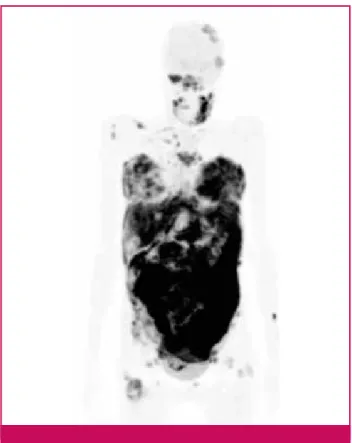
Figure 2. High avidity for 18FDG of BL-cells. Illustration of the
avidity of Burkitt’s Lymphoma-cells for the 18FDG. PET scan at
initial staging (girl, 19 years old, post transplant t(8,14)(q32;q21) pos BL, stage IV with elevated LDH serum level).
2
Contrast-enhanced chest and abdominal/pelvic CT scans are mandatory. Although BL are 18FDG-avid (Figure 2), PET scan is not formally indicated in this situation and, in clinical practice, cannot always be performed immediately.9,10 A bone marrow aspiration
with biopsy and a lumbar puncture with flow cytometry of the cerebrospinal fluid are mandatory.
Before starting treatment, cardiac evaluation (transtho-racic echocardiography), pregnancy testing in women of child-bearing age and discussion about fertility issues must be done.
Staging
Two staging systems are used: the Murphy and Hustu staging system and the Ann Arbor classification (Table 2).11,12 The first has been developed for paediatric BL
and is now also used in protocols including adolescent and young adults (AYAs). It takes into account the frequently observed extranodal involvement and in-completely resected intra-abdominal or intra-thoracic masses. This staging system is not validated in adult BL and no comparison of the prognostic value with the Ann Arbor classification is available.
Prognostic factors and treatment stratification
Prognostic factors used to stratify patients into different treatment groups vary from one co-operative group to another. Age, PS, LDH, bone marrow and central nervous system (CNS) involvement are the most frequently used.12
The LYSA-GRAALL Intergroup recognises three dif-ferent prognostic groups. Group A comprises patients with completely resected stage I or abdominal stage II
disease (Murphy and Hustu classification). Group C encompasses patients with CNS infiltration and/or bone marrow involvement. Other patients are classified as group B.
The Dana Farber Intergroup distinguishes two prog-nostic groups.13 Patients with normal LDH, good PS
(ECOG 0 or 1) and a tumour mass <10 cm are allocated to the low-risk group. Other patients are considered high-risk.
The MD Anderson Cancer Centre and the Cancer and Leukaemia Group B (CALGB) do not distinguish different prognostic groups.14,15
These stratifications determine the intensity, length and type of regimen (combination and number of cycles of systemic and intrathecal chemotherapy and the need for radiotherapy). However, it seems increasingly clear that the best prognostic marker is the chemosensitivity of the tumour.
Treatment
General considerations
Unfortunately, given the low incidence of the disease, available studies include only a limited number of patients and published randomised studies are not available. Comparing studies is difficult because dis-ease definition, stratification and median age of included patients vary.
Treatment must be started promptly (ideally within 48 hours after diagnosis) including prevention of the tumour lysis syndrome (TLS) by forced diuresis and the use of rasburicase.13 The unavoidable first step of
treatment is the application of prephase with low-dose cyclophosphamide (e.g. 200 mg/m²/day(d), d1-5) and
Table 1. Differential diagnosis between Burkitt’s lymphoma and “B-cell lymphoma, unclassifiable, with features intermediate between diffuse large B-cell lymphoma and Burkitt’s lymphoma”.1
BL B-cell lymphoma, unclassifiable, with features
intermediate between DLBCL and BL
CD10 + +
BCL6 + +/−
BCL2 − +
Ki67 >98% <90%
MYC Simple Complex
EBV +/− −
Practice Guidelines
prednisone (e.g. 60 mg/m²/d, d1-5). In comparison with standard dose of chemotherapy, the application of this prephase limits the risk of TLS by decreasing the re-lease of cytokines, particularly in case of high tumour burden, a situation that can be lethal given the aggres-siveness and chemosensitivity of the tumour. Moreover, this procedure allows finalising the staging process. Intensive short-cycle chemotherapies (ISCC) that include alkylating agents such as cyclophosphamide and cell-cycle phase-specific agents that cross the blood-brain barrier (i.e. cytarabine and methotrexate) are frequently combined but are associated with a high rate of haema-tological and non-haemahaema-tological toxicities. These regi-mens further aim to prevent CNS extension by the administration of intrathecal therapy consisting of methotrexate, cytarabine and prednisone.14,15
Low intensity chemotherapies (LIC), such as Dose Adjust-ed (DA)-EPOCH-R (etoposide, vincristine, cyclophos-phamide, doxorubicin, prednisone and rituximab) or Short Cycle (SC)-EPOCH-R, kill tumour cells by pro-longed low-concentration drug exposure.16 This type
of regimen seems as efficient and less toxic compared to ISCC and may be more suitable for frailer patients (elderly or HIV+ patients). As these regimens have no drugs that cross the blood-brain barrier, they cannot be used in case of CNS involvement.
The interval between cycles of chemotherapy must be
as short as possible and a new cycle must start as soon as haematological recovery has occurred. In this setting, the use of granulocyte-colony stimulating factors is essential.
Rituximab gives sufficient promising results to recom-mend its adjunction to chemotherapy and it may even erase the prognostic difference between young and elderly patients. However, its administration is avoided during the debulking phase given the high risk of TLS. Involved-field radiotherapy has no place in BL, except for patients with CNS involvement. In this situation, either pancranial or craniospinal irradiation (in case of leptomeningeal disease) can be applied, taking into account the high neurological and myelosuppressive toxicity rates. The recommended dose is 40 Gy, varying between 36 and 45 Gy and depending on the clinical condition. Boost radiotherapy is possible in case of localised intracerebral disease.
In case of initial CNS invasion, the number of intrathecal chemotherapy administrations is increased.
Autologous stem cell transplantation (SCT) has no place for patients in complete response (CR) and must be reserved for patients in first partial response (PR) or in chemosensitive relapse. Due to the high proliferation rate of BL, graft-versus-tumour effects observed after alloge-neic SCT are too slow to appear and to be efficient and therefore this type of transplant is not recommended.17,18
Table 2. Murphy and Ann Arbor classifications.12
2
Recommended treatment schedules for first line treatment Given the rarity of the disorder, it is highly recom-mended to include patients in clinical trials conducted by international co-operative groups.
Localised disease
In the rare cases of completely resected localised disease,
a short multi-agent chemotherapy program (e.g. CO-PADM x2) without CNS prophylaxis gives very good results and avoids toxicities associated with longer chemotherapy.19
Disseminated disease
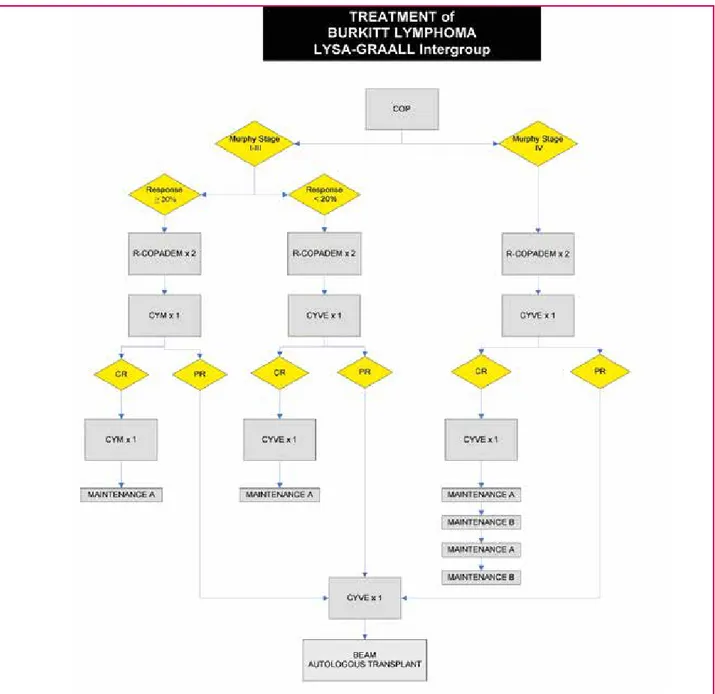
Figures 3 to 6 show different treatment schedules used Figure 3. The LYSA-GRAAL Intergroup regimen.15
CR: complete remission; PR: partial remission; COP: cyclophosphamide, vincristine, prednisone; R-COPADM: rituximab, cyclo-phosphamide, vincristine, prednisone, doxorubicin, methotrexate; CYM: HD methotrexate, cytarabine; CYVE: cytarabine, etoposide; MAINTENANCE A: prednisone, vincristine, methotrexate, cyclophosphamide, adriamycin; MAINTENANCE B: aracytine, etoposide. IT²: methotrexate & solu-medrol intrathecal; IT³: methotrexate, cytarabine & solu-medrol intrathecal; *: dosage of methotrexate depending on age and CNS infiltration ; **: number of IT injections depending on CNS infiltration; °: dosage of cytarabine depending on age; °°: dosages of cytarabine and methotrexate depending on age.
Practice Guidelines
by the main co-operative groups. ISCC used by the LYSA-GRAAL Intergroup, the German Multicentre Study Group for Adult ALL (GMALL) or the Dana Farber Institute are very efficacious with an acceptable benefit/ toxicity ratio.15,20-22
In 2012 the LYSA-GRAAL intergroup reported the pre-liminary results of the LMBA02 protocol (R-COPADM
x2 and R-CY(M)VE x2). With a median follow-up of 38 months, the 3 year EFS and OS of 128 young patients with HIV- BL were 76 and 82%, respectively.15 At the
same time, the GMALL protocol reported rates of PFS and OS at 7 years of 83 and 88%, respectively, for 134 BL patients younger than 85 years.20 The Dana Farber
protocol (R-CODOX-M x2 and R-IVAC 2x) observed Figure 4. The GMALL/NHL protocol.14
CR: complete remission; A: dexamethasone, vincristine, ifosfamide, HD-methotrexate, etoposide, cytarabine, intrathecal triple therapy; B: dexamethasone, vincristine, cyclophosphamide, HD-methotrexate, adriamycin, intrathecal triple therapy; C: dexamethasone, vindesine, HD-methotrexate, etoposide, HD-cytarabine; a: dexamethasone, reduced ifosfamide, reduced HD-methotrexate, reduced etoposide, reduced cytarabine, methotrexate intrathecal; b: dexamethasone, reduced vincristine, cyclophosphamide, reduced HD-methotrexate, adriamycin, methotrexate intrathecal; IT³: methotrexate, dexamethasone & cytarabine intrathecal.
2
PFS and OS rates of 64.6% and 72.8%, respectively in a group of 60 young patients. The 2 year EFS and OS were 83.3 and 81.5% for low-risk patients and 59.5 and 69.9% for high-risk patients.21 The National Cancer
Institute (NCI) recently published promising results obtained with the LIC DA-EPOCH-R regimen in a small cohort of 19 patients. With a median follow-up of 86 months, the PFS and OS rates were 95% and 100% respectively. The SC-EPOCH-RR regimen, reserved for HIV+ patients, was associated with PFS and OS rates of 90 and 100% respectively.22 To date, no direct
com-parison between ISCC and LIC is available. Particular situations
Elderly patients
Fit elderly patients, until the age of 75, can be treated with the ISCC regimens as long as the doses of cytarabine and methotrexate are adjusted. The DA-EPOCH-R reg-imen could be an alternative for elderly BL patients.16
With the association of rituximab and chemotherapy, the worse prognosis of older patients is less apparent.12,20
HivBL
It is recommended to use the same ISCC protocols for
patients with hivBL.23 The SC-EPOCH-RR is alternative
but the results need validation in a larger cohort.22
In addition to chemotherapy, the use of highly active anti-retroviral therapy (HAART) has clearly improved response and survival rates of these patients.16,24,25
How-ever, given the possibility of interactions between anti-retroviral and chemotherapeutic drugs, close cooperation between haematologists and infectiologists is mandatory.
Treatment recommendation BHS Lymphoproliferative Group
The Belgian Hematological Society (BHS) lymphopro-liferative subcommittee recommends an ISCC regimen for young patients with a newly diagnosed BL. These regimens can also be applied in elderly patients up to the age of 75 who are fit, as long as the doses for cytarabine and methotrexate are adjusted. A LIC as DA-EPOCH-R can be an alternative in patients older than 75 or patients less than 75 with comorbidities as long as CNS involve-ment is excluded. For HIV+ patients without important comorbidities, ISCC remains the first choice of treat-ment. SC-EPOCH-R can be an alternative in patients with a bad PS as long as the CNS is not involved.
Re-evaluation
The BHS lymphoproliferative subcommittee recom-mends performing a CT-scan in the middle and at the end of treatment. A PET scan is only indicated in case of residual masses.
Follow-up
After treatment, CT-scans are repeated every six months during the first year. A patient with sustained CR one year after treatment can be considered cured and there-fore, imaging procedures can be omitted from the second year on. Given the risk of late toxic effects, physicians must continue to follow complete blood counts and cognitive functions at regular intervals.
Relapse
Usually, relapses occur during the first year following treatment. In patients with relapsed disease, autologous SCT can result in a PFS of 30-40%. Allogeneic SCT is also an option in relapsing patients with a sibling or matched related donor; however, the role of reduced intensity conditioning (RIC) and T-cell depletion is not well defined.17,18
Conclusion
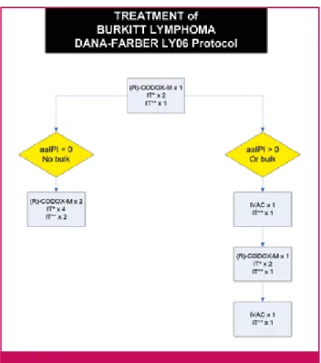
Sporadic BL and hivBL are two BL subtypes encoun-Figure 5. The DANA-Farber regimen.21,26
aa-IPI: Age-adjusted International Prognostic Index; R: rituximab; CODOX-M: cyclophosphamide, doxorubicin, vincristine, meth-otrexate; IVAC: ifosfamide, cytarabine, etoposide; IT*: cytarabine intrathecal; IT**: methotrexate intrathecal.
Practice Guidelines
tered in Belgium. Although rare, BL is a very aggressive NHL due to its high proliferation rate. Pathological and cytogenetic analyses are necessary to exclude a BLL and staging procedures must be completed rapidly to avoid delay of treatment initiation. A prephase with low-dose steroids and cyclophosphamide allows finishing initial staging, establishing chemosensitivity of the tumour and avoiding TLS. Following prephase, ‘ISCC’ or ‘LIC’, with the administration of rituximab leads to survival rates of more than 80%. However, without available randomised studies, it is quite difficult to recommend one specific therapeutic regimen above another and therefore it is highly recommended to enrol
patients in clinical studies. After treatment, patients must be followed to detect late side effects.
References
1. Swerdlow SH, et al. WHO Classification of Tumours of Haematopoietic and Lymphoid Tissues. 2008.
2. Schmitz R, et al. Burkitt’s lymphoma pathogenesis and therapeutic targets from structural and functional genomics. Nature. 2012;490:116-120.
3. Hummel M, et al. A biologic definition of Burkitt's lymphoma from transcrip-tional and genomic profiling. N Engl J Med. 2006;354:2419-30.
4. Dave SS, et al. Molecular diagnosis of Burkitt's lymphoma. N Engl J Med. 2006;354:2431-42.
5. Aukema SM, et al. Double-hit B-cell lymphomas. Blood. 2011;117:2319-31.
Figure 6. The NCI regimens.16,22
DA-EPOCH: Dose-adjusted etoposide, prednisone, vincristine, cyclophosphamide, doxorubicin; R: rituximab; SC-EPOCH: Short-cycle etoposide, prednisone, vincristine, cyclophosphamide, doxorubicin; HIV: human immunodeficiency virus; CR: complete remission.
2
6. Jaffe ES, et al. Pathology and Genetics of Tumours of Haematopoietic and Lymphoid Tissues. 2001.
7. Kluk MJ, et al. Immunohistochemical detection of MYC-driven diffuse large B-cell lymphomas. PLoS One. 2012;7:e33813.
8. Li S, et al. B-cell lymphomas with MYC/8q24 rearrangements and IGH@BCL2/ t(14;18)(q32;q21): an aggressive disease with heterogeneous histology, germinal centre B-cell immunophenotype and poor outcome. Mod Pathol. 2012;25:145-56. 9. Juweid ME, et al. Use of positron emission tomography for response assess-ment of lymphoma: consensus of the Imaging Subcommittee of International Harmonization Project in Lymphoma. J Clin Oncol. 2007;25:571-8.
10. Weiler-Sagie M, et al. (18)F-FDG avidity in lymphoma readdressed: a study of 766 patients. J Nucl Med. 2010;51:25-30.
11. Ultmann JE. Chemotherapy of non-Hodgkin-lymphoma: Introduction. Antibiot Chemother (1971). 1978;24:100-4.
12. Linch DC. Burkitt’s lymphoma in adults. Br J Haematol. 2012;156:693-703. 13. Levine AM. Challenges in the management of Burkitt's lymphoma. Clin Lymphoma 2002;3 Suppl 1:S19-S25.
14. Intermesoli T, et al. High cure rates in Burkitt’s lymphoma and leukaemia: a Northern Italy Leukaemia Group study of the German short intensive rituximab-chemotherapy program. Haematologica. 2013;98:1718-25.
15. Ribrag V, et al. Addition of Rituximab Improves Outcome of HIV Negative Patients with Burkitt’s Lymphoma Treated with the Lmba Protocol: Results of the Ran-domised Intergroup (GRAALL-Lysa) LMBA02 Protocol. (IGR sponsored LMBA02, NCT00180882) [abstract]. Blood (ASH Annual Meeting Abstracts). 2012;120. 16. Dunleavy K, et al. A prospective study of dose-adjusted (DA) EPOCH with rituximab in adults with newly diagnosed Burkitt’s lymphoma: a regimen with high efficacy and low toxicity [abstract]. Ann Oncol. 2012;19:iv83-iv84.
17. Maramattom LV, et al. Autologous and allogeneic transplantation for burkitt lymphoma outcomes and changes in utilization: a report from the centre for
inter-national blood and marrow transplant research. Biol Blood Marrow Transplant. 2013;19:173-9.
18. Ahmed SO, et al. The role of hematopoietic SCT in adult Burkitt’s lymphoma. Bone Marrow Transplant. 2013;48:617-29.
19. Gerrard M, et al. Excellent survival following two courses of COPAD chemo-therapy in children and adolescents with resected localized B-cell non-Hodgkin's lymphoma: results of the FAB/LMB 96 international study. Br J Haematol. 2008;141:840-7.
20. Hoelzer D, et al. Substantially Improved Outcome of Adult Burkitt’s Non-Hodgkin Lymphoma and Leukaemia Patients with Rituximab and a Short-Intensive Chemotherapy; Report of a Large Prospective Multicentre Trial [abstract]. Blood (ASH Annual Meeting Abstracts). 2012;120.
21. Mead GM, et al. An international evaluation of CODOX-M and CODOX-M alternating with IVAC in adult Burkitt's lymphoma: results of United Kingdom Lymphoma Group LY06 study. Ann Oncol. 2002;13:1264-74.
22. Dunleavy K, et al. Low-intensity therapy in adults with Burkitt's lymphoma. N Engl J Med. 2013;369:1915-25.
23. Petrich AM, et al. Paradigms and Controversies in the Treatment of HIV-Related Burkitt’s Lymphoma. Adv Hematol. 2012;2012:403648.
24. Oriol A, et al. High-dose chemotherapy and immunotherapy in adult Burkitt’s lymphoma: comparison of results in human immunodeficiency virus-infected and noninfected patients. Cancer. 2008;113:117-25.
25. Cortes J, et al. Hyperfractionated cyclophosphamide, vincristine, doxorubicin, and dexamethasone and highly active antiretroviral therapy for patients with acquired immunodeficiency syndrome-related Burkitt’s lymphoma/leukaemia. Cancer. 2002;94:1492-9.
26. Mohamedbhai SG, et al. Rituximab in combination with CODOX-M/IVAC: a retrospective analysis of 23 cases of non-HIV related B-cell non-Hodgkin lymphoma with proliferation index >95%. Br J Haematol. 2011;152:175-81.
Key messages for clinical practice
1. BL, characterised by an isolated translocation t(8;14)(q24;q32), is a rare but very aggressive NHL due to the extremely high proliferation rate.
2. Diagnosis and staging must be completed rapidly and treatment started within 48 hours. 3. After debulking, to avoid TLS, it is recommended to add rituximab to ISCC or LIC. Involved-field
radiotherapy has no place in BL, except for patients with CNS involvement.
4. Patients achieving CR must be followed carefully during the first year to detect recurrence of the disease. More than 80% of patients sustain their remission one year following initial treatment and are considered cured.
5. Prolonged monitoring of complete blood counts and cognitive functions are important to detect late toxicity of the applied therapies.
6. For patients in PR or with chemosensitive relapse, autologous SCT is recommended following re-induction with non-cross-resistant polychemotherapy.