Developmental variations in environmental influences including endocrine
disruptors on pubertal timing and neuroendocrine control: Revision of
human observations and mechanistic insight from rodents
Anne-Simone Parenta,b, Delphine Franssena, Julie Fudvoyea,b, Ariette Gérard a,b, Jean-Pierre Bourguignonab a Developmental Neuroendocrinology Unit, CICA Neurosciences, University of Liège, Sart-Tilman, B-4000
Liège, Belgium b Department of Pediatrics, CHU de Liège, Rue de Caillarmont 600, B-4032 Chênée, Belgium
ABSTRACT
Puberty presents remarkable individual differences in timing reaching over 5 years in humans. We put emphasis on the two edges of the age distribution of pubertal signs in humans and point to an extended distribution towards earliness for initial pubertal stages and towards lateness for final pubertal stages. Such distortion of distribution is a recent phenomenon. This suggests changing environmental influences including the possible role of nutrition, stress and endocrine disruptors. Our ability to assess neuroendocrine effects and mechanisms is very limited in humans. Using the rodent as a model, we examine the impact of environmental factors on the individual variations in pubertal timing and the possible underlying mechanisms. The capacity of environmental factors to shape functioning of the neuroendocrine system is thought to be maximal during fetal and early postnatal life and possibly less important when approaching the time of onset of puberty. © 2014 Elsevier Inc. All rights reserved.
Keywords: Puberty; Gonadotropin releasing hormone; Endocrine disruptors; Secular trends; Adolescence
1. Introduction
1.1. The classical paradigm of secular advance in human pubertal timing
Fig. 1 summarizes the data on changes in menarcheal age that are used classically to illustrate the secular trend in pubertal timing in various countries. Environmental factors have been thought to account for the reduction in menarcheal age that has been reported since 1850 till 1960 in Scandinavian countries (Tanner, 1962) and further in many European countries and USA (reviewed in Parent et al. (2003)). These findings were interpreted as a result of the improvement in life standards and socio-economical conditions (Biro et al., 2006; Dunger et al., 2006; Ong et al., 2004, 2006; Cheng et al., 2012; Himes, 2006; Roa and Tena-Sempere, 2010). A projection after 1960 of the former reduction seen in Scandinavian countries (Fig. 1) indicates that a sustained secular trend would have led to a mean menarcheal age of less than 12 yrs by the end of the 20th century. This was not the case: after 1960, the secular advancement in female pubertal timing has become less rapid or has even come to an end in countries such as Sweden, Belgium and Hungary (reviewed in Parent et al. (2003)) (Fig. 1). However, menarcheal age has shown a very rapid progression in countries like China and India (reviewed in Parent et al. (2003)) where the standard of life has improved recently. Altogether, those data are consistent with a prominent role of nutrition availability. The "critical weight/fat mass" theory proposed by Frisch and Revelle (1970) and subsequent work (Biro et al., 2006; Dunger et al., 2006; Ong et al., 2004, 2006; Cheng et al., 2012; Himes, 2006; Roa and Tena-Sempere, 2010) have put emphasis on the role of nutritional conditions based on adiposity in puberty, at the time of menarche. The discovery of leptin (Zhang et al., 1994; Campfield et al., 1995) and its prerequisite role in the neuroendocrine control of pubertal maturation and reproduction (reviewed in Sanchez-Garrido and Tena-Sempere (2013)) has added to the importance of energy balance in the prepubertal period to enable onset and progression of puberty.
The secular advancement in pubertal timing has been established following observations about mean or median age at menarche. Implicitly, the whole female pubertal process was thought to undergo similar changes though there were no data available to confirm that. The existence of similar changes for male puberty remains putative. Recent study (Goldstein, 2011) has investigated the secular trend in age at increased mortality in males,
assuming that mortality at that age is due to adolescent risk taking behaviors presumably depending on pubertal timing. Because that male adolescent mortality hump fell from an average of 21 years in 1850 to 18 years in 1960, the author concluded to a likely secular advancement in pubertal age in males. Few decades ago, the levelling off or arrested secular reduction in average menarcheal age led to the conclusion that stabilization had occurred after resetting pubertal timing to younger ages. Such a conclusion had implications on issues raised by scientists and clinicians: no further changes in pubertal timing could mean that environmental factors were stable
and that the previously defined age limits for pubertal disorders were still valid. In 1961, Thamdrup (1961) proposed the age limits of 8 years and 9 years for diagnosis of sexual precocity in girls and boys respectively. Fifty years later, those age limits have not been revised, so far, though as discussed below, the age limits for onset of puberty in the population of many countries have changed in the recent past.
1.2. Pubertal timing and preceding life periods across species
Pubertal neuroendocrine activation or reactivation of the pitui-tary-gonadal axis is essential for achievement of reproductive capacity. A leading factor in that process is Gonadotropin Releasing Hormone (GnRH) that is released by median eminence terminals of peptidergic neurons in a pulsatile manner showing increased
frequency and amplitude at the onset of puberty (Grumbach, 2002; Terasawa and Fernandez, 2001; Plant, 2008; Lomniczi et al., 2013; Ojeda and Lomniczi, 2014). This event occurs at a time in life that varies both among species and within a single species. In Fig. 2 are shown the species-related differences in relative duration of the prepubertal latency (from birth to puberty) when calculated as a percentage of lifespan for comparison purposes. Ewe (Foster et al., 1985), rat (Maeda et al., 2000) and quail (Ottinger et al., 2003) start puberty after a latency accounting for 4.8-5.7% of the lifespan as opposed to 16.3% in humans (Roelants et al., 2009) and 22.5% in baboons (Onyango et al., 2013) that is about 3-4 times longer than in non-primate species. Another less emphasized species-related difference is the variance of pubertal timing among individuals. The timing of puberty shows important differences between human individuals and the physiological range (3rd to 97th centile) of 4.8 years (Roelants et al., 2009) represents 6.25% of the life span. In the laboratory rat with a life expectancy of 2 years, the timing of puberty varies within 4-5 days (Maeda et al., 2000) accounting for an individual variance of 0.55% which is 11 times less than in humans. In sheep (Foster et al., 1985), and quail (Ottinger et al., 2003), the variance of pubertal timing represents 0.87% and 1.9% of lifespan, respectively, that is also less than in humans (Fig. 2). The baboon, a subhuman primate shows a variance of pubertal timing (Onyango et al., 2013) that is even longer than in humans, when expressed relatively to average lifespan (10.0%). These data indicate that inter-individual variations in pubertal timing may be influenced by evolution across species. These data also suggest that not only the timing i.e. the latency between birth and mean or median age at a given pubertal sign is worth being studied but also the variance i.e. the time period between the earliest and latest individuals in a reference population for occurrence of a given pubertal sign. Both parameters (latency and variance) are likely influenced by environmental factors and could even be differentially affected with different mechanisms possibly involved. In the present paper, we will review comparatively the impact of different environmental factors on pubertal timing in humans and in animal models. The variance of pubertal timing has a different magnitude across species (Fig. 2) and may not have the same significance in animals and in humans. Laboratory animals are more homogeneous in term of genetic background and are exposed to a very regulated environment. However, they are unavoidable models when it comes to study the mechanisms of neuroendocrine regulation of pubertal timing. Since some data obtained in human, non-human primates and rodents will be discussed with emphasis on neuroendocrine maturation, it is important to keep in mind that when birth takes place in rodents, maturation of the brain is less advanced than in human newborns (Clancy et al., 2007). In the rat, the onset of puberty is marked by an increase in testicular weight increase in males and vaginal opening followed by the first estrus in female rodents and (Maeda et al., 2000). The onset of puberty takes place around the time of weaning by the age of 3 weeks as evidenced from onset of increase in serum levels of gonadal hormones (Maeda et al., 2000). It is preceded by attainment of a pubertal pattern of pulsatile GnRH secretion from hypothalamic explants (Bourguignon and Franchimont, 1984; Bourguignon et al., 1990, 1992). In humans, puberty is characterized by an increase in LH and gonadal hormones. In boys, puberty is marked by an increase in testicular volume above 3 ml, consistent with Tanner G2 stage (Parent et al., 2003). In girls, the earliest manifestation of puberty is acceleration in growth velocity but commonly used markers of the timing of female puberty are the first appearance of breast development defined as Tanner B2 stage followed by menarche (Parent et al., 2003). Table 1 summarizes the different developmental stages in humans and rats.
Fig. 1. Evolution of average menarcheal age (year) in the USA and Nordic countries between 1890 and 1960
(data compiled by Tanner (1962) and further, between 1960 and 2010, in different countries in Europe, USA and around the world (updated data compiled by Parent et al. (2003)). The broken red line represents the projected reduction after 1960, based on the former changes in Scandinavian countries as reported by Tanner: mean menarcheal age would have fallen down to below 12 yrs by the end of the 20th century. In fact, after 1960, average menarcheal age has leveled off in many countries while still progressing rapidly in countries such as India or China.
Fig. 2. The length of the prepubertal latency from birth to puberty (A) and the variance of pubertal timing (B) are
represented as a percentage of lifespan in females of different species. Timing of puberty is defined here by median age at 1st ovulation. Variance of pubertal timing is calculated using the difference between upper and lower age limits for occurrence of female cycling.
Table 1: Average time limits for definition of stages of human reproductive development and approximation to
developmental stages in the laboratory rats.
Period Fetal Neonatal Infantile Juvenile/prepubertal Pubertal Post-pubertal Time limits Before birth Soon after
birth
Birth - end of lactation
End of lactation - onset of puberty Onseta - end of pubertyb
After end of puberty Female rat Gestational
day 1-21.5
PN day 1-20.5 PN day 20.5-33 PN day 33-42 >42 days Female human Gestational day 1-280 PN month 0-1
PN month 1-18 PN year 1.5-11 PN year 11-14 yrs
>14 yrs
a Vaginal opening in rats and onset of breast development in girls. b First estrus in rats and menarche in girls.
1.3. Genetic regulation of pubertal timing and environmental interactions
The activation or reactivation of GnRH secretion at puberty is regulated by a complex neuronal and glial network within the hypothalamus. The synaptic changes leading to activation of GnRH secretion involve an increase of direct excitatory input through neurons such as kisseptin and glutamate neurons and a decrease of inhibitory input from neurons such as GABA neurons (Grumbach, 2002; Terasawa and Fernandez, 2001; Plant, 2008). In addition, glial cells play a facilitatory role through production of growth factors and small molecules (Ojeda, 2010). As suggested by the complexity of this network, regulation of puberty involves redundant pathways. However, some monogenic mutations such as those affecting the GnRH receptor, Kiss1 or Kisspeptin receptor (GPR54), TAC3 or TACR3 receptor (de Roux et al., 2003; Topaloglu et al., 2010) lead to puberty failure. While mutations of those genes are associated with absence of puberty, several studies have shown that variants of more than 30 genes are associated with age at menarche in humans (Elks et al., 2010). Amongst those genes, LIN28B and the 9q31.2 locus have been identified as associated with age at menarche in several
independent genomewide studies (He et al., 2009; Ong et al., 2009; Perry et al., 2009; Sulem et al., 2009). Such genes, however, are supposed to explain very few (<1%) of the variance in age at menarche (He et al., 2009). Many of the other identified loci are associated with BMI or energy homeostasis (Elks et al., 2010). While GPR54 (Teles et al., 2008; Leka-Emiri et al., 2014), TACR3 (Leka-Emiri et al., 2014) and LIN28B (Silveira-Neto et al., 2012) mutations seem to be very uncommon or absent in patients with precocious puberty (PP), inactivating mutations in the makorin ring finger protein 3 (MKRN3) gene, have been recently identified in 2-3% of sporadic PP and even more frequently in familial PP (Abreu et al., 2013; Macedo et al., 2014; Schreiner et al., 2014) and result in invalidation of a so far unidentified inhibitory neuroendocrine mechanism.
Genetic factors are considered to explain 50-80% of the variance of age at puberty (Wehkalampi et al., 2008). Recent studies have started to unravel the possible links between environment and the genetic control of pubertal timing where epigenetic mechanisms can play an important role. Lomniczi et al. have shown that Kiss 1
expression is negatively regulated by a protein complex, the polycomb group (Ojeda and Lomniczi, 2014). At the initiation of puberty, DNA methylation of two members of this complex (Eed and Cbx7) increases, leading to a decrease of their expression and a decreased association of their proteic products with the Kiss1 promoter. Moreover, inhibition of DNA methylation prevents the peripubertal decrease of Eed and Cbx7 and their removal from the Kiss1 promoter which results in puberty failure. These recent discoveries underline the crucial role of epigenetic mechanisms in the regulation of puberty and shed a new light on the possible effects of environmental factors on those epigenetic mechanisms. The effects of the environment on the hypothalamic epigenome have not been unraveled yet but several studies have shown that the adolescent brain epigenome is affected by environmental perturbations (Morrison et al., 2013). For instance, exposure of pubertal male rats to cannabioid leads to long-term changes in gene H3K9 methylation thereby dysregulating the proenkephalin system in the nucleus accumbens (Tomasiewicz et al., 2012). Pubertal exposure to alcohol has been shown to lead to long-term changes in histone acetylation and related gene expression in the rat prefrontal cortex (Pascual et al., 2012). Thus, while recent data have shown epigenetic regulation of puberty at the hypothalamic level, the epigenetic effects of environmental factors on the hypothalamic regulation of puberty are still to be discovered.
Genome-wide studies have shown that Lin 28 is associated with age at puberty (Ong et al., 2009). Lin 28 is a RNA-binding protein that inhibits the maturation of miRNA of the Let family. Some data suggest a role of the
Lin28/Let-7 system in the hypothalamic control of puberty (Sangiao-Alvarellos et al., 2013). In male and female rats, Lin28, Lin28b, and c-Myc mRNAs (upstream positive regulator of Lin28) display very high hypothalamic expression during the neonatal period, markedly decreased during the infantile-to-juvenile transition and reach minimal levels before/around puberty in rats and monkeys. Conversely, let-7a, let-7b, mir-132, and mir-145 show opposite expression profiles (Sangiao-Alvarellos et al., 2013). In addition, manipulations disturbing puberty such as delayed puberty induced by either neonatal exposure to sex steroids or infantile exposure to constant darkness hamper these changes, indicating that the Lin28/Let-7 system could mediate some of the effects of environment on puberty. In contrast, altered puberty due to manipulation of caloric intake before weaning or between weaning and puberty does not result in comparable changes.
The data summarized in the previous paragraph suggest that the peripubertal period could be particularly sensitive to the epigenetic effects of the environment on the hypothalamic regulation of puberty. It appears that the environment also affects epigenetic pathways much earlier during development and can lead to
predisposition to some disease later in life. For instance, in vitro and in vivo models have established that epigenetic modifications caused by in utero exposure to endocrine disrupting chemicals (EDCs) can induce alterations in gene expression that may persist throughout life. Using heterozygous yellow agouti mice (Avy) in which the agouti gene encodes a molecule that promotes follicular melanocytes to produce yellow pigment instead of black, Dolinoy et al. showed that maternal exposure to environmentally relevant doses of BPA resulted in altered DNA methylation at 2 metastable loci leading to a change of fur color. Restoration of normal methylation patterns occurred with maternal supplementation of geni-stein or methyl donors (Dolinoy et al., 2007). Maternal exposure to phthalates, another group of anti-androgenic plasticizers, has been shown to increase DNA methylation and DNA methyltrans-ferases expression in mouse testis (Wu et al., 2010). Given the sensitivity of the organism to alteration of methylation early in life, one can hypothesize that early exposure to alteration of the epigenome could lead to perturbation of the hypothalamic control of puberty later in life.
1.4. Dual critical periods (fetal/neonatal life vs prepuberty) for environmental effects
In the following sections, we will discuss how the developmental stage can influence the effects of several factors (energy availability through nutrition, stress, sex steroids, endocrine disruptors) on pubertal timing. Two critical periods will be considered. Classically, the intrauterine and neonatal periods are considered to be essential for sexual differentiation and the prepubertal period is essential for sexual maturation. Beyond this effect, Barker's observations (Barker and Osmond, 1986) led to the concept of fetal set-up of lifelong adaptive mechanisms (known as the developmental origin of health and disease) that can be relevant to the
neuroendocrine control of the onset of puberty: programming of the timing of sexual maturation as an adaptive mechanism under the early influence of environmental factors.
The effects of environment are variable, depending on several factors. For instance, prepubertal restriction or excess of food and adiposity may lead to delayed or early puberty, respectively (Biro et al., 2006; Dunger et al., 2006; Ong et al., 2004, 2006; Cheng et al., 2012; Himes, 2006; Roa and Tena-Sempere, 2010) whereas
intrauterine growth restriction is associated with early puberty (Ibanez et al., 2011, 2006) in humans. Similarly, psychosocial stress during prepuberty or puberty may cause pubertal delay or arrest (van Noord and Kaaks, 1991; Tahirovic, 1998) whereas advancement of puberty has been described in children who had experienced such stress in early postnatal life or infancy (Moffitt et al., 1992; Wierson et al., 1993). In a finalist and maybe simplistic interpretation, the data discussed in this review provide evidence that environmental clues may affect pubertal timing differently depending on the life period when they come into action. In the early phase of organization of homeostasis i.e. adaptive mechanisms, adverse conditions can be interpreted by the hypothalamic centers as a risk for species survival and are translated into need of early puberty and reproduction. Reversely, in a period closer to puberty, similar adverse conditions can be interpreted as a risk for quality and outcome of pregnancy and are translated into need of delayed puberty and reproduction. Along that interpretation, the advancement of puberty in children migrating for international adoption could result from a combination of early life adversity and late prepubertal life opulence, two opposing conditions favoring advancement of puberty. Such a concept is consistent with life history interpretation of the control of reproduction that is based upon interaction between intrinsic and extrinsic constraints as reviewed and discussed by Sloboda et al. (2011).
1.5. Issues for revision: endpoints of pubertal timing and mechanisms of environmental effects in relation to period of exposure
The classical paradigm of environmental effects on pubertal development lies on secular changes in mean or median age at a particular event of puberty such as menarche. We will review and describe detailed
characteristics of the changes in pubertal timing possibly involving different signs and events in the sequence of pubertal maturation. Our purpose is to discuss the impact of environmental factors in that process including the putative role of endocrine disruptors with emphasis on the life period when they take place and the possible neuroendocrine mechanisms.
2. Variable pattern of pubertal timing depending on pubertal endpoint
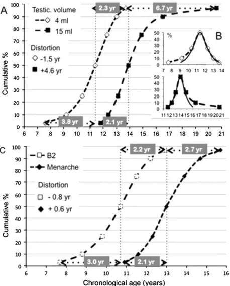
Though the mean or median age at occurrence of a given pubertal sign has been used as the sole indicator of pubertal timing in many studies, detailed analysis shows that more subtle changes occur based on the pattern of age distribution both in boys and girls. Moreover, in clinical practice, the extreme lower and upper age limits in the normal population provide important information since they are used to define early or late maturation. The distance i.e. the age difference can be calculated between the median age at occurrence of a given pubertal sign and either the 3rd or 10th cen-tile on the one hand and the 97th or 90th centile on the other hand. Such an age distance is equal on both sides of the median when the distribution is normal or Gaussian and then only, standard deviations allow determination of the normal age limits. In case of unequal (asymmetrical) distribution, the difference between the distances (median age - lower limit) and (median age - upper limit) provides an appraisal of distortion that is negative when deviation is towards younger ages and positive when it is towards older ages. Such a calculation is illustrated (Fig. 3) using the Belgian data published recently (Roelants et al., 2009). In boys, the data are represented through both the cumulative frequency (Fig. 3A) or the absolute frequency (Fig. 3B) of ages when achieving testicular volume ≥4ml and ≥ 15ml that are respectively consistent with onset and final stage of puberty. The asymmetry of the curves is obvious. In comparison with the theoretical curve of a normal distribution (Fig. 3B), the two age distribution curves are opposed by a distortion that is negative and positive for 4 and 15 ml testicular volume, respectively. These data indicate that currently, some boys tend to enter puberty earlier and some to end puberty later. In girls (Fig. 3C), the current curve of cumulative frequency for occurrence of breast (B2) seems to parallel that for menarche. Calculation of distortion at centile 3 and 97, however, reveals a negative value for B2, i.e. distribution skewed towards earliness and a positive value for menarche, i.e. distribution skewed towards lateness. Literature data on median age and upper/lower limits for occurrence of pubertal signs in boys and girls are compiled in Table 2. Using those data, we have calculated the distortion of variance at the 10th and 90th centiles of age. When studying distribution of data from 95% of the population i.e. centile 3rd to 97th (Roelants et al., 2009) such as shown in Fig. 3, it is of note that distortion is twice as much as it is using data from 80% of the population i.e. centile 10th to 90th (Table 2). This difference exists because the distortion of variance is all the more important since the extreme tail of the distribution is included. However, 3rd and 97th centile data were rarely provided in the publications and therefore calculation of distortion in Table 2 is based on 10th and 90th centile data.
Among women born in Belgium in the 1920s (Jeurissen, 1969), the median menarcheal age was 1.5 years later than in more recent cohorts and variance (10th to 90th centiles) was 4.1 years. The distortion of variance in age at menarche was negative at that time. In a Belgian cohort born in the 1960s (Vercauteren and Susanne, 1985), the median menarcheal age was advanced to 13yrs. The distance between the 10th and the 90th centile had fallen to 2.4 yrs indicating that the variance was decreasing and the distortion was still negative. In the most recent Belgian cohort born in the 1990s (Roelants et al., 2009), the median menarcheal age did no longer change while the distance between the 10th and the 90th centiles had increased to 3.1 yrs indicating that the variance was increasing and distortion became positive. Among the different countries, except the data obtained before the secular trend in menarcheal age was leveling off (van Noord and Kaaks, 1991), there were only minor
differences both in the median menarcheal age that varied between 13.0 and 13.4 yrs and in the variance i.e. the 10th to 90th centile range (Table 2). Distortion of the variance was negative in the studies involving subjects born before the 1970s (Jeurissen, 1969; Vercauteren and Susanne, 1985) and was found to be positive in several more recent studies since 1980 (Roelants et al., 2009; Talma et al., 2013; Mul et al., 2001). Consequently (Fig. 4A), there is a secular shift in distortion of variance in menarcheal age from negative to positive values during the 20th century. These data are consistent with a French report of a secular increase in proportion of women needing 5 years after menarche before attainment of regular (presumably ovulatory) cycling while there was concomitantly a secular reduction in the proportion getting menarche at 15 years or later (Clavel-Chapelon and E3N-EPIC group, 2002).
The median age at onset of breast development appears to be earlier in USA, Denmark and Greece (Aksglaede et al., 2009; Herman-Giddens et al., 1997; Papadimitriou et al., 2008) than in the Netherlands and Belgium
(Roelants et al., 2009; Mul et al., 2001). The distance between the 10th and the 90th centile (Table 2) is quite similar among the studies and a slight negative distortion of variance is reported in 3 studies (Roelants et al., 2009; Mul et al., 2001; Papadimitriou et al., 2008). As opposed to the secular trend in distortion of variance in menarcheal age, the data on age at onset of breast development in several countries show a negative distortion that seems to be more marked in the recent studies (Fig. 4A). Taken together, those observations indicate that, during the last decades of the 20th century, some female individuals tended to enter puberty earlier and some to end puberty later. This was not reflected by changes in median ages.
Table 2: Median, upper and lower age limits and distortion of age distribution for initial and final stages of
female and male puberty. N: number of subjects, CS: cross-sectional; L: longitudinal; TV: testicular volume. End-point Country year
of publ. NCS/L Year of study Centile 10 (yr) Centile 50 (yr) Centile 90 (yr) Dist. 10th to 90th (yr) Distortio n (yr) Ref. Breast stage ≥B2 Sweden 1996 138 L 1980 9.3 10.8 12.3 3.0 0 Lindgren (1996) DK 2009 1100 CS 1992 9.3 10.9 12.4 3.1 -0.1 Aksglaede et al. (2009) USA 1997 15439 CS 1992-3 8.5 10.1 11.8 3.3 +0.1 Herman-Giddens et al. (1997)
Greece 2008 311 L 1995 8.1 9.9 11.3 3.2 -0.4 Papadimitriou et al. (2008) NL 2001 3562 CS 1997 9.0 10.7 12.1 3.1 -0.3 Mul et al. (2001) Belgium 2009 4471 CS 2004 8.8 10.7 12.2 3.4 -0.4 Roelants et al. (2009) DK 2009 995 CS 2007 8.2 9.9 11.6 3.4 0 Aksglaede et al. (2009) Genital stage 5≈G2 Sweden 1996 116L 1980 9.5 11.6 13.7 4.2 0 Lindgren (1996) DK2010 824 CS 1992 10.5 11.9 13.1 2.6 -0.2 Sorensen et al. (2010) NL 2001 4019 CS 1997 7.7 11.4 12.8 5.1 -2.3 Mul et al. (2001) Belgium 2009 4219 CS 2004 8.9 11.4 13.0 4.1 -0.9 Roelants et al. (2009) DK2010 767 CS 2007 10.1 11.6 13.1 3.0 0 Sorensen et al. (2010) USA 2012 4131 CS 2005-10 6.6 10.7 13.0 6.4 -1.8 Herman-Giddens et al. (1997) TV 5=4 ml DK2010 824 CS 1992 10.8 11.9 13.0 2.2 0 Sorensen et al. (2010) NL 2001 4019 CS 1997 8.7 11.4 13.0 4.3 -1.1 Mul et al. (2001) Belgium 2009 4219 CS 2004 9.2 11.4 13.0 3.8 -0.6 Roelants et al. (2009) China 2011 18807 CS 2003-5 8.8 11.1 12.6 3.8 -0.8 Ma et al. (2011) DK2010 767 CS 2007 10.3 11.7 13.0 2.7 -0.1 Sorensen et al. (2010) Menarche Belgium 1969 1317 CS 1925-9a 12.2 14.5 16.3 4.1 -0.5 Jeurissen (1969) Belgium 1969 2254 CS 1945-9a 11.7 13.3 14.5 2.8 -0.4 Jeurissen (1969) NL2013 1882 CS 1965 11.8 13.4 14.9 3.1 -0.1 Talma et al. (2013) Belgium 1985 1048 CS 1960-5a 11.6 13.0 14.0 2.4 -0.4 Vercauteren and Susanne (1985) NL2013 3029 CS 1980 11.7 13.3 14.9 3.2 0 Talma et al. (2013) DK 2009 1100 CS 1992 11.8 13.4 15.1 3.3 +0.1 Aksglaede et al. (2009) NL 2001 3562 CS 1997 11.7 13.2 14.9 3.2 +0.2 Mul et al. (2001) NL2013 30C8 CS 1997 11.8 13.2 14.9 3.1 +0.3 Talma et al. (2013)
Belgium 2009 4471 CS 2004 11.6 13.0 14.7 3.1 +0.3 Roelants et al. (2009) DK 2009 995 CS 2007 11.8 13.1 15.1 3.3 +0.1 Aksglaede et al. (2009) NL2013 2138 CS 2009 11.5 13.0 14.5 3.0 0 Talma et al. (2013) TV s=12 ml NL 2001 4019 CS 1997 11.8 13.7 15.4 3.6 -0.2 Mul et al. (2001) Belgium 2009 4219 CS 2004 12.1 13.5 15.3 3.2 +0.4 Roelants et al. (2009) China 2011 18807 CS 2003-5 11.9 13.7 16.7 4.8 +3.0 Ma et al. (2011) Genital stage Sweden 1996 116L 1980 13.6 15.1 16.6 3.0 0 Lindgren (1996) G5 NL 2001 4019 CS 1997 13.7 15.3 19.5 5.8 +2.6 Mul et al. (2001) Belgium 2009 4219 CS 2004 13.5 15.3 18.7 5.2 +1.6 Roelants et al. (2009) a Year of birth.
Fig. 3. Cumulative frequency curves (A) and absolute frequency curve (B) of age when achieving a testicular
volume ≥4ml (beginning of puberty) or ≥15 ml (late stage of puberty) in Belgium. (C) Cumulative frequency curves of age at breast development (B2) and menarche in Belgium. In boys, the cumulative frequency curves for age at testicular volume ≥4 ml and ≥15 ml (A) seems to be parallel in the rapidly ascending phase but are divergent both in the early and late parts. The two absolute frequency curves (B) illustrate those divergences with opposed changes at the two edges. Calculation of distortion is performed at centile 3 and 97 age points through the difference between the age distances (centile 3-50) and (centile 97-50). Distortion is negative for 4 ml testicular volume and positive for 15 ml. In girls (C), the curve of cumulative frequency of occurrence of breast (B2) seems to parallel that for menarche but the two curves show also divergent patterns at the two edges. Calculation of distortion at centile 3 and 97 reveals a negative value for B2 and a positive value for menarche.
In boys, the median age at onset of genital development (G2 stage) or early pubertal increase in testicular volume (≥4ml) is similar among the different publications except G2 stage in USA (Herman-Giddens et al., 2012). As opposed to the relatively homogeneous variance of onset of puberty in girls, the variance in males is extremely different among the studies: the distance between the 10th and the 90th varies between 2.2 yrs in Denmark (Sorensen et al., 2010) and 6.4 yrs in USA (Herman-Giddens et al., 2012). Distortion of variance is negative in most studies and can attain as much as - 2.3 yrs (Mul et al., 2001). Though there are few studies available, a secular trend towards negative distortion of variance in age at onset of male puberty is suggested (Fig. 4B). While the median age at final pubertal milestones (testicular volume of 12 ml or G5 stage) was very similar among the studies, the variance i.e. the distance between the 10th and the 90th was highly variable among the studies (3.0-5.8 yrs) and distortion was positive and elevated in several studies (Roelants et al., 2009; Mul et al., 2001; Ma et al., 2011). Taken together (Fig. 4B), these data indicate that recent variations in male pubertal timing involve few or no change in median age but a trend towards negative or positive distortion for initial or final pubertal stages, respectively. Such a distorted variance appears to be greater and much more variable among the studies in boys than in girls.
The opposing changes in timing for initial and final endpoints of puberty and the greater variations in boys than in girls lead to revise the concept that current changes involve prominently females and advancement of pubertal timing. Those variations may reflect contemporary effects of environmental factors on pubertal timing.
Fig. 4. The negative or positive distortion of the 10th to 90th centile distribution of age at B2 i.e. onset of breast
development and age at menarche (A) or age at occurrence of G2 and G5 stages of genital development in boys (B) are plotted against the year of the study in different countries. A secular trend towards negative distortion appears for the initial pubertal stages (B2 and G2) as opposed to a trend towards positive distortion for the final stages (menarche and B5).
3. Period-dependent impact of environmental factors and mechanisms
In the following sections, we will discuss how the developmental stage can influence the effects of several factors on pubertal timing: energy availability through nutrition, stress, sex steroids, and endocrine disruptors. Though sex steroids do not belong to environmental factors per se, they need to be addressed because endocrine disruptors are thought to exert most of their effects through interaction with sex steroid receptors and
metabolism.
3.1. Disturbances of energy availability
3.1.1. Critical windows of exposure to abnormal energy availability and timing of puberty in humans
Consistent with Barker's hypothesis of developmental origins of health and diseases (Barker and Osmond, 1986), energy availability in prenatal life can affect age at onset of puberty. Most studies have been carried out in girls and considered age at menarche as the major endpoint. A positive correlation is found between birth weight and age at menarche. Cooper et al. (1996) have shown in a cohort of 1471 girls born in 1946 that the girls who have the lowest birth weight have the earliest age at menarche. Adair has similarly shown a significant positive relationship between birth weight and age at menarche in a longitudinal study on 997 girls born in 1983-1984 in the Philippines (Adair, 2001). Here, a rapid postnatal growth potentiates the effects of birth size. However, birth weight and age at puberty do not always appear to be significantly correlated, possibly because the link is not simply causal and many other factors are confounding. It is possible that common factors determine both low birth weight and early timing of puberty. Some studies do not find any significant correlation between birth length or weight and age at puberty but identify other prenatal or perinatal influencing factors that are related to nutrition: based on pubertal self-assessment in 4000 girls between age 8 and 14, Maisonet et al. find that pre-pregnancy maternal obesity, maternal smoking and primiparity are associated with an earlier age at onset of puberty (Maisonet et al., 2010). It is not possible, however to determine whether each of these prenatal factors has a comparable effect on programming of pubertal timing or whether those factors are closely linked and
trigger each other.
More recently, early postnatal weight gain has appeared to be more strongly linked to onset of puberty in girls than birth weight itself. A prospective study of 2715 girls indicates that low birth weight is associated with menarche before the age of 12 but a positive association between weight gain during the first 9 months of life and age at menarche is also found and statistically much more significant (Ong et al., 2009). Faster weight gain during the first 9 months of life is also positively correlated with fat mass index, body weight and body mass index at puberty (Ong et al., 2009). Other population cohort studies have similarly shown that rapid early postnatal weight gain predicts an early onset of puberty (Dunger et al., 2006; dos Santos Silva et al., 2002). Those results suggest that in addition to prenatal life, early postnatal life could be another window during which variations in nutrition availability program subsequent growth and development. Early nutrition within that window might be an important determinant of both age at puberty and risk of childhood obesity.
Childhood BMI is significantly associated with onset of puberty since heavier children reach puberty earlier (Kaplowitz et al., 2001 ; Rosenfield et al., 2009). In fact, these observations have been used to substantiate the advance in age at breast development that has been reported in the USA during the 1990s (Herman-Giddens et al., 1997). Most of the reports about an association between peripu-bertal weight and puberty onset are cross-sectional and carried out in girls with age at menarche as a marker of puberty. These methodological aspects make a formal association between body weight and puberty difficult to ascertain. Some longitudinal studies, however, have reported a similar association. Aksglaede et al. have found that BMI at 7 years is a significant predictor of age at pubertal growth spurt in a cohort of more than 150,000 boys and girls (Aksglaede et al., 2009). The heaviest prepubertal children enter puberty earlier than the lightest group. Secular trend towards early onset of puberty is found in all BMI categories suggesting that the obesity epidemic is not solely responsible. Another longitudinal study in Sweden show that BMI increase between age 2 and 8 years (adiposity rebound) is positively associated with earlier growth spurt in boys and girls (He and Karlberg, 2001). Recently, Biro confirmed the important impact of BMI on breast development in girls in the USA (Biro et al., 2013). Taken together, those epidemiological studies point towards three windows of sensitivity for a link between energy availability/adiposity and pubertal development: prenatal life, early postnatal life and childhood. Currently, it remains difficult to determine whether those periods represent three independent windows or whether they are part of a continuum. An independent regulation in each of those periods is consistent with opposing effects of prenatal and postnatal weight gain, along with the life history interpretation as discussed above (Sloboda et al., 2011).
The mechanisms involved in the connection between weight gain, adiposity and pubertal timing may be direct and/or indirect. Frish and Revelle have hypothesized that a critical fat mass was necessary for completion of puberty (Frisch and Revelle, 1970). Although challenged later (Ellison, 1982), this hypothesis set the scene for the discovery of peptides responsible for integrating energy availability and the control of reproduction. In the 1990s, leptin has appeared as one of the major factors allowing the reproductive system to sense the magnitude of energy reserves. In humans, leptin is necessary for the onset of puberty but cannot trigger early puberty indicating a permissive role for this hormone (Sanchez-Garrido and Tena-Sempere, 2013). As discussed above, children with intrauterine growth retardation have been shown to be predisposed to subsequent catch up in weight, childhood obesity and early puberty. Several mechanisms have been proposed to explain the association between rapid weight gain and early puberty. Rapid infancy/childhood weight gain has been associated with increased levels of adrenal androgens at age 8 (Ong et al., 2004). Such premature adrenarche could precipitate pitui-tary-gonadal maturation though both processes are found to be dissociated in several conditions, suggesting that physiological levels of adrenal androgens do not influence neuroendocrine maturation (Ducharme and Collu, 1982). In disorders with excess in adrenal androgens like some forms of congenital adrenal hyperplasia,
occurrence of secondary central puberty indicates possibly advanced neuroendocrine maturation due to abnormally high levels of sex steroids (Dacou-Voutetakis and Karidis, 1993). Hyperin-sulinemia has been reported in children with a rapid weight gain which is known to lead to decreased levels of sex hormone binding globulin and increased sex steroids bioavailability (Holly et al., 1989). Increased adiposity could also lead to increased aromatase activity and conversion of androgens to estrogens (de Ridder et al., 1992). Catch-up growth in children with low birth weight is associated with a rise in serum leptin (Jaquet et al., 1999) and IGF-1
(Stevens et al., 2014). Rodent models indicate that neonatal leptin could play a crucial role in the development of some neural connections controlling GnRH secretion (Bouret, 2004) and leptin is known to facilitate GnRH secretion peripubertally (Sanchez-Garrido and Tena-Sempere, 2013). IGF-1 is another factor affected by nutrition and that could be able to promote GnRH secretion (Wolfe et al., 2014). In a recent cohort study, mother's age at menarche predicts faster weight gain during infancy in the offspring (Ong et al., 2007). This suggests that the mechanism linking faster infancy weight gain to earlier menarche could include transgener-ational factors, genetic or epigenetic. Gene-environment interactions are hypothesized to account for the recent changes in distribution of age at menarche and adult obesity. Some studies have provided evidence for genetic influences. For instance, the fat mass and obesity-associated gene (FTO) has been associated with a decrease in
age at menarche and increased BMI (Elks et al., 2010). Recent genome-wide association and candidate sin-gle-nucleotide polymorphism studies have identified several loci for age at menarche that are associated with obesity (Elks et al., 2010; den Hoed et al., 2010; Fernandez-Rhodes et al., 2013). The search for known biological pathways shared by loci influencing both adiposity and menarche is still ongoing. Loci that increase adiposity and decrease age at menarche may have particularly strong effects on adult-onset diseases related to prolonged estradiol and fatness exposure such as breast cancer and cardiovascular diseases.
3.1.2. Abnormal energy availability and timing of puberty: mechanisms of action in rodent models
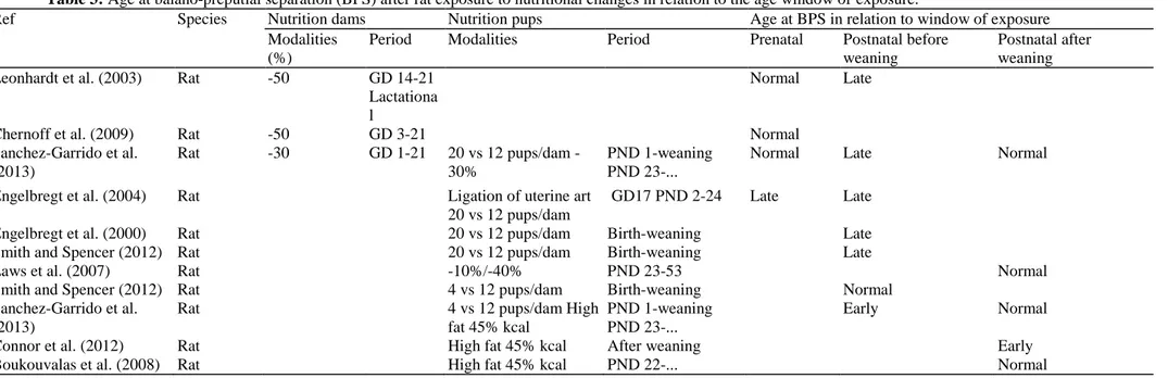
3.1.2.1. Prenatal food restriction. Several rodent models have been used to study the effects of prenatal or early
postnatal food restriction on puberty onset (Tables 3 and 4). Prenatal energy restriction is obtained through either decreased food availability to the pregnant dam or ligation of the uterine artery and postnatal food restriction through increased size of the litters (Tables 3 and 4).
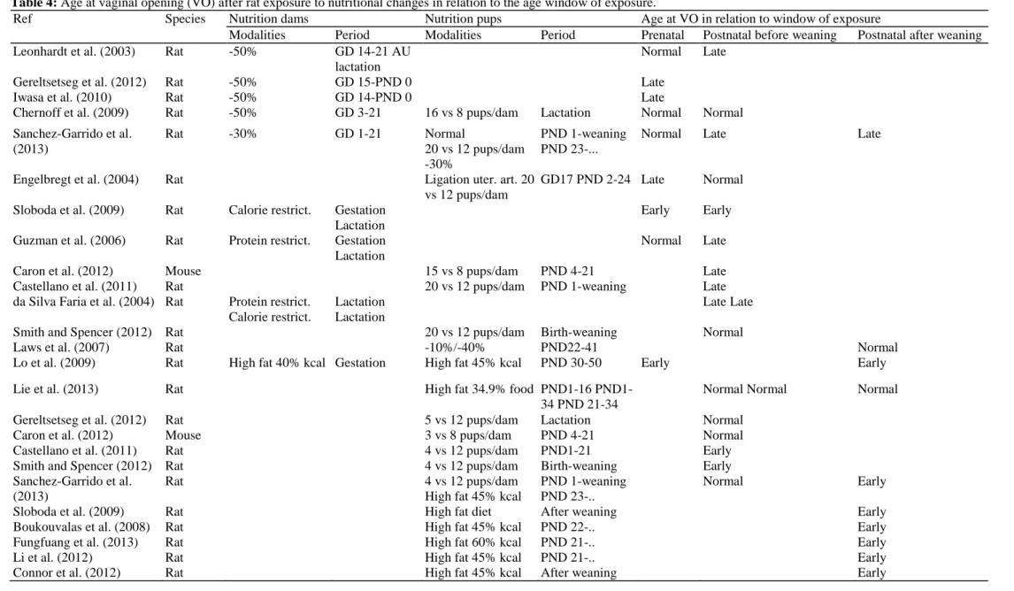
While small birth weight for gestational age is associated with early onset of puberty in humans, prenatal underfeeding usually leads to late onset of puberty in female rodents (Table 4). Maternal food restriction (Leonhardt et al., 2003; Iwasa et al., 2010) or uterine artery ligation (Engelbregt et al., 2000) are associated with late vaginal opening or first estrus. Some authors have reported no effect of maternal food restriction on pubertal development while a single study (Sloboda et al., 2009) found an acceleration of pubertal onset. Such
discrepancies could be due to differences in food restriction, birth weight or postnatal weight gain. In males, prenatal underfeeding does not affect age at balano-preputial separation (Table 3).
The hypothalamic control of GnRH secretion could be affected by prenatal food restriction in females. After maternal food restriction in rats (50% of daily food intake), Iwasa et al. have shown that the pups have delayed puberty and lower hypothalamic levels of Kiss1 mRNA on postnatal day 16 and 20 (Iwasa et al., 2010). Pups exposed to prenatal food restriction also show decreased levels of hypothalamic Kiss1 mRNA while serum leptin are increased, suggesting a central resistance to leptin (Iwasa et al., 2010). The chronic intracerebral
administration of kisspeptin peripubertally in pups malnourished prenatally restores normal onset of puberty (Iwasa et al., 2010), suggesting that prenatal food restriction could reversibly affect the kisspeptin system. Whether leptin acts directly on arcuate kisspeptin neurons to mediate regulation of GnRH secretion is still controversial (Donato et al., 2011). We have shown that prenatal food restriction (70% of daily food intake) decreases the stimulatory effect of leptin on GnRH secretion from rat hypothalamic expiants obtained from female pups at 15 days of age (Franssen et al., 2013). Those data indicate that prenatal food restriction could alter the hypothalamic sensitivity to leptin. In our model, we do not observe any significant effect of prenatal undernutrition on leptin receptor mRNA levels. In addition, serum leptin levels at 15 days of age in the animals exposed to prenatal food restriction are not different from controls (Franssen et al., 2013). Thus, the decreased effects of leptin are likely not explained neither by a decreased hypothalamic expression of its receptor nor by variations in serum leptin levels but could be explained by alterations of leptin intracellular signaling pathways.
3.1.2.2. Early postnatal food restriction. Caron et al. and Castellano et al. have shown that postnatal food
restriction by increasing the litter size leads to delayed puberty with late vaginal opening and late first estrus cycle in mice (Caron et al., 2012; Castellano et al., 2011). Altered neonatal nutrition leads to alterations in the development of brain circuits responsible for the regulation of puberty and fertility (Castellano et al., 2011). Pups that have been undernourished during early postnatal life display a reduction in the density of kisspeptin fibers compared with normally fed animals. That reduction persists later in life and is associated with decreased fertility (Caron et al., 2012). The most severely affected projections are those originating from the arcuate nucleus and projecting to the median preoptic nucleus (Caron et al., 2012). This indicates that arcuate nucleus circuits are highly plastic and respond to food availability during early postnatal life. The neuronal projections originating from the AVPV and contacting GnRH neural develop before birth (Polston and Simerly, 2006) and are not affected by postnatal undernutrition. Thus, decreased food availability is more likely to cause profound adverse organizational effects when taking place during the development of the nucleus controlling the specific physiological process. Leptin levels are also affected by postnatal nutrition. Bouret et al. have shown that postnatal leptin levels were blunted in the mice from undernourished litters (Bouret et al., 2007). One can speculate that the neural circuits, including the kisspeptin system, that develop during the first three weeks of life could be affected by the reduced levels of leptin neonatally. Further evidence of an early leptin role comes from the capacity of early postnatal leptin treatment to prevent occurrence of obesity in adult rats born with
intrauterine growth restriction (Vickers et al., 2005). It is possible that early growth catch up following refeeding provides endogenous hormonal regulators such as leptin and IGF-1 that can influence hypothalamic organization and maturation.
3.1.2.3. Peripubertal food restriction. Food restriction during the peripubertal period leads to a delay of pubertal
maturation in males and females (Tables 3 and 4) indicating that critical weight and fat reserves need to be reached in order to complete pubertal development (Frisch and Revelle, 1970). Severe fasting has been shown to
decrease hypothalamic Kiss1 mRNA expression and kisspeptin immunoreactivity as well as lowering circulating LH in pubertal rats (Castellano et al., 2010, 2005). In pubertal rats, fasting also inhibits the hypothalamic expression of the gene coding for neurokinin B and its receptor (Navarro et al., 2012). Neurokinin B appears to be co-expressed with Kiss1 in the arcuate nucleus in several species (Lehman et al., 2010) and to be required for normal pubertal maturation (Topaloglu et al., 2010). Those data indicate that neurokinin B and kisspeptin could cooperate in the metabolic control of the neuroendocrine mechanism of puberty. In 2013, Owen et al. have shown that fibroblast growth factor 21 (FGF21), a growth factor secreted by the liver in response to short-term fasting, delayed puberty by suppressing the preovulatory gonadotropin surge (Owen et al., 2013). This effect is exerted through inhibition of vasopressin in the suprachiasmatic nucleus resulting in loss of the stimulatory input to kisspeptin neurons in the AVPV (Owen et al., 2013). Other metabolic hormones have been shown to regulate pubertal activation and to signal energy sufficiency or insufficiency. Among them, ghrelin is an anorectic factor secreted by the stomach. Chronic administration of ghrelin leads to delayed puberty in male and female rats (Tena-Sempere, 2008). It is an inhibitory signal for LH secretion in rodents, sheep and non-human primates (Tena-Sempere, 2008; Iqbal et al., 2006; Vulliémoz et al., 2004). Reduction in the frequency of GnRH pulses has also been reported in immature rats following systemic administration of ghrelin (Lebrethon et al., 2007). This effect can be inhibited by an NPY Y5R antagonist (Lebrethon et al., 2007). This suggests that ghrelin effects on GnRH neuron are mediated, at least in part, by afferent neurons such as NPY neurons. Recent evidence suggests that ghrelin is able to suppress Kiss1 gene expression at discrete hypothalamic areas (Forbes, 2006). These actions of Ghrelin on the hypothalamic and the pituitary might contribute to the suppression of gonadotropin levels in conditions of persistent negative energy balance, in which ghrelin levels are commonly elevated. A growing body of evidence has supported the initial hypothesis that insulin is involved in the signaling of satiety to the central nervous system (Niswender et al., 2004). Such physiological effect is consistent with the phenotype of mice harboring a specific deletion of the insulin receptor within neuronal cells (Brüning et al., 2000). These mice have a phenotype very similar to that of mice lacking a functional leptin receptor with hyperphagic obesity and central hypogonadism secondary to functional GnRH deficiency. Using rat primary hypothalamic neurons in cell culture, Burcelin et al. could demonstrate that insulin stimulates in vitro both the expression and the secretion of GnRH (Burcelin et al., 2003). Both insulin receptor mRNA and protein are expressed in a clonal cell line expressing GnRH (Salvi et al., 2006). Taken together, these data suggest that peripheral insulin can modulate the activity of hypothalamic GnRH neurons.
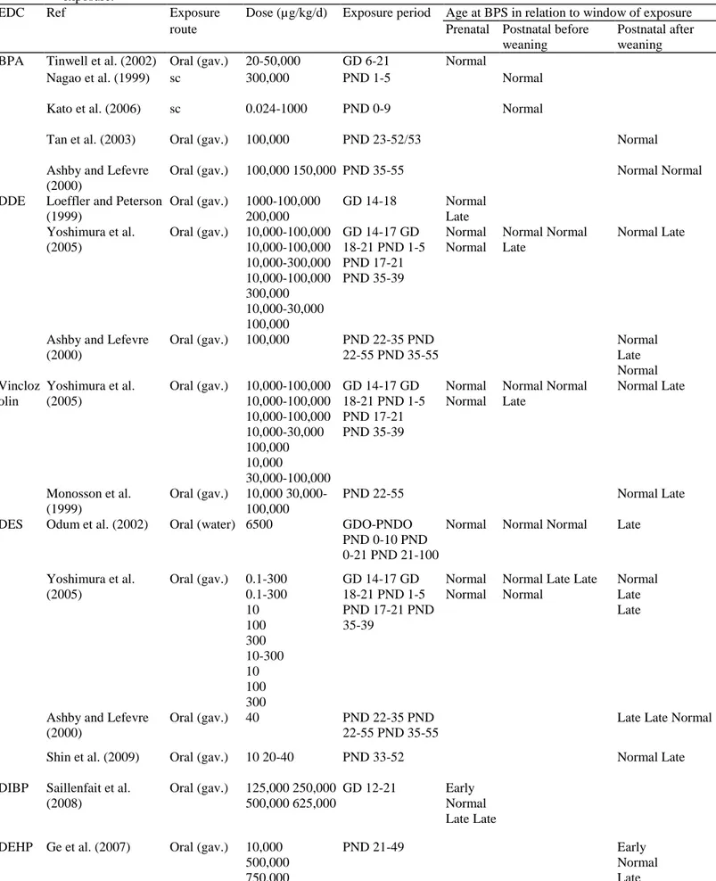
Table 3: Age at balano-preputial separation (BPS) after rat exposure to nutritional changes in relation to the age window of exposure.
Ref Species Nutrition dams Nutrition pups Age at BPS in relation to window of exposure
Modalities (%)
Period Modalities Period Prenatal Postnatal before
weaning
Postnatal after weaning
Leonhardt et al. (2003) Rat -50 GD 14-21
Lactationa l
Normal Late
Chernoff et al. (2009) Rat -50 GD 3-21 Normal
Sanchez-Garrido et al. (2013) Rat -30 GD 1-21 20 vs 12 pups/dam -30% PND 1-weaning PND 23-...
Normal Late Normal
Engelbregt et al. (2004) Rat Ligation of uterine art
20 vs 12 pups/dam
GD17 PND 2-24 Late Late
Engelbregt et al. (2000) Rat 20 vs 12 pups/dam Birth-weaning Late
Smith and Spencer (2012) Rat 20 vs 12 pups/dam Birth-weaning Late
Laws et al. (2007) Rat -10%/-40% PND 23-53 Normal
Smith and Spencer (2012) Rat 4 vs 12 pups/dam Birth-weaning Normal
Sanchez-Garrido et al. (2013)
Rat 4 vs 12 pups/dam High
fat 45% kcal
PND 1-weaning PND 23-...
Early Normal
Connor et al. (2012) Rat High fat 45% kcal After weaning Early
3.1.2.4. Food excess or high fat diet. Studies addressing the impact of early postnatal overfeeding are scarce and
report divergent observations depending on the species or strain as well as fat content of the diet. Using small litters (3-5 pups) in order to cause overnutri-tion, some authors did not observe any effect on pubertal onset in females (Gereltsetseg et al., 2012; Sanchez-Garrido et al., 2013) while others have reported earlier puberty (Castellano et al., 2011; Smith and Spencer, 2012). In this case, earlier puberty is accompanied by an increase in serum leptin levels as well as increased expression of kiss1 mRNA expression in the whole hypothalamus. These data indicate that persistent energy excess during early postnatal development might contribute to a precocious activation of the hypothalamic control of puberty. Independently of the calorie amount, quality of the diet might affect the onset of puberty (Boukouvalas et al., 2008; Fungfuang et al., 2013; Li et al., 2012). High fat diet during gestation or after weaning induces early onset of puberty in females though weight is not significantly different from the control group. There seems to be a strong sexual dimorphism since the timing of male puberty does not seem to be affected by a high fat diet following weaning (Sanchez-Garrido et al., 2013). In females, high fat diet induces advanced maturation of pulsatile LH secretion associated with an accelerated maturation of
Kiss1 and neurokinin B mRNA expression in the arcuate nucleus (Li et al., 2012). 3.2. Stress
3.2.1. Critical windows of exposure to stress and timing of puberty in human
It is well established that stress factors have an inhibitory impact on the neuroendocrine regulation of
reproduction in women (Warren and Fried, 2001 ) as in adult nonhuman female primates (Bethea et al., 2008). Strenuous physical exercise, emotional stress or weight loss are associated with hypothalamic amenorrhea (Warren and Fried, 2001 ; Bethea et al., 2008). The impact of stress on timing of puberty is particularly complex to study. Few studies have been able to isolate specific factors involving disturbances of food intake, energy expenditure, economical circumstances or such as chronic disease or psychological stresses. Moreover, many of those studies bear on a limited sample size without control population. Once again, puberty has mostly been studied retrospectively in female subjects with menarche being the main endpoint since it appears to be the most reliable event to be investigated using questionnaires. The effects may vary depending on the nature of the stressors as well as the timing of exposure. Stress in early childhood seems to be associated with earlier
menarche (Moffitt et al., 1992; Wierson et al., 1993) whereas exposure shortly before or during puberty has been associated with later onset of menarche (van Noord and Kaaks, 1991; Tahirovic, 1998). Puberty has been found to be delayed during the 1944-1945 Dutch famine study (van Noord and Kaaks, 1991) and the Balkan War (Tahirovic, 1998). In these conditions, the impact of psychological stress during war is confounded by nutritional factors associated with famine.
Children with chronic disease are known to enter puberty later. The prevalence of chronic conditions among adolescents is difficult to assess but is estimated to be between 10% and 15% (Suris et al., 2004). Delayed puberty is common to most chronic illnesses but more frequent in those characterized by malnutrition and chronic inflammation such as bowel disorders or cystic fibrosis. Possible mechanisms will be discussed below based on animal studies. Suppression of the HPG axis and delayed puberty caused by activation of the HPA axis has also been demonstrated in highly trained runners and gymnasts (Klentrou and Plyley, 2003). The impact of physical training on puberty depends on a variety of factors including the type of exercise, the time of onset and the intensity of training. Still, here as for the other conditions discussed under the stress "umbrella", it is obvious that several factors related to nutrition, emotion and activity are intricate and make particularly complex the elucidation of the pathophysiological mechanisms.
Adversity experiences earlier in life, during childhood, have been shown to affect pubertal timing. Most of those studies point to an earlier onset of puberty following psychological stress during childhood. Longitudinal investigations suggest that factors indexing problematic early environments such as marital conflicts, father absence, negative parenting practices or lower socioeconomic status are associated with a younger age at menarche (Moffitt et al., 1992; Wierson et al., 1993). As already discussed for nutritional issues, life history models have proposed that the period between birth and 7 years of age is a period of increased sensitivity to environmental cues. A disturbed environment could result in accelerated reproductive development via signals that resources are limited or uncertain, thus threatening species due to likely reduced reproductive lifespan (Ellis, 2004).
3.2.2. Stress and timing of puberty: mechanisms of action
The mechanisms involved in the effects of stress on puberty might vary depending on the time of occurrence. We will focus initially on the effect of stress taking place during puberty and then consider the mechanisms involved in alteration of puberty caused by prenatal or neonatal stress. There is convincing evidence that corticotrophin-releasing factor (CRF) plays a crucial role in the stress-induced inhibition of GnRH secretion. CRF reduces LH serum concentrations in women during the late follicular and midluteal phases of the cycle
(Barbarino et al., 1989). LH release has been shown to be inhibited by CRF in rodents and monkeys as well. Moreover, the suppression of LH secretion caused by various stresses is prevented by administration of a CRH antagonist (Li et al., 2005; Cates et al., 2004). The timing of puberty is affected by CRF since
intracerebroventricular injection of CRF during 14 days, starting on postnatal day 28, delays puberty in female rats while its antagonist causes early puberty (Kinsey-Jones et al., 2010). However, the precise neuroanatomical regions of CRF signaling to influence GnRH secretion are still to be established. The role of the paraventricular nucleus (PVN) is controversial. Although PVN CRF mRNA increases in response to stress, it appears that electrolytic lesions of the PVN fail to prevent the inhibition of LH caused by various stresses. The preoptic area (POA) is characterized by synaptic connections between CRF and GnRH neurons (MacLusky et al., 1988) and GnRH neurons express CRF receptors (Jasoni et al., 2005). This suggests an anatomical substrate for a direct functional interaction between the GnRH and the CRF system. Moreover, injection of CRF in the POA inhibits LH release (Rivest et al., 1993). Recently, several stressors such as restraint, hypoglycaemia and
lipopolysaccharides have been shown to downregulate kiss1 and GPR54 mRNA expression in the medial preoptic area and the arcuate nucleus in adult female rats (Kinsey-Jones et al., 2009). The involvement of kisspeptin in the mediation of the stress effect on puberty is still to be determined.
Glucocorticoids have also been shown to increase the inhibitory action of the RFamide-related peptide (RFRP) on GnRH secretion (Kirby et al., 2009). RFRP has been described as an inhibitor of GnRH secretion in quails, rodents and monkeys (Tsutsui et al., 2000; Clarke et al., 2009; Ubuka et al., 2009). The recent study by Kirby et al. (2009) highlights a possible inhibition of GnRH secretion by peripheral corticoids after acute and chronic stress. This effect is supported by the presence of glucocorticoid receptors in 53% of the cells expressing RFRP (Kirby et al., 2009).
Except for prenatal underfeeding as discussed above, very few studies have focused on the effect of prenatal stress on pubertal development in human. Anoxia is among the most frequent causes of stress at birth that may turn into cerebral palsy. In a cross-sectional study, this condition has been found to involve both early onset and late completion of puberty (Worley et al., 2002). However, there are numerous confounding factors such as reduced activity and feeding problems in addition to the functional impairment of brain areas that may vary among the patients. Rodent data indicate that fetal exposure to glucocorticoids may be an important determinant of sexual maturation. In the offspring of mothers subjected to stress (Politch and Herrenkohl, 1984) or exposed to ACTH or corticoids (Smith and Waddell, 2000) neonatally or during pregnancy, late puberty is observed. Though fetal exposure to maternal corticoids is limited by placental metabolism (Burton and Waddell, 1999), high maternal glucocorticoids concentrations associated with stress are expected to reach the fetus. The mechanisms through which perinatal stress affects pubertal development are still to be elucidated. Prenatal maternal stress increases offspring HPA axis sensitivity, anxiety and cognitive deficits, suggesting alteration of neurodevelopment (Howerton and Bale, 2012). The mechanisms by which early life experience, especially stress, is able to program brain development seem to involve epigenetic modulation of individual genes or large gene clusters. As an example, adult male mice exposed to early prenatal stress show altered expression and DNA methylation in CRF, glucocorticoid receptors (Mueller and Bale, 2008; Nemeroff, 1992) or BDNF (Boersma et al., 2013). Since recent studies have shown that methylation plays a pivotal role in the onset of puberty in female rodent (Ojeda and Lomniczi, 2014), one can hypothesize that early exposure to stress could alter methylation-mediated programming of puberty.
Table 4: Age at vaginal opening (VO) after rat exposure to nutritional changes in relation to the age window of exposure.
Ref Species Nutrition dams Nutrition pups Age at VO in relation to window of exposure
Modalities Period Modalities Period Prenatal Postnatal before weaning Postnatal after weaning
Leonhardt et al. (2003) Rat -50% GD 14-21 AU
lactation
Normal Late
Gereltsetseg et al. (2012) Rat -50% GD 15-PND 0 Late
Iwasa et al. (2010) Rat -50% GD 14-PND 0 Late
Chernoff et al. (2009) Rat -50% GD 3-21 16 vs 8 pups/dam Lactation Normal Normal
Sanchez-Garrido et al. (2013) Rat -30% GD 1-21 Normal 20 vs 12 pups/dam -30% PND 1-weaning PND 23-...
Normal Late Late
Engelbregt et al. (2004) Rat Ligation uter. art. 20
vs 12 pups/dam
GD17 PND 2-24 Late Normal
Sloboda et al. (2009) Rat Calorie restrict. Gestation Lactation
Early Early
Guzman et al. (2006) Rat Protein restrict. Gestation Lactation
Normal Late
Caron et al. (2012) Mouse 15 vs 8 pups/dam PND 4-21 Late
Castellano et al. (2011) Rat 20 vs 12 pups/dam PND 1-weaning Late
da Silva Faria et al. (2004) Rat Protein restrict. Calorie restrict.
Lactation Lactation
Late Late
Smith and Spencer (2012) Rat 20 vs 12 pups/dam Birth-weaning Normal
Laws et al. (2007) Rat -10%/-40% PND22-41 Normal
Lo et al. (2009) Rat High fat 40% kcal Gestation High fat 45% kcal PND 30-50 Early Early
Lie et al. (2013) Rat High fat 34.9% food 16
PND1-34 PND 21-PND1-34
Normal Normal Normal
Gereltsetseg et al. (2012) Rat 5 vs 12 pups/dam Lactation Normal
Caron et al. (2012) Mouse 3 vs 8 pups/dam PND 4-21 Normal
Castellano et al. (2011) Rat 4 vs 12 pups/dam PND1-21 Early
Smith and Spencer (2012) Rat 4 vs 12 pups/dam Birth-weaning Early
Sanchez-Garrido et al. (2013)
Rat 4 vs 12 pups/dam
High fat 45% kcal
PND 1-weaning PND 23-..
Normal Early
Sloboda et al. (2009) Rat High fat diet After weaning Early
Boukouvalas et al. (2008) Rat High fat 45% kcal PND 22-.. Early
Fungfuang et al. (2013) Rat High fat 60% kcal PND 21-.. Early
Li et al. (2012) Rat High fat 45% kcal PND 21-.. Early
3.3. Endocrine disrupters
3.3.1. Critical windows for the effects of sex steroids on puberty
Though sex steroids do not belong to environmental factors per se, their action needs to be addressed because endocrine disruptors are thought to exert most among their effects through interaction with sex steroid receptors, metabolism and effects. In primate and ovine species, pubertal timing is sexually dimorphic, enabling to study the effects of sex steroids on gender differences in pubertal timing. When female monkey fetuses have been exposed to high androgen levels, menarche is delayed by 4-6 months (Goy et al., 1988). Likewise, fetal lamb exposure to testosterone shifts pubertal timing from a female to a male pattern (Kosut et al., 1997). More insight into the developmental differences in the role of androgens comes from the use of flutamide, an androgen receptor blocker. Prenatal flutamide treatment during early gestation accelerates pubertal timing in male monkeys (Herman et al., 2006). Indirect evidence that this could involve hypothalamic effects arises from the stimulation of LH levels and pulse frequency after flutamide treatment for 3 days in adult men (Urban et al., 1988). Finally, the naturally occurring condition of androgen insensitivity due to androgen receptor mutation in XY humans is associated with a female pattern of peak height velocity at puberty (Zachmann et al., 1986). Taken together, those data are consistent with a prenatal role of androgens in setting pubertal timing.
The hypothalamic targets of perinatal steroids are still incompletely known. Recent rodent studies have underlined the role of perinatal sex steroids in programming the sexual differentiation of Kisspeptin expression in the AVPV (reviewed in Poling and Kauffman (2013)). At later developmental stages, such as puberty, AVPV Kisspeptin neurons may also be further regulated by E2 to exhibit a typical female pattern. The ARC kisspeptin population is not markedly sexually dimorphic in adulthood but this population displays sexually dimorphic characteristics during the neonatal and juvenile periods (reviewed in Poling and Kauffman (2013)). The sex difference in neonatal ARC Kiss1 levels could be due to activational effects caused by temporary variations in circulating sex steroids (reviewed in Poling and Kauffman (2013)).
The window of exposure during development can matter for the effects of sex steroids. While testosterone could reduce LH pulse frequency in both orchidectomized and ovariectomized adult monkeys (Plant, 1986), such a treatment does not affect LH in orchidectomized fetal monkey but reduces LH in the ovariectomized fetal monkey (Ellinwood et al., 1982). By contrast to the delay in pubertal timing caused by androgens prenatally, neonatal treatment of orchidectomized monkeys with testosterone for a year suppresses LH and withdrawal of treatment is followed by an early pubertal resurgence of LH secretion in 2/3 animals (Fraser et al., 2005). This observation is consistent with that of delayed breast development in a girl with a virilizing adrenal tumor who showed very rapid breast development and menarche within 6 months after surgical removal of the tumor (Bourguignon et al., 2015).
The most prevalent human model of fetal or fetal and postnatal androgen exposure is congenital adrenal
hyperplasia. This is a complex model however since, after diagnosis and initiation of treatment, further exposure to possible androgen excess as well as to glucocorticoid excess depends on the management. A less
heterogeneous condition could be the premature increase in adrenal androgens or premature adrenarche. In this condition where the exposure to unusually high levels of androgens is presumably in the prepubertal period, early menarche has been reported as a function of the severity of intrauterine growth retardation (Ibanez et al., 2006). Treatment with metformin prevents the advancement of menarche (Ibanez et al., 2011) indicating that the determinant of pubertal timing in that condition may be related to prenatal nutritional status (see Section 3.1.2.1) and postnatal insulin sensitivity with no or minor role of adrenal androgens. In summary, the sensitivity to organizing effects of testosterone (directly or after aromatization) centrally is maximal prenatally and neonatally. The central sensitivity to negative feedback effects which is greater in the male than in the female, already operates perinatally and is maximal subsequently, before puberty.
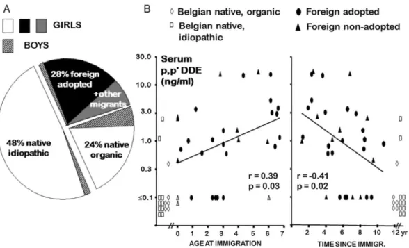
Fig. 5. (A) Etiological distribution of central precocious puberty in 145 Belgian patients. (B) Serum levels of
p,p-DDE, a derivative of DDT in different patients with sexual precocity. The foreign migrating patients with
sexual precocity are adopted or non-adopted. Serum levels of p,p-DDE in migrating patients are represented in relation to age at immigration and time since immigration. In 26 foreign patients with precocious puberty, the mean serum concentration of p,p-DDE was 10 times higher than the limit of detection, whereas the levels were below this limit in 13 among 15 Belgian native patients. Adapted from data published in
Krstevska-Konstantinova et al. (2001).
Table 5: Effects of prenatal/early postnatal versus prepubertal exposure to EDCs on timing of breast
development in girls.
EDCs Timing of breast development Period of exposure
Prenatal/early postnatal Prepubertal DDE (+DDT) Early
Normal Late
Krstevska-Konstantinova et al. (2001 ) Wolff et al. (2008)
Dioxins Early Normal
Late Leijs et al. (2008) Den Hond et al. (2002)
Table 6 : Effects of prenatal/early postnatal versus prepubertal exposure to EDCs on timing of menarche in girls.
EDCs Timing of menarche Period of exposure
Prenatal/early postnatal Prepubertal DDE (+DDT) Early
Normal Late
Vasiliu et al. (2004) Ouyang et al. (2005)
Denham et al. (2005)and Ozen et al. (2012)
PCBs Early Denham et al. (2005)
Normal Late Vasiliu et al. (2004) and Yang et al. (2005)
Dioxins Early
Normal Leijs et al. (2008) and Warner et al. (2004) Den Hond et al. (2002) Late
Phytoestrogens Early Adgent et al. (2012) Kim et al. (2011)
Normal Late Strom et al. (2001) and Giampietro et al. (2004)
Table 7 : Effects of prenatal/early postnatal versus prepubertal exposure to EDCs on pubertal timing in boys.
EDCs Pubertal timing Period of exposure
Prenatal/early postnatal Prepubertal
DDE (+DDT) Early Den Hond et al. (2011)
Normal Gladen et al. (2000) Grandjean et al. (2012)
Late Grandjean et al. (2012)
PCBs Early Den Hond et al. (2011)
Normal Mol et al. (2002) and Warner et al. (2004) Grandjean et al. (2012) Late Guo et al. (2004) and Herman et al. (2006) Den Hond et al. (2002)
Phthalates Early Mouritsen et al. (2013)
Normal Late Rais-Bahrami et al. (2004) Mieritz et al. (2012)
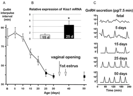
Fig. 6. (A) Evolution of mean GnRH interpuise interval throughout development in rats when studied ex vivo
using individual female hypothalamic expiants (mean ± SD, n = 5 animals per age group). (B) Relative expression of Kiss1 mRNA in the hypothalamus of female rats at 15 and 25 days of age. (C) Representative profiles of GnRH secretion from hypothalamic expiants obtained from female rats throughout development. The graphs illustrate the decrease of GnRH interpulse interval between PND 5 and 25, before the onset of puberty. Adapted from Bourguignon and Franchimont (1984), except Kiss1 mRNA data.