HAL Id: hal-02401474
https://hal.archives-ouvertes.fr/hal-02401474
Submitted on 16 Nov 2020HAL is a multi-disciplinary open access archive for the deposit and dissemination of sci-entific research documents, whether they are pub-lished or not. The documents may come from teaching and research institutions in France or abroad, or from public or private research centers.
L’archive ouverte pluridisciplinaire HAL, est destinée au dépôt et à la diffusion de documents scientifiques de niveau recherche, publiés ou non, émanant des établissements d’enseignement et de recherche français ou étrangers, des laboratoires publics ou privés.
Spatially-resolved localization and chemical speciation of
nickel and zinc in Noccaea tymphaea and Bornmuellera
emarginata
Antony van der Ent, Kathryn Spiers, Dennis Brueckner, Guillaume
Echevarria, Mark Aarts, Emmanuelle Montargès-Pelletier
To cite this version:
Antony van der Ent, Kathryn Spiers, Dennis Brueckner, Guillaume Echevarria, Mark Aarts, et al.. Spatially-resolved localization and chemical speciation of nickel and zinc in Noccaea tymphaea and Bornmuellera emarginata. Metallomics, Royal Society of Chemistry, 2019, 11, pp.2052-2065. �10.1039/C9MT00106A�. �hal-02401474�
1
Spatially-resolved localization and chemical speciation of nickel
1
and zinc in Noccaea tymphaea and Bornmuellera emarginata
2 3 4
Antony van der Ent1,2, Kathryn Spiers3, Dennis Brueckner3,4,5, Guillaume Echevarria2, 5
Mark G. M. Aarts6, Emmanuelle Montargès-Pelletier7 6
7
1Centre for Mined Land Rehabilitation, Sustainable Minerals Institute,
8
The University of Queensland, Australia. 9
10
2Laboratoire Sols et Environnement, UMR 1120, Université de Lorraine, France.
11 12
3Photon Science, Deutsches Elektronen-Synchrotron DESY, Germany.
13 14
4Department of Physics, University of Hamburg, Germany.
15 16
5Faculty of Chemistry and Biochemistry, Ruhr-University Bochum, Germany.
17 18
6Laboratory of Genetics, Wageningen University and Research, The Netherlands.
19 20
7Laboratoire Interdisciplinaire des Environnements Continentaux, CNRS,
21
Université́ de Lorraine, UMR7360, France. 22
23 24 25 26
2
ABSTRACT
27
Hyperaccumulator plants present the ideal model system for studying the physiological regulation 28
of the essential (and potentially toxic) transition elements nickel and zinc. This study used 29
synchrotron ray Fluorescence Microscopy (XFM) elemental imaging and spatially resolved X-30
ray Absorption Spectroscopy (XAS) to elucidate elemental localization and chemical speciation of 31
nickel and zinc in the hyperaccumulators Noccaea tymphaea and Bornmuellera emarginata. It turns 32
out that in the leaves of N. tymphaea nickel and zinc have contrasting localization, and whereas 33
nickel is present in vacuoles of epidermal cells, zinc occurs mainly in the mesophyll cells. In the 34
seeds Ni and Zn are similarly localized and strongly enriched in the cotyledons in N. tymphaea. Ni 35
is strongly enriched in the tip of the radicle of B. emarginata. Noccaea tymphaea has an Fe-rich 36
provascular strand network in the cotyledons of the seed. The chemical speciation of Ni in the intact 37
seeds of N. tymphaea is unequivocally associated with carboxylic acids, whereas Zn is present as 38
the phytate species. The spatially resolved spectroscopy did not reveal any spatial variation in 39
chemical speciation of Ni and Zn within the N. tymphaea seed. The dissimilar ecophysiological 40
behaviour of Ni and Zn in N. tymphaea and B. emarginata raises questions about the evolution of 41
hyperaccumulation in these species. 42
43
Key words: capsule, cotyledons; hyperaccumulator; seed; translocation.
44 45
3 1. INTRODUCTION 46 47
Hyperaccumulators are unusual plants that accumulate particular metals or metalloids in their living 48
tissues to levels that may be hundreds to thousands of times greater than is normal for most 49
plants1,2. Hyperaccumulator plants have the unique ability to take up and detoxify exceptional 50
concentrations of metals without any signs of toxicity. Plants have been found to hyperaccumulate a 51
wide range of elements, including nickel (Ni), manganese (Mn), cadmium (Cd), copper (Cu), cobalt 52
(Co), selenium (Se), arsenic (As), thallium (Tl) and Zn2,3. This contrasts with 'normal plants' that
53
have a tightly controlled regulation of essential transition elements (Cu, Fe, Ni, Mn, Zn) to avoid 54
either deficiency or toxicity. Hyperaccumulator plants represent the most extreme example of 55
adaptation to a surplus of metal transition elements in their environment, and are therefore ideal 56
model systems for understanding the physiological regulation of essential and potentially toxic, 57
non-essential transition elements4,5,6. In many Ni hyperaccumulator species, Ni occurs in mixtures 58
of citrate and malate complexes that vary in different parts of the plants7,8,9. Hyperaccumulation
59
results from adaptations of the metal regulation mechanisms shared by all higher plants10. Hence, 60
insights into the mechanisms of hyperaccumulation may be applied to improve the uptake and 61
accumulation of deficient elements, such as iron (Fe) and zinc (Zn), in economically important food 62
crops. These insights may also be applied to limit uptake of potentially phytotoxic elements, such as 63
nickel (Ni) in food crops. The extreme metal accumulation capability of hyperaccumulator plants 64
spawned the concept of phytoextraction for remediating contaminated soils, which has attracted 65
much research effort11. Hyperaccumulator plants also have potential for use in phytotechnologies 66
such as biofortication, phytoremediation and phytomining, the latter utilizes hyperaccumulators as 67
‘metal crops’ to sequester Ni or other metals in harvestable biomass that can then be used to 68
produce fine Ni chemicals12,13. 69
70
Nickel is the most recent element shown to be essential for higher plants14,15, due to its key role for 71
the activity of urease, an enzyme widely distributed in higher plants16, and playing a crucial role in
72
nitrogen remobilization from senescing leaves and during seed germination. However, excess Ni 73
induces oxidative and genotoxic stresses that are deleterious to plant growth17. Therefore, every 74
plant species needs to regulate Ni homeostasis according to its needs. Apart from its function in 75
urease activation, other physiological functions of Ni are poorly understood in higher plants6,18. 76
Although Ni is an essential micronutrient, its physiological requirement is extremely low. It is 77
shown that 0.1 mg Ni kg-1 is sufficient for seed germination and plant growth15,19. Hence, Ni 78
deficiency in naturally-grown plants rarely occurs, and the only known case is for pecan20. The
79
molecular mechanisms involved in the regulation of Ni homeostasis are not well known even in 80
4
model species such as Arabidopsis thaliana. Nickel can be transported from the soil and inside 81
plants by several families of metal transporters (e.g. ZIP/IRT, IREG, YSL21,22,23). Since plants 82
normally only require minute amounts of Ni, no Ni specific transporter has so far been identified to 83
account for enhanced Ni uptake from soil. In non-accumulator species, this function is most likely 84
performed by one of the ZIP family Zn/Fe/Mn uptake transporters e.g. the Zn-deficiency induced 85
expression of AtZIP4 can be repressed by supplying Ni2+. Preliminary results suggest that one of 86
these transporters has developed more affinity for Ni2+ in Ni hyperaccumulators than in
non-87
accumulators21,23. The transporter TgMTP1 of the CDF family was originally suspected to mediate
88
Ni storage in the vacuole of the Ni hyperaccumulator Noccaea goesingense24. More recent studies 89
suggested that transporters of the IREG/Ferroportin family, localized on the vacuolar membrane, 90
are involved in the storage of Ni in non-accumulators and hyperaccumulators21,25. Studies on Zn 91
and Cd hyperaccumulation in Brassicaceae species (e.g. Arabidopsis halleri and N. caerulescens) 92
revealed that Zn and Cd hyperaccumulation traits are correlated with high and constitutive 93
expression of genes involved in metal transport, in the biosynthesis of metal chelators and in 94
cellular defences to oxidative stresses26,27,28. These changes in gene expression are often the 95
combined effect of gene duplication and altered promoter activity29,30. 96
97
The genus Noccaea has at least 23 species that hyperaccumulate Ni, a further 10 that 98
hyperaccumulate Zn, three that hyperaccumulate Cd and one that hyperaccumulates Pb31,32,33,34,35. 99
Noccaea caerulescens (J.Presl & C.Presl) F.K.Mey. (Thlaspi caerulescens J.Presl & C.Presl) is 100
unique in consisting of different ecotypes with distinct metal tolerance and hyperaccumulation 101
abilities36. While calamine, ultramafic and non-metallicolous populations can hyperaccumulate Zn 102
and Ni, or Cd, when supplied36, they differ in their ability to tolerate these metals, often depending 103
on the metal concentrations at their site of origin37,38. Zinc is taken up by ZIP-like plasma 104
membrane located Zn-transporters. Rather than storing excess Zn in vacuoles of root cells, which 105
most non-accumulator species do, the Zn2+ is loaded into the xylem, by HMA4, as in Arabidopsis 106
halleri (L.) O'Kane & Al-Shehbaz29, thus translocated to the leaves, where it is stored in mesophyll
107
and epidermal vacuoles. Ultramafic populations are known39and converted into genetically
108
homogeneous lines by recurrent inbreeding for characterization of Zn, Ni and Cd accumulation and 109
tolerance properties. The variation in metal tolerance and hyperaccumulation is heritable and 110
independent of each other40,41,42. The calamine and non-metallicolous populations have been 111
investigated in more detail than the ultramafic populations. So far most of the analysis of ultramafic 112
N. caerulescens involved the accession from Monte Prinzera, in the Italian Apennine mountain 113
range36. This accession has been subject to several proteomics studies, trying to correlate local
114
adaptation to specific protein expression44 and was included in transcriptome studies as well45. Ni, 115
5
and also Zn, xylem loading in N. caerulescens is facilitated by high histidine levels43 although the 116
genes involved have not yet been identified. 117
Noccaea tymphaea (Hausskn.) F.K.Mey. (synonyms: Thlaspi tymphaeum Hausskn. and Thlaspi 118
goesingense Halácsy) is distributed in Albania, Bosnia and Herzegovina, Greece, Macedonia. It 119
occurs on montane ultramafic soils, where it can accumulate high foliar Ni (up to 11 800 µg g-1) and 120
relatively low foliar Zn (up to 179 µg g-1)31. Bornmuellera emarginata (Boiss.) Rešetnik 121
(synonyms: Leptoplax emarginata (Boiss.) O. E. Schulz, Peltaria emarginata (Boiss.) Hausskn.)46
122
is endemic to ultramafic soils in Greece, with a discontinuous distribution from Pindus mountains, 123
Mt. Smolikas, and the island of Euboea. Some specimens were also sampled in Syria and are kept at 124
Paris Herbarium (P) showing a disjunct distribution pattern across the Eastern Mediterranean. It is a 125
strong Ni hyperaccumulator that can accumulate up to 34 400 µg g-1 foliar Ni47. 126
127
Currently little is known on storage and acquisition of Ni in seeds and during germination. In 128
Noccaea praecox (Wulfen) F.K.Mey. (synonym: Thlaspi praecox Wulfen.) Cd was mobilised to the 129
shoots during germination, but not to the roots48. Scanning electron microscopy with energy 130
dispersive spectroscopy (SEM-EDS) was undertaken on the seeds of Noccaea pindica (Hausskn.) 131
Holub (synonym of Thlaspi pindicum Hausskn.) and the results showed that Ni accumulated in the 132
micropylar area opposite the radicle and in the epidermis of cotyledons49. Little information is 133
available about the elemental distribution in other hyperaccumulating genera of the Brassicaceae, 134
and no study yet has focussed on B. emarginata except the SEM-EDS observation of herbarium 135
specimen air-dried leaves which showed accumulation of Ni in the epidermis cells except in the 136
vicinity of stomata50. Knowledge about the ecophysiology of Ni and Zn in reproductive organs of 137
hyperaccumulator plants is especially scare. In order to provide a more general view about the 138
ecophysiology of hyperaccumulation, including the reproduction organs and first phases of life of 139
these plants, this study aimed to elucidate the distribution and chemical speciation of Ni and Zn in 140
the seeds and siliques of N. tymphaea and B. emarginata originating from their native habitats in 141
Greece. X-ray Fluorescence Microscopy (XFM) has substantial explanatory power for advancing 142
the understanding of the ecophysiology of hyperaccumulation9. In order to determine the
143
distribution and spatially-resolved chemical speciation of Ni and Zn in both species, we make use of 144
the singular ability of the Maia detector system51,52 to perform ultra-rapid X-ray elemental mapping 145
and spatially resolved X-ray Absorption Spectroscopy (XAS) on live/fresh samples. 146
147
2. MATERIALS AND METHODS
148 149
2.1 Collection of plant tissues and soils
6
Whole, live plants of N. tymphaea were collected in Greece (at the Katara Pass, 39°48'00.5"N 151
21°10'60.0"E, altitude 1690 m. a.s.l.) growing in natural ultramafic soils. Intact seeds capsules were 152
collected from B. emarginata in their native habitat Greece (near Trigona, 39°47'29"N, 21°25'32"E, 153
altitude 830 m. a.s.l.). The soils of the collection locality are described in detail eslwhere53. The 154
plants were potted in natural soil from the habitat and brought alive to the P06 beamline (PETRA 155
III Synchrotron, DESY Campus, Hamburg Germany) for the experiments described below. 156
157
2.2 Chemical bulk analysis of tissue samples
158
Plant tissue samples for bulk chemical analysis were first dried on silica gel and then dried at 70°C 159
for five days in a drying oven. They were subsequently ground and digested using 4 mL HNO3
160
(70%) in a microwave oven (Milestone Start D) for a 45-minute programme and diluted to 30 mL 161
with ultrapure water (Millipore 18.2 MΩ·cm at 25°C). Finally, they were analysed with ICP-AES 162
(Thermo iCAP 7400) for Cd, Ni, Co, Cr, Cu, Zn, Mn, Fe, Mg, Ca, Na, K, S, P. 163
164
2.3 Preparation of tissue samples for X-ray fluorescence microscopy
165
The seeds and seed capsules could be investigated in their native state without any sample 166
preparation. The intact seeds were mounted between two sheets of Ultralene thin film (4 µm) 167
stretched over a Perspex frame magnetically attached to the x-y motion stage at atmospheric 168
temperature (~20°C). However, in order to reveal the internal distribution of Ni, Zn and other 169
elements inside roots, stems and leaves, cross-sections were prepared. The samples were hand cut 170
with a stainless-steel razor blade (‘dry knife’), mounted between two sheets of 4 µm Ultralene thin 171
film in a tight sandwich to limit evaporation, and analysed within 15 minutes after excision. X-ray 172
micro-fluorescence was performed at high speed to keep the scan time to a minimum. Since the 173
penetration depth of the X-rays is greater than the thickness of a cell layer, the information obtained 174
from thick sections is a combined distribution originating from more or less superimposed cell 175
layers. The semi-thick sections (~200 µm) correspond to 3–4 cell layers. As such the obtained data 176
do not reveal subcellular distribution, but nevertheless show the tissue-level distribution (e.g. 177
epidermal cells, mesophyll, vascular bundles, etc.). 178
179
2.4 X-ray fluorescence microscopy
180
The X-ray fluorescence microscopy (XFM) experiments were undertaken at beamline P06 at 181
PETRA III at DESY (Deutsches Elektronen-Synchrotron). The undulator beam was 182
monochromatised using either a Si(111) channel-cut crystal or a double-crystal monochromator, 183
depending on beamline mode for each part of the experiment. A Kirkpatrick-Baez mirror pair was 184
used to focus the incident beam. The X-ray flux of the focussed beam was in the order of 1010 185
7
photons/s 54. X-ray fluorescence was detected using the Maia detector system in backscatter 186
geometry52,55. The large solid-angle (1.2 steradian) of the Maia detector is particularly suited to 187
biological samples such as these as it allows detection of a good proportion of the fluoresced signal, 188
allowing a reduction of the radiation dose and thus reducing potential damage to a specimen51. 189
190
The 2D µXRF measurements carried out in the microprobe of P06 at DESY were performed with a 191
beam size of 720 × 780 nm at an incident energy of 11 keV. The single-slice tomography 192
measurements of the N. tymphaea seed were carried out with a beamsize of 400 × 450 nm at a 193
photon energy of 15 keV using the same beamline endstation and general setup. Scanning 194
parameters were a step size of 2 µm, a dwell time of 1 ms and an angular range of 452 projections 195
covering 360° in 2 subscans. As the seed was naturally dehydrated a cryo-stream was not employed. 196
For the XRF 2D and tomography scans, the pixel size chosen was larger than the focused beam size 197
as a result of necessary compromises due to time constraints. 198
199
2.5 Synchrotron X-ray Absorption Spectroscopy (XAS)
200
Ni and Zn K-edge XAS spectra of the plant tissue samples and standards were recorded in 201
fluorescence mode with the Maia detector. The X-ray beam energy was calibrated using either a Ni 202
or Zn metal foil recorded in transmission, where the first peak of the first derivative was assumed to 203
be 8333 or 9659 eV, respectively. The seeds (~1.5 × 0.3 mm) were scanned at 1.6 µm pixels with a 204
20 ms per pixel dwell time. In addition to these µXRF elemental images on these seeds, spatially 205
resolved XANES spectra were collected as image ‘stacks’ of µXRF maps, each with 15 µm pixels 206
and a 12 ms per pixel dwell time, at 170 increasing energies, spanning the energy range 8183–9082 207
eV over the Ni edge at 8.333 keV, and spanning the energy range 9486-9858 eV over the Zn K-208
edge at 9659 eV. 209
210
Several Ni2+ and Zn2+ standards were prepared by adding organic ligands in calculated molar excess 211
(1:5) to Ni2+ and Zn2+ to ensure the formation of organo-metallic complexes. The selection of the
212
ligands was based on previous reports of Ni and Zn complexation in hyperaccumulator plants7,9,57,58.
213
Aqueous standards were prepared from Ni(NO3)2 or Zn(NO3)2 salts respectively in ultrapure water
214
(Millipore) with the following ligands: malate, citrate, oxalate, phytate, and histidine. 215
Supplementary references from previous experiments were also used to increase the number of 216
reference spectra, such as Ni in aqueous solution (Ni-aqueous), Ni-citrate with a metal:ligand ratio 217
equal to 1 (Ni-citrate) and Zn sulfate. The solutions were diluted to 10 mM [Ni or Zn2+] before 218
analysis. The pH of the standards was checked, and adjusted to 6. The aqueous standards were then 219
applied to filter paper (Whatmann), allowed to dry and enveloped in Kapton tape before scanning. 220
8
221 222
9
2.6 Data processing and analysis
223
The XRF event stream was analysed using the Dynamic Analysis method59,60 as implemented in 224
GeoPIXE61,62. GeoPIXE provides quantitative first-order self-absorption corrected maps of 225
projected areal elemental density – maps of elemental content. Conversion of X-ray counts to 226
concentration was performed through analysis of Ni and Co XRF reference foil scans (Micromatter, 227
Canada). The samples were each considered of a uniform thickness and either hydrated or dry, with 228
respective thicknesses and compositions of 1000 µm and C7.3O33H59N0.7S0.15 for the (hydrated)
229
whole leaf and leaf, stem and root cross-sections, and 1500 µm and C31O15H51N2S0.8 for the (dry)
230
whole seeds and capsules. Assuming a uniform thickness for the seeds introduces further 231
approximations to the measurements of seeds and seed capsules, however, as these have a generally 232
flattened oblate cross-section, the approximation was considered appropriate. 233
234
Reconstruction of the single-slice tomographic data was performed using a maximum-likelihood 235
expectation-maximization (MLEM) algorithm. 236
237
PCA analysis was performed on the XANES stacks using the MANTiS package. The extracted 238
XANES data were reduced using standard normalization procedures performed with Bruce Ravel 239
and Matthew Newville programs ATHENA and ARTEMIS63,64. Spectra were background 240
subtracted and normalized. The XANES signals obtained were fitted as linear combinations of the 241
standard spectra collected on solutions to evidence the main organic ligands involved in metal 242
complexation. The number of standards was constrained to be at a maximum of three. The sum of 243
components was released and not forced to be equal to 1. The selection of the linear combination 244
was made on the basis of the indicators of fitting quality (chi2, r-factor and reduced chi2), and the 245
number of components was finally set to 2 as a third component did not improve the fitting quality. 246
247
3. RESULTS
248 249
3.1 Localization of Ni and Zn in N. tymphaea root, stem and leaf cross-sections
250
Elemental maps of the root cross-sections (Fig. 1) show that Ni is concentrated mainly in the 251
epidermis, in phloem bundles and pericycle. Zinc is mainly enriched in the phloem bundles. In the 252
stem cross-section (Fig. 2) Ni is enriched in the collenchyma and the phloem bundles, as well as in 253
the xylem. 254
255
In the whole leaves of N. tymphaea, Ni is mainly localised in the leaf blade, with increasing 256
concentrations towards the margins. The whole leaf elemental map of Ni (Fig. 3 and Suppl. Fig 1) 257
10
provides intriguing insight in the distribution of Ni at the tissue-level. The circular features are 258
suggestive of major enrichment in the apoplast surrounding large epidermal cells. In contrast, Zn is 259
distributed primarily surrounding the secondary vasculature across the leaf blade. Calcium is clearly 260
depleted in the veins, but high in the interveinal regions. The distribution of Co (not shown) mirrors 261
that of Ni, with enrichment in the leaf margins, and towards the leaf apex. Potassium is enriched 262
throughout the leaf blade, but highest near the petiole. 263
264
In the leaf cross-section of N. tymphaea Ni and Zn have contrasting localizations (Fig 4 and Suppl. 265
Fig 2). Whereas Ni is present in vacuoles of epidermal cells, Zn occurs mainly in the mesophyll 266
cells, especially on either side of the central vascular bundles (Fig. 4). Calcium is depleted in the 267
vascular bundles, but strongly enriched in the mesophyll and also in the epidermal region. 268
Potassium is enriched in the epidermal cells and in the phloem bundles of the primary vein and also 269
in secondary veinlets. 270
271
3.2 Localization of Ni and Zn in seed capsules of N. tymphaea and B. emarginata
272
The intact N. tymphaea siliques contain 8–10 seeds attached to the central ovary (Fig. 5). Nickel is 273
strongly enriched in vascular bundles of the ovary connected via the hilum onto the seeds. Nickel 274
occurs also at the base of the style (micropyle). Zinc is highest in the micropylar region, in the 275
vascular bundles, and in the seeds in radicles. Nickel and Zn appear to be similarly highly enriched 276
in the cotyledons (Fig. 7). Calcium is highest in the margins of the seed capsules and in the style, 277
whereas K is highest in the central vascular bundles of the ovary. These patterns are similar in 278
another N. tymphaea silique (Supp. Fig. 3), but Ni and Zn differ in that Zn is particularly 279
accumulated in the radicles of the seeds, whereas Ni is more broadly enriched in the seeds. The 280
distribution of Co (not shown) again mirrors that of Ni with enrichment mainly in the vascular 281
bundles of the ovary, and in connecting tissues to the seeds. 282
283
The intact silique of B. emarginata contains just a single seed (Fig. 6 and Suppl. Figs. 4 and 5). 284
Nickel is strongly enriched on the outer margins of the capsules, as well as in the micropylar region 285
and style. Nickel appears enriched in the whole of the seeds, as well as in the hilum. As B. 286
emarginata is not a Zn hyperaccumulator, the Zn content is low and its distribution is 287
unremarkable. Calcium is strongly enriched in a peripheral region around the seed margin, likely 288
the wings. Finally, K is especially high in the hilum and vascular bundles leading into the seed 289
capsule and seed. 290
291 292
11
3.3 Localization of Ni and Zn in the seeds of N. tymphaea and B. emarginata
293
The distribution of Ni and Zn is similar and strongly enriched in the cotyledons in N. tymphaea as 294
shown in the 2D maps (Fig. 7 and Suppl. Fig. 6). In contrast, in B. emarginata they are strongly 295
enriched in the tip of the radicle (Fig. 8). The enrichment of Fe in the provascular strands of the 296
cotyledons and in the hypocotyl) is clearly visible in the network, and Fe is highest in hotspots in 297
the hilum of the micropylar area. 298
299
The tomographic reconstructions of the N. tymphaea seeds confirm the observations from the 2D 300
maps and show that Ni is localised in the vacuoles (the round solid outlines of vacuoles are clearly 301
visible) of the cotyledon epidermal cells, and similarly in the epidermal cells of the hypocotyl (Fig. 302
9). The virtual slice is looking tangentially showing the two cotyledons on either side (i.e. from the 303
narrow plane) and the hypocotyl on the top right (Suppl Figs. 7, 8 and 9). Nickel is also enriched in 304
the testa (seed coat). Nickel is depleted in the vascular bundles in the cotyledons. In contrast, Zn is 305
enriched more or less evenly throughout the cotyledons, albeit slightly higher in the hypocotyl. It 306
cannot be ascertained whether Zn is present on the vacuoles. The distribution of Fe in the 307
provascular strands (note ‘hollow’ features) of the hypocotyl and cotyledons is clearly visible. 308
309
3.4 Spatially-resolved chemical speciation of Ni and Zn in the seeds of N. tymphaea
310
The chemical speciation of Ni in the intact seeds of N. tymphaea is unequivocally associated with 311
carboxylic acids (Fig. 10). The spectra extracted from the different regions of the seed (regions 312
determined on the basis of PCA) were strictly identical to each other. Qualitative comparison with 313
reference spectra suggested the predominance of Ni-malate species, confirmed by the linear 314
combination results (Ni evidenced being at 80% complexed with malate). A smaller contribution of 315
Ni-histidine complex can be discerned with the fitting. In the case of Zn (Fig. 11), Zn-phytate 316
species were dominating Zn XANES spectra. In both Ni and Zn, spatially resolved spectroscopy did 317
not reveal any spatial variation in chemical speciation within the seed. 318
319
4. DISCUSSION
320 321
Until recently detection systems for synchrotron XFM were not sufficiently fast to analyse fresh 322
and hydrated plant tissue because the long dwell times caused excessive radiation damage66. The 323
unparalleled ability of the Maia X-ray detection system to undertake very fast measurements (per-324
pixel dwell times as low as 1 ms and total scan times of less than 20 minutes for leaf cross-sections) 325
makes it possible to analyse live plants and fresh plant materials65. There are limitations to this
326
approach, however, including the fact that the elemental maps give information from different 327
12
depths combined into one plane (in the case of whole plant leaves). The only approach that avoids 328
most or all sample preparation artefacts are cryotechniques which preserve both the distribution, the 329
chemical form and the concentration of all elements in situ66. However, such techniques are not 330
always available or not operable due to technical constraints, for example a cryo-stream can only 331
cool samples smaller than 2 mm in diameter, and only one synchrotron facility (the BioNano 332
Probe67) has a fully enclosed cryogenic chamber for large samples. In order to map elemental 333
distribution within tissues, cross-sectioning is necessary, either physical or virtual by using 334
tomographic methods. Sectioning of fresh plant material using a ‘dry knife’ method (as done in this 335
study) avoids the loss of water-soluble ions (Ni2+, Zn2+), but may result in smearing of cell sap over 336
the sample surface. Such artefacts were, however, not observed in this study and the elemental maps 337
show intact inflated vacuoles (interpreted from the K maps). Seeds and seed capsules are unique 338
among plant organs/tissues in that they are inherently dehydrated, and therefore, can be analyses “as 339
is” in microanalytical experiments. 340
341
Previous studies on the distribution of Ni and Zn in hyperaccumulator plants have shown that in 342
Hybanthus floribundus subsp. adpressus (Violaceae) seeds the highest Ni concentrations were in 343
the cotyledons, followed by the embryonic axis. In Pimelea leptospermoides (Thymelaeaceae) 344
seeds Ni was preferentially localised in the embryonic axis, and in N. caerulescens, Zn was highest 345
in cotyledons68. Nickel was also concentrated in the epidermis of the cotyledons in N. caerulescens 346
seeds69, whereas in N. pindica seeds Ni was concentrated in the micropylar area and in the 347
epidermis of cotyledons50. In Stackhousia tryonii (Celastraceae) seeds the highest Ni concentrations 348
were in the pericarp70. In Pycnandra acuminata (Sapotaceae) seeds the highest Ni concentration 349
were in the endosperm and mesocarp71. Similarly, in Biscutella laevigata (Brassicaceae) seeds, the 350
highest concentration of Zn was in the endosperm72. Finally, in Berkheya coddii (Asteraceae) Ni 351
was localised in the lower epidermis, margins of cotyledons, and the pericarp in the micropylar 352
area73,74. The diversity in location of Ni and Zn seeds of various hyperaccumulator plants reflects 353
the variety of phylogenetic origins and distinct physiologies of hyperaccumulator plants. In this 354
study we observed Ni to be localised in the vacuoles of the cotyledon and hypocotyl epidermal cells 355
in N. tymphaea seeds, which agrees with the findings for other Noccaea species previously studied. 356
During germination the seedling relies primarily on its Fe stores before it develops a root to take up 357
Fe from the soil75. Arabidopsis thaliana stores Fe in vacuoles of the root endodermis and around the 358
pro-vasculature in the cotyledons76. The Fe-rich provascular strand network of the cotyledons 359
occurs in N. tymphaea too. 360
13
The European Noccaea caerulescens is among the most intensively-studied hyperaccumulator taxa 362
globally, and used as a model for the genetics, ecology and molecular biology of metal 363
hyperaccumulation38,39,45,77. The other taxa in the genus, such as N. goesingense and N. praecox, but 364
also the various taxa in the Alyssum genus, are much less studied79,80, 81, 82. N. tymphaea, and taxa 365
from other genera (B. emarginata), have so far obtained little attention, mostly because they grow in 366
a rather confined and remote area in the Balkan region of Europe. Most Noccaea species can 367
hyperaccumulate Zn, whereas many taxa also hyperaccumulate Ni5,83,84, but since most species have
368
only been sampled in the field and not been re-grown on Zn or Ni containing soil, this issue is far 369
from resolved. We set out to determine whether the distribution and chemical speciation of Ni and 370
Zn differed in N. tymphaea and B. emarginata, both sampled from ultramafic soil. Although we 371
expected important differences, due to the differing physiological functions (potentially toxic for Ni 372
and essential for Zn) of these elements, the results show that Ni and Zn behave remarkably similar 373
in N. tymphaea and B. emarginata. The chemical speciation of Ni is univocally associated with low 374
molecular weight carboxylic ligands (likely malate), as in most hyperaccumulator species studied to 375
date7,9,58,85. Specifically, in N. caerulescens and B. emarginata X-ray absorption spectroscopy 376
showed that citrate was found as the predominant ligand for Ni in stems, whereas in the leaves 377
malate was predominant7. In contrast, Zn was associated with phytates in the seeds. In the 378
ultramafic soils of which N. tymphaea and B. emarginata grow, Ni is present at 20–50-fold higher 379
concentrations, which explains concentrations differences in the plant shoots. Under these 380
conditions N. tymphaea is not a Zn hyperaccumulator (foliar Zn reaches up to 362 µg g-1). Based on 381
the predominant Ni and Zn hyperaccumulation properties found in the current Noccaea spp., we 382
hypothesize that the genus evolved from a Ni adapted and probably Ni hyperaccumulating ancestor. 383
Some species managed to escape from ultramafic soil and develop as Zn hyperaccumulators on 384
non-metallicolous soils, with a few, e.g. N. caerulescens later adapting to and (re-)colonizing 385
calamine and ultramafic soils. Noccaea tymphaea may represent a taxon that never left the 386
ultramafic conditions and remained adapted to Ni hyperaccumulation. 387
388
This study has shown that XFM can successfully be applied to help answering questions about the 389
mechanisms of trace element hyperaccumulation, providing elemental distribution and chemical 390
speciation in fresh/live hyperaccumulator plant tissues. . The dissimilar ecophysiological behaviour 391
of Ni and Zn in N. tymphaea and B. emarginata raises questions about the evolution of 392
hyperaccumulation in these species. Given that Zn accumulation is constitutive in Noccaea spp. 393
occurring in non-metalliferous populations, Ni hyperaccumulation may have evolved as an 394
adaptation when plants colonised ultramafic soils. In comparison, B. emarginata is not a Zn 395
hyperaccumulator, and only hyperaccumulates (Ni) on metalliferous soils. 396
14
Acknowledgements
397
We acknowledge DESY (Hamburg, Germany), a member of the Helmholtz Association HGF, for 398
the provision of experimental facilities. Parts of this research were carried out at PETRA III, 399
including beamtime granted within the in-house research program of DESY, and we would like to 400
thank Jan Garrevoet and Gerald Falkenberg for assistance in using P06. The research leading to this 401
result has been supported by the project CALIPSOplus under the Grant Agreement 730872 from the 402
EU Framework Programme for Research and Innovation HORIZON 2020. A. van der Ent was the 403
recipient of a Discovery Early Career Researcher Award (DE160100429) from the Australian 404 Research Council. 405 406 REFERENCES 407 408
1 T. Jaffre T, R. R. Brooks, J. Lee and R. D. Reeves, Sebertia acuminata: A Hyperaccumulator of 409
Nickel from New Caledonia. Science, 1976, 193(4253), 579–80. 410
411
2 R. D. Reeves, Tropical hyperaccumulators of metals and their potential for phytoextraction. Plant 412
Soil, 2003, 249(1):57–65. 413
414
3 A. van der Ent, A. J. M. Baker, R. D. Reeves, A. J. Pollard and H. Schat, Hyperaccumulators of 415
metal and metalloid trace elements: Facts and fiction. Plant Soil, 2013, 362, 319–334. 416
417
4 A. J. Pollard, K. D. Powell, F. A. Harper and J.A.C. Smith, The Genetic Basis of Metal 418
Hyperaccumulation in Plants. Crit. Rev. Plant Sci., 2002, 21, 539–566. 419
420
5 U. Krämer, Metal hyperaccumulation in plants. Annu. Rev. Plant Biol., 2010, 61, 517–534. 421
422
6 T. H. B. Deng, A. van der Ent, Y-T. Tang, T. Sterckeman, G. Echevarria, J. L. Morel and R. L. 423
Qiu. Nickel hyperaccumulation mechanisms: a review on the current state of knowledge. Plant Soil, 424
2018, 423(1–2), 1–11. 425
426
7 E. Montargès-Pelletier, V. Chardot, G. Echevarria, L. J. Michot, A. Bauer and J-L. Morel. 427
Identification of nickel chelators in three hyperaccumulating plants: an X-ray spectroscopic study. 428
Phytochemistry, 2008, 69, 1695–1709. 429
15
8 D. L. Callahan, U. Roessner, V Dumontet, AM De Livera, A Doronila, AJM Baker, et al., 431
Elemental and metabolite profiling of nickel hyperaccumulators from New Caledonia. 432
Phytochemistry, 2012 81(C), 80–9. 433
434
9 A. van der Ent, D. L. Callahan, B. N. Noller, J. Mesjasz-Przybylowicz, W. J. Przybylowicz, A. 435
Barnabas and H. H. Harris, Nickel biopathways in tropical nickel hyperaccumulating trees from 436
Sabah (Malaysia). Sci. Rep., 2017, 7, 41861. 437
438
10 S. Clemens. How metal hyperaccumulating plants can advance Zn biofortification. Plant Soil, 439
2016, 411, 111–120. 440
441
11 R. L. Chaney, M. Malik, Y-M. Li, S. L. Brown, E. P. Brewer, J. S. Angle, et al. 442
Phytoremediation of soil metals. Curr. Opin. Biotechnol., 1997, 8(3), 279–84. 443
444
12 Y-M. Li, R. L. Chaney, E. Brewer, R. Roseberg, J. S. Angle, A. J. M. Baker A, et al. 445
Development of a technology for commercial phytoextraction of nickel: economic and technical 446
considerations. Plant Soil, 2003, 249(1), 107–15. 447
448
13 A. van der Ent, A. J. M. Baker, R. D. Reeves, R. L. Chaney, C. W. N. Anderson, J. A. Meech, et 449
al. Agromining: farming for metals in the future? Environ. Sci. Technol., 2015 49(8), 4773–80. 450
451
14 N. E. Dixon, C. Gazzola, R. L. Blakeley and B. Zerner, Jack bean urease (EC 3.5.1.5). 452
Metalloenzyme. Simple biological role for nickel. J. Am. Chem. Soc., 1975, 97(14), 4131–3. 453
454
15 P. Brown, R. Welch and E. Cary, Nickel: a micronutrient essential for higher plants. Plant 455
Physiol., 1987, 85(3), 801. 456
457
16 M. E. Hogan, I.E. Swift and J. Done, Urease assay and ammonia release from leaf tissues. 458
Phytochemistry 1983, 22, 663–667. 459
460
17 I.V. Seregin and A. D. Kozhevnikova, Physiological role of nickel and its toxic effects on higher 461
plants. Russ. J. Plant Physiol., 2006, 53(2), 257–77. 462
463
18 R. M. Welch. The biological significance of nickel. J. Plant Nutri., 1981, 3(1-4), 345–56. 464
16
19 J. Gerendás, J. C. Polacco, S. K. Freyermuthm and B. Sattelmacher, Significance of nickel for 466
plant growth and metabolism. J. Plant Nutr. Soil Sci., 1999, 162, 241–256 467
468
20 B. W. Wood, C. C. Reilly and A. P. Nyczepir, Mouse-ear of pecan: A nickel deficiency. 469
HortScience, 2004 39(6), 1238–1242. 470
471
21 G. Schaaf, A. Honsbein, A. R. Meda, S. Kirchner, D. Wipf and N. von Wiren, AtIREG2 472
Encodes a Tonoplast Transport Protein Involved in Iron-dependent Nickel Detoxification in 473
Arabidopsis thaliana Roots. J. Biol. Chem., 2006, 281(35), 25532–40. 474
475
22 D. Gendre, P. Czernic, G. Conéjéro, K. Pianelli, J-F. Briat, M. Lebrun, et al. TcYSL3, a member 476
of the YSL gene family from the hyper-accumulator Thlaspi caerulescens, encodes a 477
nicotianamine-Ni/Fe transporter. Plant J., 2006, 49(1):1–15. 478
479
23 S. Nishida, C. Tsuzuki, A. Kato, A. Aisu, J. Yoshida and T. Mizuno, AtIRT1, the Primary Iron 480
Uptake Transporter in the Root, Mediates Excess Nickel Accumulation in Arabidopsis thaliana. 481
Plant Cell Physiol., 2011, 52(8), 1433–42. 482
483
24 M. W. Persans, K. Nieman and D. E. Salt, Functional activity and role of cation-efflux family 484
members in Ni hyperaccumulation in Thlaspi goesingense. PNAS, 2001, 98(17), 9995–10000. 485
486
25 S. Merlot, L. Hannibal, S. Martins S, L. Martinelli, H. Amir, M. Lebrun, et al. The metal 487
transporter PgIREG1 from the hyperaccumulator Psychotria gabriellae is a candidate gene for 488
nickel tolerance and accumulation. J. Exp. Bot., 2014, 65(6), 1551–64. 489
490
26 A. G. L. Assunção, P. D. C. Martins, S. De Folter, R. Vooijs, H. Schat and M. G. M. Aarts. 491
Elevated expression of metal transporter genes in three accessions of the metal hyperaccumulator 492
Thlaspi caerulescens. Plant, Cell & Environ., 2001, 24, 217–226. 493
494
27 J. P. Hammond, H. C. Bowen, P. J. White, V. Mills, K.A. Pyke, A. J. M. Baker, et al. A 495
comparison of the Thlaspi caerulescens and Thlaspi arvense shoot transcriptomes. New Phytol., 496
2006, 170(2), 239–60. 497
498
28 M. Hanikenne, C. Nouet, Metal hyperaccumulation and hypertolerance: a model for plant 499
evolutionary genomics. Curr. Opin. Plant Biol., 2011, 14(3), 252–9. 500
17
501
29 M. Hanikenne, I. N. Talke, M. J. Haydon, C. Lanz, A. Nolte, P. Motte, et al. Evolution of metal 502
hyperaccumulation required cis-regulatory changes and triplication of HMA4. Nature, 2008 503
453(7193), 391–5.
504 505
30 A. R. Craciun, C-L. Meyer, J. Chen, N Roosens, R. De Groodt, P. Hilson, et al. Variation in 506
HMA4 gene copy number and expression among Noccaea caerulescens populations presenting 507
different levels of Cd tolerance and accumulation. J. Exp. Bot., 2012, 63(11), 4179–89. 508
509
31 R. D. Reeves and R. R. Brooks Hyperaccumulation of lead and zinc by two metallophytes from 510
mining areas of Central-Europe. Environ. Pollut., 1983, 31, 277–285. 511
512
32 R. R. Brooks. 1998. Geobotany and hyperaccumulators. In: R. R. Brooks, ed. Plants that 513
hyperaccumulate heavy metals. Wallingford, UK: CAB International, 55–94. 514
515
33 E. Lombi, F. Zhao, S. Dunham and S. McGrath, Cadmium accumulation in populations of 516
Thlaspi caerulescens and Thlaspi goesingense. New Phytol., 2000, 145(1):11–20. 517
518
34 K. Vogel-Mikuš, D. Drobne and M. Regvar, Zn, Cd and Pb accumulation and arbuscular 519
mycorrhizal colonisation of pennycress Thlaspi praecoxWulf. (Brassicaceae) from the vicinity of a 520
lead mine and smelter in Slovenia. Environ. Pollut., 2005, 133, 233–242. 521
522
35 A. Mohtadi, S. M. Ghaderian and H. Schat, A comparison of lead accumulation and tolerance 523
among heavy metal hyperaccumulating and non-hyperaccumulating metallophytes. Plant Soil, 524
2012, 352(1-2):267–76. 525
36 A. G. L. Assunção, W. M. Bookum, H. J. M. Nelissen, R. Vooijs, H. Schat and W. H. O. Ernst, 526
Differential metal-specific tolerance and accumulation patterns among Thlaspi caerulescens 527
populations originating from different soil types. New Phytol., 2003, 159, 411–419. 528
529
37 T-H-B. Deng, C. Cloquet, Y-T. Tang, T. Sterckeman, G. Echevarria, N. Estrade, J-L. Morel and 530
R-L Qiu, Nickel and Zinc Isotope Fractionation in Hyperaccumulating and Nonaccumulating 531
Plants. Environ. Sci. Technol., 2014, 48, 11926–11933. 532
18
38 C. Gonneau, N. Genevois, H. Frérot, C. Sirguey and T. Sterckeman, Variation of trace metal 534
accumulation, major nutrient uptake and growth parameters and their correlations in 22 populations 535
of Noccaea caerulescens. Plant Soil, 2014, 384, 271–287. 536
39 C. Gonneau, N. Noret, C. Godé, H. Frérot, C. Sirguey, T. Sterckeman and M. Pauwels, 537
Demographic history of the trace metal hyperaccumulator Noccaea caerulescens (J. Presl and C. 538
Presl) F. K. Mey. in Western Europe. Mol. Ecol., 2017, 26(3), 904–922. 539
540
40 A. X. Deniau, B. Pieper, W. M. Ten Bookum, P. Lindhout, M. G. M. Aarts and H. Schat, QTL 541
analysis of cadmium and zinc accumulation in the heavy metal hyperaccumulator Thlaspi 542
caerulescens. Theor. Appl. Genet., 2006, 113(5), 907–20. 543
544
41 A. G. L. Assunção, B. Pieper, J. Vromans, P. Lindhout, M. G. M. Aarts and H. Schat, 545
Construction of a genetic linkage map of Thlaspi caerulescens and quantitative trait loci analysis of 546
zinc accumulation. New Phytol., 2006, 170(1), 21–32. 547
548
42 J. P. Xing, R. F. Jiang, D. Ueno, J. F. Ma, H. Schat, S. P. McGrath and F. J. Zhao, Variation in 549
root-to-shoot translocation of cadmium and zinc among different accessions of the 550
hyperaccumulators Thlaspi caerulescens and Thlaspi praecox. New Phytol., 2008, 178, 315–325. 551
552
43 A. D. Kozhevnikova, I. V. Seregin, N. T. Erlikh, T. A. Shevyreva, I. M. Andreev, R. Verweij, et 553
al. Histidine-mediated xylem loading of zinc is a species-wide character in Noccaea caerulescens. 554
New Phytol., 2014, 203(2), 508–19. 555
556
44 G. Visioli, S. Vincenzi, M. Marmiroli and N. Marmiroli, Correlation between phenotype and 557
proteome in the Ni hyperaccumulator Noccaea caerulescens subsp. caerulescens. Environ. Exper. 558
Bot., 2012, 77, 156–164. 559
560
45 P. Halimaa, Y-F. Lin, V. H. Ahonen, D. Blande, S. Clemens, A. Gyenesei, E. Häikiö, S. O. 561
Kärenlampi, A. Laiho, M. G. M. Aarts, et al. Gene expression differences between Noccaea 562
caerulescens ecotypes help to identify candidate genes for metal phytoremediation. Environ. Sci. 563
Technol., 2014, 48, 3344–3353. 564
565
46 Resetnik, I., Schneeweiss, G.M., Liber, Z., 2014. Two new combinations in the genus 566
Bornmuellera (Brassicaceae). Phytotaxa 159, 298–3. 567
19
47 R.D. Reeves, R.R. Brooks and J.R. Press, Nickel accumulation by species of Peltaria Jacq. 569
(Cruciferae). Taxon, 1980, 29, 629–633. 570
571
48 K. Vogel-Mikuš, P. Pongrac, P. Kump, M. Nečemer, J. Simčič, P. Pelicon, M. Budnar, B. Povh 572
and M. Regvar, Localisation and quantification of elements within seeds of Cd/Zn 573
hyperaccumulator Thlaspi praecox by micro-PIXE. Environ. Pollut., 2007, 147, 50–59. 574
575
49 G. K. Psaras and Y. Manetas, Nickel localization in the seeds of the hyperaccumulator Thlaspi 576
pindicum Hauskn. Ann. Bot., 2001, 88, 513–516. 577
578
50 G. K. Psaras, Th. Constantinidis, B. Cotsopoulos and Y. Manetas, Relative abundance of nickel 579
in the leaf epidermis of eight hyperaccumulators: Evidence that the metal is excluded from both 580
guard cells and trichomes. Ann. Bot., 2000, 86, 73–78. 581
582
51 C. G. Ryan, R. Kirkham, R. M. Hough, G. Moorhead, D. P. Siddons, M. D. de Jonge, D. J. 583
Paterson, G. De Geronimo, D. L. Howard and J. S. Cleverley, Elemental X-ray imaging using the 584
Maia detector array: The benefits and challenges of large solid-angle. Nucl. Instrum. Methods Phys. 585
Res. A, 2010, 619, 37–43. 586
587
52 D. P. Siddons, R. Kirkham, C. G. Ryan, G. De Geronimo, A. Dragone, A. J. Kuczewski, Z. Y. 588
Li, G. A. Carini, D. Pinelli, R. Beuttenmuller, et al. Maia X-ray Microprobe Detector Array System. 589
J. Phys. Conf. S., 2014, 499, 012001–10. 590
591
53 Bani, A., Echevarria, G., Mullaj, A., Reeves, R. D, Louis Morel, J L., Sulçe, S., 2009. Nickel 592
Hyperaccumulation by Brassicaceae in Serpentine Soils of Albania and Northwestern Greece. 593
Northeastern Naturalist 16, 385–404. 594
595
54 U. Boesenberg, C. G. Ryan, R. Kirkham, D. P. Siddons, M. Alfeld, J. Garrevoet, et al. Fast X-596
ray microfluorescence imaging with submicrometer-resolution integrating a Maia detector at 597
beamline P06 at PETRA III. J. Synchrotron Rad., 2016, 18, 1–11. 598
599
55 R. Kirkham, P. A. Dunn and A. J. Kuczewski. The Maia Spectroscopy Detector System: 600
Engineering for Integrated Pulse Capture, Low-Latency Scanning and Real-Time Processing. AIP 601
Conf. Proc., 2010, 1234, (240). 602
20
56 C. G. Ryan, D. P. Siddons, R. Kirkham, Z. Y. Li, M. D. de Jonge, D. J. Paterson, A. Kuczewski, 604
D. L. Howard, P.A. Dunn, G. Falkenberg, et al. Maia X-ray fluorescence imaging: Capturing detail 605
in complex natural samples. J. Phys. Conf. S., 2014, 499, 012002–12. 606
57 R. Tappero, Microspectroscopic study of cobalt speciation and localization in hyperaccumulator 607
Alyssum murale. PhD thesis. 2009, 1–158. 608
609
58 D. H. McNear, R. L. Chaney and D. L. Sparks. The hyperaccumulator Alyssum murale uses 610
complexation with nitrogen and oxygen donor ligands for Ni transport and storage. Phytochemistry, 611
2010, 71, 188–200. 612
613
59 C. G. Ryan and D. N. Jamieson. Dynamic analysis: on-line quantitative PIXE microanalysis and 614
its use in overlap-resolved elemental mapping. Nucl. Instrum. Methods Phys. Res. B, 1993, 77, 203– 615
214. 616
617
60 C. G. Ryan. Quantitative trace element imaging using PIXE and the nuclear microprobe. Int. J. 618
Imaging Syst. Technol., 2000, 11(4): 219–230. 619
620
61 C. G. Ryan, D. R. Cousens, S. H. Sie and W. L. Griffin. Quantitative analysis of PIXE spectra in 621
geoscience applications. Nucl. Instrum. Methods Phys. Res. B, 1990, 49, 271–276. 622
623
62 C. G. Ryan, B. E. Etschmann, S. Vogt, J. Maser, C. L. Harland, E. van Achterbergh, et al. 624
Nuclear microprobe – synchrotron synergy: Towards integrated quantitative real-time elemental 625
imaging using PIXE and SXRF. Nucl. Instrum. Methods Phys. Res. B, 2005, 231(1-4), 183–8. 626
627
63 M. Newville. EXAFS analysis using FEFF and FEFFIT. J. Synchrotron Radiat., 8, 96–100, 628
2001. 629
630
64 B. Ravel and M. Newville. ATHENA, ARTEMIS, HEPHAESTUS: Data Analysis for x-ray 631
absorption spectroscopy using IFEFFIT. J. Synchrotron Radiat. 2005, 4, 537-41. 632
633
65 P. M. Kopittke, T. Punshon, D. J. Paterson, R. V. Tappero, P. Wang, F. P. C. Blamey, A. van der 634
Ent and E Lombi, Synchrotron-Based X-Ray Fluorescence Microscopy as a Technique for Imaging 635
of Elements in Plants. Plant Physiol., 2018, 178(2), 507–523. 636
21
66 A. van der Ent, W. J. Przybyłowicz, M. D. de Jonge, H. H. Harris, C. G. Ryan, G. Tylko, D. J. 638
Paterson, A. D. Barnabas, P. M. Kopittke and J. Mesjasz-Przybyłowicz, X-ray elemental mapping 639
techniques for elucidating the ecophysiology of hyperaccumulator plants. New Phytol., 2017, 640
218(2): 432–452.
641 642
67 S. Chen, J. Deng, Y. Yuan, C. Flachenecker, R. Mak, B. Hornberger, Q. Jin, D. Shu, B. Lai, 643
Maser J., et al. The Bionanoprobe: hard X-ray fluorescence nanoprobe with cryogenic capabilities. 644
J. Synchrotron Radiat., 2013, 21, 66–75. 645
646
68 A. G. Kachenko, N. P. Bhatia, R. Siegele, K. B. Walsh and B. Singh, Nickel, Zn and Cd 647
localisation in seeds of metal hyperaccumulators using µ-PIXE spectroscopy. Nucl. Instrum. 648
Methods Phys. Res. B, 2009, 267, 2176–2180. 649
650
69 M. Mattarozzi, G Visioli, AM Sanangelantoni and M. Careri, ESEM-EDS: In vivo 651
characterization of the Ni hyperaccumulator Noccaea caerulescens. Micron, 2015, 75, 18–26. 652
653
70 N. P. Bhatia, I. Orlic, R. Siegele, N. Ashwath, A. J. M. Baker and K. B. Walsh, Elemental 654
mapping using PIXE shows the main pathway of nickel movement is principally symplastic within 655
the fruit of the hyperaccumulator Stackhousia tryonii. New Phytol., 2003, 160, 479–488. 656
657
71 S. Sagner, R. Kneer, G. Wanner, J. Cosson, B. Deus-Neumann and M. Zenk, 658
Hyperaccumulation, complexation and distribution of nickel in Sebertia acuminata. 659
Phytochemistry, 1998, 47, 339–347. 660
661
72 J. Mesjasz-Przybyłowicz, K. Grodzińska, W. J. Przybyłowicz, B. Godzik and G. Szarek-662
Łukaszewska, Nuclear microprobe studies of elemental distribution in seeds of Biscutella laevigata 663
L. from zinc wastes in Olkusz, Poland. Nucl. Instrum. Methods Phys. Res. B, 2001, 181, 634–639. 664
665
73 W. J. Przybyłowicz, C.A. Pineda, V. M. Prozesky and J. Mesjasz-Przybylowicz, Investigation of 666
Ni hyperaccumulation by true elemental imaging. Nucl. Instrum. Methods Phys. Res. B, 1995, 104, 667
176- 181, 1995. 668
669
74 S. Groeber, W. J. Przybyłowicz, G. Echevarria, E. Montargès-Pelletier, A. D. Barnabas and J. 670
Mesjasz-Przybyłowicz, Fate of nickel and calcium in seedlings of the hyperaccumulator Berkheya 671
coddii during germination. Biol. Plant., 2015, 59, 560–569. 672
22
673
75 E. L. Bastow, V. S. G. de la Torre de, A. E. Maclean, R. T. Green, S. Merlot, S. Thomine and J. 674
Balk, Vacuolar Iron Stores Gated by NRAMP3 and NRAMP4 Are the Primary Source of Iron in 675
Germinating Seeds. Plant Physiol., 2018,177, 1267–1276. 676
677
76 S. Kim, T. Punshon, A. Lanzirotti, L. Li, J. M. Alonso, J. R. Ecker, J. Kaplan and M. L. 678
Guerinot, Localization of iron in Arabidopsis seed requires the vacuolar membrane transporter 679
VIT1. Science, 2006, 314, 1295–1298. 680
681
77 A. G. L. Assunção, H. Schat and M. G. M. Aarts, Thlaspi caerulescens, an attractive model 682
species to study heavy metal hyperaccumulation in plants. New Phytol., 2003, 159, 351–360. 683
684
78 D. Blande, P. Halimaa, A. I. Tervahauta, M. G. M. Aarts and S. O. Kärenlampi, De novo 685
transcriptome assemblies of four accessions of the metal hyperaccumulator plant Noccaea 686
caerulescens. Scientific Data, 2017, 4, 160131 687
688
79 R. D. Reeves and A. J. M. Baker, Studies on metal uptake by plants from serpentine and non-689
serpentine populations of Thlaspi goesingense Hálácsy (Cruciferae). New Phytol., 1984, 98, 191– 690
204. 691
692
80 U. Krämer, R. D. Smith, W. W. Wenzel, I. Raskin and D. E. Salt, The Role of Metal Transport 693
and Tolerance in Nickel Hyperaccumulation by Thlaspi goesingense Halacsy. Plant Physiol., 1997, 694
115, 1641–1650.
695 696
81 D. E. Salt, Nickel hyperaccumulation in Thlaspi goesingense: a scientific travelogue. In: Vitro 697
Cellular & Developmental Biology-Plant 2001, 37, 326–329. 698
699
82 H. Küpper, E. Lombi, F. J. Zhao, G. Wieshammer and S. P. McGrath, Cellular 700
compartmentation of nickel in the hyperaccumulators Alyssum lesbiacum, Alyssum bertolonii and 701
Thlaspi goesingense. J. Exper. Bot., 2001, 52, 2291–2300. 702
703
83 M. A. Koch and D. German, Taxonomy and systematics are key to biological information: 704
Arabidopsis, Eutrema (Thellungiella), Noccaea and Schrenkiella (Brassicaceae) as examples. 705
Front. Plant Sci., 2013, 4, 267 706
23
84 S. I. Taylor and M. R. Macnair, Within and between population variation for zinc and nickel 708
accumulation in two species of Thlaspi (Brassicaceae). New Phytol., 2006, 169, 505–514. 709
710
85 D. L. Callahan, A. J. M. Baker, S. D. Kolev and A. G. Wedd, Metal ion ligands in 711
hyperaccumulating plants. J. Biol. Inorg. Chem. 2006, 11, 2–12. 712
24 FIGURE CAPTIONS 714 715
Figure 1. Elemental µXRF maps of fresh Noccaea tymphaea root hand cut section. The maps
716
measure 4.6 × 3.2 mm (460 × 316 pixels). The elemental image was acquired in 10-µm step size 717
with 5 ms dwell per pixel, 11.0 keV, incident beam, showing K, Ca, Ni and Zn maps. Abbreviations 718
annotated of anatomical features: C cortex, Xy xylem, Ph phloem. 719
720
Figure 2. Elemental µXRF maps of fresh Noccaea tymphaea stem hand cut section. The maps
721
measure 1.72 × 1.78 mm (430 × 444 pixels). The elemental image was acquired in 4-µm step size 722
with 7 ms dwell per pixel, 11.0 keV, incident beam, showing K, Ca, Ni and Zn maps. Abbreviations 723
annotated of anatomical features: C cortex, Xy xylem, Ph phloem. 724
725
Figure 3. Elemental µXRF maps of fresh Noccaea tymphaea whole mature leaf. The maps measure
726
12.55 × 9.28 mm (502 × 371 pixels). The elemental image was acquired in 25-µm step size with 10 727
ms dwell per pixel, 11.0 keV, incident beam, showing K, Ca, Ni and Zn maps. 728
729
Figure 4. Elemental µXRF maps of fresh Noccaea tymphaea leaf hand cut section. The maps
730
measure 4.45 × 0.91 mm (890 × 181 pixels). The elemental image was acquired in 5-µm step size 731
with 12m ms dwell per pixel, 11.0 keV, incident beam, showing K, Ca, Ni and Zn maps. 732
Abbreviations annotated of anatomical features: UE epidermis, LE epidermis, PM palisade 733
mesophyll, SM spongy mesophyll, Xy xylem, Ph phloem. 734
735
Figure 5. Elemental µXRF maps of Noccaea tymphaea intact silique. The maps measure 4.91 ×
736
10.02 mm (327 × 668 pixels). The elemental image was acquired in 15-µm step size with 10 ms 737
dwell per pixel, 11.0 keV, incident beam, showing K, Ca, Ni and Zn maps. 738
739
Figure 6. Elemental µXRF maps of Bornmuellera emarginata intact silique. The maps measure 7.5
740
× 6.52 mm (375 × 326 pixels). The elemental image was acquired in 20-µm step size with 20 ms 741
dwell per pixel, 11.0 keV, incident beam, showing K, Ca, Ni and Zn maps. 742
743
Figure 7. Elemental µXRF maps of Noccaea tymphaea intact whole seed. The maps measure 1.83
744
× 1.15 mm (1143 × 717 pixels). The elemental image was acquired in 1-µm step size with 20 ms 745
dwell per pixel, 11.0 keV, incident beam, showing K, Ca, Ni and Zn maps. 746
25
Figure 8. Elemental µXRF maps of Bornmuellera emarginata intact whole seed. The maps
747
measure 4.52 × 3.73 mm (903 × 746 pixels). The elemental image was acquired in 5-µm step size 748
with 20 ms dwell per pixel, 11.0 keV, incident beam, showing K, Ca, Ni and Zn maps. 749
750
Figure 9. Single-slice tomography µXRF maps of Noccaea tymphaea intact whole seed. The
751
elemental image was acquired in 2-µm step size with 1 ms dwell per pixel, with 15.0 keV as the 752
energy of the incident beam, showing Compton, Fe, Ni and Zn K maps. 753
754
Figure 10. Nickel speciation within the Noccaea tymphaea seed. A Principal Component Analysis
755
(PCA) was performed on the stack of fluorescence scans, deciphering 4 regions of interest (A and 756
B) from which XANES spectra were extracted. 2 supplementary spectra were extracted from the 757
whole seed (white dotted line on picture A) and from the tip of the hypocotyl (black dotted line on 758
picture A). Panel C shows the corresponding XANES, compared to Ni-malate and Ni-histidine 759
spectra. Panel D displays the linear combination fitting (red dotted line) for one spectrum. 760
761
Figure 11. Zinc speciation within the Noccaea tymphaea seed. A PCA was performed on the stack
762
of fluorescence scans, deciphering 4 regions of interest (A and B) from which XANES spectra were 763
extracted. 2 supplementary spectra were extracted from the whole seed (white dotted line on picture 764
A) and from the tip of the hypocotyl (black dotted line on picture A). Panel C presents the different 765
spectra and compares them to Zn-phytate spectrum recorded in the same conditions on P06. 766
Spectrum 3 displays a high background level, preventing a correct interpretation and was discarded. 767
Panel D shows the linear combination fitting (red dotted line) and the fitting residual (green line) for 768
one spectrum. 769
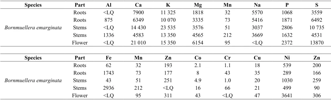
26
Table 1. Macro and trace element concentrations in roots, stems and flowers of Bornmuellera emarginata (values in µg g-1 dry weight) with ICP-AES.
<LQ is lower than the limit for quantification.
Species Part Al Ca K Mg Mn Na P S Bornmuellera emarginata Roots <LQ 7900 11 325 1818 32 5570 1068 3559 Roots 875 6349 10 070 3335 73 5416 1871 6492 Stems <LQ 14 430 23 535 3576 51 3037 2806 10 735 Stems 1336 4583 13 350 4565 212 3669 1632 4531 Flower <LQ 21 010 15 350 6154 95 <LQ 2372 13870 Species Part Fe Mn Zn Co Cr Cu Ni Zn Bornmuellera emarginata Roots 62 32 193 2.1 1.1 18 539 200 Roots 1743 73 177 8 43 35 289 166 Stems 43 51 251 4.9 1.0 20 1030 259 Stems 2936 212 <LQ 16 66 21 499 90 Flower <LQ 95 311 43 <LQ 47 3641 306
27
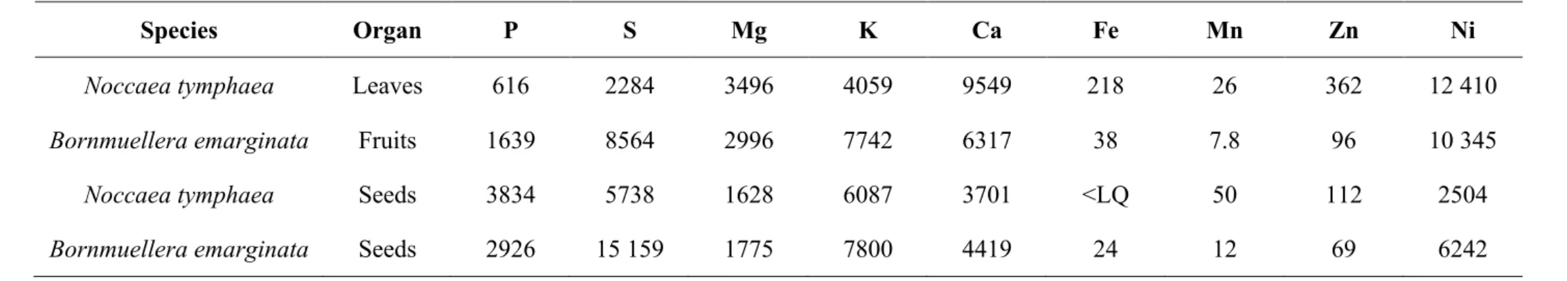
Table 2. Macro and trace element concentrations in leaves, fruits, and seeds of the actual Noccaea tymphaea and Bornmuellera emarginata samples
used for XFM elemental mapping (values in µg g-1 dry weight) with ICP-AES. <LQ is lower than the limit for quantification.
Species Organ P S Mg K Ca Fe Mn Zn Ni
Noccaea tymphaea Leaves 616 2284 3496 4059 9549 218 26 362 12 410
Bornmuellera emarginata Fruits 1639 8564 2996 7742 6317 38 7.8 96 10 345
Noccaea tymphaea Seeds 3834 5738 1628 6087 3701 <LQ 50 112 2504