ÉTUDE DE MATÉRIAUX HYBRIDES À BASE DE
NANOPARTICULES À CONVERSION ASCENDENTE ET
POLYMÈRES: DE NANOVECTEURS À FILMS MINCES
par
Jun Xiang
Thèse présentée au Département de chimie en vue de l’obtention du grade de docteur ès sciences (Ph.D.)
FACULTÉDESSCIENCES UNIVERSITÉDESHERBROOKE
STUDY
OF
UPCONVERSION
NANOPARTICLE/POLYMER
HYBRID
MATERIALS:
FROM
NANOVECTORS
TO
THIN
FILMS
by
Jun Xiang
A Thesis
Presented to the Département of Chimie
in Partial Fulfillment of the Requirements for the Degree of Doctor of Philosophy (Ph.D.)
FACULTÉDESSCIENCES UNIVERSITÉDESHERBROOKE
Le 30 Juin 2018
le jury a accepté la thèse de Monsieur Jun Xiang dans sa version finale.
Membres du jury
Professeur Yue Zhao Directeur de recherche Département de chimie
Professeur Fiorenzo Vetrone Évaluateur externe
INRS-EMT Université du Québec
Professeur Pierre D. Harvey Évaluateur interne Département de chimie
Professeur Serge Lacelle Évaluateur interne Département de chimie
Professeur Gessie Brisard Président-rapporteur Département de chimie
SOMMAIRE
La recherche réalisée dans cette thèse porte sur le design, la synthèse et l’investigation de trois nouveaux types de matériaux hybrides à base des polymères et des nanoparticules à conversion ascendante (NPCA). Ces matériaux sont sensibles à la lumière proche infrarouge (PIR) en raison de la NPCA qui absorbe la lumière ayant une longueur d’onde de 980 nm et émette la lumière dans le visible et l’ultraviolet (UV). Pour deux systèmes étudiés, leur application potentielle comme nanovecteur de médicament dont la libération est déclenchée par un rayonnement PIR est démontrée. Quant à l’étude de l’autre système sous forme de film mince, elle permet de démontrer que la méthode d’assemblage couche-par-couche est efficace pour organiser les NPCA dans un film conjointement avec des nanoparticules d’or (NPAu). Les résultats obtenus valident les nouvelles stratégies ou méthodologies proposées, lesquelles sont générales et offrent de nouvelles perspectives dans le développement et l’exploitation pour applications des matériaux hybrides de NPCA/polymère.
Dans le premier projet (chapitre 1), l’auto-assemblage d'un copolymère dibloc amphiphile sur la surface de NPCA est réalisée pour donner naissance à micelle dont le noyau contient une seule NPCA. À l’aide de polymérisation radicalaire par transfert d'atome initiée en surface de NPCA, le copolymère dibloc couvre la NPCA, avec poly(4,5-diméthoxy-2-nitrobenzyl méthacrylate) (PNB) comme bloc interne et hydrophobe et poly(méthoxy polyéthylène glycol monométhacrylate) (POEG) comme bloc extérieur et hydrophile. Sous une exposition au laser de 980 nm, la lumière UV émise par la NPCA est absorbée par PNB, ce qui entraîne le clivage des groupes o-nitrobenzyle et la formation de groupes acide carboxylique. L'hydrophilie accrue du copolymère résultant de la réaction photochimique déclenchée par la lumière PIR perturbe le nanovecteur et ainsi conduit à la libération de la doxorubicine (DOX).
Dans le second projet (chapitre 2), les NPCA sont complexées avec un polyélectrolyte UV-labile par interaction électrostatique entre charges de signes opposés, ce qui permet le
couvrement de chaque NPCA par une couche polymère. Un médicament modèle chargé, la fluorescéine (FLU), est encapsulé dans la couche de polyélectrolyte via un processus de co-assemblage. De même, sous l’irradiation laser de 980 nm, la lumière UV émise par la NPCA est absorbée par des groupes latéraux photolytiques du polyélectrolyte, ce qui entraîne le clivage des groupes o-nitrobenzyle et la formation de groupes acide carboxylique. Par conséquent, une partie des charges positives portées par le polyélectrolyte est convertie en espèces chargées négativement, et cette inversion perturbe l'équilibre entre les composants chargés et conduit à la libération de molécules de FLU. Dans le dernier projet (chapitre 3), la méthode couche-par-couche est utilisée pour assembler les NPCA dans un film mince de manière contrôlée, en ajustant notamment l’ordre de dépôt de quatre bicouches NPCA/polyélectrolyte et quatre bicouches NPAu/polyélectrolyte. Les spectres d'absorption et les spectres d'émission à conversion ascendante de ces films minces hybrides plasmoniques et sensibles au PIR révèlent que l'organisation spatiale déterminée par les séquences de dépôt influence l'interaction entre ces deux types de nanoparticules et ainsi leurs propriétés optiques.
Mots-clés: nanoparticules à conversion ascendante, polymères UV-sensibles, nanovecteurs, matériaux hybrides, auto-assemblage de polymères, assemblage couche-par-couche
ABSTRACT
The research carried out in this thesis involves the design, synthesis, and investigation of three new types of hybrid materials based on polymers and upconversion nanoparticles (UCNP). These materials are sensitive to near-infrared light (NIR) because UCNP can absorb light with a wavelength of 980 nm then emit light in the visible and ultraviolet (UV) light region. For the first two systems studied, their potential application as a drug nanovector whose release is triggered by NIR radiation is demonstrated. As for the other system in the form of a thin film, it demonstrates that the layer-by-layer assembly is an effective method for organizing UCNP in a film together with gold nanoparticles (AuNP). The obtained results validate the proposed new strategies or methodologies, which are general and offer new perspectives in the development and exploration of UCNP/polymer hybrid materials applications.
In the first project (Chapter 1), the self-assembly of an amphiphilic diblock copolymer on the surface of UCNP is carried out to form a micelle with single UCNP encapsulation inside. Using surface-initiated atom transfer radical polymerization, UCNP is covered by the diblock copolymer with poly(4,5-dimethoxy-2-nitrobenzyl methacrylate) (PNB) as the inner and hydrophobic block and poly(methoxy polyethylene glycol monomethacrylate) (POEG) as the outer and hydrophilic block. Under 980 nm laser exposure, the UV light emitted by the UCNP is absorbed by PNB, resulting in the cleavage of o-nitrobenzyl groups and formation of carboxylic acid groups. The increased hydrophilicity of the copolymer resulting from the photochemical reaction triggered by the NIR light disturbs the nanovector and thus leads to the release of doxorubicin (DOX).
In the second project (Chapter 2), UCNP are complexed with a UV-labile polyelectrolyte by electrostatic interaction between charges of opposite sign, which allows the covering of each UCNP by a polymer layer. A charged model drug, fluorescein (FLU), is encapsulated in the polyelectrolyte layer via a co-assembly process. Also, under the 980 nm laser irradiation, the UV light emitted by the UCNP is absorbed by photolytic side groups of the
polyelectrolyte, resulting in cleavage of o-nitrobenzyl groups and formation of carboxylic acid groups. As a result, a portion of the positive charges carried by the polyelectrolyte is converted to negative charges, and this inversion disrupts the balance between the charged components and leads to the release of FLU molecules.
In the last project (Chapter 3), the layer-by-layer method is used to assemble UCNP into a thin film in a controlled manner, in particular by adjusting the deposition order of four UCNP/polyelectrolyte bilayers and four AuNP/polyelectrolyte bilayers. The extinction spectra and the upconversion emission spectra of these plasmonic and NIR-sensitive hybrid thin films reveal that the spatial organization determined by the deposition sequences influences the interaction between these two types of nanoparticles and thus their optical properties.
Keywords: upconversion nanoparticles, UV-responsive polymers, nanovectors, hybrid materials, polymer self-assembly, layer-by-layer assembly
ACKNOWLEDGEMENT
First and foremost, I would like to express my most sincere gratitude to my supervisor Prof. Yue Zhao for his guidance, patience, encouragement, and support during the whole period of my Ph.D. studies at Université de Sherbrooke. I appreciate him to give me the valuable opportunity to do research that I am really interested in, and always support me and believe in me whenever I meet problems. I really learned a lot from Prof. Zhao over the past years. I believe his wide academic experience and rigorous scientific attitude will be my lifetime wealth. Also, I thank him for giving me the financial support which allows me to finish my Ph.D. program.
I would like to thank two members of my doctoral committee, who are also in the jury of my thesis, Prof. Gessie Brisard and Prof. Serge Lacelle, as well as all the professors and staff working in the Department of Chemistry, Université de Sherbrooke for their kind help. I would like to thank Prof. Fiorenzo Vetrone (Université du Québec) and Prof. Pierre D. Harvey for serving in the jury of my thesis. I also would like to thank Mr. Paul-Ludovic Karsenti for his help in taking the upconversion luminescence spectra.
I acknowledge the following people, my colleagues and dearest friends, who made my four years’ stay in Sherbrooke full of joys and memories: Mrs. Xia Tong, Prof. Dongchen He, Dr. Li Wang, Dr. Feng Shi, Dr. Qiang Yan, Dr. Olivier Boissière, Dr. Hu Zhang, Dr. Bing Yu, Dr. Weizheng Fan, Dr. Guo Li, Dr. Hongji Zhang, Dr. Shangyi Fu, Dr. Shengwei Guo, Dr. Xin Zhao, Dr. Rong Yang, Dr. Juan Xuan, Dr. Dehui Han, Dr. Xili Lu, Dr. Hui Xiao, Dr. Xiancong Lu, Dr. Xiaorong Wang, Dr. Di Gao, Dr. Lei Hu, Dr. Aurélie Lespes, Dr. Hojjat Seyedjamali, Mr. Damien Habault, Mr. Feijie Ge, Mr. Farhad Farnia, Mr. Liangliang Dong, Mrs. Amélie Augé, Mr. Ricardo Da Silva Lemos, Mrs. Laura Mourot, Mr. Zhichao Jiang, Mrs. Yaoyu Xiao, and Mr. Lu Yin.
I would like to give my thanks and respect to the Ministère de I'Éducation, du Loisir et du Sport du Québec and Chinese Scholarship Council (CSC) for awarding me a scholarship to support my living and study in Canada.
I also would like to thank all the members of the Service d’éducation du Consulat Général de la République Populaire de Chine à Montréal, especially Ms. Suli Yan, for all their kind helps.
I express my sincere thanks to my family for their exceptional care and encouragement during my study in Canada, especially my parents, my parents-in-law, my wife Yutong Xie, my little son Vincent Xiang and my younger brother Xingxing Xiang. Without your encouragement and accompany, none of this would have been possible.
Finally, I would like to acknowledge the following organizations for their financial support: Natural Sciences and Engineering Research Council of Canada (NSERC), Le Fonds de recherche du Québec: Nature et technologies (FQRNT), Centre for Self-Assembled Chemical Structures (CSACS) and La Fondation de Université de Sherbrooke.c
TABLE OF CONTENT
SOMMAIRE ... IV ABSTRACT ... VI ACKNOWLEDGEMENT ... VIII TABLE OF CONTENT ... X LIST OF FIGURES ... XIII LIST OF TABLES ... XXIV
INTRODUCTION ... 1
I.1 A Brief Introduction to UCNP ... 2
I.2 From Hydrophobic to Hydrophilic: Coating of UCNP with Polymer Materials ... 6
I.2.1 Synthesis of Hydrophilic Polymer-capped UCNP ... 7
I.2.2 Post-modification of UCNP with Hydrophilic Polymers ... 8
I.3 Nanovectors based on UCNP/Polymers for NIR Light-Triggered Drug Release ... 13
I.3.1 Covalent Conjugation of Payloads with UCNP/Polymer Nanovectors ... 18
I.3.2 Non-covalent Conjugation of Payloads with UCNP/Polymers Nanovectors ... 20
I.4 Preparation of Thin Films with UCNP by LbL Assembly ... 28
I.5 Objectives of the Thesis ... 31
CHAPTER 1 NEAR-INFRARED LIGHT-TRIGGERED DRUG RELEASE FROM UV-RESPONSIVE DIBLOCK COPOLYMER-COATED UPCONVERSION NANOPARTICLES WITH HIGH MONODISPERSITY ... 34
1.1 About the Project ... 34
1.2 Contributions ... 35
1.3 Paper Published in Journal of Materials Chemistry B 2018, 6, 3531. ... 36
1.3.1 Abstract ... 37
1.3.2 Introduction ... 38
1.3.3 Experimental ... 41
1.3.5 Conclusions ... 58
1.3.6 Supporting Information... 64
1.4 Summary of the Project ... 71
CHAPTER 2 NANOCOMPLEXES OF UV-SENSITIVE POLYELECTROLYTE AND UPCONVERSION NANOPARTICLES FOR NEAR-INFRARED LIGHT-TRIGGERED PAYLOAD RELEASE ... 72
2.1 About the Project ... 72
2.2 Contributions ... 73
2.3 Paper Published in ACS Applied Materials & Interfaces 2018, 10, 20790. ... 74
2.3.1 Abstract ... 75
2.3.2 Introduction ... 76
2.3.3 Experimental Sections ... 79
2.3.4 Results and Discussion ... 86
2.3.5 Conclusions ... 98
2.3.6 Supporting Information... 106
2.4 Summary of the Project ... 126
CHAPTER 3 SPATIAL ORGANIZATION AND OPTICAL PROPERTIES OF LAYER-BY-LAYER ASSEMBLED UPCONVERSION AND GOLD NANOPARTICLES IN THIN FILMS ... 127
3.1 About the Project ... 127
3.2 Contributions ... 128
3.3 Paper Published in Journal of Materials Chemistry C 2016, 4, 9343. ... 129
3.3.1 Abstract ... 130
3.3.2 Introduction ... 131
3.3.3 Experimental ... 133
3.3.4 Results and Discussion ... 137
3.3.5 Conclusions ... 145
3.3.6 Supporting Information... 150
CHAPTER 4. GENERAL DISCUSSION AND PERSPECTIVE ... 157
4.1 General Discussion ... 157
4.2 Future Studies ... 161
CONCLUSIONS ... 167
LIST OF FIGURES
INTRODUCTION
Figure 1. (a) Excitation-state absorption (ESA). (b) Energy transfer upconversion (ETU). .. 3 Figure 2. (a) A schematic diagram of upconversion processes in a Yb3+-Tm3+ codoped
system upon excitation with a 980 nm laser. Yb3+ and Tm3+ serve as the sensitizer and
activator, respectively. (b) UCL spectrum of NaYF4:20%Yb,0.5%Tm nanocrystals. The
term symbol 2S+1LJ used to label the f states is according to the Russel-Saunders notation and refers to the total spin quantum number (S), the total orbital quantum number (L) and the total angular momentum quantum number (J), respectively. ... 4 Figure 3. Applications of RE doped UCNP. Adapted with permission from [2]. ... 5 Figure 4. Synthesis of water-soluble UCNP via an unconventional method. Reproduced with
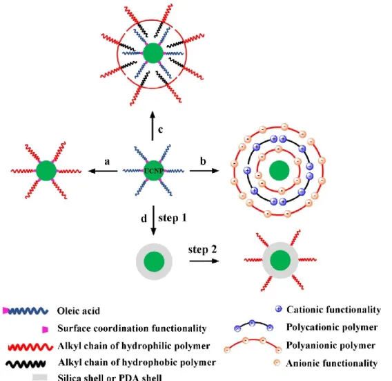
permission from [15]. ... 7 Figure 5. Schematic illustration of the routes for post-modification of oleic acid-coated
UCNP with hydrophilic polymers: (a) ligand exchange, (b) LbL assembly, (c) ligand attraction and (d) silica-shell or mussel inspired PDA coating (two-step method). ... 10 Figure 6. (a) The pharmacokinetic profile of conventional drug formulation. Adapted from
[103]. (b) Serval doses are needed to maintain the concentration in the therapeutic window for a longer duration. Adapted from [104]. ... 13 Figure 7. Tissue penetration depth of light with different wavelengths. UV light can only
penetrate hundreds micrometer. By contrast, NIR light can deeply penetrate into tissues (several centimeters). Reproduced from [118] with permission... 14 Figure 8. Four types of UCNP-assisted photoreactions. Reproduced with permission from
[118]. ... 16 Figure 9. Photoisomerization mechanism of o-nitrobenzyl alcohol derivatives into an
o-nitrosobenzaldehyde, releasing a carboxylic acid. Adapted with permission from
Figure 10. (a) Surface modifications of SiO2-coated UCNP for incorporating hydrophilic
polymer chains with drug molecules. (b) The initiation of the release of drug molecules under 980 nm light irradiation. Reproduced from [135] with permission. ... 19 Figure 11. (a) Chemical structure of the designed conjugated polyelectrolyte. (b) Schematic
illustration of 980 nm light regulated initiation of the photosensitizer to generate ROS for PDT and DOX release for chemotherapy. Reproduced from Ref. [137] with permission from The Royal Society of Chemistry. ... 20 Figure 12. Schematic illustration of drug release mechanisms from nanovectors based on
UCNP-assisted photoreactions of photoresponsive polymers: (a) shift of hydrophobic-hydrophilic balance, (b) cleavage of junctions, (c) main chain degradation, (d) gel-sol transition and (e) variation of surface charge. ... 21 Figure 13. (a) Schematic illustration of using 980 nm light to trigger dissociation of polymer
micelles. (b) Photolysis reaction of the polymer. (c) UV-vis spectra of resulted micelles and UCL spectra of UCNP and micelles. TEM images of micelles before (d) and after (e) NIR light excitation. Reprinted with permission from [138]. Copyright (2018) American Chemical Society. ... 23 Figure 14. (a) The degradation of light-sensitive nanoparticles after absorbing UC
luminescence and simultaneously initiating the release of guest molecules. (b) UV-vis spectra of light-sensitive nanoparticles and UCL spectra of neat UCNP. (c) Photodegradation mechanism of photoresponsive polymers. Reproduced with permission from [143]. ... 24 Figure 15. (a) Synthesis route of designed polyelectrolyte and its photolytic process. (b)
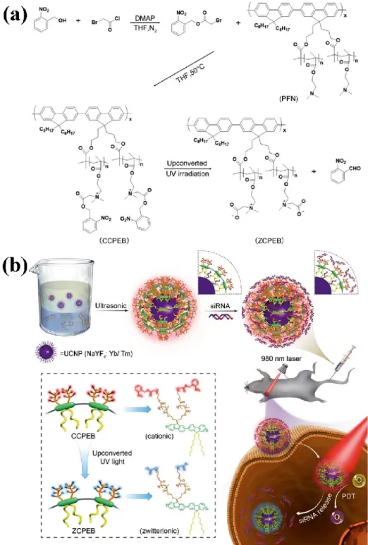
Schematic illumination of the nanotherapeutic system for combing PDT with siRNA therapy. Reproduced with permission from [144]. ... 26 Figure 16. (a) Chemical structure of the photocleavable cross-linker (PhL). (b)
Oleate-capped UCNP. (c) UCNP@SiO2. (d) (3-glycidyloxypropyl) trimethoxysilane
and chitosan (CH) modified UCNP. (e) Encapsulating FITC-BSA inside the CH shell and formation of hydrogel-coated UCNP. (f) NIR light-triggered photodegradation of hydrogel and thus drug release. Reproduced with permission from [145]. ... 27
Figure 17. (a) Synthetic route: (I) TEOS and CTAB for coating mesoporous silica layer, (II) the introduction of long alkyl chains, (III) DOX loading and self-assembly. (b) Schematic illustration of multifunctional nanocomposites and NIR light-initiated drug release. Reproduced from Ref. [146] with permission from The Royal Society of Chemistry. ... 28 Figure 18. (a) Schematic illustration of the NIR light-triggered hydrophobic-to-hydrophilic
transition of polymer chains. (b) An illustration of NIR-controlled combination chemotherapy, PDT and imaging. Reproduced with permission from [147]. ... 29 Figure 19. (a) Schematic illustration of immersive LbL assembly on a planar substrate. (b)
The charge characteristics of the films after deposition step. Reproduced with permission from Ref [160]. ... 30
CHAPTER 1
Figure 1. (a) Schematic illustration of UCNP coated with self-assembled micelle of an amphiphilic diblock copolymer and use of NIR light excitation of UCNP to induce the micelle disruption and the release of encapsulated drug molecules. (b) Chemical structure of the diblock copolymer and NIR light-triggered photocleavage of nitrobenzyl groups. ... 39 Figure 2. TEM images for UCNP (a), UCNP-Br (b), photoresponsive polymer-grafted
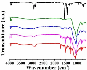
UCNP (UCNP@PNB) (c), and diblock copolymer grafted UCNP (UCNP@PNB-b-POEG) (d). Scale bar is 40 (white) or 100 nm (black). ... 46 Figure 3. FTIR spectra of (a) UCNP, (b) UCNP-NH2, (c) UCNP-Br, (d) UCNP@PNB, and (e)
UCNP@PNB-b-POEG. ... 48 Figure 4. Absorption spectrum of UCNP@PNB-b-POEG aqueous solution (black), and
upconversion emission spectra under 980 nm excitation for neat UCNP in hexane (blue), UCNP@PNB in DMF (green), and UCNP@PNB-b-POEG in H2O (red). Inset is a
photograph of an aqueous solution of UCNP@PNB-b-POEG... 48 Figure 5. (a) Effect of temperature on the hydrodynamic diameter (DH, solid cycle) and
polydispersity index (PDI, hollow cycle) of UCNP@PNB-b-POEG in H2O (1 mg/mL).
DOX-UCNP@PNB-b-POEG (red) in H2O at 25 oC. (c) Absorption spectra of DOX
(blue), UCNP@PNB-b-POEG (black, 0.1 mg/mL), and DOX-UCNP@PNB-b-POEG (red, 0.1 mg/mL). Inset photos are the aqueous solutions of nanoparticles before (left) and after (right) DOX encapsulation. ... 49 Figure 6. Absorption spectra of UCNP@PNB-b-POEG (0.05 mg/mL, 1 mL) recorded after
various times of 365 nm UV irradiation (1 mW/cm2) showing photocleavage reaction of
PNB in H2O. ... 51
Figure 7. (a) Schematic of the setup used to detect species diffusing from the nanocarrier solution through a dialysis membrane into aqueous solution filled in the cuvette induced by 980 nm laser irradiation (diffusing molecules are either photocleaved nitrosobenzaldehyde or released drug molecules). (b) Absorption spectral change over time for nitrosobenzaldehyde molecules cleaved by NIR light exposure of UCNP@PNB-b-POEG (4 W). (c) Plot of absorbance at 375 nm in (b) vs. time. The times at which the NIR laser is turned on or turned off are indicated. (d) UV-vis spectra, (e) DLS hydrodynamic diameter (DH) measurement, and (f) FTIR spectra of
UCNP@PNB-b-POEG before (black) and after (red) NIR light irradiation. ... 53 Figure 8. Spectral changes of DOX-UCNP@PNB-b-POEG (0.1 mg/mL, 2.5 mL) under UV
light (365 nm, 1 mW/cm2) irradiation: (a) absorption spectra, (b) emission spectra (λ ex =
480 nm), and (c) change in fluorescence intensity (at 585 nm) vs. irradiation time. (d) Using the setup in Fig. 7a, change in the normalized fluorescence intensity of DOX (at 585 nm) in the aqueous solution underneath the dialysis cup containing DOX-UCNP@PNB-b-POEG solution (0.1 mg/mL, 0.5 mL) vs. time, only 15 min of UV light irradiation being applied after 16 h as indicated. In all experiments, the solution is still without stirring. ... 56 Figure 9. (a) Absorption spectral change over time for the aqueous solution separated from
DOX-UCNP@PNB-b-POEG solution (3 mg/mL, 0.4 mL) through a dialysis membrane (see the setup in Fig. 7a) before and after 980 nm laser exposure (4 W). (b) Change in the absorbance at 497 nm over time with data from (a), the times at which the NIR laser being turned on or off are indicated. ... 57
Figure S1 Surface modification of UCNP through a bottom-up strategy: (1) silica layer coating, (2) immobilization of ATRP initiators, (3) growth of hydrophobic block that is a UV-responsive polymer (PNB), and (4) growth of hydrophilic block (POEG). The
UCNP and thin silica layers are depicted as red and black rods, respectively. ... 64
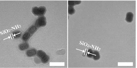
Figure S2. TEM images for UCNP-NH2. Scale bar is 60 nm. ... 64
Figure S3. TEM image of UCNP-Br. Scale bar is100 nm. ... 65
Figure S4. TEM images of UCNP@PNB. Scale bar is 60 nm. ... 65
Figure S5. TEM images of UCNP@PNB-b-POEG. Scale bar is 200 nm. ... 65
Figure S6. TGA analysis of UCNP-NH2, UCNP-Br, UCNP@PNB and UCNP@PNB-b-POEG. ... 66
Figure S7. SEC curve of PNB-b-POEG cleaved by HF etching of UCNP-b-POEG. ... 66
Figure S8. The emission spectra of the neat UCNP (black) in hexane and UCNP-Br (red) in DMF. ... 70
CHAPTER 2 Figure 1. (a) Schematic illustration of near-infrared (NIR) light-triggered polymer layer disruption and drug release from the nanovector of UCNP@silica@polyelectrolyte self-assembled through electrostatic interactions. (b) Polymer chemical structure, used UCNP (NaLuF4:18%Yb,0.5%Tm@NaYF4) and the NIR light-triggered photocleavage reaction of the photolabile polyelectrolyte. ... 77
Figure 2. TEM images and the size distributions as revealed by DLS for UCNP@silica (a and c) and UCNP@silica@PPE (b and d). The inset photos show the corresponding aqueous solution of UCNP@silica (in c) and UCNP@silica@PPE (in d). ... 87
Figure 3. (a) DH vs. molar ratio of PPE chains to UCNP@silica obtained from DLS measurements in H2O. The inset is an enlarged plot over the region of interest. (b) The effect of molar ratio of PPE chains to UCNP@silica on the zeta potential of the resulting UCNP@silica@PPE after complexation (pH 6.6). ... 88 Figure 4. (a) UV-vis absorption spectra of USP-1, USP-2 and USP-3. The three USP samples
have the same molar ratio of PPE chains to UCNP@silica (135:1) but differ in the mass ratio of FLU to UCNP@silica used for the preparation of FLU-loaded
UCNP@silica@PPE nanocomplexes (1:1 for USP-1, 2.2:1 for USP-2, and 4.5:1 for USP-3) (Table 1). (b) UV-vis absorption spectra of FLU-UCNP@silica@PPE (0.4 mg/mL, from USP-3 to USP-7) and free FLU. The samples of USP-4, USP-5, USP-6 and USP-7 have the same mass ratio of FLU to UCNP@silica used for the loading as USP-3 (4.5:1) but different molar ratios of PPE to UCNP@silica (0 for USP-4, 17:1 for USP-5, 34:1 for USP-6, and 67:1 for USP-7) (Table 1). The inset picture shows an aqueous solution of USP-3 (0.4 mg/mL). ... 89 Figure 5. (a) The absorption spectrum (black continuous line) of a mixture of UCNP@silica
and PPE as well as its UCL spectrum (red dotted line) under 980 nm laser irradiation. The emitted UV light (325-375 nm) by UCNP in the mixture displays a decrease in intensity as compared to the UCL spectrum of neat UCNP (blue dashed line). The inset photograph shows the mixture emitting visible light where the 980 nm laser beam passes through the solution. (b) The setup used to detect released molecules diffusing through a dialysis membrane into aqueous solution underneath (Tris buffer, 10 mM, pH 7.4) upon NIR light irradiation (~29 W/cm-2, 980 nm). ... 91
Figure 6. (a) Absorption spectral change over time for the cleaved molecules from UCNP@silica@PPE (5 mg/mL, 0.5 mL) triggered by a CW 980 nm laser (~29 W/cm-2)
using the setup in Figure 5b. (b) Plot of absorbance at 350 nm vs. time. “ON” labels indicate the beginning of the NIR light irradiation experiment in the following two hours, while “OFF” labels mean the switching off the NIR light laser in the following one hour. ... 92 Figure 7. (a) DH of UCNP@silica@PPE after three hours of NIR irradiation. (b) FT-IR
spectra of UCNP@silica@PPE before (black) and after (red) 980 nm illumination for three hours... 93 Figure 8. (a) Absorption spectral changes over time from the bottom solution of USP-6 (0.4
mL in dialysis cup; molar ratio of PPE chains to UCNP@silica is 34:1). (b) Plot of absorbance at 490 nm vs. time, “ON” labels indicate the beginning of the NIR light irradiation experiment in the following one hour, while “OFF” labels mean the
switching off the NIR light laser in the following thirty minutes. The control test is for
the solution without NIR light irradiation... 95
Figure 9. Plots of absorbance vs. time for three FLU-UCNP@silica@PPE samples differing only in the amount of polyelectrolyte (Table 1). For USP-3 (molar ratio of PPE chains to UCNP@silica is 135:1) and USP-7 (the ratio is 67:1), “ON” labels indicate the beginning of the NIR light irradiation experiment in the following two hours, while “OFF” labels mean the switching off the NIR light laser in the following one hour. For USP-6, the “ON” and “OFF” are the same as in Figure 8b. For all control tests (open symbols), the same solutions were not subjected to NIR irradiation... 96
Scheme S1. The synthetic route for the UV-labile polyelectrolyte PPE. ... 106
Figure S1. 1H NMR spectrum of 5-hydroxy-2-nitrobenzyl alcohol (2). ... 106
Figure S2. 13C NMR spectrum of 5-hydroxy-2-nitrobenzyl alcohol (2). ... 107
Figure S3. 1H NMR spectrum of (5-(3-(dimethylamino) propoxy)-2-nitrophenyl) methanol (3). ... 107
Figure S4. 13C NMR spectrum of (5-(3-(dimethylamino) propoxy)-2-nitrophenyl) methanol (3). ... 108
Figure S5. 1H NMR spectrum of 5-(3-(dimethylamino)propoxy)-2-nitrobenzyl methacrylate (4). ... 108
Figure S6. 13C NMR spectra of 5-(3-(dimethylamino)propoxy)-2-nitrobenzyl methacrylate (4). ... 109
Figure S7. 1H NMR spectra and main peak labeling for: (a) 5 and (b) PPE. The ratio of integrated areas of methyl hydrogen (CH3-O, 3.37 ppm) and ethyl hydrogen (Ph-CH2-O, 5.34 ppm) peaks in Fig. S7a indicates that the NB units account for ~44% in number. The ratio of integrated areas of nitro aromatic hydrogen peak (NO2-Ph-H, 8.16 ppm) and overlapped peaks of methyl hydrogen (N-CH3, 3.27 ppm) and methyl hydrogen (CH3-O, 3.37 ppm) in Fig. S7b indicates that the degree of quaternization is ~93%. 110 Figure S8. TEM image for UCNP (NaLuF4:18%Yb,0.5%Tm@NaYF4). ... 111
Figure S9. FTIR spectra of UCNP@silica before (a) and after (b) the removal of CTAB. In comparison with the FTIR spectrum of nanoparticles with CTAB, the disappearance of
the characteristic bands at approximately 2926 and 2854 cm-1 assigned to the
asymmetric (νas) and symmetric (νs) stretching of C―H in CTAB molecules after
washing indicates the successful elimination of surfactant. ... 111 Figure S10. Size histogram for UCNP@silica from TEM observations (N>100). ... 112 Figure S11. TEM images for UCNP@silica@PPE (molar ratio of PPE chains to
UCNP@silica is 135:1). ... 112 Figure S12. Size histogram for UCNP@silica@PPE from TEM observations (N>80). .... 113 Figure S13. Hydrodynamic diameter (DH) and polydispersity index (PDI) of UCNP@silica
in H2O (~1.1 mg/mL). ... 113
Figure S14. TGA analysis of UCNP@silica, PPE, and UCNP@silica@PPE (molar ratio of PPE chains to UCNP@silica is 122:1, which is obtained from the feed ratio used for preparing this nanocomplex). ... 116 Figure S15. The absorption spectral change of PPE (0.1 mg/mL in H2O) over 365 nm
exposure time (80 mW/cm2). ... 117
Figure S16. Zeta potential of the UCNP@silica@PPE nanocomplex (molar ratio of PPE chains to UCNP@silica is 135:1) changes over NIR irradiation time. Due to the small beam of the 980 nm laser, it’s hard to monitor the zeta potential for the solution in the dialysis cup during the illumination. Instead, 10 μL of the nanocomplex solution (1 mg/mL) in an Eppendorf tube was irradiated by the 980 nm laser and then diluted for the measurements... 118 Figure S17. Normalized UV-vis spectra of bottom solutions for three
FLU-UCNP@silica@PPE samples after 360 min. ... 118 Figure S18. TEM image for Er-UCNP (NaYF4:18%Yb,2%Er@NaYF4). Er-UCNP have an
average dimension of 46 (L) × 42 (W) nm. ... 120 Figure S19. (a) TEM image, (b) size distribution from DLS measurement and (c) zeta
potential for Er-UCNP@silica (1 mg/mL in H2O, pH 6.6). The average thickness of
silica layer is 9 nm. ... 121 Figure S20. (a) TEM image and (b) size distribution from DLS measurement (1 mg/mL, H2O)
Figure S21. (a) UCL spectra of Er-UCNP and UCNP. (b) The absorption (continuous line) and UCL spectrum (dotted line) of the mixture of Er-UCNP@silica and PPE. Also shown is the UCL spectrum of neat Er-UCNP in hexane. Note: the composition of the mixture is PPE (2 mL, 0.1 mg/mL) and Er-UCNP@silica (0.2 mg). The inset photograph shows that the aqueous solution of Er-UCNP@silica (1 mg/mL) emits visible light (mainly green light) where the 980 nm laser beam passes through the solution... 122 Figure S22 UV-vis absorption spectra of FLU loaded Er-UCNP@silica@PPE as a function
of molar ratio of PPE chains to Er-UCNP@silica while keeping the mass ratio of FLU to Er-UCNP@silica at 4.5:1. ... 124 Figure S23. (a) Absorption spectral changes over time from the bottom solution of
FLU-Er-UCNP@silica@PPE. (b) Plot of absorbance at 490 nm vs. time, “ON” labels indicate the beginning of 980 nm laser exposure in the following one hour, while “OFF” labels mean the switching off the NIR laser in the following thirty minutes. The control test is for the solution without NIR light irradiation. For comparison, the data obtained with FLU-UCNP@silica@PPE (USP-6) in Figure 8b are also shown. ... 125
CHAPTER 3
Figure 1. Schematic illustration of using the LBL assembly method to fabricate multilayer nanocomposite film on quartz/PDDA substrate: (a) multilayer film of UCNP (NaYF4:Yb,Tm@NaYF4); and (b) multilayer film comprising UCNP and AuNP
spatially organized in different ways. Note: the deposition is on both sides of the substrate but only one side is shown in the sketch; n is the number of bilayers (nanoparticles with polyelectrolyte form one bilayer); from left to right corresponds to the 1st bilayer on the substrate to the nth bilayer. ... 132
Figure 2. (a) TEM image of NaYF4:Yb,Tm@NaYF4. (b) Upconversion luminescence spectra
of (UCNP-PAA/PAH)n (n is the number of bilayers, n = 0-10) formed on quartz/PDDA
substrate upon 980 nm excitation at 2.55 kW cm-2. (c) Plots of intensity of the major
emission peaks in (b) vs. number of bilayers. (d) SEM image of (UCNP-PAA/PAH)11.
Figure 3. (a) TEM image of AuNP. (b) Extinction spectra of single AuNP/PAH ((Au|)1)
bilayer built up with different dip-coating time and AuNP solution. (c, d) SEM images of one (AuNP/PAH)1 bilayer at different dip-coating times: (c) 15 min; (d) 1 h. ... 140
Figure 4. Extinction spectra (a, c, e) and emission spectra (980 nm excitation, CW laser at 11.77 kW cm-2) (b, d, f) for LBL assembled films comprising UCNP-PAA/PAH (U|)
and AuNP/PAH (Au|) bilayers built up with different deposition sequences giving rise to different spatial organizations of the two types of nanoparticles: (U|U|U|U|Au|Au|Au|Au|) (a and b); (U|U|Au|Au|U|U|Au|Au|) (e and f); and (U|Au|U|Au|U|Au|U|Au|) (e and f). ... 141 Figure 5. SEM images of three thin films: (a) (U|U|U|U|Au|Au|Au|Au|); (b)
(U|U|Au|Au|U|U|Au|Au|); and (c) (U|Au|U|Au|U|Au|U|Au|). Red ring in (b) indicates one UCNP. ... 144 Figure S1. TEM image of UCNP functionalized with poly(acrylic acid) (PAA). ... 151 Figure S2. FT-IR spectra of UCNP-OA (oleic acid) and UCNP-PAA. ... 151 Figure S3. UV-vis-NIR extinction spectrum of (UCNP-PAA/PAH)11. ... 152
Figure S4. UCL emission spectra of (UCNP-PAA/PAH)10 or 11 LBL films (ex=980 nm, 2.55
kW cm-2): (UCNP-PAA/PAH)
10 (blue); (UCNP-PAA/PAH)10 kept in the air for 16 h
(red); (UCNP-PAA/PAH)11 (black). ... 152
Figure S5. UCL emission spectra of (UCNP-PAA/PAH)11 film (ex=980 nm, 2.55 kW cm-2):
0 h (black); after keeping in the air for 11 d (red). ... 153 Figure S6. UCL emission spectra for a 4-bilayer UCNP and three UCNP/AuNP multilayers
containing the same number of UCNP bilayers and prepared using different LBL deposition sequences. The spectra were recorded under the same 980 nm excitation intensity of 2.55 kW/cm2. ... 153
Figure S7. Linear fit of the plot of absorbance at 513 nm vs. number of AuNP bilayers. . 154 Figure S8. The power distribution of 980 nm laser across the calibrated beam. The plot was
fitted by Gauss curve (red line)... 155
Figure 20. Schematic illustration of drug release systems based on UCNP and photoresponsive polymers with photo-tunable LCST or UCST. (a) The basic design principle is the synthesis of photoresponsive polymers (random or diblock copolymers) with temperature sensitivity. Release of drugs from several types of systems: (b) polymer micelles, (c) vesicles and (d) thin films. ... 165 Figure 21. (a) UV or NIR light-triggered photolysis reaction of poly(NIPAM-co-ONB),
resulting in the shift of LCST from below 37 oC to above 37 oC. Plots of solution
transmittance vs. temperature for poly(NIPAM-co-ONB) (b and c) and poly(NIPAM-co-ONB) grafted UCNP. (d) (5 mg/mL) before (black) and after (red) UV irradiation in PBS saline solution (10 mM, pH 7.4). ... 166
LIST OF TABLES
CHAPTER 1
Table S1. Basic information of nanoparticles. ... 67 Table S2. Useful information from TGA analysis. ... 69
CHAPTER 2
Table 1. Preparation conditions for samples of FLU-UCNP@silica@PPE nanocomplexes84 Table S1. Analysis of the USP-3 sample. ... 114 Table S2. Mass of UCNP@silica and UCNP@silica@PPE at room temperature. ... 116 Table S3. Analysis of the UCNP@silica@PPE nanocomplex in Figure S14. ... 117 Table S4. Analysis of the molar ratio of PPE chains to Er-UCNP@silica in nanocomplexes of
Er-UCNP@silica@PPE. ... 123
CHAPTER 3
Table S1. Size and Zeta Potential of Nanoparticles ... 150 Table S2. Linear Regression (y = a + b·x) of the Plots of Luminescence Intensity (y) vs.
Number of Bilayer (x) for Different Emission Peaks. ... 150 Table S 3. The Relationship between the Output Power of 980 nm Laser and its Real Power
INTRODUCTION
In the past decade, researches on rare-earth (RE) doped upconversion nanoparticles (UCNP) for constructing novel functional materials have witnessed an explosive growth (1-3). Particularly, these inorganic nanoparticles (NPs) have promising applications in the fields of biomedicine (4-6), energy conversion (7-9), and security patterns (10-12). As one kind of advanced material, UCNP provide innovative solutions or ideas for some major problems in numerous areas. For instance, in the bioimaging field, UCNP can be utilized in multiplexing and multimodal imaging for deep bio-tissues owing to their excitation wavelength located in the biological transparency window, high stability, low cytotoxicity and non-photobleaching properties (13, 14). Another example is that integration of UCNP into organolead halide perovskite solar cells can enhance the power conversion efficiency by extending the spectral absorption in the near-infrared (NIR) light region (15). These are attributed to the distinctive non-linear optical property of UCNP, which has the capability of converting multiple low-energy photons (NIR light) into high-energy photons (ultraviolet (UV) or visible light). Hence, these fascinating nanocrystals can act as antennas to bridge NIR and UV/visible light, making NIR light-initiated photoreactions and expansion of the spectral absorption range possible. In section 1.1, a brief introduction to UCNP is presented.
Integration of UCNP with organic polymer materials for applications in, for instance, drug delivery, anti-counterfeiting materialsand solar cells, have attracted much research interest around the world (16-21). The reasons for their combination are multiple, such as endowing UCNP with water dispersity, improving compatibility with polymer matrix, loading guest molecules and offering stimuli-responsive properties (pH, temperature, light, and so on). In the first two works of this thesis, we designed two nanovectors capable of NIR light-triggered payload release, which involves the surface modification of UCNP with polymer materials. Thus, the surface coating strategies to endow UCNP with water dispersity by hydrophilic polymers are summarized in section 1.2. In section 1.3, an
overview of recent progress in the development of nanovectors based on UCNP/polymers for the application of NIR light-triggered cargo release is presented.
In the preparation of UCNP/polymers hybrid materials, knowledge on how to organize these two types of materials for potential uses is crucial. Fundamentally, it is desirable to find a generalized and straightforward approach to organize UCNP in the polymer matrix. To this end, the third work accomplished in this thesis is about using the layer-by-layer (LbL) assembly technique to organize UCNP and gold nanoparticles (AuNPs, also extensively explored with polymers) through polyelectrolytes. Therefore, a brief introduction to the preparation of UCNP/polyelectrolytes thin films by LbL assembly is presented in section 1.4. The introduction will be ended with a statement of the objective of the thesis work.
I.1 A Brief Introduction to UCNP
RE doped UCNP (<100 nm) are inorganic nanocrystals with trivalent lanthanide ions as dopants. Their outstanding property is the conversion of low-energy photons (NIR light) into high-energy photons (visible or UV light) through sequential absorption of multiple photons, which is attributed to the ladder-like electronic transition levels of RE metallic ions and long luminescence lifetime (up to millisecond). Although lanthanide-doped UC bulk materials have been known for several decades, the first investigation on UCNP (5-20 nm) with good photoluminescence property was reported in 2000 (22). The reason is that the surface quenching effect caused by the high surface-to-volume ratio of NPs leads to the reduction of emission centers (23). After an extended period of research, it is now known that the type of host matrix, the concentration of dopant and the phase construction should be carefully adjusted to obtain UCNP with high quantum yield (QY) of upconversion luminescence (UCL). It should be mentioned that the most frequently used host matrix is NaREF4, which has relatively low phonon energy, adequate thermal and environmental
Three primary mechanisms explain the UC process of UCNP: excitation-state absorption (ESA), energy transfer upconversion (ETU) and photon avalanche (PA). Noteworthy, PA is not often observed in UCNP because the observation of this phenomenon needs exceptionally high pumping intensity (25). Also, PA mechanism is restricted within a few typical ions, like Pr3+, limiting their further exploration. Consequently, most UC processes
involve ESA and ETU. In ESA, only one RE ion participates in the UC process. When one electron stays in a metastable excited state as shown in Figure 1a, it can be further populated to a higher excitation state after absorbing another photon of the incident light. The UCL emissions can be detected when the pumped electrons at higher energy levels return to their ground states. However, this process has a relatively low efficiency due to the low absorption cross-sections of lanthanide trivalent ions. By contrast, ETU process involves two different lanthanide ions (sensitizer and activator). The sensitizer is responsible for harvesting the light energy, and the activator takes charge of emitting high-energy photons. Figure 1b shows a typical ETU process. At the beginning, electrons of the sensitizer and activator are both populated in their metastable intermediate excitation states. Then the energy is transferred from the sensitizer to the adjacent activator through a non-radiative process, which allows the activator to populate to a higher energy state. UC emissions take place when the activator drops to the ground state in a radiative manner. By Figure 1. (a) Excitation-state absorption (ESA). (b) Energy transfer upconversion (ETU).
carefully modulating the concentration of these two ions, UC emissions from the as-synthesized UCNP can be readily observed by a NIR light laser excitation at a low power density through the ETU process.
As mentioned above, sensitizer and activator are often co-doped into the same host matrix to obtain high quality of UCNP. So far, the Yb3+/Tm3+ (sensitizer/activator) dual ions-doped
β-NaREY4 have been extensively investigated (26, 27). In Figure 2a, the population process
together with the UCL mechanism between Yb3+ and Tm3+ are depicted. The UCL takes
place when the populated electrons go back to the ground state by a radiative manner. We can see the emission bands from the UPL spectrum in Figure 2b. UCNP have narrow emissions in the 250-550 nm region, with UV light at 291, 341 and 361 nm, blue light at 451 and 475 under excitation with a continuous-wave (CW) 980 nm diode laser. These emission bands can be attributed to the electron transitions from 1I
6 → 3H6 (294 nm), 1I6 → 3F
4 (345 nm), 1D2 → 3H6 (368 nm), 1D2 → 3F4 (450 nm) and 1G4 → 3H6 (475 nm).
Figure 2. (a) A schematic diagram of upconversion processes in a Yb3+-Tm3+ codoped
system upon excitation with a 980 nm laser. Yb3+ and Tm3+ serve as the sensitizer and
activator, respectively. (b) UCL spectrum of NaYF4:20%Yb,0.5%Tm nanocrystals. The term
symbol 2S+1LJ used to label the f states is according to the Russel-Saunders notation and refers to the total spin quantum number (S), the total orbital quantum number (L) and the total angular momentum quantum number (J), respectively.
Due to the rapid growth of nanotechnology, a variety of methodologies have realized the preparation of UCNP with excellent photoluminescence property at low power density excitation in the past decade. Primarily, the solution-based synthetic routes (hydro-/solvothermal synthesis, thermolysis method, emulsion method, etc.) are powerful for the convenient and reproducible synthesis of high-quality UCNP (28). For example, their sizes can be precisely tuned below 10 nm without sacrificing their brightness or stability through the thermolysis method (29). These technologies significantly promote the exploration of the usage of UCNP in functional materials over the last few years. As shown in Figure 3, smart materials based on UCNP have large potential applications in diverse fields, such as bioimaging, anti-counterfeiting, displaying and so on (2). Owing to the large anti-stokes shift, narrow emission bands, non-photobleaching and chemical stability of UCNP, these chemical and optical properties make them act as excellent candidates for the biomedical uses, such as bioimaging, drug delivery, and photodynamic therapy (PDT). It is expected that UCNP will be transferred from the academic stage into real-world applications shortly.
I.2 From Hydrophobic to Hydrophilic: Coating of UCNP with Polymer Materials
In general, UCNP are synthesized by a thermolysis approach, which produces UCNP with high crystallinity, high UCL efficiency and low polydispersity. Unfortunately, the as-synthesized UCNP can only be dispersed in non-polar organic solvents (e.g., cyclohexane) due to their surface hydrophobic ligands, like oleic acid (OA) and oleylamine (OM). These organic ligands significantly restrict UCNP applications. So, it is necessary to modify their surface for preparing novel functional materials. For instance, decoration of UCNP can improve their dispersity and compatibility in composites (20, 30), which is favorable for fabricating materials with high transparency (31). Moreover, particularly in the biomaterials fields, adjusting the surface of UCNP from hydrophobic to hydrophilic is required because of the aqueous environment in biological tissues. Besides the modified UCNP should also be biocompatible, biodegradable and multi-functional (protein conjunction, drug loading and release) in some cases to meet various needs. Undoubtedly, surface coating of UCNP is the first step to explore their further usage. Several comprehensive reviews related to this topic have been published in the past few years (32-34).
So far, various kinds of materials, including inorganic (e.g., silica) and organic materials (small organic molecules or polymers), have been successfully coated onto the surface of UCNP. Among them, polymers have received considerable attention due to their light-weight, low-cost, flexibility and easy process (35-37). Especially in the biomedical fields, some specific polymers, like polyethylene glycol (PEG)-coated NPs can attenuate nonspecific uptake by cells and reduce plasma protein adsorption, thereby resulting in increased circulation half-life in the bloodstream (1, 38-41). Therefore, the combination of polymers and UCNP shows considerable prospects for potential applications. A variety of strategies have been put forward for regulating UCNP surface from hydrophobic to hydrophilic by using polymer materials. They can be broadly divided into two categories. One is the direct synthesis of hydrophilic polymer-capped UCNP and the other is post-functionalization of UCNP with hydrophilic polymers.
I.2.1 Synthesis of Hydrophilic Polymer-capped UCNP
To combine UCNP with a hydrophilic polymer, the frequently used method is hydro- or solvothermal approach. Specifically, polar solvents (like H2O or diethylene glycol) are
employed to produce UCNP at elevated temperature (typically < 250 oC) under high
pressure for which an autoclave is often utilized. Before reaction, the polymer is added as an additive to form coordination with precursors. During the formation of nanocrystals, it acts as a macromolecular surfactant to coat and stabilize NPs, and simultaneously modulate Figure 4. Synthesis of water-soluble UCNP via an unconventional method. Reproduced with permission from [15].
the growth kinetics. The polymers can be polyethylenimine (PEI) (42), polyacrylic acid (PAA) (43), polyvinylpyrrolidone (PVP) (44), PEG (45) and so on.
Lin and co-workers reported an unconventional method in 2016 (15). As shown in Figure 4, they prepared a star-like polymer micelle with PAA as inner blocks and PEG as outer blocks. Due to the strong coordination interactions between the RE metallic ions and carboxyl groups, the precursors preferentially enriched in the inside PAA blocks. During the reaction, the outer PEG chains prevented the agglomeration while the PAA chains were buried in the nanocrystals. After purification, the as-synthesized UCNP with a high degree of monodispersity and highly pure phase can be used without further modification.
I.2.2 Post-modification of UCNP with Hydrophilic Polymers
For the methods belonging to this category, hydrophobic ligands-capped UCNP with high UC luminesce efficiency are first synthesized via thermolysis method and then post-modified with polymer materials. Like the hydrothermal method, thermolysis method is also a solution-based process. In comparison with the synthesis process of the hydrothermal method, its significant advantage is that the synthesis of UCNP can be carried out under normal pressure at elevated temperature (generally > 250 oC) without the
requirement of expensive instruments. Also, the reaction is readily monitored during the reaction, which makes it accessible. Once hydrophobic ligands-capped UCNP are obtained, their surfaces are further modified by various kinds of approaches. Here, a brief survey on the main strategies related to polymers are presented, including ligand exchange, layer-by-layer (LbL) assembly, ligand attraction (amphiphilic polymer encapsulation), silica shell assisted coating and mussel-inspired polydopamine (PDA) coating.
For the ligand exchange method (Figure 5a), the underlying principle is the strong coordination interactions between RE metallic ions on the surface and organic groups (such as ―C=O, ―NH2 and ―SH) in polymers. Hence, polymers with these specific functional
groups can be utilized to replace the initially capped ligand, like poly(allylamine) (46), polyethylenimine (PEI) (47), PAA (10, 48-50) and multidentate thiolate-grafting
polymer (51). To enhance the exchange efficiency, polydentate or excess monodentate polymer ligands should be added. In a typical example, Van Veggel and co-workers successfully transferred oleate-stabilized UCNP from toluene to water by ligand exchange (52). First, UCNP were dispersed into a dimethylformamide/dichloromethane (1:1, v/v) solution of PVP. After refluxing at 100 oC for six hours, OA was successfully
replaced by PVP (10,000 Da). Notably, Murray et al. reported a generalized and simple method to replace OA by nitrosonium tetrafluoroborate (NOBF4) (53). After addition of this
salt, the resulting UCNP dispersions in DMF are stable for several years without any aggregation or precipitation. Owing to the weak coordination interaction between RE ions and BF4-, the surface of UCNP can be further coated with polymer materials, like
polypyrrole (PPy) (54) and phosphate-PEG (55). Furthermore, Qian and co-workers prepared hydrophilic polymer brushes grafted UCNP by surface-initiated atom transfer radical polymerization (SI-ATRP) (56). To replace OA with 2-bromo-2-methylpropionic acid (BMPA), excess BMPA was added to a chloroform solution containing UCNP. The experiment was carried out in a relatively mild condition (40 oC, 2 h). After the replacement
of ligand, the anchored BMPA was utilized to initiate in situ growth of hydrophilic polymer brushes. The thickness of brushes can be easily tuned from 4 to 11.8 nm by adjusting reaction time. Due to the various choices of monomers, this grafting-from method enriches the family of polymers on the surface of UCNP. It should be mentioned that the drawback of ligand exchange is that the surrounding water may dramatically quench the UCL intensity of UCNP (57, 58).
LbL assembly is a generalized and facile approach utilizing electrostatic interactions to modify substrates. It involves sequential adsorption of oppositely charged polyelectrolytes onto a substrate. (Figure 5b) (48, 50, 59-61). Li et al. designed a fluorescence resonant energy transfer (FRET) system based on UCNP via LBL assembly (62). The as-synthesized ethylenediaminetetraacetic acid (EDTA)-capped UCNP carries negative surface charges at pH 8.5 in an aqueous solution. Then the sequential adsorption of polycations, poly(allylamine hydrochloride) (PAH) and polyanions, poly(styrene sulfonate) (PSS) was
surface charge alternations in this process. Finally, biotin was conjugated to UCNP/PAH/PAA/PAH through the cross-linking reaction between carboxylic groups of biotin and primary amine groups on the polyelectrolyte functionalized phosphors. This system was successfully applied to detecting trace amounts of avidin (from 0.5 to 370 nM). The primary advantage for coating surface via LBL assembly is that it permits preparation of coated objects at any size and shape. Moreover, the coated surface is extraordinarily uniform and the thickness of polyelectrolyte layer can be precisely controlled (63, 64).
Figure 5. Schematic illustration of the routes for post-modification of oleic acid-coated UCNP with hydrophilic polymers: (a) ligand exchange, (b) LbL assembly, (c) ligand attraction and (d) silica-shell or mussel inspired PDA coating (two-step method).
Ligand attraction is a simple method conferring water-solubility without removing hydrophobic ligands of UCNP (65-72). UCNP can be encapsulated into the hydrophobic domain of polymer micelles through van der Waals forces between water repelling ligands and polymer chains, while the hydrophilic corona ensures water-solubility (Figure 5c). In 2015, Jiang et al. designed and synthesized a NIR light-triggered payloads release platform based on UCNP/photoresponsive polymer composites (73). A novel amphiphilic random copolymer was synthesized with photoremovable moieties as side groups. This polymer was capable of co-assembling with hydrophobic UCNP to obtain core-shell structured micelles. Upon irradiation with 980 nm light, the photocleavage reaction in the hydrophobic domain induced the dissociation of micelles (photocleavage of the N―O bond in side groups resulted in the generation of sulfonic acid groups), resulting in the release of dye molecules (Nile Red). The significant advantage of ligand attraction is the availability of exceptionally stable UCNP and the preservation of photoluminescence property of UCNP (still in the hydrophobic environment) (74). However, one of the most significant shortcomings is the uncontrolled encapsulation of two or more UCNP into one single micelle, resulting in heterogeneity and high UCNP content.
Silica-shell assisted polymer coating represents an important method for decorating UCNP with polymer materials. In a two-step method (Figure 5d), it requires a coating of an amorphous silica layer first onto UCNP followed by a further polymer coating. As is known, surface silanization is also a versatile and facile way to modify NPs (such as Fe3O4, QDs
and AuNPs) with low cytotoxicity, excellent chemical stability, biocompatibility, and water-solubility (75-78). By modulating the hydrolysis and condensation of silane couple agents (like TEOS and APTES), the thickness of silica can be controlled ranging from a few nanometers to tens of nanometers either by the Stöber process or reverse microemulsion method (79-83). Moreover, the primary advantage of surface silanization is that it offers numerous functional groups (―SH, ―NH2, ―CH=CH2, etc.) for further
modification (drug loading or growth of polymers). Hence, the introduction of polymers can be accomplished either by grafting from or grafting onto approaches (84-86). Lin et al.
based on UCNP and polymer (86). A uniform mesoporous silica layer (mSiO2) was coated
on the surface of UCNP via a microemulsion system. After removal of template surfactant (CTAB), double bonds were introduced into the cavities and surface of UCNP@mSiO2.
Then poly[(N-isopropylacrylamide)-co-(methacrylic acid)] was synthesized by a photo-induced polymerization. By regulating the extension/collapse state of polymer brushes through temperature or pH, the release of antitumor drug doxorubicin exhibited an “on-off” release pattern. It should be noted that the silica shell has excellent optical transparency, thus resulting in very low or undetectable the influence of silica coating on the UCL intensity (87-89).
Mussel-inspired PDA coating is a recently developed coating technique (90). It is reported that PDA can modify most substrates attributed to the auto-oxidation of dopamine at a weak alkaline pH by the dissolved oxygen. Since then, PDA has been widely used in different areas, such as flame retardant nanocoating (91), nanocatalysts (92) and Li-ion batteries (93, 94). Besides PDA exhibits high biocompatibility and water-solubility, which significantly expands its uses in biomaterials areas (95-97). Like silica coating, PDA offers a flexible platform for further modifications owing to the reaction between catechol or quinone moieties of PDA and nucleophiles (such as thiols and amines) (Figure 5d) (98). Recently, Liu et al. designed and prepared nanoplatforms based on PDA coated UCNP for
in vivo multimodality imaging and chemo-photothermal synergistic therapy (99). Various
thickness of PDA (3, 5 and 8 nm) was coated onto the surface of UCNP by a microemulsion system. Then amino-terminated PEG was grafted onto UCNP@PDA by a Schiff base reaction or Michael-type addition. This exceptionally photostable (808 nm, >100 min under 1.3 W/cm2) PEGylated UCNP@PDA exhibited excellent photothermal
conversion capability (200 s from 30 oC to 50 oC when doped lanthanide Gd content
reached 100 ppm) in aqueous solution. After loading DOX, this new nanoplatform shows the tremendous synergistic interaction between photothermal therapy and the enhanced chemotherapy when combined with 808 nm irradiation. Noteworthy, in comparison with silica coating, thick PDA layer (dark-brown color) may substantially affect the upconversion emissions (particularly in the UV region) due to the strong absorption of PDA
layer (100, 101). Therefore, the thickness of PDA should be carefully controlled in the synthesis. Moreover, anti-oxidation agents may be added as well (101).
Though diverse types of methods are available as listed above, a judicious choice of approach is the key to the success of future applications. When we select a modification strategy, simplicity, controllability and accessibility should be considered. In addition, assisted platforms (silica or PDA) and appropriate use of environment (e.g., aqueous solution) are also relevant (57, 102). In all, whatever the method used, a vital prerequisite for surface coating is the preservation of high upconversion emissions, which is beneficial for their future applications.
I.3 Nanovectors based on UCNP/Polymers for NIR Light-Triggered Drug Release
In traditional chemotherapy, anticancer drugs diffuse nonspecifically during biodistribution. To produce a therapeutic response with these drugs, their concentrations in plasma should remain in a proper range defined as the therapeutic window. As shown in Figure 6a, the concertation of drug molecules should be over the minimum effective level (desired-response) to cause therapeutic effect and below the toxic concentration (adverse Figure 6. (a) The pharmacokinetic profile of conventional drug formulation. Adapted from [103]. (b) Serval doses are needed to maintain the concentration in the therapeutic window for a longer duration. Adapted from [104].
distinguish the healthy cells and the cancer cells, the level of concentration should always keep a balance between killing cancer cells and sparing the healthy cells. In addition, the patients should retake the medicines to maintain the therapeutic level due to the fast decay of drug concentration as time goes on (Figure 6b) (104), leading to reduced compliance in patients.
In contrast to the nonspecifically targeted drug delivery in traditional chemotherapy, nanovectors (<500 nm) have the ability to transfer payloads to the targeted site. They can selectively accumulate in tumor lesions through the enhanced permeability and retention (EPR) effect or conjugation with targeting ligands, resulting in improved pharmacokinetic and pharmacodynamics (105). Development of stimuli-responsive nanovectors allows us to release cargoes in a controllable manner under external stimulation (pH, temperature, light, enzyme, etc.). Though pH- and thermo-sensitive nanovectors are well known, it is difficult for them to become a universal stimulating source because the microenvironment varies from individual to individual (106-109). Furthermore, it is hard to modulate the concentration of chemicals to be within a therapeutic/toxicity range owing to the fixed release profiles at the specific environment. In comparison, light is a preferred stimulus Figure 7. Tissue penetration depth of light with different wavelengths. UV light can only penetrate hundreds micrometer. By contrast, NIR light can deeply penetrate into tissues (several centimeters). Reproduced from [118] with permission.
because it offers spatiotemporal convenience to release species in a non-contact way with remote and instant control, which reduces the possibility of implant complications caused by using a scalpel (110-113). Moreover, the combination of different wavelength of light allows us to achieve multiple complex therapies. Last but not the least, parameters like irradiance, beam diameter, and duration of exposure, are easily adjusted to tune the drug release profiles.
For endowing nanovectors with light-sensitivity, the combination of organic photosensitive chromophores and organic materials is a simple and frequently utilized approach. The reason for using organic materials is that their drug loading capacity tends to be high. Under light irradiation, the photoresponsive moieties in the nanovectors change structure or chemical composition. Noteworthy, the wavelength of the excitation light for biomedical applications is crucial. The photodamage and penetration depth in live organisms should be considered. In the conventional design, UV and visible light are frequently applied but indeed they have poor penetration depth in live bio-tissues and UV light may cause cancer issues particularly for those with chronic diseases (114-117). The tissue penetration by the light of different frequencies is shown in Figure 7 (118). Empirically, the longer the wavelength is, the better the penetration capability and the less the photodamage are. Taking NIR light as an example, it can penetrate deeply in live organisms because its wavelength lies in the “biological transparency window”, which makes it exhibit less absorption and scattering by water and hemoglobin, leading to deep penetration capability and minimal phototoxicity (119). However, the majority of photoresponsive moieties do not absorb NIR light directly. Traditionally, this issue can be resolved by simultaneous two-photon absorption (TSA) of chromophores (120, 121). This non-linear optical process allows us to excite a molecule from the ground state to high electronic state with the absorption of two photons (identical or different frequency). However, this process requires exceptionally high excitation power density (range from 106 to 109 W/cm2) due to small
TSA cross-sections. Moreover, this ultrahigh power density very often kills cells (boiling). Another promising solution is using UCNP as transducers to convert NIR light into UV
excitation power density (<103 W/cm2) is many orders of magnitude lower than that of TSA.
This distinct property allows inexpensive diode lasers to induce photoreactions.
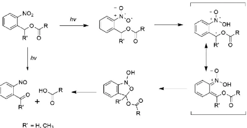
Up to now, UCNP have been applied to initiate photoreactions, such as photoisomerization, photopolymerization, photolysis and photocoupling (Figure 8) (118, 122-126). Among them, UCNP-assisted photolysis reactions have been widely employed for numerous applications, like the patterning of biomaterials (127), cell adhesion (126) and siRNA release (128). In these systems, o-nitrobenzyl (ONB) alcohol derivatives are the most frequently used organic photolytic chromophores (80, 115, 129, 130). Because these ONB derivatives, as photolabile groups, have been well-studied and readily available (131, 132). The cleavage mechanism is based on the photo-induced isomerization of ONB into
o-nitrosobenzaldehyde (Figure 9) (132). Their absorption band of ONB groups (280-360
nm) overlaps with the upconverted UV emissions from Tm3+-doped UCNP. In a system
comprised of Tm3+-UCNP and ONB photolytic chromophores, the cleavage reaction can be
triggered by the NIR light.
Figure 8. Four types of UCNP-assisted photoreactions. Reproduced with permission from [118].
Constructing NIR light-sensitive nanovectors with UCNP and polymer materials for drug delivery has generated intense interest in the past seven years. These elaborately designed generally include three parts: UCNP, polymers and payloads. As a core component, Tm3+-doped UCNP are commonly used with the capability of emitting UV and visible
lights upon NIR light irradiation (980 nm). In general, the UV upconversion photons are used to initiate photoreactions and the visible upconversion emissions are employed for fluorescence imaging. As for polymer materials, they can be hydrophilic ones, amphiphilic random or diblock copolymers, or nanogels prepared by various polymerization techniques. In most cases, they possess light-sensitivity by incorporating organic photosensitive units. So far, numerous types of payloads (drug molecules, siRNA, large biomolecules, etc.) have been encapsulated into the structures of nanovectors with UCNP and polymers. After assembling these three parts, the release of cargos is always caused by UCNP-assisted photoreactions of photocaged drug molecules or photoresponsive polymers.
Regarding the ways for incorporating cargos, these nanovectors are broadly classified into two categories. One is that guest molecule is conjugated to nanovectors through covalent bonding. The other is that payloads are entrapped into nanovectors through non-covalent interactions. In the following sections, a brief survey of them is presented.
Figure 9. Photoisomerization mechanism of o-nitrobenzyl alcohol derivatives into an
I.3.1 Covalent Conjugation of Payloads with UCNP/Polymer Nanovectors
In this category, guest molecules are linked into the structure of nanovectors (UCNP/polymer) through chemical modifications. The significant advantage is that the amount of drug in these nanovectors can be precisely tuned. Noteworthy, drug molecules must keep their therapeutic effect during chemical modifications. Upon exposure to NIR light, UC photons are utilized to induce cleavage of anchor points, resulting in the release of active drugs. Two typical examples are given in this section regarding the position of payloads in the structure of nanovectors.
Payloads can be covalently bonded to UCNP (133-135). Rubio-Retama et al. designed a nanovector based on PEGylation of SiO2-coated UCNP (Figure 10) and the investigation of
their toxicity was carried out at the cellular level (135). The hydrophilic polymer chains, PEG5000, were grafted onto the SiO2-coated UCNP. Then a potent cytotoxic drug,
doxorubicin (DOX), was covalently attached to the surface with a photolabile group of an
o-nitrobenzyl alcohol molecule. In the absence of the NIR light stimulus, these NPs
exhibited little cytotoxicity to Hela cells. By contrast, upon NIR light irradiation, the active drug molecules cleaved from these NPs dramatically reduced the cells viability.
Payloads can also be integrated into the structure of polymers (13, 136, 137). As shown in Figure 11, Liu and co-workers designed and synthesized a multifunctional nanovector (combination of chemotherapy and PDT) based on conjugated polyelectrolyte encapsulated UCNP (137). First, the chemotherapeutic drug, DOX, was covalently linked to a conjugated polyelectrolyte (acting as a surface coating agent and photosensitizer) through a UV-cleavable ONB linker. Then this polyelectrolyte and OA-capped UCNP were assembled through ligand attraction. The UV and visible upconversion emissions emitted by UCNP were utilized to control DOX release and initiate the polymer photosensitizer to generate reactive oxygen species (ROS), respectively. In comparison with the control group, their results showed that the combination therapy upon 980 nm irradiation could exhibit enhanced inhibition of cell growth.
![Figure 3. Applications of RE doped UCNP. Adapted with permission from [2].](https://thumb-eu.123doks.com/thumbv2/123doknet/5419696.126683/29.918.162.793.666.1018/figure-applications-doped-ucnp-adapted-permission.webp)
![Figure 8. Four types of UCNP-assisted photoreactions. Reproduced with permission from [118]](https://thumb-eu.123doks.com/thumbv2/123doknet/5419696.126683/40.918.216.753.244.580/figure-types-ucnp-assisted-photoreactions-reproduced-permission.webp)