HAL Id: dumas-01887588
https://dumas.ccsd.cnrs.fr/dumas-01887588
Submitted on 15 Oct 2018HAL is a multi-disciplinary open access archive for the deposit and dissemination of sci-entific research documents, whether they are pub-lished or not. The documents may come from teaching and research institutions in France or abroad, or from public or private research centers.
L’archive ouverte pluridisciplinaire HAL, est destinée au dépôt et à la diffusion de documents scientifiques de niveau recherche, publiés ou non, émanant des établissements d’enseignement et de recherche français ou étrangers, des laboratoires publics ou privés.
The skull vibration-induced nystagmus test as vestibular
examination in children with sensorineural hearing loss
Cindy Colombé
To cite this version:
Cindy Colombé. The skull vibration-induced nystagmus test as vestibular examination in children with sensorineural hearing loss. Human health and pathology. 2018. �dumas-01887588�
AVERTISSEMENT
Ce document est le fruit d'un long travail approuvé par le
jury de soutenance et mis à disposition de l'ensemble de la
communauté universitaire élargie.
Il n’a pas été réévalué depuis la date de soutenance.
Il est soumis à la propriété intellectuelle de l'auteur. Ceci
implique une obligation de citation et de référencement
lors de l’utilisation de ce document.
D’autre part, toute contrefaçon, plagiat, reproduction illicite
encourt une poursuite pénale.
Contact au SID de Grenoble :
bump-theses@univ-grenoble-alpes.fr
LIENS
LIENS
Code de la Propriété Intellectuelle. articles L 122. 4
Code de la Propriété Intellectuelle. articles L 335.2- L 335.10
http://www.cfcopies.com/juridique/droit-auteur1
UNIVERSITÉ GRENOBLE ALPES UFR DE MÉDECINE DE GRENOBLE
Année : 2018
THE SKULL VIBRATION-INDUCED NYSTAGMUS TEST AS VESTIBULAR EXAMINATION IN CHILDREN WITH SENSORINEURAL HEARING LOSS
THÈSE
PRÉSENTÉE POUR L’OBTENTION DU TITRE DE DOCTEUR EN MÉDECINE DIPLÔME D’ÉTAT
Cindy COLOMBÉ
THÈSE SOUTENUE PUBLIQUEMENT À LA FACULTÉ DE MÉDECINE DE GRENOBLE*
Le 28 Septembre 2018
DEVANT LE JURY COMPOSÉ DE Président du jury :
Monsieur le Professeur Christian Adrien RIGHINI Membres :
Monsieur le Professeur Sébastien SCHMERBER, directeur de thèse Monsieur le Professeur Dominique PLANTAZ
Madame le Docteur Joëlle TROUSSIER Monsieur le Docteur Georges DUMAS
*L’UFR de Médecine de Grenoble n’entend donner aucune approbation ni improbation aux opinions émises dans les thèses ; ces opinions sont considérées comme propres à leurs auteurs.
6
REMERCIEMENTS
A MES MAITRES ET MEMBRES DU JURY
Monsieur le Professeur Christian Adrien RIGHINI, président du jury
Vous me faites l’honneur de présider cette thèse.
Je vous suis reconnaissante pour l’enseignement que vous m’avez apporté tout au long de mon internat.
Votre rigueur clinique et chirurgicale, vos connaissances théoriques qui font « mouche » à la visite professorale du vendredi sont un exemple pour chacun d’entre nous.
Je vous sais gré de m’avoir intégrée dans l’équipe COLL au sein de l’Institute for Advanced Biosciences afin de réaliser mon Master 2.
Soyez assuré de mon profond respect.
Monsieur le Professeur Sébastien SCHMERBER, directeur de thèse
Vous me faites l’honneur de diriger cette thèse.
Je vous suis reconnaissante de l’étendue des connaissances théoriques et chirurgicales que vous nous transmettez, et de votre confiance quotidienne au bloc opératoire. Je vous remercie pour votre disponibilité et votre enthousiasme quand il est question d’otologie, mais aussi de sujets plus généraux de culture générale ou de politique.
Soyez assuré de mon profond respect.
Monsieur le Professeur Dominique PLANTAZ
Vous me faite l’honneur de juger ce travail.
Quand je vous ai demandé de participer à l’évaluation de mon travail de thèse, vous avez tout de suite accepté alors même que l’on ne se connaissait pas et que nous n’avions jamais travaillé ensemble. Je suis fière que vous fassiez parti de mon jury de thèse.
7 Je vous témoigne toute ma gratitude et mon profond respect.
Madame le Docteur Joëlle TROUSSIER
Je te suis infiniment reconnaissante d’avoir accepté de mener ce travail avec moi et de m’avoir accompagnée à chaque étape de ma formation dans ton domaine de prédilection, l’audiophonologie de l’enfant. Tu es quelqu’un de remarquable et d’exemplaire. Ta bonne humeur et ton optimisme sont si contagieux, qu’on referait le monde…
Monsieur le Docteur Georges DUMAS
Je vous suis reconnaissante d’accepter de juger ce travail, qui porte sur le sujet de toute une carrière professionnelle qui vous est cher.
Je vous remercie d’être toujours disponible pour les « avis vertigineux » que l’on vous demande et votre curiosité passionnée dans le domaine de la vestibulométrie force l’admiration.
8
A MES MAITRES ET AUTRES MEDECINS QUI ONT MARQUE MON PARCOURS
En ORL au CH de Chambéry : aux Docteurs Patrick MANIPOUD à l’esprit paternaliste,
rigoureux mais bienveillant, Christophe GUICHARD à l’esprit vagabond mais attentionné, Patrick NICOLLET l’esprit humoriste et artistique, mais aussi aux Docteurs SAUMUR et GUIRAUD, vous m’avez tous beaucoup apporté lors de mon premier semestre d’ORL.
En ORL au CHU de Grenoble : au Professeur Emile REYT, avec qui j’ai fait mes premiers
pas au CHU, merci d’avoir été disponible, rigoureux, et de m’avoir transmis vos connaissances en rhinologie. Je vous suis profondément reconnaissante. A Alexandre KARKAS notre idole et mentor, parti trop vite de Grenoble, nous te regrettons énormément toi, tes « bijoux » et les fous rires au bloc. Je suis si heureuse que tu aies trouvé l’âme sœur et que tu sois épanoui dans ta vie personnelle. A Ihab ATALLAH, j’admire ce que tu as appris aux USA et que tu développes ici à Grenoble et ta capacité de travail. Merci d’être toujours disponible pour nous. J’espère que tu auras une grande carrière. A Anne Rivron, merci de poursuivre l’exploration vestibulaire de l’enfant. A Alice HITTER, tu nous manques.
Au CHU de Grenoble : A Julie VILLA, merci d’être toujours disponible et à l’écoute pour
discuter de nos patients. A Arnaud ATTYE, merci pour ton aide précieuse en imagerie de la tête et du cou. Au Docteur Florence TAHON pour sa disponibilité, au Docteur Kamel BOUBAGRA.
Au CH d’Annecy Genevois : en ORL : au Docteur Bernard FONLUPT, j’ai commencé à
comprendre l’oreille grâce à vos dessins. Vous êtes un grand personnage. Aux Docteurs BODINO, PAOLETTI, ANGEL. En CMF : au Docteur Leslie NOYELLES.
9
En Neurochirurgie au CHU de Grenoble : à Monsieur le Professeur Emmanuel GAY, je
vous admire pour votre humanisme, votre abnégation, votre talent chirurgical et votre capacité à tout gérer sans jamais vous plaindre. Vous êtes un chef de service incroyable. Aux Professeurs Olivier PALOMBI et Stephan CHABARDES, aux Docteurs Eric SEIGNEURET, Dominique HOFFMAN, Benjamin NICOT, ma deuxième famille professionnelle. A Laurent SELEK qui ne peut lire ces mots mais qui est si présent dans notre mémoire.
En CMF au CHU de Grenoble : au Professeur Jean-Philippe GIOT, aux Docteurs Béatrice
MORAND, merci pour ta confiance et ton enseignement, Aurélie VIGNERON, j’ai aimé travailler avec toi, Emma ORSET, plus rapide que l’éclair, Juliette COUDURIER la reine des rhinoplasties, Marine HITIER la déesse des paupières, Isé MARZLOFF.
Aux anesthésistes d’ORL : aux Docteurs Yves PRA à l’humour ravageur, Marie-Pierre
ABOUSSOUAN à l’efficacité redoutable, Soumaya BEN REJEB si douce et calme, Jean-François ROGER, venez en ORL.
Aux ORL libéraux : un immense merci au Docteur ATTARD d’avoir été si disponible et si
bienveillant, au Docteur TIXIER pour sa confiance et sa gentillesse, aux Docteurs Nils MOREL et Thomas LEQUEUX pour leur accueil chaleureux dans leur cabinet, au Docteur Edouard CHAUVET pour sa pédagogie passionnée et son accueil chaleureux dans son cabinet, aux Docteurs GUICHARD et CUISNIER pour leur accueil à Romans, au Docteur PENISSON pour les fous rires aux congrès et son accueil à Voiron.
A Jean-Pierre ATTALI pour son enseignement chirurgical durant mon externat, et surtout son humour, sa culture, et sa passion pour l’histoire.
10
11
12
13
Préambule
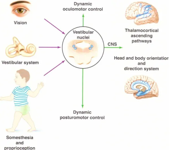
Aristote définit cinq sens, que nous connaissons bien en oto-rhino-laryngologie, pour en étudier au moins trois sur cinq, que sont l’ouïe, l’odorat et le goût. L’équilibre constitue le 6ème sens oublié par Aristote, et le 4ème sens auquel le médecin ORL est confronté chaque jour. Il résulte d’une intégration centrale multi-sensorielle complexe (Figure 1).
Figure 1. L’équilibre résulte de l’intégration multi-sensorielle au sein des noyaux vestibulaires, des informations afférentes provenant des organes récepteurs de la vue, l’équilibre, et la proprioception. Le résultat complexe de cette intégration est envoyé vers les structures motrices et corticales du système nerveux central. Schéma emprunté à S. Wiener-Vacher, Les monographies amplifon, n°62.
14
1- Bilan vestibulaire de l’enfant 1-1 Interrogatoire
L’interrogatoire des parents et de l’enfant permet de préciser les antécédents néonataux (prématurité), familiaux (surdités génétiques et syndromiques), les troubles ophtalmologiques et neurologiques, l’âge de la marche, la présence de chute dans l’enfance, la présence et l’histoire d’une surdité et éventuellement la symptomatologie vertigineuse, l’existence de céphalées, nausées, vomissements.
1-2 Examen oto-neuro-vestibulaire clinique
Tout enfant reçoit un examen otologique, vestibulaire et neurologique pour orienter le diagnostic vers un pathologie centrale ou périphérique.
1-2.1 L’examen postural et le développement moteur
La première des évaluations consiste à regarder l’enfant marcher et jouer dans la salle d’attente. Ceci donne un aperçu sur l’équilibre global et la coordination des mouvements. Ensuite, un bilan complet est possible, utilisant diverses évaluations standardisées portant sur le développement posturo-moteur ainsi que sur l’équilibre (Figure 2).
L’examen le plus sensible pour dépister une atteinte vestibulaire bilatérale est l’appui unipodal les yeux fermés (le test est normal si le maintien est supérieur à 4 secondes). A partir de 4 ans, il est possible d’évaluer l’équilibre d’un enfant au moyen d’un test standardisé : le Modified Clinical Test of Sensory Interaction in Balance (CTSIB-M) (Figure 3).
18 Figure 2. Cahier de passation et de profil du développement moteur fonctionnel du jeune enfant de 0 à 12 mois (Vaivre-Douret, 1997, Elsevier, Paris). Il existe également un cahier pour le jeune enfant de 13 à 48 mois.
19 Figure 3. Le Modified Clinical Test of Sensory Interaction in Balance (CTSIB-M) est un test standardisé permettant d’évaluer la fonction d‘équilibre chez l’enfant à partir de 4 ans. Il se compose de 4 items : 1-debout les yeux ouverts sur une surface ferme ; 2-debout les yeux fermés sur une surface ferme ; 3-debout les yeux ouverts sur une surface molle ; 4-debout les yeux fermés sur une surface molle. L’item 4 est le plus difficile et le plus sensible pour révéler un déficit vestibulaire.
1-2.2 Examen de l’oculomotricité
L’évaluation de la stabilisation du regard est menée sur un enfant dont la tête est immobile. Dès le début, il est possible de rechercher la présence d’un gaze nystagmus qui correspond à l’apparition d’un nystagmus dans le regard légèrement excentré de 20° à 20 cm. La poursuite
20 oculaire d’un jouet permet de révéler chez l’enfant présentant une poursuite saccadique, une atteinte cérébelleuse. L’étude de la convergence et de la divergence permettent de révéler un trouble ophtalmologique associé à la surdité, conduisant à un bilan orthoptique et ophtalmologique pédiatrique complet.
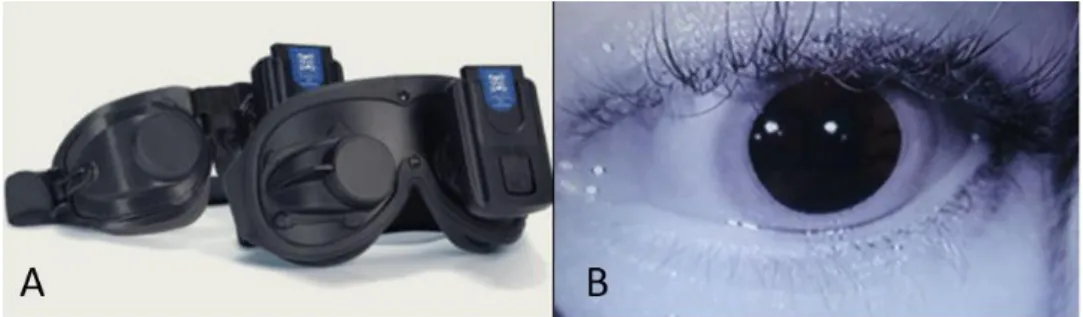
Le nystagmus correspond à un mouvement oculaire alternant phases rapide et lente, dont la phase lente indique le côté déficitaire, et la phase rapide, par convention le sens du nystagmus et le vestibule le plus actif. La recherche d’un nystagmus spontané ou provoqué n’est pas toujours aisée chez l’enfant. Si le nystagmus est d’origine périphérique, il est inhibé par la fixation oculaire. Pour le révéler, il est possible d’utiliser des lunettes de Frenzel rendant la vision floue, ou des lunettes de vidéoscopie, permettant d’observer un nystagmus spontané dans le noir sans fixation oculaire (Figure 4).
Figure 4. A : lunettes de vidéoscopie. B : visualisation en direct, sur un écran, de l’œil filmé par caméra infrarouge.
1-2.3 L’examen otologique
Chaque enfant bénéficie d’une otoscopie et d’une audiométrie adaptée à son âge, voire d’otoémissions acoustiques.
21
1-2.4 L’examen neurologique
En fonction de l’âge du patient, on évalue les réflexes archaïques (Moro, Babinski…), ostéotendineux, la somesthésie, la tonicité, la motricité fine et la marche. L’examen des douze paires crâniennes est essentiel.
1-2.5 Explorations fonctionnelles vestibulaires standards a- Le test au fauteuil rotatoire
Il évalue la réactivité des canaux semi-circulaires (CSC) latéraux à basse et moyenne fréquence.
Le test pendulaire permet de tester les deux CSC latéraux à une fréquence de 0.05 Hz. La compensation centrale va rapidement masquer une asymétrie vestibulaire.
L’impulsion rotatoire selon un axe vertical permet d’appliquer une accélération brève et une décélération angulaire de 40°/s² suivie d’une vitesse de rotation constante à 60°/s, produisant respectivement une excitation et une inhibition des 2 CSC latéraux. Le réflexe vestibulo-oculaire (RVO) peut être enregistré par vidéo-nystagmographie ou électro-nystagmographie (Figure 5). Une atteinte vestibulaire se traduit par une absence ou une diminution de la réponse nystagmique du côté atteint. Le processus de compensation centrale va également rapidement atténuer une asymétrie vestibulaire.
22 Figure 5. Le test au fauteuil rotatoire. A : fauteuil rotatoire manuel. B : disposition de l’examen au fauteuil rotatoire chez l’enfant avec port de lunettes de vidéoscopie.
b- Les potentiels évoqués otolithiques myogéniques cervicaux (PEOMc)
Les PEOMc permettent d’évaluer le réflexe vestibulo-collique ou sacculo-collique via la fonction du saccule, le nerf vestibulaire inférieur et le noyau vestibulaire, enregistré du côté ipsilatéral dans le muscle sterno-cléido-mastoïdien pendant une stimulation acoustique (Figure 6). Le cPEOM est une onde biphasique comprenant une onde positive P13 et une onde négative N23. Son amplitude est dépendante de l’amplitude electromyographique du muscle SCM. Une absence de cPEOM ou une diminution de plus de 40% par rapport au côté controlatéral signifie qu’il existe une atteinte des voies otolithiques vestibulo-spinales.
23 Figure 6. Les potentiels évoqués otolithiques myogéniques cervicaux. A : disposition des électrodes et technique de réalisation des PEOM cervicaux chez un très jeune enfant. B : résultats de PEOMc normaux montrant une onde biphasique.
c- Le video Head Impulse Test (VHIT)
Le VHIT est une version enregistrée du Head Impulse Test (HIT) clinique, permettant d’obtenir un gain objectif et reproductible du RVO pour les 6 canaux semi-circulaires, à haute fréquence. Le système Synapsys® VHIT Ulmer (Figure 7) ne comporte pas de lunettes et permet donc de tester les enfants à partir de l’âge de 3 mois. Des valeurs normales chez l’enfant ont récemment été publiées par Wiener-Vacher SR & Wiener S.
24 Figure 7. Le video Head Impulse Test. A : système VHIT, Synapsys (Marseille, France). B : disposition d’une salle de VHIT lors d’un examen chez l’enfant. Utilisation d’un petit jouet lumineux pour attirer le regard chez les enfants plus jeunes. C : résultats de VHIT chez une enfant, montrant à gauche le canalogramme et à droite le tableau résumé des gains du réflexe vestibulo-oculaire (VOR).
d- Le test calorique
Le test calorique détermine la réponse des CSC latéraux aux basses fréquences à la suite d’une irrigation successive des conduits auditifs externes à l’eau froide 30°C et l’eau chaude 44°C pendant 30 secondes. Cette stimulation induit un nystagmus correspondant à une réponse vestibulo-oculaire.
Ce test est assez mal toléré chez l’enfant, mais il permet d’apprécier un déficit vestibulaire ancien et totalement compensé. En revanche c’est un test comparatif entre les 2 côtés
25 stimulés. Il est moins reproductible chez l’enfant, d’autant plus chez les petits enfants du fait des nombreux artefacts (yeux fermés, mouvements, agitation, fatigue).
A l’issue de ce bilan, une fiche synthétique « bilan vestibulaire de l’enfant » est remplie (Figure 8)
26
Figure 8. Fiche de synthèse du bilan vestibulaire de l’enfant réalisé en consultation
e- Posturographie
La posturographie statique et dynamique assistée par ordinateur (Figure 9) permet d’évaluer l’équilibre statique et dynamique au moyen d’une plateforme de force. La première condition, teste l’équilibre statique et permet de déterminer le COP (centre de pression). Les résultats représentent le balancement du corps. Dans la deuxième condition, la posture du sujet est perturbée par des mouvements de la plateforme, impliquant sa vision, son vestibule et sa proprioception pour regagner l’équilibre. Ceci permet à la machine de définir l’organisation sensorielle du sujet (SOT Sensory Organization Test) et de calculer le score d’équilibre (ES Equilibrium Score). Ce test n’est possible qu’à partir de 6 ans. Des valeurs normatives ont été établies par Ferber-Viart et al.
27
Figure 9. Posturographie. Appareil de posturographie, exemple chez la personne âgée.
2- Le test vibratoire osseux (TVO) de Dumas
En 1935, Von-Bekesy découvre qu’une vibration au niveau du crâne entraîne des réflexes et des impressions de mouvement qu'il attribue à la stimulation des récepteurs vestibulaires. En 1973, Lücke applique une vibration de 100 Hz sur les mastoïdes d’un patient présentant un déficit vestibulaire unilatéral et remarque l’apparition d’un nystagmus induit. Depuis 1999 et Hamann et Schuster, Dumas ne cessent d’explorer et de développer l’utilisation du test vibratoire osseux que l’on appelle maintenant test de Dumas.
2-1- Technique
Une vibration de 100 Hz est appliquée pendant 5 à 10 secondes, perpendiculairement sur les deux mastoïdes et le vertex du patient, par l’examinateur tenant le vibrateur dans sa main dominante (Figure 8). Le nystagmus induit par la vibration peut être visualisé à l’aide de
28 lunettes de Frenzel, ou plus adéquatement avec des lunettes de vidéoscopie ou enregistrées en vidéonystagmographie 2D ou 3D.
2-2- Stimulation
Il existe plusieurs types de vibrateurs. Celui commercialisé par Synapsys® (France) permet une vibration à 30, 60 et 100 Hz. Idéalement le vibrateur utilisé présente une surface circulaire de 20 mm de diamètre recouverte d’un caoutchouc mince.
2-3- Localisation
La stimulation la plus efficace est celle réalisée au niveau des processus mastoïdiens, sauf lorsqu’il existe une troisième fenêtre, auquel cas la stimulation au vertex donne une réponse plus intense. Une stimulation au vertex permet la réalisation d’un Weber vestibulaire.
2-4- Interprétation
Lorsqu’il existe un déficit vestibulaire unilatéral total, le test vibratoire osseux révèle un nystagmus horizontal ou torsionnel, dont la phase rapide bat du côté opposé au côté lésé, quelque soit le côté stimulé. Le nystagmus s’arrête lorsque la stimulation s’arrête. Ce test est reproductible et ne montre pas de phénomène d’habituation.
29 Figure 8. Le test vibratoire osseux. A : Vibrateur V-VIB 3F (Synapsys, Marseille, France) générant 3 fréquences de vibration (30, 60 et 100 Hz). B : Installation et technique d’application du vibrateur sur la mastoïde gauche d’un enfant assis sur un fauteuil de consultation.
30
Problématique de l’étude
L’analyse exhaustive de la fonction vestibulaire d’un enfant est une évaluation longue, difficile, contraignante, qui requiert une équipe entraînée, spécialisée, flexible, et du matériel coûteux, dont certains accessoires doivent être adaptés à l’enfant. Le test vibratoire osseux de Dumas est un examen simple, rapide, global, réalisable en 30 secondes au fauteuil de consultation, sans contre-indication ni effet secondaire.
L’objectif de cette étude était d’évaluer le test vibratoire osseux de Dumas chez l’enfant sourd en le comparant aux autres examens vestibulaires standards à savoir l’évaluation posturale standardisée, les potentiels évoqués otolithiques myogéniques, le test au fauteuil rotatoire et le video head impulse test.
31
Article
The Skull Vibration-Induced Nystagmus Test as vestibular examination in children with sensorineural hearing loss
C. Colombe1,2, G. Dumas1,5, J. Troussier1,3, S. Schmerber1,2,3,4
1. Grenoble Alpes University Hospital, Department of Otolaryngology- Head and Neck Surgery, 38000 Grenoble, France
2. Grenoble Alpes University, UFR de Médecine, 38000 Grenoble, France
3. Alpes Reference Center of Cochlear Implants
4. Univ. Grenoble Alpes, Brain Tech Lab INSERM UMR 1205, F-38000 Grenoble, France
5. EA-3450 DevAH Développement, Adaptation et Handicap. Faculté de Médecine et UFR STAPS Université de Lorraine. Villers les Nancy- France
Corresponding author : Cindy Colombé, Department of Otolaryngology-Head and Neck surgery, BP 217, Grenoble Alpes University Hospital, France, Email : ccolombe@chu-grenoble.fr;
32
Abstract :
Objectives: Vibration to the mastoid can induce a nystagmus termed skull vibration induced nystagmus (SVIN). This recent first-line indicator of asymmetrical vestibular function quickly reveals a vestibular asymmetry in unilateral vestibular lesion patients and is used as a vestibular Weber Test. We aimed to assess the usefulness and tolerance of the SVIN test (SVINT), in children with moderate to profound sensorineural hearing loss.
Methods: A prospective monocentric study of children with moderate to profound unilateral or bilateral deafness, was conducted between October 2017 and May 2018. The SVINT was performed on each mastoid and vertex, first and double-blind by 2 clinicians, separately; the Modified Clinical Test of Sensory Interaction in Balance (CTSIB-M), the rotary chair testing, the video Head Impulse Test (VHIT) and the cervical Vestibular Evoked Myogenic Potentials (cVEMP) were proceeded the same day, blinded from the SVINT result. The parents' written consent was collected.
Results: A total of 23 children, aged from 7 months to 14 years were included. Five had unilateral and 18 bilateral deafness. The main etiologies were genetic non-syndromic (5), genetic syndromic (3), malformation (6), unknown (4), CMV (2), meningitis (2), neonatal hypoxia (1).
Sensitivity and specificity were respectively 46% and 100%, Negative and positive predictive values were 59% and 100%, concordant coefficient kappa was 1. Clinical tolerance was good even in young children.
Conclusion: The SVINT is a rapid and easy procedure to assess vestibular function as a bedside examination. Compliance is good even in infants.
Keywords: Skull vibration-induced nystagmus test, balance, vestibular function, children, sensorineural hearing loss.
33
Introduction
The Skull Vibration Induced Nystagmus Test (SVINT) is a bedside, non-invasive, quick, global (canalar and otolithic) and easy to perform test, which acts as a vestibular Weber test. The SVINT stimulates the vestibule at high frequencies, ranging from 30 to 100 Hz. Studies showed the interest of vibratory stimuli on forehead and mastoïd processes to detect asymmetrical vestibular disorders (1–4). But several studies has demonstrated that the SVINT is more sensitive at 100 Hz on each mastoid processes and vertex (5–7).
Although daily used in adults and children, the SVINT has never been studied in this last population.
Vestibular impairments are twice more frequent in children with sensorineural hearing loss (SNHL) because of anatomical, physiological and histological similarities between the cochlear and vestibular receptor-organs (8–10). In addition, the extend and severity of vestibular loss is directly linked to the etiology and severity of the SNHL (11).
Therefore, complete vestibular assessment is an integral part of the clinical examination of children with sensorineural hearing loss. Usually, vestibular assessment consists in standardized tests of static and dynamic balance function evaluation, and functional multifrequency vestibular explorations namely cervical vestibular-evoked myogenic potentials (cVEMP), Rotary Chair Testing (RCT), video head impulse test (VHIT) and caloric test (CT).
There are several standardized tests of static and dynamic balance function assessment, such as the modified clinical test of sensory interaction in balance (CTSIB-M) and the Bruininsk-Oseretsky Test of Motor Proficiency BOT2 (12,13). The CTSIB-M is quite simple and fast, since it includes 4 steps of 30 seconds each,. In 2013, Park et al. showed that the mCTSIB
34 could be used instead of the Sensory Organization Test (SOT) of computerized dynamic posturography (CDP) in screening to distinguish normality from abnormality in dizzy adult patients with unilateral vestibulopathy (14). In a case control study of 31 children aged 4.1 to 12.1 years, Dannenbaum et al. noted that the CTSIB score was significantly reduced in children with global developmental delay compared to children with typical development, and the test-retest reliability was also good (15). So it was decided to use it as postural and balance standardized assessment.
Cervical VEMP reveals an inhibitory otolith vestibulo-spinal reflex recorded in the ipsilateral sternocleidomastoid (SCM) muscle in response to loud acoustic stimuli. CVEMP is a reliable method to measure the otolith function and it is not influenced by central compensation processes. Normal values in children have been listed in few recent publications (16,17). P13 and N23 latencies slightly increase with age (18).
Rotary chair testing (RCT) has been an essential diagnostic tool for vestibular evaluation since the early 20th century when Róbert Bárány pioneered both the application of rotational chair testing and caloric testing for vestibular assessment. It assesses a simple reflex behavior, the vestibulo-ocular reflex (VOR), which involves only three neurons from input to output. As it overcomes many of the weaknesses of caloric testing and is very well tolerated in children, many clinicians acknowledge it to be the gold standard for pediatric vestibular assessment. The procedure can be performed successfully, using either sophisticated computerized motorized chairs, or manual rotation. VOR normal values in children have been fewly studied (19–22).
The VHIT tests the 6 semicircular canals individually with high frequency stimuli (23–25). Recording ocular and head movements by a remote camera, the software can detect catch-up
35 saccades and calculate the VOR gain. The VHIT system detect the covert saccades too, which are not detected with the HIT maneuver. Recently, Wiener et al. has investigated results of VHIT gain in healthy children (26).
Caloric testing can be contraindicated in êdiatric population due to tympanic perforation, chronic otitis media or external meatus malformation. Moreover, it assesses only one of the five vestibular receptor-organs of the inner ear, and a very small part of the vestibular system’s dynamic frequency range. This test is not always reliable, and it is non physiologic. As it is poorly tolerated by children, there are limited data available for pediatric testing. Our team is not used to caloric testing in infants and young children, so we decided to consider only the VHIT and rotary chair testing.
This global evaluation is therefore restrictive, time-consuming, onerous and challenging for any non-specialized team. This appears all the more laborious in the pre-operative cochlear implant (CI) children, especially in profound congenital deaf infants, who undergo a lot of tests during their early life.
Indeed, since the advent of cochlear implantation thirty years ago, the techniques and indications have been developing, improving rehabilitation of hearing impairments particularly in young children with severe to profound congenital or early-onset sensorineural hearing loss (SNHL) in prelingual stage.
Few studies involving children have proved that cochlear implantation leads in 20-70% of vestibular impairment and 9-10% of complete vestibular loss (11,12,27–29). Nevertheless, there are increasing evidences advocating that cochlear implantation would have a beneficial effect on vestibular function in children (30).
36 Thus, a cochlear implantation should be supervised before and after surgery by a complete vestibular assessment.
A thorough vestibular examination suitable for children is required. The SVINT appears to be a promising test in pediatric population and presents no contraindication in children.
The aim of this study was to assess how the SVINT could detect vestibular disorders in children with sensorineural hearing loss, compared to the standard battery of vestibular tests (standardized postural evaluation, rotary chair testing, video head impulse test and cervical vestibular-evoked myogenic potentials). The sensitivity, specificity and compliance of each test was also explored.
37
Materials & Methods
Population
Twenty-three children with unilateral or bilateral mild to profound sensorineural hearing loss and consulting at the Department of Otorhinolaryngology at the Grenoble Alpes University Hospital (France) were included prospectively in the study between November 2017 and May 2018. Children with axial deficit, oculomotor deficit or cerebellar ataxia were excluded. All children and parents were counseled regarding study participation. Verbal understanding and written consent were obtained.
All children underwent otoscopy, audiometry and vestibular examination including the SVINT, the CTSIB-M, the RCT, the cVEMP and the VHIT.
The SVINT was performed by a senior and a junior, first, and double blinded from the other tests, which were realized the same day, helped by a confirmed nurse.
The Skull vibration induced nystagmus test (SVINT or Dumas’ Test)
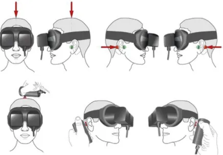
As described by Dumas et al. (4), the SVINT consists in applying a 100-Hz-bone-conducted vibration to either mastoid process and vertex with a V-VIB 3F-stimulator (Synapsys, Marseille, France) perpendicularly to the skin, during 10 seconds for each location (Figure 1
38 Figure 1. Skull vibration-induced nystagmus test technique in clinical practice. Principle of stimulation: the examiner can face the subject, as in the first example. The vibrator cylindrical contact is applied perpendicularly to the designated surface (red spot) with a pressure of about 10 N or 1 kg on the vertex or each mastoid process [level to the external acoustic meatus (Green spot)]. The examiner uses the other hand to maintain and immobilize the subject’s head. The same type of stimulation can be performed with the examiner behind the subject (second example situation). Stimulation must avoid the mastoid tip to prevent from muscular vibration radiation and proprioceptive involvement. Schema borrowed from Dumas et al. Frontiers in Neurology, 2017.
Figure 2. The vibration-induced nystagmus complements other vestibular tests in the vestibule multifrequency analysis. Place of the SVINT in the currently known frequency spectrum of the vestibular system. This graph summarizes the complementarity of vestibular tests, introduces the concept of the optimal vestibular compensation zone for the horizontal canal and the bone conduction stimulation frequencies necessary to obtain ocular vestibular-evoked myogenic potentials (oVEMP) and cervical evoked myogenic potentials (cVEMP). Adapted from Chays et al. (72) modified by
Dumas (university PhD thesis 2014). VOR: Vestibular ocular reflex; EMG: Electromyography; HST:
Head shaking test; VLF, very low frequencies; LF, low F; MF, middle F: HF, high F; VHF, very high F.
39 Children were placed in seated position, on the chair directly, or on the lap of one parent. Nystagmus was observed with videoscopy glasses or even easily without glasses depending on the child compliance. Children were conditioned, applying the vibrator first on their hand or on their parents’ hand as a game. The SVINT was considered positive when bone stimulation triggered a reproducible and sustained nystagmus without direction change. This nystagmus beats in the same direction irrespective of which mastoid is stimulated and stops at stimulation offset.
Result, feasibility, compliance and presence of crying were noted in the observation book by the senior and the junior.
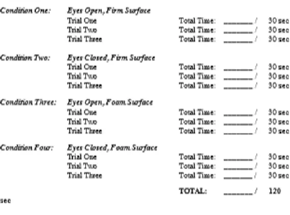
The Standardized postural evaluation by CTSIB-M
The CTSIB-M is a modification of the CTSIB that eliminates the use of the visual conflict dome (31). It includes conditions 1,2,4, and 5 of the original CTSIB.
To perform the test, patients stand with their hands at their side and perform the following 4 conditions (Supplementary data figure 6):
1) Stand on firm surface with the eyes open (condition 1) 2) Stand on firm surface with the eyes closed (condition 2)
3) Stand on compliant surface (foam) with the eyes open (condition 3) 4) Stand on compliant surface (foam) with the eyes closed (condition 4)
Patient performance is timed for 30 seconds. If a patient is unable to maintain the position for 30 seconds he is provided with 2 additional attempts. The scores of the 3 trials are averaged. The best score is 120 seconds.
40 Total score (Modified CTSIB) = Average Time Condition 1 (if > 1 trial required) + Average Time Condition 2 (if > 1 trial required) + Average Time Condition 3 (if > 1 trial required) + Average Time Condition 4 (if > 1 trial required)
The Cervical Vestibular Evoked Myogenic Potential (cVEMP) testing
Otolithic function was assessed with air conducted cVEMP. A 100 and 110-dB 500-Hz tone burst, was administered through headphones (Telephonics TDH-39P) with 100 sweeps averaged and 2 recording runs for each side.
An active electrode was positioned on the mid part of both sternocleidomastoid (SCM) muscles, the ground electrode on the forehead and the reference electrode over the upper part of the manubrium. A contralateral head turn was used to activate the sternocleidomastoid muscle contraction during stimulation.
Younger children were seated on the parent’s lap face to face. During the stimulation, the parent supported the lower part of the child’s trunk, so that the child had to straighten up to reach a toy. The toy was presented to the opposite side of the stimulation so that the child turned his head to the opposite side of the stimulation (about 45°).
All measures were recording on Neuro-Audio.net software (Neurosoft Society, Ivanovo, Russia). Electromyography of the SCM muscle was displayed on screen computer and permitted a visual feedback of the intensity of the muscle contraction. Cervical VEMP responses were considered as either present or absent within an age-appropriate range of latencies of the P13 (8.5-14 ms) and N23 (15-23 ms) waveforms (17,32). A 50% decrease of
the amplitude of one side comparing to the other side, corresponded to a unilateral hypofunction.
41
The Rotational chair testing (RCT)
Standard rotary chair was used to produce vertical-axis rotation, which directly assess lateral semicircular canal function. Vestibular ocular reflex (VOR) was measured, subjects sitting in a rotational chair and wearing videoscopy goggles, while the chair was manually rotated (0,5Hz) 10 rounds symmetrically clockwise and counter-clockwise. Children under 6 years of age were sitting on their parents’ lap, who maintained goggles and their head, straight plated against their chest. Presence of nystagmus during the rotation, and duration of the nystagmus in seconds after the net stop were noted for each side in the observation book. Results were considered abnormal if either a significant asymmetry was consistently detected in the VOR response between clockwise and counter-clockwise rotation or the VOR was absent with rotation in at least one direction. Normative data suggest that pediatric responses are similar to those of adults and perhaps even more robust in infants (22).
The Video Head Impulse Test
The VHIT were all carried out with Synapsys® (Marseilles, France) VHIT Ulmer system in children usually following the same protocol as that for adults (23). Children under the age of 5-6 years sat on their parents’ lap, face to the camera, one meter away from it, and their attention was attracted on a flashing toy or a cell phone at a distance of 1.3 m. Then the same right-handed clinician applied a brief and unpredictable head turn of 10-20° in the horizontal plane, and the vertical plane of left anterior right posterior (LARP) canals and vertical right anterior and left posterior (RALP) canals. The software automatically set aside trials outside the right plane or of inadequate velocity. VOR gain of each canal were automatically calculated.
42
Statistical analysis
The SVINT was assessed in all children, compared to the CTSIB-M, the cVEMP, the RCT and the VHIT. Sensitivity (Se), specificity (Sp), positive and negative predictive values (PPV, NPV) and likelihood ratios were determined with 95% confidence intervals.
Concordant coefficient kappa of Cohen was calculated to determine the concordance of the SVINT between the 2 clinicians (the senior and junior). All analyses were performed with RStudio software.
43
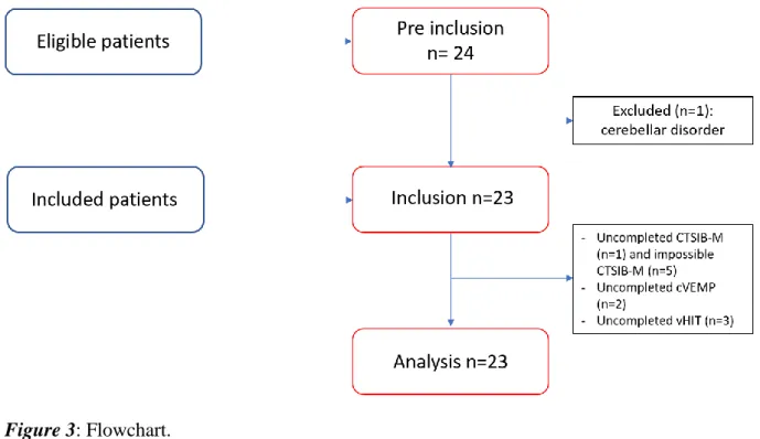
Results
Twenty-three patients were included in the study (Figure 3), 16 (70%) females and 7 (30%) males with mean age of 5.3 years old (ranged from 7 months to 14 years old).
Figure 3: Flowchart.
Heighteen (78%) children had bilateral hearing loss and 5 (22%) had unilateral hearing loss. Height children were assessed before cochlear implantation, 2 after a bilateral implantation. Etiologies of deafness are represented in Figure 4. The predominant etiologies of deafness were genetic (8 patients) (5 non-syndromic and 3 syndromic) and malformation (6 patients).
44 Figure 4. Different etiologies of deafness. Genetic non-syndromic, genetic syndromic, malformation, unknown, meningitis, CMV: Cytomegalovirus, Hypoxia.
Characteristics of the population are given in Table 1. Seven (30%) had partial unilateral vestibular deficit, 3 (13%) had complete unilateral deficit, 3 (13%) had partial bilateral deficit, none had complete bilateral deficit (Table 1).
Deficit unilateral bilateral TOTAL
partial n=7 n=3 n=10
complete n=3 n=0 n=3
TOTAL n=10 n=3 n=13
Table1. Vestibular deficit in the studied population.
SVINT results
SVINT was positive in 6 (26.1%) patients. Among these patients, 1 (17%) had unilateral deafness, 3 (50%) had asymmetrical hearing loss and 2 (33%) had symmetrical bilateral
45 hearing loss. The SVINT corresponded to 2 (33%) partial unilateral vestibular deficits, 3 (50%) complete unilateral vestibular deficits and 1 (17%) partial unilateral vestibular deficit (Table 2).
Deficit Positive SVINT
Partial unilateral n=2
Complete unilateral n=3
Partial bilateral n=1
Complete bilateral n=0
TOTAL n=6
Table 2. SVINT results compared to the battery of test results. The battery of tests permitted classifying vestibular deficit into partial unilateral, complete unilateral, partial bilateral and complete bilateral vestibular deficit.
The etiologies corresponding to a positive SVINT were malformation (2), CMV (2), meningitis (1) and unknown (1). The sensitivity and specificity of the SVINT, compared to the whole battery of tests, were respectively 46% and 100%. Positive and negative predictive values were respectively 100% and 59%. The positive and negative likelihood ratios were respectively infinite and 0.54 (Table 3).
The concordant coefficient of kappa between the fellow and the senior was 1.
CTSIB-M results
The standardized balance evaluation using CTSIB-M was completed only in children aged from 4 years old. The test was impossible in 4 cases, due to the impossibility for children to stand on, and partially realized (only eyes opened) in 1 case, a 3-year-old girl. The test showed an abnormal result (score lower than 120) in 4 (21%) cases. The sensitivity and
46 specificity were respectively 30% and 88%. The positive and negative predictive values were respectively 75% and 50%. The positive and negative likelihood ratios were 2.40 and 0.80.
CVEMP results
Cervical VEMP was completed in 21 (91%) patients. One was too tired to sustain her head and the test was not reliable, and one refused to perform the test. The 2 tests could not be performed again later. Among the complete tests, 10 (48%) showed a unilateral vestibular deficit, 1 (4%) a bilateral deficit and 10 (48%) were normal. The sensitivity and specificity of the CVEMP were respectively 92% and 100%, the positive and negative predictive values were 100% and 79%, the positive and negative likelihood ratios were Infinite and 0,08.
RCT results
The rotary chair testing was completed in all children. 16 (69%) had normal results, 5 (22%) had a unilateral vestibular deficit and 2 (9%) had a bilateral vestibular deficit. The sensitivity and specificity of RCT were respectively 38% and 80%, positive and negative predictive values were 71% and 50%, positive and negative likelihood ratios were 1.92 and 0.77.
VHIT results
The VHIT was performed in all children, but 4 (17%) were incomplete due to agitation of the child or artefacts (mainly for the anterior canal). Fourteen (61%) had normal VHIT result, 7 (30%) had a bilateral vestibular deficit and 2 (9%) had a unilateral vestibular deficit. Among children presenting a unilateral vestibular deficit, 1 had a partial unilateral hyporeflexia and 1
47 had a complete unilateral vestibular hyporeflexia. Among children with bilateral vestibular deficit, 2/7 had a symmetrical hyporeflexia and 5/7 had an asymmetrical vestibular areflexia or hyporeflexia. Three children with a bilateral vestibular deficit had a partial unilateral areflexia and 2 had a partial asymmetrical hyporeflexia. The sensitivity and specificity of the VHIT were respectively 69% and 100%, the positive and negative predictive values were 100% and 71%, the positive and negative likelihood ratios were Infinite and 0.31 (Table 3).
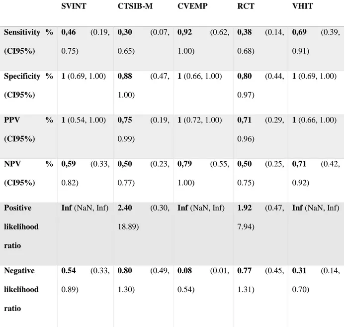
SVINT CTSIB-M CVEMP RCT VHIT
Sensitivity % (CI95%) 0,46 (0.19, 0.75) 0,30 (0.07, 0.65) 0,92 (0.62, 1.00) 0,38 (0.14, 0.68) 0,69 (0.39, 0.91) Specificity % (CI95%) 1 (0.69, 1.00) 0,88 (0.47, 1.00) 1 (0.66, 1.00) 0,80 (0.44, 0.97) 1 (0.69, 1.00) PPV % (CI95%) 1 (0.54, 1.00) 0,75 (0.19, 0.99) 1 (0.72, 1.00) 0,71 (0.29, 0.96) 1 (0.66, 1.00) NPV % (CI95%) 0,59 (0.33, 0.82) 0,50 (0.23, 0.77) 0,79 (0.55, 1.00) 0,50 (0.25, 0.75) 0,71 (0.42, 0.92) Positive likelihood ratio
Inf (NaN, Inf) 2.40 (0.30, 18.89)
Inf (NaN, Inf) 1.92 (0.47, 7.94)
Inf (NaN, Inf)
Negative likelihood ratio 0.54 (0.33, 0.89) 0.80 (0.49, 1.30) 0.08 (0.01, 0.54) 0.77 (0.45, 1.31) 0.31 (0.14, 0.70)
Table 3 : comparative results of the sensitivity, specificity, positive predictive value (PPV), negative predictive value (NPV), positive and negative likelihood ratios of the skull vibration-induced nystagmus test (SVINT), modified clinical test of sensory interaction in balance (CTSIB-M), cervical
48 vestibular-evoked myogenic postentials (CVEMP), rotary chair testing (RCT) and video head impulse test (VHIT). Inf (infinite), NaN (Not a number).
SVINT versus CTSIB-M, CVEMP, RCT and VHIT
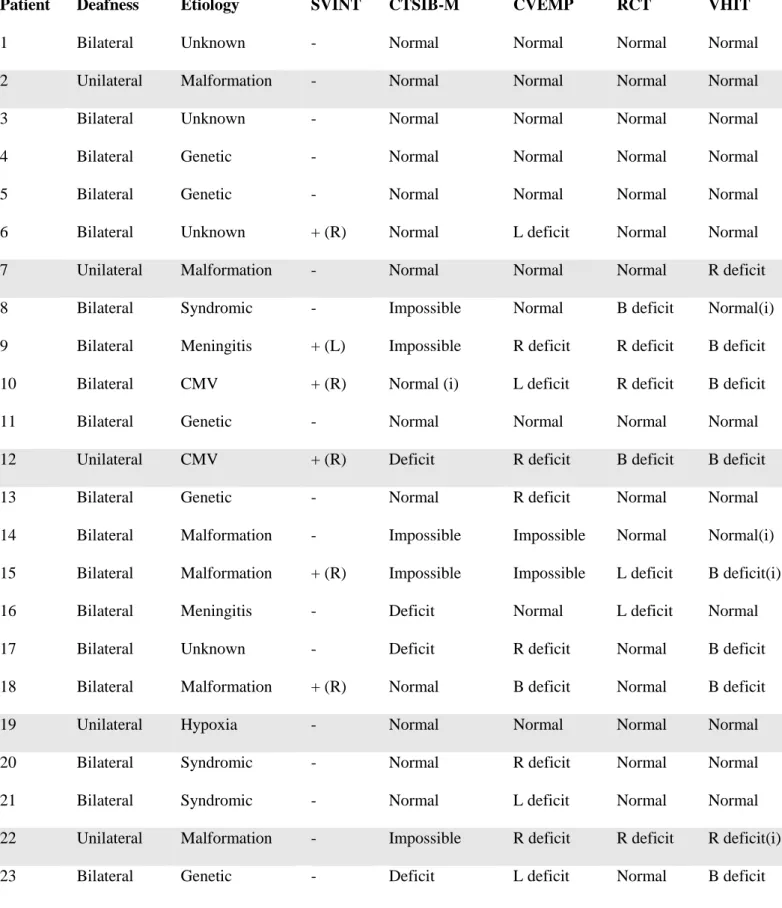
CTSIB-M: Only 1 of the 4 abnormal cases corresponded to a positive SVINT.
CVEMP: Three (14%) had a positive SVINT with an ipsilateral deficit, 1 (5%) had a contralateral deficit on SVINT and 1 a bilateral deficit on CVEMP. In total, 5/6 children having a positive SVINT had an abnormal CVEMP. The last one didn’t perform the CVEMP test because she was too tired.
RCT: Two children had normal RCT results although a positive SVINT, 2 had correlated unilateral deficit in SVINT and RCT while 1 had an opposite result in SVINT and RCT, and 1 had a bilateral deficit in RCT with a positive SVINT.
VHIT: Among the 5 children with bilateral vestibular deficit on VHIT, and positive SVINT, all had asymmetrical hypo or areflexia. One child had a unilateral partial hyporeflexia on VHIT, with a negative SVINT. One child had a normal VHIT result although a negative SVINT. This child had an ipsilateral vestibular deficit on CVEMP.
49 Patient Deafness Etiology SVINT CTSIB-M CVEMP RCT VHIT
1 Bilateral Unknown - Normal Normal Normal Normal
2 Unilateral Malformation - Normal Normal Normal Normal
3 Bilateral Unknown - Normal Normal Normal Normal
4 Bilateral Genetic - Normal Normal Normal Normal
5 Bilateral Genetic - Normal Normal Normal Normal
6 Bilateral Unknown + (R) Normal L deficit Normal Normal
7 Unilateral Malformation - Normal Normal Normal R deficit
8 Bilateral Syndromic - Impossible Normal B deficit Normal(i)
9 Bilateral Meningitis + (L) Impossible R deficit R deficit B deficit
10 Bilateral CMV + (R) Normal (i) L deficit R deficit B deficit
11 Bilateral Genetic - Normal Normal Normal Normal
12 Unilateral CMV + (R) Deficit R deficit B deficit B deficit
13 Bilateral Genetic - Normal R deficit Normal Normal
14 Bilateral Malformation - Impossible Impossible Normal Normal(i)
15 Bilateral Malformation + (R) Impossible Impossible L deficit B deficit(i)
16 Bilateral Meningitis - Deficit Normal L deficit Normal
17 Bilateral Unknown - Deficit R deficit Normal B deficit
18 Bilateral Malformation + (R) Normal B deficit Normal B deficit
19 Unilateral Hypoxia - Normal Normal Normal Normal
20 Bilateral Syndromic - Normal R deficit Normal Normal
21 Bilateral Syndromic - Normal L deficit Normal Normal
22 Unilateral Malformation - Impossible R deficit R deficit R deficit(i)
23 Bilateral Genetic - Deficit L deficit Normal B deficit
Table 4. Comparative results for each test. The SVINT is rated as positive (+) or negative (–) and nystagmus side is conventionally the rapid phase. R (right), L (left), B (bilateral), (i) impossible.
50
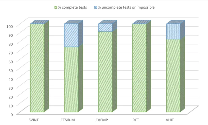
Compliance
The realization of the SVINT showed 4 (17%) children crying, but they also cried for the other tests. All could perform the test so the compliance of the SVINT was 100%. The CTSIB-M, CVEMP, RCT and VHIT had a compliance respectively equal to 74%, 91%, 100% and 83% (Figure 5).
Figure 5. Compliance with testing for each test.
0 10 20 30 40 50 60 70 80 90 100
SVINT CTSIB-M CVEMP RCT VHIT
51
Discussion
In our study, we demonstrated that the SVINT was well tolerated in children. Its sensitivity was low (46%) but its specificity was very high (100%) and its reproducibility was almost perfect (kappa=1).
The prevalence of unilateral SNHL was 21,7% although other studies had a prevalence of 30-40% of moderate to profound unilateral SNHL. It concerned the population of hearing screening American newborns. The difference can easily be explained by the little number of children included in our study and the demographic differences between United States and France (33,34).
The distribution of different etiologies was quietly similar to that of other studies. We found a majority of malformation and genetic deafness. Like other studies, the unknown etiology was predominant too, due to parents’ refusal of genetic analysis (33,35).
The anatomical and embryological similarities between vestibular and cochlear receptor-organs support the finding that cochlear and vestibular disorders are often associated. We found that 56% of children with mild to profound SNHL had a vestibular deficit, which is globally consistent with the literature, except for Shinjo et al. who found vestibular disorders in 85% of children tested before cochlear implantation (36). More than 1/3 of children with SNHL had vestibular and balance disorders according to Cushing et al. (28). Thierry et al. found 50% of abnormal vestibular evaluations in deaf children using CI (29). On the other hand, our results are not consistent with other findings concerning bilateral vestibular loss. Indeed, in our study, 3 (13%) children (i.e. 23% of children presenting a vestibular disorder) had partial bilateral vestibular loss but we did not bring out complete bilateral vestibular loss. These results are consistent with Wiener-Vacher et al. who estimated it around 20% but differ
52 from the results published by Oyewumi et al. who found 40% of total bilateral vestibular loss in his cohort (10,37). This difference is explained by the type of population who was mainly constituted by children with profound SNHL using CI in their study, and the etiologies of deafness, because it is well known that meningitis, CMV and malformations of the inner ear lead to more vestibular impairments than other etiologies (10).
Our study is the first one analyzing the results of the SVINT in pediatric population. Vestibular and balance disorders are twice more frequent in children with SNHL so that we included prospectively children with mild to profound SNHL. We demonstrated that the SVINT was relatively sensitive but especially quite specific. The sensitivity of the SVINT, equal to 46% in this study, was lower than other studies in adults.
According to Dumas et al. the sensitivity of the SVINT was respectively 98% in total unilateral vestibular disorders, and 75% in partial unilateral vestibular disorders. The specificity was 98%.(3,4,38) Xie at al. showed the sensitivity and specificity of the SVINT are respectively 81% and 100% whereas Nuti and Mandala found 75% and 100% (39,40). These results are quite different in children. This can be explained by the little number of asymmetric vestibular loss in our study: surely, we found 10 (43%) unilateral vestibular deficit and most of them were partial deficit. We only found 6 children having a positive SVINT result. Undeniably, this is explained by the fact that the SVINT is positive, i.e. reveals a nystagmus, only when the asymmetry between the two vestibules is greater than 50%.
Dumas et al. showed that the sensitivity of the SVINT increases with the degree of vestibular
loss (41). In addition, we know that the VOR gain is relatively stable after 2 months of age and no significant difference in VOR was demonstrated with the stage of development (19,21,22,42). But is there a maturation of VOR for high frequencies that could explain the lower sensitivity of SVINT in children? There are results providing evidence that physical
53 maturation of the skull contributes to infant-adult differences in bone conduction attenuation (43). Even slight, this difference could explain the poorer results of the SVINT in children. But, it does not exist any study investigating the maturation of VOR gain for high frequencies in children.
However, our SVINT results were surprising too, especially in children who had a unilateral malformation of the inner ear and a negative SVINT result. Despite this, we can think that malformation of the inner ear does not affect necessarily the vestibule. Indeed, several children presenting a unilateral malformation of the inner ear not only had a negative SVINT, but also normal VHIT and CVEMP. For instance, one of children had a unilateral cochlea malformation with cophosis; the complete vestibular assessment, including CTSIB-M, CVEMP, RCT and VHIT, was normal. It demonstrated how specific the SVINT is. Indeed, the specificity of the SVINT was excellent in our study. All the positive tests were associated with a vestibular deficit, ipsilateral to the slow phase of the evoked nystagmus or bilateral. Even though Dumas et al. found a specificity of the SVINT equal to 98%, our results might be shaded by an increased number of patients. We could extrapolate saying cochlear malformation does not influence vestibular function, but we need more subjects to prove it. Positive SVINT were found in 2 malformations of the inner ear, 2 CMV neonatal infection, 1 meningitis infection and 1 unknown etiology. It is admitted that malformations, CMV and meningitis infections are providers of the most serious vestibular impairments. This makes sense that the SVINT is positive in these specific cases, which are often severe asymmetrical deficits.
The other tests CTSIB-M, CVEMP, RCT and VHIT, respectively showed sensitivity equal to 30%, 92%, 38% and 69%, and specificity of 88%, 100%, 80%, 100%, which is completely consistent with the literature (44,45).
54 The CTSIB-M has been modified for children as Pediatric Clinical Test of Sensory Interaction on Balance. This test assesses only static posture, and not dynamic postural function. It cannot be performed under 4 years old, but studies showed the test is better than the sensory organization test calculated in posturography (14). The Bruininks Oseretsky Test of Motor Proficiency II (BOT-2) balance subset is a relevant test, age-matched, static and dynamic, including 9 separate tasks. It shows large differences between children with SNHL requiring cochlear implantation (CI) compared with age-matched controls (13,28). But Oyewumi et al. suggested that the one-foot standing eyes closed subtask should be able to identify children at risk of total bilateral vestibular loss with excellent sensitivity and specificity (37).
Sensitivity of cervical VEMP could be increased using bone conduction CVEMP. It is especially useful in infants who often have otitis media or t-tube, even though we did not meet this problem in our study. Despite this, reliability was poorer for children due to the SCM contraction.
Feasibility analysis showed that the compliance of the SVINT and the RCT were 100%, whereas those of the CTSIB-M, CVEMP and VHIT were respectively 74%, 91% and 83%. Certain children with neurological deficit or children under 4 years old cannot perform all the standard tests. Here SVINT can be useful to screen a unilateral vestibular disorder.
55
Conclusion
This prospective study on a pediatric population has shown that the SVINT or Dumas’ test was easy, fast, and very well tolerated in children, and could be used at any age. It is a global test, able to detect a vestibular deficit of the otolithic and canal function with a sensitivity of 46% and an excellent specificity of 100%, with a good inter-examiner concordance of 1.
This test was not a good screening test for vestibular dysfunction alone. It could be performed at first-line on a consultation chair, and secondarily supplemented by CVEMP and VHIT, providing additional information on the receptivity of the vestibule at very high frequencies.
57 References
1. Hamann K-F, Schuster E-M. Vibration-Induced Nystagmus – A Sign of Unilateral Vestibular Deficit. ORL. 1999;61(2):74–9.
2. Ohki M, Murofushi T, Nakahara H, Sugasawa K. Vibration-Induced Nystagmus in Patients with Vestibular Disorders. Otolaryngol-Head Neck Surg. 2003 Sep;129(3):255–8. 3. Dumas G, De Waele C, Hamann KF, Cohen B, Negrevergne M, Ulmer E, et al. Le test vibratoire osseux vestibulaire. Ann Otolaryngol Chir Cervico-Faciale. 2007 Sep;124(4):173– 83.
4. Dumas G, Curthoys IS, Lion A, Perrin P, Schmerber S. The Skull Vibration-Induced Nystagmus Test of Vestibular Function—A Review. Front Neurol [Internet]. 2017 Mar 9 [cited 2017 Mar 14];8. Available from:
http://journal.frontiersin.org/article/10.3389/fneur.2017.00041/full
5. Dumas G, Lion A, Curthoys IS, Perrin P, Schmerber S. Skull vibration induced nystagmus frequency and location optimization in severe unilateral lesion patients and in anterior canal dehiscences. Neurophysiol Clin Neurophysiol. 2016 Nov 1;46(4):255–6. 6. Dumas G, Michel J, Lavieille JP, Ouedraogo E. [Semiologic value and optimum stimuli trial during the vibratory test: results of a 3D analysis of nystagmus]. Ann
Oto-Laryngol Chir Cervico Faciale Bull Soc Oto-Oto-Laryngol Hopitaux Paris. 2000 Nov;117(5):299– 312.
7. Zamora EG, Araújo PE-S, Guillén VP, Gamarra MFV, Ferrer VF, Rauch MC, et al. Parameters of skull vibration-induced nystagmus in normal subjects. Eur Arch
Otorhinolaryngol. 2018 Aug;275(8):1955–61.
8. O’Reilly R, Grindle C, Zwicky EF, Morlet T. Development of the Vestibular System and Balance Function: Differential Diagnosis in the Pediatric Population. Otolaryngol Clin North Am. 2011 Apr;44(2):251–71.
9. O’Reilly RC, Morlet T, Cushing SL. Basic Mechanisms. In: Manual of pediatric balance disorders [Internet]. Available from: https://g.co/kgs/8b3dUZ
10. Wiener-Vacher S, Quarez J, Priol A. Epidemiology of Vestibular Impairments in a Pediatric Population. Semin Hear. 2018 Aug;39(03):229–42.
11. Cushing SL, Gordon KA, Rutka JA, James AL, Papsin BC. Vestibular End-Organ Dysfunction in Children With Sensorineural Hearing Loss and Cochlear Implants: An Expanded Cohort and Etiologic Assessment. Otol Neurotol. 2013 Apr;34(3):422–8.
12. Kelly A, Liu Z, Leonard S, Toner F, Adams M, Toner J. Balance in children following cochlear implantation. Cochlear Implants Int. 2018 Jan 2;19(1):22–5.
13. Cushing SL, Chia R, James AL, Papsin BC, Gordon KA. A Test of Static and Dynamic Balance Function in Children With Cochlear Implants: The Vestibular Olympics. Arch Otolaryngol Neck Surg. 2008 Jan 1;134(1):34.
58 14. Park MK, Kim K-M, Jung J, Lee N, Hwang SJ, Chae SW. Evaluation of
uncompensated unilateral vestibulopathy using the modified clinical test for sensory interaction and balance. Otol Neurotol Off Publ Am Otol Soc Am Neurotol Soc Eur Acad Otol Neurotol. 2013 Feb;34(2):292–6.
15. Dannenbaum E, Horne V, Malik F, Villeneuve M, Salvo L, Chilingaryan G, et al. Vestibular Assessments in Children With Global Developmental Delay: An Exploratory Study. Pediatr Phys Ther Off Publ Sect Pediatr Am Phys Ther Assoc. 2016;28(2):171–8. 16. Monobe H, Murofushi T. Vestibular neuritis in a child with otitis media with effusion; clinical application of vestibular evoked myogenic potential by bone-conducted sound. Int J Pediatr Otorhinolaryngol. 2004 Nov;68(11):1455–8.
17. Sheykholesami K, Kaga K, Megerian CA, Arnold JE. Vestibular-Evoked Myogenic Potentials in Infancy and Early Childhood: The Laryngoscope. 2005 Aug;115(8):1440–4. 18. Welgampola MS, Rosengren SM, Halmagyi GM, Colebatch JG. Vestibular activation by bone conducted sound. J Neurol Neurosurg Psychiatry. 2003;74(6):771–778.
19. Fincchio D, Preston A. The vestibulo-ocular reflex and visual vestibular interactions. :14.
20. Goodkin F. The development of mature patterns of head-eye coordination in the human infant. Early Hum Dev. 1980 Dec 1;4(4):373–86.
21. Eviatar L, Eviatar A. The normal nystagmic response of infants to caloric and perrotatory stimulation. The Laryngoscope. 1979 Jul;89(7 Pt 1):1036–45.
22. Wiener-Vacher SR, Ledebt A, Bril B. Changes in otolith VOR to off vertical axis rotation in infants learning to walk. Preliminary results of a longitudinal study. Ann N Y Acad Sci. 1996 Jun 19;781:709–12.
23. Murnane O, Mabrey H, Pearson A, Byrd S, Akin F. Normative Data and Test-Retest Reliability of the SYNAPSYS Video Head Impulse Test. J Am Acad Audiol. 2014 Mar 1;25(3):244–52.
24. Ulmer E, Bernard-Demanze L, Lacour M. Statistical study of normal canal deficit variation range. Measurement using the Head Impulse Test video system. Eur Ann Otorhinolaryngol Head Neck Dis. 2011 Nov;128(5):278–82.
25. Lehnen N, Ramaioli C, Todd NS, Bartl K, Kohlbecher S, Jahn K, et al. Clinical and video head impulses: a simple bedside test in children. J Neurol. 2017 May;264(5):1002–4. 26. Wiener-Vacher SR, Wiener SI. Video Head Impulse Tests with a Remote Camera System: Normative Values of Semicircular Canal Vestibulo-Ocular Reflex Gain in Infants and Children. Front Neurol [Internet]. 2017 Sep 7 [cited 2018 Jun 6];8. Available from: https://www.ncbi.nlm.nih.gov/pmc/articles/PMC5594068/
27. Jacot E, Van Den Abbeele T, Debre HR, Wiener-Vacher SR. Vestibular impairments pre- and post-cochlear implant in children. Int J Pediatr Otorhinolaryngol. 2009
59 28. Cushing SL, Papsin BC, Rutka JA, James AL, Gordon KA. Evidence of Vestibular and Balance Dysfunction in Children With Profound Sensorineural Hearing Loss Using Cochlear Implants: The Laryngoscope. 2008 Oct;118(10):1814–23.
29. Thierry B, Blanchard M, Leboulanger N, Parodi M, Wiener-Vacher SR, Garabedian E-N, et al. Cochlear implantation and vestibular function in children. Int J Pediatr
Otorhinolaryngol. 2015 Feb;79(2):101–4.
30. Gnanasegaram JJ, Parkes WJ, Cushing SL, McKnight CL, Papsin BC, Gordon KA. Stimulation from Cochlear Implant Electrodes Assists with Recovery from Asymmetric Perceptual Tilt: Evidence from the Subjective Visual Vertical Test. Front Integr Neurosci [Internet]. 2016 Sep 13;10. Available from:
https://www.ncbi.nlm.nih.gov/pmc/articles/PMC5020067/
31. Cohen H, Blatchly CA, Gombash LL. A study of the clinical test of sensory interaction and balance. Phys Ther. 1993 Jun;73(6):346–51; discussion 351-354.
32. Kelsch TA, Schaefer LA, Esquivel CR. Vestibular Evoked Myogenic Potentials in Young Children: Test Parameters and Normative Data: The Laryngoscope. 2006 Jun;895– 900.
33. Morton CC, Nance WE. Newborn Hearing Screening — A Silent Revolution. N Engl J Med. 2006 May 18;354(20):2151–64.
34. Vila P, Lieu JEC. Asymmetric and Unilateral Hearing Loss in Children. Cell Tissue Res. 2015 Jul;361(1):271–8.
35. Usami S-I, Kitoh R, Moteki H, Nishio S-Y, Kitano T, Kobayashi M, et al. Etiology of single-sided deafness and asymmetrical hearing loss. Acta Otolaryngol (Stockh).
2017;137(sup565):S2–7.
36. Shinjo Y, Jin Y, Kaga K. Assessment of vestibular function of infants and children with congenital and acquired deafness using the ice-water caloric test, rotational chair test and vestibular-evoked myogenic potential recording. Acta Otolaryngol (Stockh). 2007
Jan;127(7):736–47.
37. Oyewumi M, Wolter NE, Heon E, Gordon KA, Papsin BC, Cushing SL. Using Balance Function to Screen for Vestibular Impairment in Children With Sensorineural Hearing Loss and Cochlear Implants: Otol Neurotol. 2016 Aug;37(7):926–32.
38. Dumas G, Perrin P, Schmerber S. Nystagmus induced by high frequency vibrations of the skull in total unilateral peripheral vestibular lesions. Acta Otolaryngol (Stockh). 2008 Jan;128(3):255–62.
39. Xie S, Guo J, Wu Z, Qiang D, Huang J, Zheng Y, et al. Vibration-Induced Nystagmus in Patients with Unilateral Peripheral Vestibular Disorders. Indian J Otolaryngol Head Neck Surg. 2013 Dec;65(4):333–8.
40. Nuti D, Mandalà M. Sensitivity and specificity of mastoid vibration test in detection of effects of vestibular neuritis. Acta Otorhinolaryngol Ital. 2005 Oct;25(5):271–6.
60 41. Dumas G, Karkas A, Perrin P, Chahine K, Schmerber S. High-Frequency Skull
Vibration-Induced Nystagmus Test in Partial Vestibular Lesions: Otol Neurotol. 2011 Oct;32(8):1291–301.
42. Sakaguchi M, Taguchi K, Sato K, Akahira T, Netsu K, Katsuno S, et al. Vestibulo-ocular reflex and visual vestibulo-Vestibulo-ocular reflex during sinusoidal rotation in children. Acta Oto-Laryngol Suppl. 1997;528:70–3.
43. Mackey AR, Hodgetts WE, Small SA. Maturation of bone-conduction transcranial and forehead attenuation using a measure of sound pressure in the ear canal. Int J Audiol. 2018 Apr 3;57(4):283–90.
44. Fife TD, Colebatch JG, Kerber KA, Brantberg K, Strupp M, Lee H, et al. Practice guideline: Cervical and ocular vestibular evoked myogenic potential testing: Report of the Guideline Development, Dissemination, and Implementation Subcommittee of the American Academy of Neurology. Neurology. 2017 Nov 28;89(22):2288–96.
45. Hamilton SS, Zhou G, Brodsky JR. Video head impulse testing (VHIT) in the pediatric population. Int J Pediatr Otorhinolaryngol. 2015 Aug;79(8):1283–7.
61 Supplementary data
Figure 6. CTSIB-M english version. The total score correspond to the sum of the conditions 1, 2, 3 and 4.