Rapid quantification of 2-hydroxypropyl-b-cyclodextrin in liquid
pharmaceutical formulations by
1
H nuclear magnetic resonance
spectroscopy
Gilles Dufour
a,⇑, Brigitte Evrard
a,1, Pascal de Tullio
b,1a
Laboratory of Pharmaceutical Technology and Biopharmacy, Department of Pharmacy, Centre Interfacultaire de Recherche du Médicament (CIRM), University of Liege, 4000 Liège, Belgium
b
Laboratory of Medicinal Chemistry, Department of Pharmacy, Centre Interfacultaire de Recherche du Médicament (CIRM), University of Liege, 4000 Liège, Belgium
a r t i c l e
i n f o
Article history:
Received 22 January 2015
Received in revised form 27 February 2015 Accepted 3 March 2015
Available online 20 March 2015 Keywords: 1H NMR Cyclodextrins Quantification Pharmaceutical solutions Quality assessment
a b s t r a c t
Quantification of 2-hydroxypropyl-b-cyclodextrin (2-HP-b-CD) is not yet described in United States Pharmacopeia (USP) and European Pharmacopeia (EP). A useful quality control tool is therefore needed for the specific quantification in finished liquid pharmaceutical products, especially for formulations containing 2-HP-b-CD as an active ingredient. A new technique is also mandatory for the development of future formulations in which 2-HP-b-CD concentration could influence the properties of these formula-tions. Here, we described the use of1H NMR for the rapid quantification of 2-HP-b-CD directly into pharmaceutical solutions without any extraction or separation steps. This technique was successfully applied to different pharmaceutical solutions comprising an i.v. solution (budesonide/2-HP-b-CD complex), an eye drop solution (IndocollyreÒ) and an oral solution (SporanoxÒ). Specificity, linearity, pre-cision (repeatability and intermediate prepre-cision), trueness, limits of quantification (LOQs) and accuracy were used as validation criteria.
Ó 2015 Elsevier B.V. All rights reserved.
1. Introduction
Cyclodextrins (CDs) are cyclic oligosaccharides made up of linked
a
-1,4-glucopyranose units which form a truncated cone-like struc-ture comprising a hydrophobic cavity and a hydrophilic external part. Cyclodextrins are useful excipients widely used in pharmaceu-tical formulations as complexing agents, especially for their ability to interact with poorly water soluble drugs (BCS Class II and IV) in order to increase their apparent water solubility and therefore their oral bioavailability (Arun, 2008; Brewster and Loftsson, 2007; Del Valle, 2004; Nitalikar et al., 2012). Cyclodextrins can also be used to modify drug stability, to reduce undesirable drug side effects such as gastrointestinal drug irritation, to mask undesirable taste and to control the drug release from modified-release devices (Carrier et al., 2007; Ono et al., 2011; Szejtli and Szente, 2005). Moreover, CDs can be used as active ingredients in the treatment of pathologies such as Niemann-Pick disease (Camargo et al., 2001) thanks to their abilities to interact with cholesterol. In order to improve theirproperties, some substituted CDs have been developed
(Del Valle, 2004; Stella and He, 2008). Among them, 2-hydrox-ypropyl-b-cyclodextrin (2-HP-b-CD) is found in several pharmaceutical formulations such as eye drops, oral solutions or I.V. solutions (Loftsson and Brewster, 2010). According to United States Pharmacopeia and European Pharmacopeia guidelines, the characterization and quantification methods of pharmaceutical excipients should be defined. Given that 2-HP-b-CD carbohydrates contain no chromophores, and that the molar mass is an average mass related to the average substitution degree, classical LC-UV ana-lytical techniques are not applicable for their dosage. More specific approaches were therefore described for the quantification of CDs by using evaporative light scattering detection, mass spectrometry, refractive index detection, pulsed amperometry, colorimetric detec-tion or based on cyclodextrin fluorescence enhancement (Agueros et al., 2005; Hammes et al., 2000; Hui Jiang et al., 2014; Szeman et al., 2002), but to the best of our knowledge, none of them was developed for the quantification of 2-HP-b-CD directly into com-mercialized liquid pharmaceutical formulations without any extrac-tion or separaextrac-tion steps. Such technique is essential for a rapid and efficient quality control of CDs concentration in formulations when they are used as excipient and more importantly as active ingredient. Moreover, a useful technique is also required for phar-maceutical development of formulations containing cyclodextrins. http://dx.doi.org/10.1016/j.ejps.2015.03.005
0928-0987/Ó 2015 Elsevier B.V. All rights reserved.
⇑Corresponding author at: CHU, Tower 4, 2nd floor, Laboratory of Pharmaceutical Technology and Biopharmacy, Department of Pharmacy, University of Liege, Avenue de l’hôpital, 1, 4000 Liège, Belgium. Tel.: +32 43664306; fax: +32 43664302.
E-mail address:gilles.dufour@ulg.ac.be(G. Dufour).
1 Equally supervised this work.
Contents lists available atScienceDirect
European Journal of Pharmaceutical Sciences
For example, dry powder for inhalation containing cyclodextrins, for which the CD concentration is known to influence the flow proper-ties of the powder (Vozone and Cabral Marques, 2002), could be ana-lyzed by using this technique further to their dissolution in H2O.
Furthermore, some interesting abilities of cyclodextrins, such as permeability enhancer, could be evaluated by quantifying cyclodex-trin content before and after their passage through a cell layer (Matilainen et al., 2008). Since Nuclear Magnetic Resonance (NMR) spectroscopy is one of the most widely used techniques for under-standing the interaction between CD and guest compounds (Bertholet et al., 2005; Higashi et al., 2009; Lis-Cieplak et al., 2014; Malet-Martino and Holzgrabe, 2011; Schneider et al., 1998; Vogt and Strohmeier, 2012; Yang et al., 2009) and to evaluate the number of substituted glucopyranose units (molar substitution), we aim to apply1H nuclear magnetic resonance spectroscopy (1H NMR) to
quantify 2-HP-b-CD in pharmaceutical solutions. Under controlled conditions,1H NMR spectroscopy is considered as quantitative given
that the intensity of a signal is directly related to the amount of reso-nant nuclei (Holzgrabe et al., 2005).1H NMR spectroscopy could
therefore be used for precise quantification of a specific substance (Pauli et al., 2012). However, due to specificities of 2-HP-b-CD such as uncertainty in the molecular weight and in the substitution degree and the relative broadness of the NMR signals, the applica-tion of quantitative NMR (q-NMR) could be difficult. Moreover, due to the presence of water in liquid pharmaceutical solutions, a sequence with water signal suppression has to be selected to mini-mize the water signal for a direct analysis. This could affect the abso-lute quantification of 2-HP-b-CD, especially for signals close to the water signal. Taking into account these problems, we decided to use NMR as a universal and highly specific detector but not as a pri-mary method. Then, a validation process is needed to quantify 2-HP-b-CD in pharmaceutical formulations with a high accuracy. It is widely known that all free hydroxyl groups could be affected by chemical modifications and thus can exhibit differential sub-stitution patterns. Obviously, this approach therefore requires the use of 2-HP-b-CD coming from the same batch to prepare the stan-dard solutions and did not lead to a generic method. Nevertheless, this work demonstrates that NMR could be a valuable, easy and con-venient method to quantify, within the pharmaceutical require-ments, CDs in pharmaceutical formulations. Indeed, the simplicity and the rapidity of sample preparations and the absence of any extraction or separation steps represent a major improvement for a direct dosage in complex mixtures.
2. Materials and methods 2.1. Chemicals and solutions
2-HP-b-CD (molar substitution = 0.64) and b-CD were kindly donated by Roquette (Lestrem, France) and 2-HP-
c
-CD was kindly donated by ISP Global Technologies (Germany). Budesonide was obtained from INDIS (Aartselaar, Belgium). IndocollyreÒ (Bausch& Lomb) eye drop solution (indomethacin) and SporanoxÒ
(Janssen-Cilag) oral solution (itraconazole) were purchased in a local pharmacy. Phosphate Buffer Saline (PBS) was provided by Lonza (Verviers, Belgium). Trimethylsilyl-3-propionide acid-d4 (TMSP) and deuterium oxide (99.96% D) were purchased from Eurisotop (Gif-sur-Yvette, France). Certified Maleic acid and phos-phate buffer powder was provided by Sigma–Aldrich (Karlsruhe, Germany). Ultrapure water (18.2 MX/cm resistivity) was produced by a Milli-QÒsystem (Millipore).
2.2. Calibration standards
Stock solutions were individually prepared by dissolving 250 mg of 2-HP-b-CD in 50 ml of Milli-QÒ water. A Mettler
Toledo (Schwerzenbach, Switzerland) AT261 was used to weight 2-HP-b-CD (precision: 10
l
g). All weights were corrected for the water content after its measurement by Karl-Fischer titration. Calibration standards at seven concentration levels within the range 0.05–5 mg/ml (0.05; 0.1; 0.25; 0.5; 0.75; 1 and 5 mg/ml) were obtained by dilution of the stock solution with Milli-QÒwater. At 500
l
l of these solutions, 100l
l of D2O buffer, 100l
lof a 35 mM solution of maleic acid and 10
l
l of TMSP were added for NMR analysis. Three independent solutions of each concentra-tion were prepared and analyzed three times.2.3. Validation standards
Validations standards were prepared in the same way as the calibration standards. Six concentrations (0.05; 0.1; 0.5; 0.75; 2 and 5 mg/ml) were analyzed three times for three series of experi-ments. Validation standards are considered as true values by consensus.
2.4. NMR measurements
All samples were recorded at 298 K on a Bruker Avance spec-trometer operating at 500.13 MHz for the proton signal acquisi-tion. The instrument was equipped with a 5 mm TCI cryoprobe with a Z-gradient. A 1D NOESY-presat sequence was used in order to minimize the water signal. TMSP was used for the zero calibra-tion. The NOESY-presat experiment used a RD-90°-t1-90°-tm-90°-acquire sequence with a relaxation delay of 4 s, a mixing time of 100 ms and a fixed t1 delay of 20
l
s. The water suppression pulse was placed during the relaxation delay (RD). The number of tran-sients was typically 32. The acquisition time was fixed to 3.2769001 s and a quantity of 4 dummy scans was chosen. The data were processed with the Bruker TOPSPIN 2.1 software with a standard parameter set (SI = 64K, LB = 0.30). The phase and base-line corrections were performed manually over the entire spectral range. Integrations were done in manual mode for the choice of integration limits (generally without the 13C satellites whenpresent) and if needed the Bias and slope functions were used to correct the integral calculation.
2.5. Validation of the method
The validation of the method was performed on three series of experiments. The following criteria were tested: specificity (com-pared to b-cyclodextrin and hydroxypropyl-
c
-cyclodextrin), lin-earity, precision (repeatability and intermediate precision), trueness, limits of quantification (LOQs) and accuracy. Total error was used as decision criterion for the validation process (Boulanger et al., 2009; Hubert et al., 2004; Rozet et al., 2011). The acceptance limits were set at ±7.5% and the minimum proba-bility to obtain future results within these limits was set at b= 95% (b-expectation limits). All validation results were com-puted using the e-novalÒsoftware (Arlenda, Liege, Belgium).2.6. Application of the method
The validated method was applied on different liquid pharma-ceutical formulations. A budesonide/2-HP-b-CD complex IV solution was prepared by dissolving 0.15 g of 2-HP-b-CD in 10 ml of PBS (10 mM) and by adding budesonide (100
l
g/ml). A Mettler Toledo (Schwerzenbach, Switzerland) AT261 was used to weight 2-HP-b-CD and budesonide (precision: 10l
g). After a mixing of 24 h at 350 rpm, the solution was sterile filtered through a 0.22l
m filter unit. Two other liquid pharmaceutical formulations containing 2-HP-b-CD were purchased in a local pharmacy. They consisted in an eye drop containing indomethacin and an oral solution containingitraconazole both in complex with 2-HP-b-CD. These formulations were diluted with Milli-QÒwater in order to obtain concentrations
of 2-HP-b-CD which are comprised of somewhere between the upper and lower limits of the calibration curve. At 500
l
l of these solutions, 100l
l of D2O buffer, 100l
l of a 35 mM solution of maleicacid and 10
l
l of TMSP were added for NMR analysis. 3. Results and discussionThe general aim of our research was the setting of a fast and simple method for the dosage of 2-HP-b-CD directly into a pharma-ceutical liquid formulation which could also be used during the development of new formulations containing 2-HP-b-CD. The first step in this development was the validation of a1H NMR method
for the analysis of 2-HP-b-CD in water solution without extracting steps. In order to quantify 2-HP-b-CD with a good sensitivity and specificity, the wider methyl signal (doublet at 1.1 ppm) from the hydroxypropyl group was chosen (Fig. 1A). The Noesy-presat sequence was selected to allow the reduction of water signal. The signal at 1.1 ppm (Fig. 1B) was then integrated and compared to the signal of maleic acid used as internal reference and cali-brated to its number of proton (2). Given that overlapping sub-stances with a signal close to 1.1 ppm, such as propylene glycol, could be present in a liquid pharmaceutical formulation, the broad signal at 5.2 ppm (Fig. 1B) corresponding to the proton H1, could also be used. However, due to the proximity of this signal with the water residual signal and to the pre-saturation process, the proportionality between this integral and integral of maleic acid signal could be altered. Then, caution should be taken to ensure a good and reproducible pre-saturation process. Depending on the formulation, the signal at 1.1 ppm or the signal at 5.2 ppm can therefore be used for the specific quantification of 2-HP-b-CD. European pharmacopeia describes the use of the methyl signal at 1.1 ppm and glycosidic signals between 5 and 5.4 ppm for the molar substitution measurement. However, due to the influence of the pre-saturation process on signal intensity, we did not use the signal at 5 ppm which is close to the water residual signal and preferred the signal at 5.2 ppm. Therefore, due to the use of this specific NMR pre-saturation sequence and to the characteristic of 2-HP-b-CD (variation in substitution degree and molecular weight), qNMR standard requirements could be difficult to apply in this case, even if the measured T1 of the molecule is <1 s. Indeed, the loss of accuracy induced by the water signal suppres-sion is not compatible with pharmaceutical requirements in terms of quantification, especially for formulations containing CDs as an active ingredient. It is also important to note that the molar sub-stitution of 2-HP-b-CD influence the signal integration, especially for the signal at 1.1 ppm. Indeed, given that the signal integration is proportional to the number of resonant nuclei, an increase or decrease in the proportion of substituted glucopyranose units modify this integration. Calibration curves must be established with CDs coming from the same batch and therefore having the same mean substitution degree and molecular mass than the cyclodextrin to be analyzed. For usual concentrations of CDs (see Section3.2), only 32 scans have to be accumulated to obtain a good S/N ratio. This led to a very rapid analysis time (few minutes). However, according to lower concentration, a higher number scans could be used to decrease the lower limit of quantification (LLOQ). 3.1. Validation method
According to ICH Q2 (R1) guidelines (International Conference on Harmonization (ICH) of Technical Requirements for Registration of Pharmaceuticals for Human Use, 2005), several val-idation criteria were evaluated as described in Section2.5. In order
to evaluate the reliability of the results, an accuracy profile based on tolerance intervals was used (Rozet et al., 2007). Tolerance intervals or b-expectation tolerance intervals defined an interval space where each future result will fall with a specific probability (b). This tolerance interval is defined for each validation standard concentration level on the basis of their estimated intermediate precision, standard deviation and bias. The accuracy profile is defined by plotting the upper tolerance limit on the one hand and the lower tolerance limit on the other. While this accuracy profile stays within the previously set acceptance limits, the method is considered as validated. This approach gives us the guar-antee that each further measurement of an unknown sample is included within tolerance limits at the 5.0% level (b-expectation limit = 95%).
3.1.1. Method specificity
1H NMR spectra of 2-HP-b-CD led to different signals from 1.1 to
5.2 ppm (Fig. 1B). Theoretically, all these signals could be used for the quantification of 2-HP-b-CD but given that the signal at 1.1 ppm (corresponding to the methyl of the hydroxypropyl group) is specific to cyclodextrins possessing a hydroxypropyl group and appears as the wider signal, it was preferably used. The analysis of the entire spectra and its comparison with spectra obtained from others CDs such as b-cyclodextrin and 2-HP-
c
-CD allows ver-ifying the specificity of the method (Fig. 2). Indeed, the signal at 3.8 ppm is clearly different between these cyclodextrins. Moreover, the signal at 1.1 ppm is lacking in non hydroxypropyl substituted CDs and the spectral zone close to 5 ppm is clearly dif-ferent between b-cyclodextrin and the two others cyclodextrins. The specificity of the method concerning the excipients and active ingredients present in liquid formulations could be ensured by1HNMR in the case of simple formulations without any other signal in the 1.1 or 5.2 ppm areas such as IndocollyreÒor by 2D COSY NMR
for more complicated ones such as SporanoxÒoral solution (see
Section3.2).
Fig. 1. Chemical structure of 2-hydroxypropyl-b-cyclodextrin; R = hydroxypropyl (A)1
H NMR spectrum of water solution spiked with 2-hydroxypropyl-b-cyclodex-trin (B). The signal at 1.1 ppm corresponding to the methyl of the hydroxypropyl group and the signal at 5.2 ppm corresponding to the H1 proton are enlarged.
3.1.2. Response function
The response function is the relationship existing between the response signal and the concentration of the analyte in the sample.
In order to determine the most accurate calibration curve, different models were applied. The calibration curve of the integrated signal at 1.1 ppm was built with seven concentration levels within the Fig. 2. Comparison of1
H NMR spectra between 2-hydroxypropyl-beta-cyclodextrin, 2-hydroxypropyl-gamma-cyclodextrin and beta-cyclodextrin.
Fig. 3. Upper pictures: Accuracy profiles obtained by considering weighed (1/X) quadratic regression for the signal at 1.1 ppm (left) and by considering a linear regression after square root transformation for the signal at 5.2 ppm (right). Relative errors of the back-calculated concentrations, represented by green dots, are spread around the relative bias (red line) and comprised of somewhere between the beta-expectation tolerance limits, represented by dashed lines. Lower pictures: Observed-estimated plot (linearity profile) obtained by considering weighed (1/X) quadratic regression for the signal at 1.1 ppm (left) and by considering a linear regression after square root transformation for the signal at 5.2 ppm (right). The plain line is the identity line: Y = X. The dashed limits correspond to the Accuracy Profile i.e. the b-expectation tolerance limits expressed in absolute values. The dotted curves represent the acceptance limits expressed in the concentration unit. The method is considered as valid within the range for which the dashed curves are within the dotted acceptance limits. (For interpretation of the references to color in this figure legend, the reader is referred to the web version of this article.)
range of 0.05–5 mg/ml. Validations standards were prepared in the same way at six concentration levels. Each of them was analyzed in triplicate in three series of experiments in order to evaluate inter and intra assay precision. According to the calibration curve, val-idation standard concentrations were back-calculated to deter-mine the mean relative bias, as well as the standard deviation for repeatability and intermediate precision. On this basis, several accuracy profiles were evaluated to select the calibration curve which provided the most appropriate response function. The cali-bration model is selected based on the accuracy index, the dosing range, the precision and the trueness. To this end, the weighted (1/ X) quadratic regression presents the best compromise between these parameters and was therefore chosen. The calibration curve obtained from this regression model is defined by this equation: Y = a + bX + cX2 where Y is the analytical response and X is the
introduced concentration (in mg/ml). For the integration of the sig-nal at 5.2 ppm, the calibration curve was built with five concentra-tion levels within the range of 0.25–5 mg/ml. Validaconcentra-tion standards were prepared in the same way at four concentration levels within the range of 0.5–5 mg/ml. The best accuracy profile is based on a linear regression after square root transformation (sqrt) of both concentration and response. The calibration curve obtained from this regression model is defined by this equation: sqrt(Y) = a + b sqrt(X) where Y is the analytical response and X is the introduced concentration (in mg/ml). Due to the use of a pre-saturation sequence, a simple linear regression does not give an appropriate estimation of the cyclodextrin concentration (especially for signals close to the water signal and for low concentrations in cyclodex-trin) as displayed inFig. 4.
3.1.3. Trueness
Trueness is related to the closeness of agreement between a mean experimental value resulting from several test results and a conventionally accepted value (reference value). Trueness there-fore gives information on systematic error. According to the appro-priate model which was previously determined, validation standard concentrations were back-calculated to determine true-ness expressed in terms of relative bias (%) at each concentration level of the validation standards. As shown inTable 1, trueness never exceeded 1.7% for the validation with the signal at 1.1 ppm and 2.7% for the validation with the signal at 5.2 ppm.
3.1.4. Precision
Precision is related to the closeness of agreement between mea-surements from multiple sampling of a same homogenous sample.
Precision therefore gives information on random error. The latter could be evaluated at two different levels: repeatability and intermediate precision. Precision is described in terms of relative standard deviation (RSD) values at each concentration level of the validation standards. Results are computed in Table 1. Relative biases related to repeatability and intermediate precision never exceeded 2.5% for the analytical method based on the signal at 1.1 ppm and 2.2% for the analytical method based on the signal at 5.2 ppm which demonstrates the high precision of the validated methods.
Fig. 4. Accuracy profiles obtained by considering linear regression for the signal at 1.1 ppm (left) and for the signal at 5.2 ppm (right). Relative errors of the back-calculated concentrations, represented by green dots, are spread around the relative bias (red line) and are not comprised of somewhere between the beta-expectation tolerance limits, represented by dashed lines, for lower concentrations. (For interpretation of the references to color in this figure legend, the reader is referred to the web version of this article.)
Table 1
Validation criterion for1
H NMR quantification of 2-HP-b-CD in water by using two different signals. Validation criteria 1 H NMR spectroscopy of 2-HP-b-CD (peak at 1.1 ppm) 1 H NMR spectroscopy of 2-HP-b-CD (peak at 5.2 ppm) Response function
Weighted (1/X) Linear regression (calibration
curve)
Quadratic Regression After square root transformation Trueness
(Conc (mg/ml)/rel bias (%))
Level 1 4.975/ 0.5078 5.042/0.8480 Level 2 2.003/0.1427 2.050/2.494 Level 3 0.7456/ 0.5884 0.7699/2.650 Level 4 0.4918/ 1.638 0.5018/0.3648 Level 5 0.09944/ 0.5554 / Level 6 0.05008/0.1632 / Precision
(Repeatability (RSD%)/Intermediate precision (RSD%)) Level 1 2.425/2.481 1.469/1.808 Level 2 1.776/1.861 1.254/1.254 Level 3 1.232/1.877 1.751/1.751 Level 4 1.782/1.990 2.009/2.109 Level 5 1.666/1.852 / Level 6 2.052/2.449 / Accuracy
b-expectation tolerance limits (%)
Level 1 6.638/5.622 4.134/5.830 Level 2 4.509/4.795 0.5734/5.561 Level 3 6.552/5.375 1.633/6.934 Level 4 6.791/3.514 4.911/5.640 Level 5 5.494/4.383 / Level 6 6.449/6.772 / Linearity Slope 0.9956 1.007 Intercept 0.0001833 0.01359 R2 0.9991 0.9994
3.1.5. Accuracy and linearity
Accuracy is related to the closeness of agreement between the test value and the value which is considered as a conventional reference. Accuracy therefore gives information on total error (sys-tematic and random errors). Upper and lower bounds of b-expecta-tion tolerance intervals at the 5% level were calculated at each concentration level and did not exceed the previously set accep-tance limits as shown inFig. 3. The two methods are therefore con-sidered as validated within the range between 0.05 and 5 mg/ml for the signal at 1.1 ppm and between 0.5 and 5 mg/ml for the sig-nal at 5.2 ppm. The linearity of an asig-nalytical method is the ability within a definite range to obtain results directly proportional to the concentration of the analyte in the sample. A linear regression model is fitted on the back-calculated concentrations as a function of the introduced concentrations in 2-HP-b-CD. As shown inFig. 3, the linearity of the model is demonstrated given that the absolute b-expectation tolerance limits are within the absolute acceptance limits. Coefficients of determination (R2) have been calculated for
both response functions (for the signal at 1.1 ppm and the signal at 5.2 ppm) and are presented inTable 1.
3.1.6. LOQs
Limits of quantification were obtained by calculating the small-est and the highsmall-est concentration beyond which the accuracy lim-its previously described go over the acceptance limlim-its. Given that the entire dosing range is comprised of somewhere between the lower and upper limits where the analytical method achieves ade-quate accuracy, the lower limit of quantification (LLOQ) and the upper limit of quantification (ULOQ) are set at 0.05 and 5 mg/ml
respectively for the signal at 1.1 ppm and set at 0.5 and 5 mg/ml respectively for the signal at 5.2 ppm. LLOQ could be lowered by increasing the number of scans during NMR experiment, but in terms of the intended use of this quantification method, it is not necessary to reach a lower concentration. Indeed, 2-HP-b-CD is always used at high concentration levels in pharmaceutical formulations (see Section3.2) in order to complex active ingredi-ents. An upper limit of quantification (ULOQ) has been determined given that the enhancement of the viscosity due to the presence of CDs could influence signal resolution and quantification. We there-fore determined ULOQs for which the viscosity enhancement does not modify the quantification of both signals.
3.2. Applications of the method
To assess the capability of our1H NMR method to specifically
quantify 2-HP-b-CD in more complicated matrices, we analyzed three different pharmaceutical preparations. Among them, we choose different kinds of formulations comprising an i.v. solution
(budesonide/2-HP-b-CD complex), an eye drop solution
(IndocollyreÒ) and an oral solution (SporanoxÒ). The budesonide/
2-HP-b-CD complex solution is a simple formulation in water with-out other excipients. The anti-inflammatory eye drop solution IndocollyreÒ contains thiomersal and arginine as excipients and
the oral solution SporanoxÒcontains various excipients such as
sor-bitol, propylene glycol and saccharin sodium salt. The aim of this analysis is to show that the integration of the signal at 1.1 ppm and/or the signal at 5.2 ppm is still possible, even when the formulation composition is varying. We expected to demonstrate Fig. 5. Upper left:1
H NMR spectrum of IndocollyreÒeye drop solution. Upper right:1
H NMR spectrum of SporanoxÒoral solution. Arrow indicates the presence of 2-HP-b-CD
under the intense signal of propylene glycol. Lower:1
that non-specific interferences are not observed in the integration areas. Given that the quantitative proportion of excipients in IndocollyreÒand SporanoxÒare not know and that the
concentra-tion of active molecules (indomethacin, itraconazole and budes-onide) characterized by a very low water solubility could not be reached without 2-HP-b-CD (solubility enhancer), the traditional calibration curve approach (where well known concentrations of 2-HP-b-CD are added to a free 2-HP-b-CD matrix) developed in the validation process could not be applied. We therefore realized calibration curves by using the standard addition method in order to take into account the matrix effect. In this method, the standard (2-HP-b-CD) is directly added in a different amount (from 0.5 to 5 mg/ml) to the samples (IndocollyreÒ, SporanoxÒ and
budes-onide/2-HP-b-CD complex). The total concentration is the com-bination of the unknown (2-HP-b-CD into the formulation) and the standard. A standard addition plot could therefore be drawn to determine the concentration of 2-HP-b-CD in these tested formulations. The point when zero concentration is added in 2-HP-b-CD corresponds to the unknown concentration and the other points correspond to the concentration after standard addition. The X-intercept corresponds therefore to the negative concentration of 2-HP-b-CD originally present in the formulation. Quantifications are based on calibration curves realized with 2-HP-b-CD with a molar substitution of 0.64 as calculated by using the European Pharmacopeia method based on the following equation: Molar sub-stitution = area of the signal at 1.1 ppm/3⁄the area of the signals
between 5 and 5.4 ppm. The substitution degree can be obtained by multiplying the molar substitution by 7. Since the molar sub-stitution of the 2-HP-b-CD used in the formulation is not known (except for the budesonide/2-HP-b-CD i.v. solution, where the molar substitution is 0.64), the quantification cannot be realized for IndocollyreÒand SporanoxÒ. Indeed, the pre-saturation signal
we used to decrease the intensity of the water signal influence the intensity of all signals and especially those close to the water signal (signals between 5 and 5.4 ppm) which impair the molar substitution measurement by using the European Pharmacopeia method. However, in order to assess the capability of our method to give a similar relation between concentration and response in different matrices, we compared the slope and the residual sum of square (RSS) of calibration curves realized by the standard addition method in IndocollyreÒand SporanoxÒ with calibration
curves obtained for 2-HP-b-CD in water. The RSS is related to the pure error and the lack of fit, or in other words, refers to the portion that is not explained by the regression model. The closer the data points are to the fitted curve, the smaller the RSS is (Norman and Smith, 1998).
3.2.1. Budesonide/2-HP-b-CD complex
As shown inFig. 5, despite the presence of budesonide, the sig-nal at 1.1 ppm corresponding to the methyl of the 2-HP-b-CD hydroxypropyl group is quantifiable and free of interference. To assess if any signals non-related to CDs could be hidden by the sig-nal of interest, we performed a 2D COSY NMR spectroscopy which is a useful technique for the detection of substances with overlap-ping signals. As in1H NMR spectroscopy, no interferences were
detected. Other budesonide signals corresponding to aromatic rings are also present but in an area between 6 and 7.5 ppm. The signal at 5.2 ppm could also be used for the quantification of 2-HP-b-CD. We chose to use the signal at 1.1 ppm and we performed three calibration curves by adding known concentrations of CD directly into the solution comprising budesonide and 2-HP-b-CD based on a weighted (1/X) quadratic regression. This i.v. solu-tion was prepared by dissolving 0.15 g of 2-HP-b-CD in 10 ml of PBS (10 mM) and by adding budesonide (100
l
g/ml). The calcu-lated concentration is 147.9 (±4.6) mg/ml which is very close to the weighted concentration of 150 mg/ml.3.2.2. IndocollyreÒ
As in the previous example, the presence of the active ingredi-ent (indomethacin) and excipiingredi-ents does not interfere with the integration of 2-HP-b-CD signals (at 1.1 and 5.2 ppm). As shown inFig. 5, small aliphatic peaks of indomethacin are present in the 1.1 ppm area but do not interfere with the 2-HP-b-CD signal integration. A 2D COSY NMR spectroscopy was undertaken and confirms the signals purity. Both signals can therefore be used and the signal at 5.2 ppm was chosen. Three calibration curves were performed by adding known concentrations of 2-HP-b-CD directly into the eye drop solution based on a linear regression after square root transformation. The slope (1.025 ± 0.05) is in accordance with the one found with a calibration curve of 2-HP-b-CD in water (1.059 ± 0.007). RSS (0.034 ± 0.015) is low which confirmed the good fitting of the regression model. Results are dis-played inTable 2.
3.2.3. SporanoxÒ
SporanoxÒsolution is composed of various excipients such as
sorbitol or propylene glycol which increase the number of signals on the spectrum. Unlike other examples, the 1.1 ppm spectral zone is not free of interference. Indeed, by analyzing the signal at 1.1 ppm, a little deformation of the shape could be observed as shown inFig. 5and highlight the putative presence of an overlap-ping substance. The 2D COSY NMR spectroscopy analysis con-firmed the presence of another substance which interferes with Table 2
Comparisons between the slope and the RSS from solutions which contain 2-HP-b-CD in water and more complicated solutions (IndocollyreÒand SporanoxÒ) regarding the
regression model. 2-HP-b-CD concentration in budesonide-2-HP-b-CD solution is calculated based on three independent measurement.
Formulation Conc in 2-HPbCD Peak used Regression model Slope RSS
(mg/ml) (±SD) (±SD)
2-HPbCD in water /a 1.1 ppm Weighted (1/X) 8.075 1.61
Quadratic regression (±0.11) (±2.27)
2-HPbCD in water /a
5.2 ppm Linear regression 1.059 0.0034
After square root transformation (±0.007) (±0.001)
Bude-2-HPbCD 147.9 (±4.6) 1.1 ppm Weighted (1/X) / /
Quadratic regression
IndocollyreÒ /b 5.2 ppm Linear regression 1.025 0.034
After square root transformation (±0.05) (±0.015) SporanoxÒ
/b
5.2 ppm Linear regression 0.9642 0.0054
After square root transformation (±0.006) (±0.0055)
aSlope and RSS comes from the validation process and are based on 9 calibration curves. b
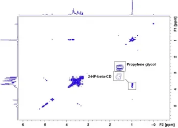
the integration of this signal (Fig. 6). This substance has been iden-tified as propylene glycol which is present in high concentration in SporanoxÒfor its ability to increase the water solubility of
itra-conazole. Deconvolution analysis (Total-Line-Shape) could be used to distinguish overlapping signals but is not applicable in this case given that signal of interest (2-HP-b-CD) is completely hidden by the much more intense signal of propylene glycol. We therefore chose to use the signal at 5.2 ppm which seems free of interference and performed three calibration curves by adding known concen-trations of 2-HP-b-CD directly to the oral solution based on a linear regression after square root transformation. As for the other formulations, the slope (0.9642 ± 0.006) is close to the slope found with a calibration curve of 2-HP-b-CD in water (1.059 ± 0.007) and the RSS (0.0054 ± 0.0055) is very low which confirmed the good fit of the regression model.
4. Conclusions
In this study, we presented for the first time a convenient method for the quantification of 2-HP-b-CD in solution based on NMR. This technique was successfully applied to various liquid pharmaceutical formulations and confirmed its ability to specifi-cally quantify 2-HP-b-CD in various and complex matrices which highlight its interest as a quality control instrument and as an interesting tool for the development of new formulations where cyclodextrin concentration could influence the properties of the formulation (such as dry powder for inhalation). Moreover, this approach fills the essential need of developing convenient analyti-cal technique for the quantification of CDs in formulations where they are used as active ingredients. This method could also be applied to the quantification of other cyclodextrins such as b-cyclodextrin and 2-HP-
c
-CD by using the same process. Even if q-NMR could not be applied in the case of CDs, this method dis-played two major advantages: samples pre-treatment and sep-aration methods are not required leading to a rapid, easy and specific approach for CDs quantification. Indeed, the NMR mea-surement only takes four minutes, while the sample preparation only requires a simple addition of deuterated water in order to apply a special pre-saturation sequence to reduce the water signal and of reference compounds. The measurement can therefore be realized directly in the pharmaceutical liquid formulation without any separation or extraction steps. Even if a generic method is dif-ficult to develop in this field, this work opens new perspectives for the quantification of excipients and cyclodextrins in complex media.Acknowledgement
Authors thank Lucas Dewalque for critical comments and the ‘‘Fond National de la Recherche Scientifique’’ – FNRS from which Pascal de Tullio is senior research associate.
References
Agueros, M., Campanero, M.A., lrache, J.M., 2005. Simultaneous quantification of different cyclodextrins and Gantrez by HPLC with evaporative light scattering detection. J. Pharm. Biomed. Anal. 39, 495–502.
Arun, R., 2008. Cyclodextrins as drug carrier molecule: a review. Sci. Pharm. 76, 567–598.
Bertholet, P., Gueders, M., Dive, G., Albert, A., Barillaro, V., Perly, B., Cataldo, D., Piel, G., Delattre, L., Evrard, B., 2005. The effect of cyclodextrins on the aqueous solubility of a new MMP inhibitor: phase solubility, 1H-NMR spectroscopy and molecular modeling studies, preparation and stability study of nebulizable solutions. J. Pharm. Pharm. Sci. 8, 164–175.
Boulanger, B., Rozet, E., Moonen, F., Rudaz, S., Hubert, P., 2009. A risk-based analysis of the AAPS conference report on quantitative bioanalytical methods validation and implementation. J. Chromatogr. B, Anal. Technol. Biomed. Life Sci. 877, 2235–2243.
Brewster, M.E., Loftsson, T., 2007. Cyclodextrins as pharmaceutical solubilizers. Adv. Drug Deliv. Rev. 59, 645–666.
Camargo, F., Erickson, R.P., Garver, W.S., Hossain, G.S., Carbone, P.N., Heidenreich, R.A., Blanchard, J., 2001. Cyclodextrins in the treatment of a mouse model of Niemann-Pick C disease. Life Sci. 70, 131–142.
Carrier, R.L., Miller, L.A., Ahmed, I., 2007. The utility of cyclodextrins for enhancing oral bioavailability. J. Control. Release: Off. J. Control. Release Soc. 123, 78–99. Del Valle, E.M.M., 2004. Cyclodextrins and their uses: a review. Process Biochem. 39,
1033–1046.
Hammes, W., Bourscheidt, C., Büchsler, U., Stodt, G., Bökens, H., 2000. Quantitative
determination of a-cyclodextrin in human plasma by liquid
chromatography/positive ion electrospray mass spectrometry. J. Mass Spectrom. 35, 378–384.
Higashi, K., Ideura, S., Waraya, H., Moribe, K., Yamamoto, K., 2009. Incorporation of salicylic acid molecules into the intermolecular spaces of c -cyclodextrin-polypseudorotaxane. Cryst. Growth Des. 9, 4243–4246.
Holzgrabe, U., Deubner, R., Schollmayer, C., Waibel, B., 2005. Quantitative NMR spectroscopy – applications in drug analysis. J. Pharm. Biomed. Anal. 38, 806– 812.
Hubert, P., Nguyen-Huu, J.J., Boulanger, B., Chapuzet, E., Chiap, P., Cohen, N., Compagnon, P.A., Dewe, W., Feinberg, M., Lallier, M., Laurentie, M., Mercier, N., Muzard, G., Nivet, C., Valat, L., 2004. Harmonization of strategies for the validation of quantitative analytical procedures. A SFSTP proposal – Part I. J. Pharm. Biomed. Anal. 36, 579–586.
Hui Jiang, R.S., Fujiwara, Hideji, De Meulder, Marc, de Vries, Ronald, Gong, Yong, Kao, Mark, Yanjanin, Nicole M., Carillo-Carasco, Nuria, Xu, Xin, Elizabeth, Ottinger, M.W., Daniel, S., Ory, Jiang, Xuntian, 2014. Development and validation of sensitive LC-MS/MS assays for quantification of HP-b-CD in human plasma and CSF. J. Lipid Res. 55.
International Conference on Harmonization (ICH) of Technical Requirements for Registration of Pharmaceuticals for Human Use, T.Q.R.V.o.A.P.T.a.M., Geneva, 2005. International Conference on Harmonization (ICH) of Technical Requirements for Registration of Pharmaceuticals for Human Use, Topic Q2 (R1): Validation of Analytical Procedures: Text and Methodology, Geneva. Lis-Cieplak, A., Sitkowski, J., Kolodziejski, W., 2014. Comparative proton nuclear
magnetic resonance studies of amantadine complexes formed in aqueous solutions with three major cyclodextrins. J. Pharm. Sci. 103, 274–282. Loftsson, T., Brewster, M.E., 2010. Pharmaceutical applications of cyclodextrins:
basic science and product development. J. Pharm. Pharmacol. 62, 1607–1621. Malet-Martino, M., Holzgrabe, U., 2011. NMR techniques in biomedical and
pharmaceutical analysis. J. Pharm. Biomed. Anal. 55, 1–15.
Matilainen, L., Toropainen, T., Vihola, H., Hirvonen, J., Jarvinen, T., Jarho, P., Jarvinen, K., 2008. In vitro toxicity and permeation of cyclodextrins in Calu-3 cells. J. Control. Release: Off. J. Control. Release Soc. 126, 10–16.
Nitalikar, M., Sakarkar, D., Jain, P., 2012. The cyclodextrins: a review. J. Curr. Pharm. Res. 10, 01–06.
Norman, R., Smith, H., 1998. Applied Regression Analysis, third ed. Wiley. Ono, N., Miyamoto, Y., Ishiguro, T., Motoyama, K., Hirayama, F., Iohara, D., Seo, H.,
Tsuruta, S., Arima, H., Uekama, K., 2011. Reduction of bitterness of antihistaminic drugs by complexation with beta-cyclodextrins. J. Pharm. Sci. 100, 1935–1943.
Pauli, G.F., Godecke, T., Jaki, B.U., Lankin, D.C., 2012. Quantitative 1H NMR. Development and potential of an analytical method: an update. J. Nat. Prod. 75, 834–851.
Rozet, E., Ceccato, A., Hubert, C., Ziemons, E., Oprean, R., Rudaz, S., Boulanger, B., Hubert, P., 2007. Using tolerance intervals in pre-study validation of analytical methods to predict in-study results. J. Chromatogr. A 1158, 111–125. Rozet, E., Marini, R.D., Ziemons, E., Boulanger, B., Hubert, P., 2011. Advances in
validation, risk and uncertainty assessment of bioanalytical methods. J. Pharm. Biomed. Anal. 55, 848–858.
Schneider, H.-J., Hacket, F., Rüdiger, V., 1998. NMR studies of cyclodextrins and cyclodextrin complexes. Chem. Rev. 98, 1755–1785.
Fig. 6. 2D COSY NMR spectroscopy of SporanoxÒ
oral solution. The two overlapping signals at 1.1 ppm (corresponding to 2-HP-b-CD and propylene glycol) are enlarged.
Stella, V.J., He, Q., 2008. Cyclodextrins. Toxicol. Pathol. 36, 30–42.
Szejtli, J., Szente, L., 2005. Elimination of bitter, disgusting tastes of drugs and foods by cyclodextrins. Eur. J. Pharm. Biopharm.: Off. J. Arbeitsgemeinschaft Pharm. Verfahrenstechnik 61, 115–125.
Szeman, J., Gerloczy, A., Csabai, K., Szejtli, J., Kis, G.L., Su, P., Chau, R.Y., Jacober, A., 2002. High-performance liquid chromatographic determination of 2-hydroxypropyl-g-cyclodextrin in different biological fluids based on cyclodextrin enhanced fluorescence. J. Chromatogr. B 774, 157–164.
Vogt, F.G., Strohmeier, M., 2012. 2D solid-state NMR analysis of inclusion in drug-cyclodextrin complexes. Mol. Pharm. 9, 3357–3374.
Vozone, C.M., Cabral Marques, H.M., 2002. Complexation of budesonide in cyclodextrins and particle aerodynamic characterization of the complex solid form for dry powder inhalation. J. Incl. Phenom. Macrocycl. Chem. 44, 5. Yang, B., Lin, J., Chen, Y., Liu, Y., 2009. Artemether/hydroxypropyl-beta-cyclodextrin
host-guest system: characterization, phase-solubility and inclusion mode. Bioorg. Med. Chem. 17, 6311–6317.