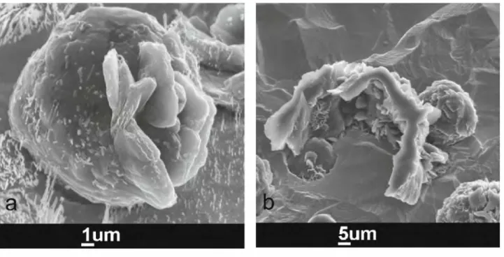
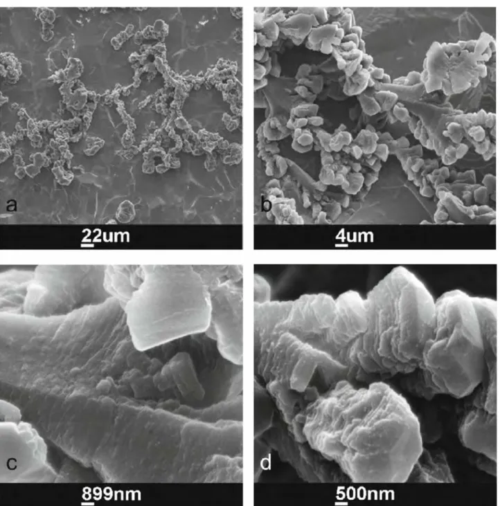
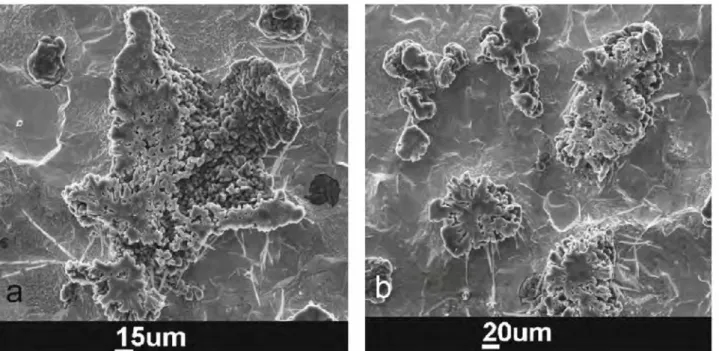
Degenerated Graphite Growth in Ductile Iron
9
0
0
Texte intégral
Figure
+4
Documents relatifs
