Thymus and T cells
Vincent Geenen, Fabienne Brilot, Isabelle Hansenne, Chantal Renard and Henri Martens
Keywords: thymus, self-tolerance, autoimmunity, neuroendocrine self-antigen, oxytocin,
type 1 diabetes, insulin-like growth factor 2
1. Introduction
Evidence for intimate interconnections between the three major systems of cell
communication, the nervous, endocrine and immune systems, has opened important novel research perspectives. Neuroimmune-endocrine interactions are now established as crucial factors for the control of body development and homeostasis. In distant species and
invertebrates, the foundations of both the neuroendocrine system and innate immunity were coexisting until now without any apparent problem. Some 300 millions years ago, in a relatively short period after agnathan fishes (e.g., hagfish and lamprey), adaptive immunity emerged in the first gnathostomes, cartilaginous fishes (e.g., shark and ray). Somatic
recombination machinery characterizes adaptive immunity and is responsible for the random generation of the huge diversity of immune receptors able to recognize infectious antigens. The emergence of this novel form of immune defenses exerted a so potent pressure that structures and mechanisms developed along the paths of lymphocyte traffic to impose immunological self-tolerance, that is, the inability of the immune system to attack the host organism. Together with the generation of diversity and memory, self-tolerance constitutes a fundamental property of the immune system. The progressive rise in the level of immune diversity and complexity also explains why self-tolerance failures (i.e., organ-specific autoimmune diseases) were increasingly detected during evolution, the maximum being currently observed in the human species. The first thymus appeared in cartilaginous fishes (chondrichthyes), concomitantly with the emergence of rudimental forms of adaptive immunity. Though some forms of tolerance induction already takes place in primary hemopoietic sites (fetal liver and bone marrow), antigen-dependent B-cell tolerance is primarily due to an absence of T-cell help. Among all lymphoid structures, the thymus is the only organ specialized in the establishment of central self-tolerance. The thymus crucially stands at the crossroad between the immune and neuroendocrine systems. In this organ responsible for thymopoiesis—T-cell generation—(Kong et al., 1998), the neuroendocrine system regulates the process of T-cell differentiation from the very early stages. In addition, T lymphocytes undergo inside the thymus a complex educative process that establishes central T-cell self-tolerance of neuroendocrine principles (Geenen et al., 1992; Martens et al., 1996). Within the thymus, a confrontation permanently occurs between previously
established neuroendocrine principles and a recent system equipped with recombination machinery promoting stochastic generation of response diversity. Contrary to a previous assumption, the thymus functions throughout life (Poulin et al., 1999; Geenen et al., 2003) and plays a fundamental role in the recovery of a competent T-cell repertoire after intensive chemotherapy or during highly active antiretroviral therapy (Mackall et al., 1995; Douek et al., 1998).
2. Development and cytoarchitecture of the thymus
In the human species, the thymus is located in the anterior mediastinum, overlying the heart and the basal blood vessels. Its general shape resembles the leaves of the thyme plant. It is
divided into two lobes surrounded by a connective capsule. By sending incomplete septa, the capsule delineates thymic lobules further divided into distinct zones: subcapsular cortex, cortex, cortico-medullary junction, and medulla. The generation of a peripheral CD4+ and CD8+ T-cell pool occurs in the thymic organ that provides the appropriate cellular
microenvironments. Thymocyte development is not a pure autonomous genetic program, but it closely depends on interactions of pre-T cells with the complex cellular network of the thymus.
Thymic epithelial cells (TECs) constitute the major component of the thymus stroma and
mainly derive from the endoderm of the third branchial pouch. In this endoderm, recent studies have identified epithelial stem cells able to form a thymus with normal phenotype and function in vivo (Bennett et al., 2002; Gill et al., 2002). Further development of this primitive epithelial rudiment depends upon an important mesenchymal contribution from the cephalic neural crest (Bockman, 1997). Some human diseases (and animal models) include a defective thymus development leading to primary immune deficiencies. DiGeorge's syndrome
associates congenital absence (or hypoplasia) of thymus, parathyroids, and defects in the heart and truncal vessels. This syndrome results from a migration failure of the cephalic neural crest. Mice in whom the homeobox gene Hoxa3 has been disrupted present thymic aplasia, parathyroid hypoplasia, and frequent defects in heart and great vessels. Wild animals with immune deficiencies most closely related to DiGeorge's syndrome are nude mice with hairlessness and lack of thymic development resulting from defect in epithelial cells. The nude phenotype is caused by mutations in nude gene on murine chromosome 11 encoding the transcription factor winged-helix nude (whn or Foxn1). In the absence of whn, the thymus anlage still develops but is filled with primitive epithelial cells that do not specialize and segregate into subregions. Other transcription factors (Pax-1, Pax-9) also contribute to the ontogeny of thymic epithelium.
Thymic nurse cells (TNCs) are large epithelial cells located in the subcapsular and outer
cortex. They contain the subcellular equipment and the enzymatic machinery required for antigen processing and presentation. Though they express various neuroendocrine markers and synthesize neuropeptides, their precise embryonic origin remains unknown.
Thymic dendritic cells (DCs), mainly located at the cortico-medullary junction, and
macrophages, without any precise topography, derive from bone marrow and constitute the
other components of the thymic stroma.
The thymus also receives sympathetic autonomic innervation (Bulloch, 1985). A number of neurotransmitters have been identified by immunocytochemistry within thymic sensory nerve fibers.
The thymus is classically described as a lymphoepithelial structure. However, it is important to understand that the lymphoid compartment is a mobile population that originate as T-cell progenitors in primitive hemopoietic sites, migrate into the thymus, and differentiate through close contacts with the surrounding TECs/TNCs, DCs and macrophages.
3. The dual role of the thymus in T-cell differentiation
The pioneering studies conducted by Miller and Osoba (1967) have demonstrated the active role played by the thymus in the generation of peripheral T lymphocytes, and its previously assumed function as an endocrine gland was less and less considered. From the primary sites
of hemopoiesis (fetal yolk sac, liver, and then bone marrow), possibly under the control of some chemokines, T-cell precursors enter into the thymus through capillaries at the cortico-medullary junction, then migrate to the subcapsular cortex (Fig. 1). An intense proliferation of T-cell blasts takes place in the outer cortex. The transmembrane molecule Notch-1 plays an important role in the induction of T-lineage restriction and commitment. Then, intimate contacts with TECs/TNCs promote the activity of the recombination-activating genes (RAG 1 and RAG 2). RAG-catalyzed generation of T cell diversity occurs through the random
recombination of gene segments encoding the variable parts of the T-cell antigen receptor (TCR) (Fugmann et al., 2000). TCR recombination and expression by thymocytes (pre-T cells) constitutes a pivotal event because, among the number of about 108 possible combinations, many of them are able to recognize self-antigens presented by the major histocompatibility complex (MHC) proteins expressed by TECs/TNCs, DCs and
macrophages. The so-called thymic negative selection represents the massive deletion of T-cell clones expressing self-reactive TCR and results either from programmed T-cell death or developmental arrest. Thymus censorship is responsible for the establishment of central
T-cell self-tolerance (Nossal, 1994). This fundamental process is very powerful since, from 100
T-cell precursors, only 1-2 T cells will leave the thymus in a state of self-tolerance and competence against foreign antigens.
The concomitant occurrence within the same organ of opposite events such as T- cell negative selection and T-cell life/diversity generation represents a prominent problem in modern biology. A number of models have been proposed to explain the paradox of thymus physiology. A first model attributed different functional roles to the cells of thymic stroma: TECs/TNCs would be mainly responsible for inducing T-cell proliferation and development, whereas thymic DCs and macrophages, as dedicated antigen-presenting cells (APCs), would mediate T-cell negative selection. Although DCs and macrophages are very efficient in inducing T-cell deletion, this model is no more valid since it is now demonstrated that TECs/TNCs are also able to present self-antigens and provoke T-cell negative selection. Medullary TECs in particular are highly competent in achieving self-tolerance. The most popular model is called the avidity model (Ashton-Rickardt et al., 1994): T cells bearing a TCR with a high affinity for self-antigen at high density will undergo clonal deletion, while those with a low-affinity TCR and/or in presence of low-density self-antigen survive and further develop. However, the affinity of a given TCR for its cognate antigen is rather low (with a KD around 10-7 M), and thus one may question the physiological meaning of lower
affinities. The model of the dual role played by the repertoire of thymic neuroendocrine-related genes recapitulates at the molecular level the paradox of thymus physiology. According to this model, thymic neuroendocrine-related precursors are the source of two distinct types of interactions with immature T cells. On the one hand these precursors deliver
ligands that are not secreted but targeted to the outer surface of TECs/TNCs from where they
can bind to neuroendocrine receptors expressed by immature T cells. A high affinity (KD
ranging from 10-11 to 10-9 M) and a low specificity characterize this cryptocrine type of signaling. On the other hand the same precursors undergo antigen processing and are the source of neuroendocrine self-peptides presented by the thymic MHC machinery. A negative signal would result from the low-affinity but highly specific binding of the complex
MHC/neuroendocrine self-antigen to its cognate TCR. According to the theory of clonal deletion, the central T-cell self-tolerance of neuroendocrine principles results from the intrathymic presentation of neuroendocrine self-antigens (Fig. 2).
In addition to deletion of reactive T-cell clones, the establishment of central thymic self-tolerance also includes the generation of regulatory T cells (TREG) that are enriched in the
CD4+ CD25+ T cell subset (Shevach, 2000; Sakagushi, 2001). It remains to be determined how far the thymic self-antigen repertoire contributes to the generation of TREG lymphocytes. 4. The nature of neuroendocrine self (Table I)
From the investigation of intrathymic expression of neuroendocrine-related self-peptide precursor genes, a series of properties were drawn to define the nature of neuroendocrine self. (1) Neuroendocrine self-antigens usually correspond to peptide sequences that have been highly conserved throughout the evolution of one given family. (2) A hierarchy characterizes their expression pattern. In the neurohypophysial family, oxytocin (OT) is the dominant peptide synthesized by TECs/TNCs from different species (Geenen et al., 1986). The binding of OT to OT receptor (OTR) expressed by pre-T cells induces a very rapid phosphorylation of focal adhesion related kinases. This event should play a major role in the promotion of
immunological “synapses” between immature T lymphocytes and thymic APCs
(TECs/TNCs, macrophages and DCs). With regard to tachykinins, neurokinin A (NKA)—but not substance P (SP)—is the peptide generated from the processing by TECs of the
preprotachykinin A (PPT-A) gene product (Ericsson et al., 1990). All the genes of the insulin family are expressed according to a precise hierarchy and topography: IGF2 (TECs/TNCs) >
IGF1 (thymic macrophages) >> INS (thymic DCs and/or medullary TECs) (Geenen and
Lefèbvre, 1998; Derbinski et al., 2001). This hierarchical pattern is meaningful since the tolerogenic response primarily concerns the dominant epitopes of a protein family (Sercarz et al., 1993). In fetal thymic organ cultures, the blockade of thymic IGF-mediated signaling, at the level of IGF ligands (in particular IGF-2) or IGF receptors, severely interferes with early stages of T-cell differentiation and proliferation. On the contrary, the treatment of fetal thymic organ cultures with antibodies against (pro)insulin does not exert any significant effect on T-cell differentiation (Kecha et al., 2000). (3) Neuroendocrine precursors are not processed in a classic fashion, but they undergo antigenic processing for presentation by, or in association with, thymic MHC proteins. (4) Some differences exist in the processing between thymic APCs and dedicated peripheral APCs. At least for some neuroendocrine self-antigens (OT and neurotensin), those differences imply that their presentation by thymic APCs is not restricted by MHC alleles like presentation of foreign antigens by peripheral APCs (Vanneste et al., 1997; Geenen et al., 1999).
During ontogeny of Balb/c mice, OT transcripts are detected on fetal day 13 (E13) both in thymus and brain, whereas vasopressin (VP) gene transcription starts on E14 in the brain and is clearly detected in the thymus only on E15. The earlier OT expression in the thymus supports the role of thymic OT in the induction of central T-cell tolerance of the neurohypophysial peptides before their appearance in the hypothalamic magnocellular
neurons (Fig. 3). The expression of the neurohypophysial receptor genes (OTR, V1R, V2R and
V3R) was investigated on murine thymic CD4- CD8- lymphoma cell lines, as well as on murine thymic T-cell subsets. OTR transcripts are detected in CD4-CD8-, CD4+ CD8+ and CD8+ cells, while a faint V3R expression is restricted to CD4+ CD8+ and CD8+ cells. V1R and
V2R expression could not be detected on any T cell subset. In fetal thymic organ cultures, a
specific OTR antagonist increases late T-cell apoptosis confirming the involvement of OT/OTR-signaling in T-cell proliferation/survival (Hansenne et al., in press).
5. The development of autoimmunity as a defect in thymic self-tolerance establishment
In 1992, a defect in thymic T-cell education to recognize and to tolerate self-antigens was hypothesized to play a pivotal role in the development of hypothalamus-specific
autoimmunity (Geenen et al., 1992; Robert et al., 1992). As already hypothesized by Burnet (1973), the pathogenesis of autoimmune diseases could result from the appearance of
“forbidden” self-reactive clones in the peripheral lymphocyte repertoire. Since the thymus is the primary site for induction of self-tolerance, thorough investigation of a defective thymic censorship should provide the scientific community with important keys to understand the mechanisms underlying the development of autoimmune responses. A number of
abnormalities of thymic morphology and cytoarchitecture have been described for several autoimmune disorders. The expression of the transcription factor autoimmune regulator (AIRE) gene is maximal in murine medullary TECs, but it is absent in the thymus epithelium of diabetic NOD mice (Heino et al., 2000). Type 1 diabetes (juvenile or insulin-dependent diabetes) is a chronic devastating disease resulting from an autoimmune response specifically oriented against pancreatic islet ß cells, the only cells secreting insulin according to the endocrine model. In accordance with the above hypothesis, INS transcripts were measured at lower levels in the thymus of human fetuses with short class I VNTR (variable number of tandem repeats) alleles, a genetic trait of type 1 diabetes susceptibility (Vafiadis et al., 1997; Pugliese et al., 1997). The expression of insulin-related genes was also analyzed in thymus, liver and brain of a common animal model of type 1 diabetes, the bio-breeding (BB rat). A defect of Igf2 expression was evidenced in the thymus of more than 80% of diabetes-prone BB rats, in close accordance with the incidence of diabetes in this rat strain (Kecha-Kamoun et al., 2001). In these rats, this tissue-specific defect in Igf2 transcription could contribute both to lymphopenia (including a lack of antigen-specific TREG cells that control autoimmune
diabetes), as well as to the absence of central self-tolerance of insulin family. As a
consequence of the defective thymic censorship, self-reactive T cells bearing TCR oriented against dominant epitopes of insulin-related peptides continuously migrate from the thymus and enrich the peripheral pool with self-reactive T cells exhibiting a potential cytotoxic power against islet ß cells. Under certain environmental influences, a molecular bridge could be installed between the target autoantigenic epitopes leading to activation of the self-reactive T-cell pool and subsequent ß-T-cell destruction (Fig. 4). Diabetes-prone NOD mice also display a defect in central tolerance (negative selection) of T lymphocytes (Kishimoto and Sprent, 2001). The transcription of genes encoding tissue-specific antigens (including Ot, Igf2, Ins2, and Npy) is markedly down-regulated in the thymus epithelium of Aire-/- mice (Anderson et al., 2002). Moreover, Aire deficiency results in an almost complete thymic failure to delete CD4+ T cells with high affinity for a pancreas-specific antigen (Liston et al., 2003). Thus, more and more arguments provide strong evidence that a defect in thymic self-tolerance plays a pivotal role in the pathogenesis and development of organ-specific autoimmunity. The origin of systemic (non-organ specific) autoimmunity is still a matter of debate and could be elucidated through further investigations of the non-specific, innate immune system.
6. Pharmacological applications and conclusions
The study of neuroendocrine gene expression and precursor processing in the thymus led to the identification of neuroendocrine self-peptides. With regard to insulin-related gene expression in the thymus, IGF-2—a prominent fetal growth factor—was identified as the dominant self-peptide precursor of the insulin family expressed in the thymus from different species, including man. This observation is in close accordance with the theory of self-recognition which, according to F.M. Burnet, is not an inherited property but is gradually acquired in the course of fetal life. Although the tolerogenic properties of neuroendocrine self-peptides remain to be further documented, they are suspected from what is known about the immunological tolerance of classic hormones. The development of specific antibodies by active immunization (i.e., experimental breakdown of self-tolerance) revealed that OT is
more tolerated than VP, and that IGF-2 is also more tolerated than IGF-1, and much more than insulin. Some cases of diabetes insipidus result from an autoimmune process against VP-producing hypothalamic neurons (infundibulohypothalamitis) (Scherbaum et al., 1985; Imura et al., 1993). Insulin is a primary autoantigen tackled by the autoimmune response observed in type 1 diabetes and this might result from its very low expression in the thymus. At the opposite, autoimmunity has never been observed against OT and IGF-2. The strong tolerance of these peptides, resulting from high expression of their genes in the thymus, may be
considered as the consequence of evolutive pressure to protect fundamental processes such as species reproduction and individual ontogeny (Geenen et al., 1999). The putative pathogenic role of forbidden self-reactive T cells against IGF-2 deserves to be further analyzed through immunization of Igf2-/- mice.
Thus although VP and insulin behaves as immunogenic auto-antigens of their respective families, OT and IGF-2 may be viewed as the tolerogenic self-peptide precursors of
neurohypophysial and insulin families, respectively. Quite provocatively, perhaps is it now appropriate to establish a distinction between an auto-antigen and a self-antigen (Fig. 5). According to this distinction, auto-antigens can be considered as “altered” self-antigens. Though they are highly homologous, i.e., evolutionarily related, they are not identical and this biochemical difference theoretically drives opposite immune responses (i.e., immunogenic vs. tolerogenic responses). This proved to be correct in a recent preclinical study. A dominant epitope of insulin (Ins B9-23) and the homologous sequence of IGF-2 (IGF-2 B11-25) compete
for binding to DQ8, a MHC class II allele conferring major susceptibility to type 1 diabetes. In young DQ8+ type 1 diabetic patients, Ins B9-23 presentation by DQ8 elicits a dominant IFN-γ secretion by isolated peripheral blood mononuclear cells, whereas presentation of IGF-2 B11-25 promotes a dominant regulatory IL-10 secretion. The powerful tolerogenic properties of
thymic self-antigens should be soon exploited to cure and/or prevent severe autoimmune diseases (such as type 1 diabetes) which constitute the heavy tribute, mainly paid by mankind, for the diversity, complexity, and efficiency of its system of immune defenses.
7. References
Anderson MA, Venanzi ES, Klein L, Chen Z, et al. (2002): Projection of an immunological self shadow within the thymus by the Aire protein. Science 298:1395-1401
Ashton-Rickardt PG, Bandeira A, Delaney JR, Van Kaer L, et al. (1994): Evidence for a differential avidity model of T-cell selection in the thymus. Cell 74:577-583
Bennett AR, Farley A, Blair NF, Gordon J, Sharp L, Blackburn CC (2002): Identification and characterization of thymic epithelial progenitor cells. Immunity 16:803-814
Bockman DE (1997): Development of the thymus. Microsc Res Tech 38: 209-215
Brilot F, Chehadeh W, Martens H, Renard C, Geenen V, Hober D (2002): Persistent infection of human thymic epithelial cells by coxsackievirus B4. J Virol 76:5260-5265
Bulloch K (1985): Neuroanatomy of lymphoid tissue: a review. In Neural Modulation of
Immunity, Guillemin R, Cohn M, Melnechuk T, eds. San Diego: Academic Press, pp.
Burnet FM (1973): A reassessment of the forbidden clone hypothesis of autoimmune diseases. Aust J Exp Biol Med Sci 50:1-9
Derbinski J, Schulte A, Kyewski B, Klein L (2001): Promiscuous gene expression in medullary thymic epithelial cells mirrors the peripheral self. Nat Immunol 2:1032-1039 Douek DC, Macfarland RD, Keiser PH, Gage EA, et al. (1998): Changes in thymic function with age and during the treatment of HIV infection. Nature 396:690-695
Ericsson A, Geenen V, Robert F, Legros JJ, et al. (1990): Expression of preprotachykinin-A and neuropeptide-Y messenger RNA in the thymus. Mol Endocr 4:1211-1218
Fugmann SD, Lee AI, Schockett PE, Villey IJ, Schatz DG (2000): The RAG proteins and V(D)J recombination: complexes, ends and transposition. Annu Rev Immunol 18:495-522 Geenen V, Legros JJ, Franchimont P, Baudrihaye M, et al. (1986): The thymus as a neuroendocrine organ: coexistence of oxytocin and neurophysin in the human thymus.
Science 232:508-511
Geenen V, Robert F, Martens H, De Groote D, Franchimont P (1992): The thymic education of developing T cells in self neuroendocrine principles. J Endocrinol Invest 15:621-629 Geenen V, Achour I, Robert F, Vandersmissen E, et al. (1993): Evidence that insulin-like growth factor 2 (IGF-2) is the dominant thymic peptide of the insulin superfamily. Thymus 21:115-127
Geenen V, Lefèbvre PJ (1998): The intrathymic expression of insulin-related genes:
implications in pathophysiology and prevention of type 1 diabetes. Diabetes/Metab Rev 14: 95-103
Geenen V, Kecha O, Brilot F, Charlet-Renard C, Martens H (1999): The thymic repertoire of neuroendocrine-related self-antigens: biological role in T cell selection and pharmacological implications. NeuroImmunoModulation 6:115-125
Geenen V, Poulin JF, Dion ML, Martens H, et al. (2003): Quantification of T cell receptor rearrangement excision circles to estimate thymic function: an important new tool for endocrine-immune physiology. J Endocrinol 176:305-311
Gill J, Malin M, Holländer GA, Boyd R (2002): Generation of a complete thymic microenvironment by MTS24+ thymic epithelial cells. Nat. Immunol. 3:635-642
Hansenne I, Rasier G, Defresne MP, Greimers R, et al. (2003): Neurohypophysial receptor gene expression by thymic T cell subsets and thymic T cell lymphoma cell lines. Dev
Immunol, in press
Heino M, Peterson P, Sillanpää N, Guérin S, et al. (2000): RNA and protein expression of the murine autoimmune regulator gene (Aire) in normal, Rel-B-deficient and in NOD mouse. Eur
Imura H, Nakao K, Shimatsu A, Ogawa Y, et al. (1993): Lymphocytic
infundibuloneurohypophysitis as a cause of central diabetes insipidus. N Engl J Med 329: 683-687
Kecha O, Brilot F, Martens H, Franchimont N, et al. (2000): Involvement of insulin-like growth factors in early T cell development: a study using fetal thymic organ cultures.
Endocrinology 141:1209-1217
Kecha-Kamoun O, Achour I, Martens H, Collette J, et al. (2001): Thymic expression of insulin-related genes in an animal model of autoimmune type 1 diabetes. Diabetes/Metab Res
Rev 17:146-152
Kishimoto H, Sprent J (2001): A defect in central tolerance in NOD mice. Nat Immunol 2: 1025-1031
Kong FK, Chen CH, Cooper MD (1998): Thymic function can be accurately monitored by the level of recent T cell emigrants in the circulation. Immunity 18:514-518
Liston A, Lesage S, Wilson J, Peltonen L, Goodnow CC (2003): Aire regulates negative selection of organ-specific T cells. Nat Immunol 4:350-354
Mackall CL, Fleisher TA, Brown MR, Andrich MP, et al. (1995): Age, thymopoiesis, and CD4+ T-lymphocyte regeneration after intensive chemotherapy. N Engl J Med 332:143-149 Martens H, Goxe B, Geenen V (1996): The thymic repertoire of neuroendocrine
self-peptides: physiological implications in T-cell life and death. Immunol Today 17:312-317 Martens H, Kecha O, Charlet-Renard C, Defresne MP, Geenen V (1998): Neurohypophysial peptides stimulate the phosphorylation of pre-T cell focal adhesion kinases.
Neuroendocrinology 67:282-289
Miller JFAP, Osoba D (1967): Current concepts of the immunological function of the thymus. Physiol Rev 47:437-520
Nossal GJV (1994): Negative selection of lymphocytes. Cell 76:229-239
Poulin JF, Viswanathan MN, Harris JM, Komanduri KV, et al. (1999): Direct evidence for thymic function in adult humans. J Exp Med 190:479-486
Pugliese A, Zeller M, Fernandez AJr, Zalcberg LJ, et al. (1997): The insulin gene is
transcribed in the human thymus and transcription levels correlate with allelic variation at the
INS VNTR-IDDM2 susceptibility locus for type 1 diabetes. Nat Genet 15:293-297
Robert FR, Martens H, Cormann N, Benhida A, Schoenen J, Geenen V (1992): The
recognition of hypothalamo-neurohypophysial functions by developing T cells. Dev Immunol 2:131-140
Vafiadis P, Bennett ST, Todd JA, Nadeau J, et al. (1997): Insulin expression in human thymus is modulated by INS VNTR alleles at the IDDM2 locus. Nat Genet 15:289-292
Sakagushi S (2001): Policing the regulators. Nat Immunol 2:283-284
Scherbaum WA, Bottazzo GF, Czernichow P, Wass JAH, Doniach D (1985): Role of autoimmunity in central diabetes insipidus. Front Horm Res 13:232-239
Sercarz EE, Lehmann PV, Ametani A, Benichou G, Miller A, Moudgil K (1993): Dominance and crypticity of T cell antigenic determinants. Annu Rev Immunol 11:729-766
Shevach EM (2000): Regulatory T cells in autoimmunity. Annu Rev Immunol 18:423-449 Vanneste Y, Ntodou-Thome A, Vandersmissen E, Charlet C, et al. (1997): Identification of neurotensin-related peptides in human thymic epithelial cell membranes and relationship with major histocompatibility complex class I molecules. J Neuroimmunol 76:161-166
8. Legends to figures
Fig. 1: Pathways of intrathymic T-cell differentiation.
From fetal liver then bone marrow, T-cell progenitors enter the thymus by capillaries at the cortico-medullary junction and migrate to the subcaspsular outer cortex where they divide intensively (T-cell blasts). Interactions between randomly rearranged TCR chains on thymocytes and self-antigen/MHC complexes presented by thymic stromal cells drive the programme of T-cell differentiation in the thymus. From 100 thymocyte precursors that enter the thymus, only 1-2 self-tolerant CD4+ helper and CD8+ cytotoxic emigrate from the thymus to establish the peripheral T-cell repertoire.
Fig. 2: The dual role in the thymus of neuroendocrine-related self-peptide genes.
Neuroendocrine gene-encoded precursors are the source of two types of interactions with pre-T cells. (1) pre-They deliver ligands, which are not secreted but targeted to the outer surface of thymic plasma cell membranes. Those ligands bind to T-cell neuroendocrine receptors leading to activation of transductory pathways such as increase of inositol triphosphate (IP3)
and phosphorylation of focal adhesion kinases (FAK). (2) The same precursors are processed into self-antigens presented by—or in association with—thymic MHC molecules. This presentation will lead to clonal deletion and/or developmental arrest T cells bearing a TCR oriented against neuroendocrine self-antigens and MHC complexes.
Fig. 3: Ontogeny of OT, VP and OTR expression in thymus and brain of Balb/c mouse.
OT transcription is already detectable on E13 in both murine thymus and brain, whereas VP
transcripts are detected in the brain later (E14), and are detected in the thymus only on E15. Though not quantitative, the profile of OTR expression seems to decrease in the thymus and to increase in the brain during fetal development.
Fig. 4: Role of the thymus in the development of the autoimmune response against islet insulin-secreting ß cells.
Together with abnormalities of thymic cytoarchitecture, the thymus-specific defect of IGF2 expression contributes both to lymphopenia (with a decreased number of TREG cells) and to
the absence of central T-cell self-tolerance of the insulin family. The influence of the
in the periphery through the establishment of a bridge between the ß-cell auto-antigen and forbidden self-reactive T cells. Coxsackievirus B4 (CVB4) infection is a major environmental factor implicated in the pathogenesis of type 1 diabetes. Three mechanisms are currently proposed to explain the CVB4 diabetogenic influence: (1) molecular mimicry between sequences of glutamic acid decarboxylase (GAD) and a structural CVB4 protein; (2) direct toxic action of CVB4 toward islet ß cells and bystander activation of autoreactive T cells; and (3) CVB4 infection of the thymic epithelium and interference with the establishment of central T-cell self-tolerance (Brilot et al., 2002).
Fig. 5: Opposite immune responses driven by insulin as an auto-antigen vs. IGF-2 as a self-antigen precursor.
As evidenced by numerous authors, insulin (Ins) is a major ß-cell specific auto-antigen tackled by the diabetogenic autoimmune response. Ins is immunogenic and the sequence B9-23
is a dominant epitope recognized by pathogenic CD4+ T cells. IGF-2 is the dominant member of the insulin family expressed in TECs of different species and this may explain why the immune tolerance of IGF-2 is so hard to break. Both Ins B9-23 and the homologous sequence
IGF-2 B11-25 bind with the same affinity to DQ8, one HLA class II allele conferring major
susceptibility to type 1 diabetes. DQ8 presentation of IGF-2 B11-25 to peripheral blood
mononuclear cells of type 1 diabetic patients elicits a dominant IL-10 secretion as revealed by Elispot analyses. The regulatory/tolerogenic properties of IGF-2 (or IGF-2 B11-25) could be
exploited in a vaccination strategy to reprogram immune self-tolerance of the insulin family.
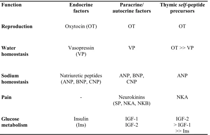
Table I. The organization of the thymic repertoire of neuroendocrine-related self-peptide
precursors/genes Function Endocrine factors Paracrine/ autocrine factors Thymic self-peptide precursors
Reproduction Oxytocin (OT) OT OT
Water homeostasis Vasopressin (VP) VP OT >> VP Sodium homeostasis Natriuretic peptides
(ANP, BNP, CNP) ANP, BNP, CNP ANP
Pain - Neurokinins (SP, NKA, NKB) NKA Glucose metabolism Insulin (Ins) IGF-1 IGF-2 IGF-2 > IGF-1 >> Ins
The fundamental principle is based upon the close homology between peripheral hormones and related thymic self-peptide genes/precursors. Tolerogenic neuroendocrine self-peptide precursors are predominantly conserved throughout evolution of their family and are the dominant members of one family that are expressed in the thymus.
9. Further reading
Anderson G, Jenkinson EJ (2001): Lymphostromal interactions in thymic development and function. Nat Rev Immunol 1:31-40
Andrew D, Aspinall R (2001): IL-7 and not stem cell factor reverses both the increase in apoptosis and the decline of thymopoiesis seen in aged mice. J Immunol 166:1524-1530 Benoist C, Mathis D (1999): T-lymphocyte differentiation and biology. In: Fundamental
Immunology, 4thed., Paul WE, ed. Philadelphia: Lippincott-Raven, pp. 367-409
Bona C, Casares S, Brumeanu TD (1998): Towards development of T-cell vaccines. Immunol
Today 19:126-133
Gainer H, Wray S (1994): Cellular and molecular biology of oxytocin and vasopressin. In:
The Physiology of Reproduction, 2nd ed., Knobil E, Neill JD, eds. New York: Raven Press,
pp. 1099-1129
Geenen V, Kecha O, Brilot F, Hansenne I, et al. (2001): Thymic T-cell tolerance of neuroendocrine functions: physiology and pathophysiology. Cell Mol Biol 47:179-188 Geenen V, Brilot F (2003) The role of the thymus in the development of organ-specific autoimmunity: a way to novel vaccines ? Ann NY Acad Sci, in press
Kamradt T, Mitchison NA (2001): Tolerance and autoimmunity. New Engl J Med 344:655-664
Klein L, Kyewski B (2000): “Promiscuous” expression of tissue antigens in the thymus: a key to T-cell tolerance and autoimmunity? J Mol Med 78:483-494
Markert ML, Boeck A, Hale LP, Kloster AL, et al. (1999): Transplantation of thymus tissue in complete DiGeorge syndrome. N Engl J Med 341:1180-1189
Mentlein R, Kendall MD (2000): The brain and thymus have much in common: a functional analysis of their microenvironments. Immunol Today 21:133-140
Savino W, Dardenne M (2000): Neuroendocrine control of thymus physiology. Endocr Rev 21:412-443
von Boehmer H, Kisielow P (1991): How the immune system learns about self. Sci Am 265: 74-81
10. Acknowledgments
Our studies are supported by the Liege University Special Research Fund, the Fondation Leon Fredericq (Liege Medical School), the National Fund for Scientific Research (Belgium), the Belgian Federation against Cancer, the Belgian Association of Diabetes, the Fondation Vaugrenier for Tolerance Research (Geneva, Switzerland), the European Association for the Study of Diabetes (Dusseldorf, Germany), and the Juvenile Diabetes Research Federation (New York, United States). Our gratitude is due to Kai W. Wucherpfennig (Dana Farber Cancer Institute, Harvard) who analyzed the binding affinity to DQ8 of Ins B9-23 and IGF-2
B11-25. This contribution is dedicated to the memory of Don C. Wiley PhD, and Joseph
Wybran MD, PhD. 11. Related websites www.nature.com/ni/special_focus/autoimmunity/index.html www.endo-society.org www.diabetes.org www.easd.org www.isnim.org www.isni2004.org www.pnirs.org www.mindbody.org