HAL Id: tel-00696553
https://tel.archives-ouvertes.fr/tel-00696553
Submitted on 25 Sep 2012
HAL is a multi-disciplinary open access
archive for the deposit and dissemination of
sci-entific research documents, whether they are
pub-lished or not. The documents may come from
teaching and research institutions in France or
abroad, or from public or private research centers.
L’archive ouverte pluridisciplinaire HAL, est
destinée au dépôt et à la diffusion de documents
scientifiques de niveau recherche, publiés ou non,
émanant des établissements d’enseignement et de
recherche français ou étrangers, des laboratoires
publics ou privés.
Characterization of the sensory processings in the barrel
cortex of the anaesthetized rat.
Luc Estebanez
To cite this version:
Luc Estebanez. Characterization of the sensory processings in the barrel cortex of the anaesthetized
rat.. Neurobiology. Ecole Normale Supérieure de Paris - ENS Paris, 2011. English. �tel-00696553�
Thèse de doctorat de l'École Normale Supérieure
Luc Estebanez
1,2Pour obtenir le grade de
Docteur de l'École Normale Supérieure
Characterization of the sensory processings in
the barrel cortex of the anaesthetized rat.
Caractérisation des traitements sensoriels
dans le cortex à tonneaux du rat anesthésié.
Thèse présentée et soutenue à Gif sur Yvette le _________2011 devant le jury composé de :
Présentée par
École Doctorale n°474
F
V
ront iè re sD
u ivan tFrontières du Vivant
Unité de Neurosciences, Information et Complexité, CNRS UPR-3293,
1 Avenue de la Terrasse, 91198 Gif-sur-Yvette, France. Ecole Normale Supérieure, Institut de Biologie de l’ENS,IBENS, Paris, F-75005 France
Résumé
Chez les rongeurs, le traitement par le cortex à tonneaux de l'information sensorielle en provenance des vibrisses est mal compris.
En effet, malgré l'aide fournie par l'organisation de ce cortex en une reproduction stricte de la topographie de l'appareil sensoriel, il a été difficile jusqu'à présent d'identifier de façon indiscutable le système de filtrage linéaire et non-linéaire qu'utilisent les neurones du cortex à tonneaux durant leur traitement des scènes tactiles auxquelles ils sont exposés. Pour mieux identifier ces traitements corticaux, nous avons développé un système de stimulation vibrissale permettant d'appliquer des déflections sur un grand nombre de vibrisses indépendamment, dans toutes les directions possibles et ce à travers une vaste gamme fréquentielle.
En utilisant ce dispositif de stimulation multivibrissale durant des enregistrements ex-tracellulaires de l'activité électrique des neurones du cortex à tonneaux de rats anesthésiés, nous avons pu identifier plus précisément le filtrage linéaire des stimulations vibrissales, qui s'avère similaire pour tous les neurones que nous avons pu enregistrer.
Par ailleurs, en explorant les aspects non-linéaires du traitement effectué par ces rones, nous avons noté qu'ils se séparent en deux familles distinctes : d'un côté des neu-rones "locaux" qui se sont avérés sensibles à des contrastes locaux dans les déflections multivibrissales. De l'autre, des neurones "globaux" capables au contraire de détecter des situations où les déflections sont similaires pour de nombreuses vibrisses.
Enfin, en effectuant d'autres enregistrements dans la couche II/III du cortex à ton-neaux, cette fois à l'aide d'un microscope deux-photons, nous avons pu noter que les neurones appartenant aux familles locales et globales étaient séparés en groupes spatiale-ment distincts et que la position spatiale des neurones était plus généralespatiale-ment étroitespatiale-ment liée à l'ensemble de leurs propriétés de filtrage des déflections vibrissales.
Abstract
The processing of whisker deflections by rodents barrel cortex neurons is still poorly understood. Indeed, to date, the support provided by the strict mapping of the spatial arrangement of the peripheral sensory apparatus onto the cortical surface has not been sufficient to settle on a reasonable model of whisker processing. In particular, at the mo-ment, the linear and non-linear filtering of whisker stimulations carried in this cortical area are unclear.
In order to tackle this problem, we developed a multiwhisker stimulator that allows the independent deflection of 24 whiskers, in any direction, over a wide frequency band. By combining this whisker stimulation device with electrophysiological recordings carried in the barrel cortex of anaesthetized rats, we could identify a family of linear fil-ters common to all recorded neurons.
In addition, we explored the non-linear responses of these neurons to spatio-temporal combinations of whisker deflections, and we observed two types of neuronal responses. In one side, "local" neurons responded to salient whisker deflections occurring on a single whisker and that contrasted with other whisker deflections. On the other side, "global" neurons were sensitive to the overall level of similarity between the deflections applied across stimulated whiskers.
Finally, we studied the functional response of the neurons found in the layer II/III of this cortex with the help of a two-photon microscope. Using this tool, we found that local and global neuron types were strongly spatially segregated. More generally, we observed a strict mapping of the functional tuning of barrel cortex neurons onto the surface of the rat barrel cortex.
Remerciements
Je tiens à remercier en tout premier lieu Daniel Shulz et Laurent Bourdieu, mes deux di-recteurs de thèse. Pour des raisons que je ne m'explique toujours pas, ils ont bien voulu soutenir, accompagner, nourrir un projet un peu fou combinant deux laboratoires, deux méthodes d'enregistrement neuronal fondamentalement différentes, un projet requiérant le développement de dispositifs techniques lourds.
Pour ce faire, ils ont mis en commun d'importants moyens financiers, et ils m'ont permis de soliciter le savoir faire des meilleurs artisans - terme empreint pour moi d'une grande noblesse - des deux laboratoires : Jean-Yves Tiercelin et Patrick Parra, mécaniciens à l'INAF ont conçu avec moi puis entièrement réalisé l'ensemble des (très nombreuses) pièces mécaniques nécessaires à l'accomplissement de mes projets. Je veux ici leur exprimer mon admiration et ma très profonde gratitude.
Gérard Paresys, électronicien à l'ENS, a lui aussi été solicité pour prendre part dans
cet improbable projet, et il a bien voulu qu'il prenne vie, même si pour cela il dût se lancer dans la conception de circuits électroniques à une échelle bien plus proche de la carte mère que du circuit RC...
Enfin, Gérard Sadoc, informaticien à l'UNIC, parlant le Delphy avec un accent parfait, a toujours résolu mes problèmes même les moins raisonnables (.dat→ .nex → .dat → .nex ?) avec une célérité et une élégance proprement déconcertantes.
Sami El Boustani voulait apprendre à "faire des expériences", on en a fait quelques
unes ensemble... J'ai appris énormément en sa compagnie, de la modélisation des champs récepteurs à la rigueur la plus extrême dans l'interprétation des résultats. J'espère qu'on fera encore un bout de chemin ensemble !
Julien Bertherat a été mon compagnon durant les très très longues journées de
micro-scopie deux-photon, où son endurence a fait merveille. Je resterai toujours ébloui par la perfection de ses craniotomies...
Je n'oublie pas Jean-François Leger, qui a conçu et réalisé avec Julien Bertherat, Lau-rent Bourdieu et Yves Kremer le microscope novateur que j'ai eu la chance d'utiliser.
Je pense aussi à tous ceux qui ont été important pour moi au laboratoire de Gif-sur-Yvette : je pense à Yves Frégac qui dirige le laboratoire et qui n'est pas avare en très bonnes idées, Alain Destexhle, Valérie Ego-Stengel, mais aussi Yves Boubenec, Olivier
Marre, Pierre Yger, Charlotte Deleuze, Pierre-Jean Arduin, Julie LeCam.
Guillaume Hucher et Aurélie Daret on été les chevilles ouvrières de mes expériences,
mais je pense aussi à tant d'autres personnes de l'UNIC avec qui j'ai partagé les JC des lundis ou qui, à des degrés divers, ont contribué au bon déroulement de ce travail de thèse. I still remember the excitement of starting my bachelor summer internship in Garrett
Stanley laboratory. This is the place where I found how mysterious and attractive the study
of animal behaviour and the whisker system could be.
I would also like to acknowledge here the deep impact of my one year stay in Carl Petersen laboratory, EPFL, prior to the onset of my PhD. There, I met some remarkable people, starting with Carl Petersen himself, an ingenuous researcher who has this special gift for asking to right question using the right method. Isabelle Ferezou, Sylvain Crochet and James Poulet were post-doc in the lab at this time. Their respective research has deeply influenced my own work. I am thus honoured that both Sylvain Crochet and James Poulet agreed to take part in my examining committee, together with Miguel Maravall, Garrett Stanley and Daniel Pressnitzer.
Finalement, comment ne pas avoir de gratitude pour Anouch, dont l'amour et le soutien m'ont porté tout au long de ces 4 années, ainsi que pour mes parents, mon frère et ma soeur, qui ont tous apporté leur pierre à ce fragile édifice...
Contents
1 Introduction: identifying the whisker stimulations processed by the barrel
cor-tex 13
1.1 A strategy for the study of cortical processing . . . 13
1.2 Barrel Cortex: beyond anatomical landmarks . . . 14
1.2.1 A labelled line sensory system? . . . 14
1.2.2 Beyond the "principal whisker" model . . . 14
1.2.3 A cortex embedded in a complex network of subcortical nuclei 16 1.3 Looking for the tuning of barrel cortex neurons . . . 17
1.3.1 Studying a sensory system without the support of intuition . . 17
1.3.2 observing free animals use their whisker system . . . 18
1.3.3 Identifying whisker stimulations by the yardstick of neuronal activity . . . 21
1.4 Beyond tuning: processing and context dependence of processing . . 25
1.4.1 Interactions be multiple sensory dimensions can define pow-erful sensory processings . . . 25
1.4.2 Are barrel cortex neurons processings also dependent on sen-sory context? . . . 26
1.5 Aims of the thesis . . . 26
2 The craft of whisker stimulation 29 2.1 Single macrovibrissa mechanical stimulation . . . 30
2.1.1 Electromagnetic stimulators . . . 30
2.1.2 Piezoelectric stimulators . . . 31
2.1.3 Electrical whisking . . . 31
2.2 Towards the faithful reproduction of arbitrary whisker stimulations . . 32
2.2.1 Controled impulse stimulations with mechanical stimulators . 32 2.2.2 Linear models of whisker stimulators for mechanical and soft-ware corrections . . . 32
2.3 Multiple whisker stimulations . . . 33
Patent: Micrometric Movement device and method for implementing same 34 Article: The Matrix: A new tool for probing the whisker-to-barrel system with natural stimuli . . . 51
3 An exploration of barrel cortex neurons linear filters 63 3.1 Barrel cortex neurons linear filters lack a well defined structure as well as consistency across experimental conditions . . . 63
3.1.1 Linear receptive fields are affected by anaesthetics . . . 64 9
3.1.2 Linear receptive fields are affected by the density of the
multi-whisker stimulation . . . 64
3.2 The sensory-motor hypothesis: coding without complex sensory re-ceptive fields . . . 65
3.3 But: are they more elaborate receptive fields hidden in the multidi-mensional whiskerpad sensory space? . . . 66
Article: Spatial structure of multiwhisker receptive fields in the barrel cortex is stimulus dependent . . . 67
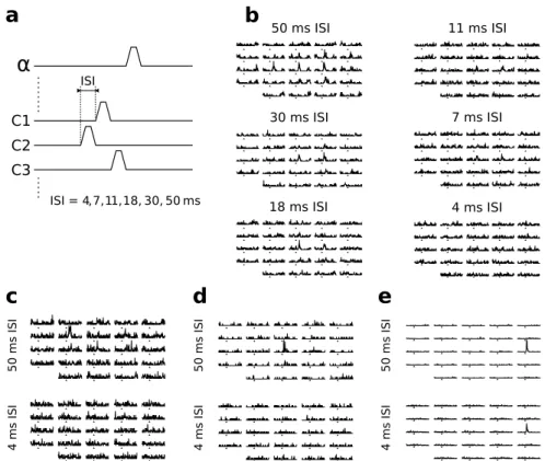
4 Non-linear facilitations and suppressions in barrel cortex neurons support context-dependent cortical processing 87 4.1 Barrel cortex functional responses depend non-linearly on the time sequence of whisker deflections . . . 87
4.1.1 A dominant suppressive interaction . . . 87
4.1.2 Functional responses are facilitated at shorter ISI . . . 88
4.1.3 Intracellular recordings suggest a competition between a divi-sive and an additive mechanism . . . 88
4.2 Non-linear interactions are amplified by increasing the number of stim-ulated whiskers . . . 89
4.2.1 Even remote whiskers strongly impact responses to consecu-tive PW stimulations . . . 89
4.2.2 Most multiwhisker stimulations lead to the suppression of PW functional responses . . . 90
4.2.3 Simultaneous multiwhisker stimulations lead to large increases in firing rate in a subset of neurons . . . 90
4.3 Non-linear multiwhisker interactions may be involved in higher order barrel cortex processing . . . 91
Article: Correlated input reveals coexisting coding schemes in a sensory cortex . . . 92
5 A topographical organization for barrel cortex neurons tunings 115 5.1 Introduction . . . 115
5.1.1 The whisker to barrel topy . . . 115
5.1.2 A directional tuning topy in the barrel . . . 116
5.1.3 Barrel cortex neurons tuning: beyond direction . . . 117
5.2 Methods . . . 119 5.2.1 Surgery . . . 119 5.2.2 Two-photon microscopy. . . 119 5.2.3 Whisker stimulation . . . 119 5.2.4 Histology . . . 120 5.3 Results . . . 120
5.3.1 A two-photon imaging setup with photon noise sensitivity . . 120
5.3.2 Minimization of photobleaching at the expense of single spike detection . . . 121
5.3.3 40Hz full frame imaging based acquisition allows high-frequency noise removal on hours long acquisitions . . . 121
5.3.4 A topographically organized cortex . . . 123
5.3.5 Local and global responses are strongly spatially segregated . . 125
5.3.6 The complex phase/orientation co-tuning is visibly organized in space. . . 126
5.4 Discussion . . . 126
6 Sensori-motor integration in the rodent barrel cortex 129
6.1 Whisker movements: whisking, and more . . . 129 6.1.1 The whisking CPG hypothesis . . . 129 6.1.2 Large modulation of the whisking basal properties during
ro-dents active environment exploration . . . 130 6.2 Cortical control on whisker movements . . . 130
6.2.1 Cortical control on whisker movements is carried by a dis-tributed network comprising at least M1 and S1 . . . 130 6.2.2 Tight cortical sensori-motor integration may support optimal
sensing strategies . . . 131 6.2.3 Towards attention-driven whisking patterns . . . 131 6.3 Roadblocks in the study of the whisker S1-M1 cortical module . . . . 132
6.3.1 Mismatching motor and sensory representations preclude the study of the interactions between these two components . . 132 6.3.2 The many functional circuits of M1 are poorly known . . . 133
Chapter 1
Introduction: identifying the
whisker stimulations processed by
the barrel cortex
1.1
A strategy for the study of cortical processing
What is the processing performed in the cortex? There seems to be little in common
be-tween the functions of primary sensory cortices [Mountcastle et al., 1957], [Hubel and Wiesel, 1959], the primary motor cortex ([Todorov, 2000] [Georgopoulos et al., 1986]) and higher area cortices such as the Broca area [Broca, 1861]. How could common pro-cessing principles be shared by such a diverse set of brain structures?
Still, to this large number of contrasted functions corresponds a very homogeneous columnar structure, classically described as a 6 layered stack ('the column') with a shared functional focus across layers - meaning in the case of the primary somatosensory cortex that contacts with the same area of the body triggered functional responses through layers [Mountcastle, 1957]. At first sight, the only notable structural heterogeneity across cortical areas is in the different thickness of specific layers of the cortex. This heterogeneity was used to tell apart cortical areas [Brodmann, 1909]: homotypic cortices show six layers of comparable thickness, while heterotypic cortices have uneven layer thicknesses. Among them, granular cortex shows a dense concentration of spiny stellate excitatory interneurons in layer IV (relaying information from the thalamus) while agranular cortex has a reduced layer IV and a much developed layer V containing large "Betz" pyramidal neurons that project directly to the spinal chord [Betz, 1874]. In accordance with these structural differences, agranular cortical areas have been linked to the planning and control of motor behaviours [Fritsch and Hitzig, 1870], while granular cortical areas have been found to be specialized in the processing of specific sensory inputs [Caton, 1875; Berger, 1929].
The specialization into sensory processing of granular areas makes them an attractive point of entry to study the microstructure and functional properties of the cortex. Indeed, in these cortical areas, a tight link exists between neuronal activity and sensory stimulation, as shown by the possibility to predict the neuronal activity of sensory cortices to a high degree with a linear/non-linear (LN) model of the sensory processing, a 'black-box' model that links the spiking activity of barrel cortex neurons with their functional input in two steps: stimuli are first projected into a sensory subspace for which the neuron encode
information (linear step), and second the firing rate of the neuron is estimated across this subspace (non-linear step). In the awake monkey primary visual cortex (V1), the variance of neurons activity that could be explained by such model ranged from 20% to 80% with a median at around 40% [Chen et al., 2007]. These high numbers were obtained despite the presence of patterns of spontaneous activity that may carry information unrelated to the immediate sensory stimulations [Kenet et al., 2003] and despite the potential impact of internally driven changes of state [Poulet and Petersen, 2008] that have been show (in barrel cortex) to affect cortical processing [Fanselow and Nicolelis, 1999; Ferezou et al., 2007].
This strong link between an external sensory input and neuronal activity is a useful leverage to study the cortical representations of information and the processing performed in these areas.
1.2
Barrel Cortex: beyond anatomical landmarks
1.2.1
A labelled line sensory system?
The study of many primary sensory cortices is made difficult by the lack of anatomical landmarks that would relate a given area of the brain with the processing of a well defined sensory input. For instance, in V1, very limited anatomical clues delineate the territory devoted to a specific part of the visual field such as the fovea (but see [Horton, 1984]).
In contrast with all other primary sensory cortices, barrel cortex is endowed with a sharp and visible anatomical representation of the corresponding peripheral sensory whiskers apparatus (fig. 1.1.a) in the form of "barrels" [Woolsey and der Loos, 1970] that can be revealed using cytochrome oxydase staining (fig. 1.1.b). Even better, the spa-tial extent of the whiskers that trigger functional responses to whisker deflections (barrel cortex spatial receptive fields [Axelrad et al., 1976]) seems to directly match this barrel organization [Welker, 1971; Welker and Woosley, 1974; Ito, 1981]. Indeed, multiunit recordings carried in a barrel show a dominant response to their corresponding whisker -the principal whisker (PW) - to -the point of providing a barrel-precise localization of an electrode track within the cortex (fig. 1.1.c, see the application of this method in
Chap-ter 3). The separation of this cortex in functionally distinct barrels is also clear in slices
when studying the spread of electrical stimulations targeting layer IV barrels [Petersen and Sakmann, 2000, 2001]. Finally, this one to one link between whiskers and barrels is so strong that suppressing a whisker at birth results in the adult rodent cortex missing the corresponding barrel [der Loos and Woolsey, 1973].
Such a link between cortical structure and the spatial organization of the periphery has made barrel cortex a popular model for the study of the properties of the cortical column. This 'column' approach culminates in the systematic study of the C2 column from slices up to the awake behaving animal, for instance in the Petersen laboratory [Ferezou et al., 2006; Crochet and Petersen, 2006; Ferezou et al., 2007; Poulet and Petersen, 2008; Lefort et al., 2009; Gentet et al., 2010; Crochet et al., 2011].
1.2.2
Beyond the "principal whisker" model
Although the principal whisker paradigm holds well at the multiunit scale, single-unit recordings often display multiwhisker receptive fields [Simons, 1978; Ito, 1981; Simons, 1985; Chapin, 1986]. Following these initial works, barrel cortex receptive fields have been often described - and are still seen by some - as made of a strong response to the
a
b
c
d
e
rostral caudal 50 ms 50 Hzf
C3 C2 50 Hz 50 ms α β γ δ A1 A2 A3 A4 B1 B2 B3 B4 C1 C2 C3 C4 D1 D2 D3 D4 E1 E2 E3 E4 Se ns or y VPMdm VPMvl TG Follicle/ whisker complex Brainstem VL Cortex POm RN BPN S1,S2 MCx BPN BG Lemniscal Extralemniscal Paralemniscal FN ZI Cer, Pn, IO TN SC S5 M oto r II/III layer IV VFigure 1.1 –The whisker sensory system. (a) Whiskers (white) inserted into the whiskerpad (black) on the left side of a rat snout. (b) 75 µm thick layer IV slice of a flattened and cytochrome oxydase processed rat barrel cortex. Arrow: electrical lesion produced by the electrode inserted in the C2 barrel following multiunit recordings. (c) Spheres: multiunit receptive fields recorded during the insertion of the electrode every 100 µm. Sphere size is proportional to the strength of the functional response to a given whisker. Pur-ple: 3D linear regression of the multiunit recording. Green: actual electrode track derived from histological reconstruction of electric lesions. Notice the match between the multiunit regression and the anatomical electrode track (d) Single unit (RSU, layer IV) receptive field explored across 24 macrovibrissae. Notice the response to the stimulation of several adjacent whiskers. Triangles: stimulus onset (e) Single unit (RSU, layer IV) receptive field obtained for rostral deflections versus caudal deflections. Notice the displacement of the single whisker triggering functional responses from C3 to C2 between these two directions. (f) Network of nuclei and cortical areas taking part into the whisker system. In alphabetical order: BG:Basal Ganglia, BPN: Brainstem Premotor Nucleus, Cer: Cerebellum, FN: Facial Nucleus, IO: Inferior Olive, MCx: Mo-tor Cortex, POm: POsterior nucleus median, Pn: Pontine nucleus, RN: Red nucleus, S1: primary Sensory cortex, S2: secondary Sensory cortex, TG: Trigeminal Ganglion, TN: Trigeminal Nucleus, VL: VentroLateral nucleus, ZI: Zona Inserta, VPMdm: dorsomedial section of the Ventral Posterior Medial nucleus, VPMvl: ventrolateral section of the Ventral Posterior Medial nucleus. Diagram modified from [Diamond et al. 2008]
principal whisker, surrounded by much weaker responses to the deflection of the adjacent whiskers [Wright and Fox, 2010]. However, even this rule of 'concentric circles' where increasingly remote whiskers trigger decreasingly strong functional response does not ac-count for the properties of many receptive fields of barrel cortex neurons such as the layer IV regular spiking unit presented in Figure 1.1.d (these results regarding multiwhisker receptive fields are further discussed in Chapter 3).
Beyond this model, many functional properties of barrel cortex neurons are still not integrated into a broader picture: a large proportion of barrel cortex was found early on to be tuned to the direction of whisker deflections [Simons, 1985; Brecht and Sak-mann, 2002; Wilent and Contreras, 2005] (fig. 1.1.e). Strongly non-linear summations of responses to the stimulation of adjacent whiskers were also observed in many stud-ies [Simons, 1985; Simons and Carvell, 1989; Brumberg et al., 1996; Goldreich et al., 1998; Shimegi et al., 1999; Ego-Stengel et al., 2005]. These different selectivities and non-linearities all question the view of the barrel cortex as a straightforward mirror of the whiskerpad contacts with objects. They are however to this day not part of a clear functional picture of barrel cortex neurons.
1.2.3
A cortex embedded in a complex network of subcortical nuclei
The circuit that supports these functional properties also turned out to be far from a la-belled line, in contrast with the "classical" picture of the system. In this classical view, the whisker system is made of a series of relays from the whiskers up to the barrel cortex. The first relay is the whisker follicle where the transduction is carried out. In this structure, whisker motion is highly reliably detected [Jones et al., 2004] by mechanoceptive neurons (their soma constitutes the trigeminal ganglion). These neurons project their axons to the trigeminal nucleus [Simons, 1985]. In turn, this nucleus relays its input to the thalamus where clearly delineated areas related to each whisker can be observed: the barreloids [Van Der Loos, 1976]. Finally, the afferent voley reaches layer IV of the barrel cortex where it is relayed separately within each of the different barrels, before being projected into layer V and II/III where convergence from multiple barrels give rise to multiwhisker receptive fields [Simons, 1985].
This simple picture of the whisker system has been attractive for researchers studying the barrel cortex since it depicts this cortical area as the only stage where multiwhisker integration of the sensory inputs is likely to occur, with a strong case being made for considering the rest of the system as a simple wiring that independently links each whiskers to its related barrel column. However, more recently, the circuit involved in the processing of whisker deflections has progressively turned into an increasingly complex network of recurrent subcortical nuclei.
Indeed, a renewed view of the whisker system has progressively emerged where mul-tiwhisker responses are already found at the trigeminal nucleus level [Veinante and De-schênes, 1999] as well as in the thalamic nuclei that are projecting their axons to the cortex, including the VPM [Armstrong-James and Callahan, 1991]. In addition, no less than three different input pathways (lemniscal, paralemniscal and extraleminscal) have been found to project to the barrel cortex. Each have different temporal filtering and spa-tial integration properties. Each project to different parts of the barrel cortex [Bureau et al., 2006; Yu et al., 2006]. The lemniscal pathway originates from the VPMdm area of the thalamus and projects into the cortical layer 4 barrels. The extralemniscal pathway originates from the VPMvl and goes to the septum in layer 4 of the barrel cortex. Finally, POm projects into the septal area of layer Va (these different pathways are reviewed in [Diamond et al., 2008; Petersen, 2007], see also fig. 1.1.f).
Within the barrel cortex, these different pathways are integrated through many cortico-cortical projections, both internal to one given barrel column [Lefort et al., 2009] and also laterals, projecting from one column to the next (reviewed in [Schubert et al., 2007]).
Finally, active movements of the whiskerpad were found to be tightly coupled with perception. Active whisker movement were first seen as a direct homologue of passive whisker deflection, thus leading to the development of "passive" electrical whisking exper-iments where rostrocaudal "whisking" movements were mimicked by electrically stimu-lating in a rhythmic manner the facial nerve [Brown and Waite, 1974; Szwed et al., 2003]. In contrast, it has now been found that whisking is correlated with an internally driven change in the state of the barrel cortex [Poulet and Petersen, 2008] that affects directly the neuronal processing of whisker stimulations [Fanselow and Nicolelis, 1999; Ferezou et al., 2007]. Further, barrel cortex is involved into the motor control of whisker movements through a direct descending motor pathway [Matyas et al., 2010].
(1) Such complexity of the whisker system network as well as (2) the large number of tuning and non-linear responses that have been observed in the barrel cortex (see sub-section 1.2.2 and Chapter 4) all suggest that barrel cortex is doing more than a simple mirroring of ongoing whisker deflections.
The elucidation of this potentially complex sensory processing rests largely on the study of the link between well chosen whisker stimulations and the resulting neuronal activity of barrel cortex neurons: to some extent, the identification of an optimal stimulus for the barrel cortex is a proxy for the identification of the processing carried out in this area. One illustration of such a direct link can be found in the study of the primary visual cortex. In this area, the identification of gabor filters as the optimal stimulus for V1 simple neurons [Hubel and Wiesel, 1962; Daugman, 1985] was quickly followed by their use in computer image analysis as one of the most efficient processing steps to identify object edges [Daugman, 1988]. This direct equivalence between optimal stimulations and sensory processing units has been formalized in the reverse correlation analysis of sensory systems [De Ruyter Van Steveninck and Bialek, 1988], a methodology that we thoroughly used in the work presented in Chapter 4.
In the following, we review the stimulations that best trigger functional responses in the barrel cortex, and we analyse the corresponding views of the processing going on in the barrel cortex. We argue that in contrast with the visual cortex, the actual processings carried in the barrel cortex have yet to be identified and that so far only the "envelop" of this processing has been identified.
1.3
Looking for the tuning of barrel cortex neurons
1.3.1
Studying a sensory system without the support of intuition
The whisker system has no human equivalent. The sensitivity of tactile hairs has been compared with the contact with fingers [Carvell and Simons, 1990] and a tentative replica of the whisker system for humans has been created to gain insight into the sensations mediated by this organ [Saig et al., 2010]. Still, no solid human intuition of the range or type of sensory stimulations available to the rat can be obtained on such grounds.
Comparing the study of the whisker system with the study of the visual system makes this shortcoming even more striking. The study of vision was supported by intuitions arising from human visual perception and its well developed psychophysics, including the knowledge that the visual system perceives spatial shape, that it is sensitive to binocular effects and even more basically that it is sensitive to a certain range of time and spatial
frequencies. In addition, the existence of a Human visual system ensured the availability of sensory stimulators tailored to match well the sensory range of mammalian visual systems (including cats and non-human primates): screens, that is, in their recent incarnations, television and computer screens.
In contrast, the lack of any intuition regarding even an estimate of the range of 'sensi-ble' whisker stimulations adds up to a very limited set of anatomical indications regarding the "proper" use of whiskers as a sensory organ: Caudal whiskers are thick and seem strong, but are easily bent when strained [Carvell and Simons, 1990]. They can actually be exposed to a variety of stimuli including sand-papers [Guic-Robles et al., 1989] with-out showing clear signs of fatigue. In addition to this lack of clear mechanical boundaries regarding possible tactile stimulations, whiskers are inserted into the intricate muscular architecture of the whiskerpad [Dorfl, 1982; Simony et al., 2010] that allow fast and large scale changes in their position, and blood sinus surrounding each whisker follicle may modulate the damping and rigidity of the system over a wide range of values [Vin-cent, 1913; Fraser et al., 2006]. Overall, the general characteristics of relevant sensory stimulations appear hard to grasp on the basis of the mechanical structure of the whisker system.
In this condition, it may be useful to turn to the active functioning of the system and to find out the stimulations that are indeed taken into account and processed by the whisker system. This question can be addressed at several levels, that each correspond to widely different mindsets and experimental settings : in freely behaving animals, what are the spontaneous uses of whiskers by animals endowed with this sensory modality? Dur-ing trained behaviours, what are the type of stimulations that the animal can efficiently discriminate? When recording neuronal activity in the barrel cortex of non-behaving
an-imals, what range of whisker deflections makes the barrel cortex fire?
1.3.2
observing free animals use their whisker system
An ethological view on whisker systems across animal species
Whisker systems are found in virtually all mammals, regardless of their environment (sea, ground, air) with the exception of Humans (even monkeys have whiskers [Hayward, 1975]). Whisker systems are thought to share a common phylogenetic origin [Brecht et al., 1997]. As such, and despite the focus of this study specifically on the whisker sys-tem of the rat, it may be useful to go through the diversity of whisker uses across mammals since such a survey may unveil whisker functions that are also relevant to the rat albeit less prominent than in other, more specialized mammalian species.
Among the best described whisker systems, the harbor seal uses its whiskers for hunt-ing by trackhunt-ing the water turbulences produced by preys [Dehnhardt and Ducker, 1996; Dehnhardt et al., 1998, 2001].
Also using its whiskers to identify fluid turbulences, the bat makes use of sensory hairs implanted on its wings to measure the instantaneous air flow in which it flies, thus assessing the risk of a stall. By removing these hairs, the bat loses much of its ability to perform acrobatic flight [Sterbing-D'Angelo et al., 2011].
Fluid flow analysis thus appears across species as an unintuitive but major role of whisker systems. Although no direct study has shown this role to be well developed in rodents, we hypothesize that at least limited capabilities of this sort are present in this species since studies have been already carried describing functional response of the neu-rons of the rat barrel cortex to directed air blows on the whiskers [Welker, 1971; Sosnik et al., 2001; Ahissar et al., 2001]. Supporting this hypothesis is the behaviour of the
ter Shrew, a mammalian species that shares many homoplasies with rodents and that uses its whisker system for prey hunting both on the ground and underwater. Rodents - like the shrew - may well be capable of sensing both object contacts in the ground and fluid flow in water and in the air.
Another major difference between whisker systems is between 'whisking' and 'non-whisking' animals. Rodents as well as some other mammals such as shrews can perform so called whisking: active and rhythmic movement of their whiskers in the rostrocaudal axis at 5-15Hz (rat), 10-40Hz (mice) [Jin et al., 2004], and around 25Hz in shrews [Anjum et al., 2006]. Rats whisk during the tactile exploration of novel objects [Moreno et al., 2010] and are capable of efficient tactile discriminations while carrying out this active whisker displacement [Carvell and Simons, 1990] associated with a marked internally driven change in cortical state [Poulet and Petersen, 2008].
However, although cats have whiskers and perform nocturnal hunting (likely relying heavily on their whiskers in this condition) and are indeed endowed with cortical neu-rons capable of functional responses to whisker stimulations [Schultz et al., 1976], they cannot whisk, similar in this to many other mammals such as dogs. Such examples of non-whisking whisker systems add to the interest of studying the rat whisker system in a situation where whisking does not take place, such as in an anaesthetized preparation. The relevance of this non-whisking situation has also been recently fully established by the development of a non-whisking rat discrimination task [Adibi and Arabzadeh, 2011]. It is also being studied in Daniel Shulz laboratory through a novely discrimination task (Boubenec et al., in preparation).
The use of whiskers by behaving rodents
By performing behavioural experiments comparing rats with all whisker intact versus all whiskers cuts it was shown that rats depend strongly on their whiskers to solve mazes, and that whiskers are an important organ to help carry out tactile discrimination [Vincent, 1912]. However, it was not until the late twentieth century that a full demonstration was made of the ability of rats to perform tactile discrimination between different levels of texture roughness with their full whiskerpad [Guic-Robles et al., 1989; Carvell and Simons, 1990] and down to only two whiskers in the case of large scale features or even a single whisker in the case of smaller scale features that produce different whisker vibration frequencies [Carvell and Simons, 1995].
If whiskers are necessary and sufficient to tell apart two textures, it means that the whisker motion resulting from these two textures differs in critical "features" that are used as a discrimination criterion by the system.
One frequent strategy to identify such features has been to look at the most prominent aspects of the mechanics of individual whisker, starting with ex-vivo whiskers, plucked and glued on a stand, and filmed with high speed videography. In such studies, whiskers displayed a marked ringing when touching objet. Ringing was also found, to a lesser extend, in the awake animal [Neimark et al., 2003; Hartmann et al., 2003]. Following these initial studies, a ringing based model was proposed where each whisker, due to its different length, would vibrate to analyse the frequency spectrum of the contacted object, similar to the way a cochlea works [Moore, 2004].
Although it has been shown that some barrel cortex neurons are indeed capable of cod-ing in a phase-precise manner high frequency sinusoidal deflections [Ewert et al., 2008], this property is unlikely to be used to code free-ringing at the natural frequency of the whisker for several reasons. First, in studies of the freely behaving animal, ringing per se does not seem to trigger neurons firing [Jadhav et al., 2009]. Second, the ringing
quency of the whisker changes with the position of its contact with objects [Szwed and Shulz, 2007], thus making unlikely a code based on the intrinsic ringing frequency of whiskers.
More recently, the increased use of automated extraction of whisker movements from high speed cameras movies [Knutsen et al., 2005] has lead to a careful examination of whisker micromotions resulting from the rat contacting textures [Ritt et al., 2008; Wolfe et al., 2008]. The main observation extracted from this work is that "stick and slip" motion events are prominent. These events (taking place in the course of about 20ms) are two step processes where the whisker is first stopped in its sweeping by a contact with the texture, and after a few milliseconds is then suddenly released, thus leading to a dampened oscillation at the ringing frequency. Interestingly, these features not only are frequently found during free whisker contacts with objects, but also turn out to efficiently trigger functional response, both in the urethane anaesthetized trigeminal ganglion [Lottem and Azouz, 2009] and in the awake cortex [Jadhav et al., 2009].
It is interesting to note that comparable features have been found when looking for optimal filters across the whisker system by using the reverse correlation approach in the penthobarbital anaesthetized trigeminal ganglion [Jones et al., 2004] as well as in the urethane anaesthetized VPm [Petersen et al., 2008] and in the urethane anaesthetized cortex [Maravall et al., 2007] as well as in our own work on the isoflurane anaesthetized rat (see Chapter 3).
However - in contrast with the 'ringing cochlea' hypothesis, the stick and slip hy-pothesis does not propose any specific role for the spatial organization of the whiskerpad beyond the fact that multiple whiskers sample simultaneously a wider space than a single whisker. This lack of function does not seem to be a reasonable assumption in the view of the highly structured and reproducible geometry of the whiskerpad across individuals [Towal et al., 2011] and across mammalian species [Ahl, 1986]. Indeed, such a stable structure across species is necessarily strongly selected by evolutionary pressure and is thus likely to take part in a valuable biological function, beyond simple spatial pavement. What could be this important function? One consequence of the row/column or-ganization is that sequential deflection of whiskers when touching the edge of an object mainly differ in the dephasing between whisker rows [Sachdev et al., 2001]: such delayed correlation between consecutive whiskers may be used by the animal to compute kinetic properties of the touched object.
In addition to the analysis of object kinetics, we hypothesize that the analysis of the correlation between multiple whisker contacts results in different levels of cross-whisker correlation in function of the scale - granulometry - of the contacted objects. Tactile objects with a characteristic size smaller that the intervibrissal distance would lead to uncorrelated adjacent whisker deflections, while increasingly large-scale textures would drive adjacent whiskers in an increasingly correlated fashion. This hypothesis has been our working model to define multiple whisker stimulations in the study of their analysis by barrel cortex neurons (see Chapter 3).
In the view of the potential implication of interwhisker statistics in the coding of sev-eral parameters of the tactile scene, it is surprising to note that barely no study has in-vestigated the characteristics of multiple-whisker stimulations taking place when animals touch textures and objets: tracking multiple whiskers in an awake behaving animal si-multaneously makes experiments difficult. Still, the few studies that succeed in this feat [Von Heimendahl et al., 2007; Ritt et al., 2008; Wolfe et al., 2008] did not take that op-portunity to describe the statistics of the multiple whisker contacts, instead limiting the multiwhisker analysis to the coherence between whisker movements when no physical contact occured [Wolfe et al., 2008].
1.3.3
Identifying whisker stimulations by the yardstick of neuronal
ac-tivity
So far, we have seen that whisker-based behaviours reveal the type of whisker stimulations they favour. These stimulations provide a valuable estimate of the space of 'ethological' whisker stimulations that should be further considered. However, to identify within this ethological range the stimulations encoded by barrel cortex neurons, the activity of barrel cortex neurons should be recorded and directly related to whisker movements.
The 'simplest' strategy to build a link between whisker stimulations and neuronal ac-tivity is to let the animal freely behave and to relate post-hoc the stimulations sensed by the animal and neuronal activity. Such an approach is hindered by both practical and theoretical drawbacks. Indeed, it is difficult to track the precise movements of whiskers in freely behaving animals, and electrophysiological recordings in this setting also are demanding.
Still, beyond technical issues, the general lack of well defined unitary events in 'natu-ral stimulations' hinders forward correlation (PSTH) approaches. Indeed, these analyses are based on the assumption that spikes code the functional response to a well defined stimulation event. Three noteworthy exceptions that have been studied using forward correlation approaches in the freely behaving animal are the onset of a single whisker contact with a piezoelectric sensor [Crochet and Petersen, 2006; Curtis and Kleinfeld, 2009], the whisking cycle as reported by electromyography or high speed videography [Fee et al., 1997; Crochet and Petersen, 2006; Curtis and Kleinfeld, 2009] and sick and slip whisker deflections occuring during the whiskers contacts with a texture [Jadhav et al., 2009]. For all three types of events, barrel cortex neurons of awake freely behaving ani-mals were elegantly shown to produce a marked modulation of their firing rate. Still, due to the lack of experimental control on the type of stimulations applied by the animal on its whisker, it does not seem possible to characterize much further the animal neuronal selectivity, for instance to different shapes of stick and slips, or for different directions of whisker contact.
In contrast, reverse correlation approaches may appear at first sight better adapted to analyse the link between the discharge of a neuron and uncontrolled self -stimulations. Indeed reverse-correlation analysis is based on the statistical analysis of the ensemble of stimulations that occurred just before the onset of spikes. Spike triggered average (STA) is the analysis that results from averaging the spike triggered ensemble, while spike triggered covariance (STC) amounts to performing a PCA of the spike triggered ensemble. Noise sensory stimulations are used in these experiments to explore in an unbiased manner the widest possible stimulus ensemble.
However, although 'natural' stimulations look a lot like noise to the unexperienced eye, their statistical properties do not match the requirements of classical reverse correlation analysis methods such as STA or STC [Schwartz et al., 2006]. Indeed, such stimuli do not present equally all possible stimulus frequencies, and they also tend to be particularly rich in higher order statistics [Simoncelli and Olshausen, 2001]. Both characteristics can induce strongly bias in the output of reverse correlation algorithms. More complex and computationally intensive methods should be used in such situations [Sharpee et al., 2004] to alleviate this caveat. Anyhow, all reverse correlation methods require a very large number of spikes (more than 50 per stimulus dimension), meaning that one should often collect several thousand spikes from a neuron to attain an adequate characterization of its functional properties - such a requirements is unlikely to be attained in the awake behaving animal.
Controlled stimulations in the awake animal
Such limits to the examination of the stimulus selectivity of barrel cortex neurons in the freely behaving animal can only be an enticement to better control the stimulation applied to the animal whiskerpad. One of the slightest possible departure from the 'awake behav-ing' animal is to control whisker stimulation in the awake animal. We could successfully developed such a behavioural setting during a one year stay at Carl Pertersen laboratory (EPFL) just prior to the onset of my PhD.
Water scheduled head fixed mice were trained to associate a deflection of their re-maining C2 whisker with the immediate and short term availability of water on a port next to their mouth ((fig. 1.2.a)). The buildup of this behavioural association was as-sessed by measuring licking patterns through the crossing of an optical gate by the mouse tongue ((fig. 1.2.b)). Mice maintained a clear focus on the task through the 10 minute daily training sessions (fig. 1.2.c).
The end product of this training was a low latency (240 ms) licking response of the animal to whisker stimulations (fig. 1.1.d), with latencies showing a progressive decrease through training sessions (fig. 1.1.e), while the ability of the animal to specifically con-dition its licking on stimulation, as measured by the ratio between whisker stimulation triggered and non-trigged licking (behavioural index) steadily increased (fig. 1.1.f).
Further development of this behaviour and the study of the corresponding activity of barrel cortex neurons are being currently carried out in Carl Petersen laboratory. Impor-tantly, training sessions are being shortened so as to limit the 'overtraining' effects that is likely to have made the task barrel cortex independent (fig. 1.1.g)
To perform whisker stimulations on an awake behaving animal, we applied on the only untrimmed whisker, C2, a ferromagnetic paste made of iron powder mixed with high viscosity grease, and we applied brief magnetic pulses on this actuator through a strong electromagnet set below the mouse. This method was derived from a previoulsy designed whisker stimulation with a metal particle glued on the whisker [Melzer et al., 1985; Ferezou et al., 2006] with the added aim of being able to let the whisker of the mouse intact training days after training days. The choice of this technique made whisker stimulations independent of any whiskerpad movement performed by the mouse (thus opening the sensory-motor loop without the need for surgery or anaesthesia). However, only limited control could be exerted on the whisker: the stimulation past exerted a con-stant downward deflection on the whisker. In addition, the impulse stimulation resulted in a marked whisker ringing at an unnatural frequency dictated by the loading of the whisker with paste. The stimulation could only produce a dorso-ventral deflection. Finally, such magnetic stimulation could not be applied independently on more than a single whisker at a time. All these limitations make awake whisker stimulations of limited use for the in depth characterization of the sensitivity of the whisker system. We expect similar issues to affect most strategies that could be used to control whisker stimulations without fully controlling whisker movements.
Controlled stimulations in the anaesthetized animal
A radical departure from the freely behaving awake animal approach is to favour a high degree of control on whisker stimulations to the expense of the potentially active func-tioning of the whisker system. The use of anaesthetics has long been - and is still - the preferred method to set the animal in a passive and pain-free state where both surgical procedures and arbitrary whisker stimulations can be carried out. However, the infusion of such chemicals in the animal body affects many of the properties of the whisker system:
experiment time (s)
a
b
c
d
e
g
normal lesioned no stimulation
f
Figure 1.2 –The stimulus detection task carried in the head-fixed mouse. (a) General preparation. The mouse is sitting in a tube (1). The headpost (2) is secured with glue and dental cement (2) on the mouse skull and is screwed (3) on a head fixation system (4) during behaviour training sessions. A tube closure (5) prevents the mouse from removing the magnetic stimulation paste (6) from the whisker with its paws. Magnetic pulses generated by a coil (7) deflect the stimulated whisker that has been fitted with a magnetic past. Immediately following magnetic stimulations, water can be obtained during a fixed time by licking on a tube (8) lying next to the mouse mouth. Licks on the tube are are optically detected and used as the direct measurement of the task learning. (b) Example of optical monitoring of lick patterns (top) and their threshold based detection (bottom). (c) Steady licking across 10 min training sessions. (d) Peristimulus lick histogram obtained from a trained animal. Estimated licking latency is 240 ms. (e) Mean latency to lick progressively decreases across training sessions. (f) Ratio between licking trigged by a whisker stimulation and an untrigged licking (behaviour index) across training sessions. (g) Average behaviour index across all 4 trained animals with magnetic past on the whisker (left), following a lesion of the barrel cortex (right) and when no magnetic past was applied on the whisker.
- All active movements are removed, thus breaking the sensory-motor properties of the whisker system, including whisking but also motor feedback in response to passive whisker stimulations [Ferezou et al., 2007].
- The cortical state is deeply affected by anaesthesia. Isoflurane and halothane, two volatile anaesthetics, can induce a wide range of cortical states, from III-1 to IV, depending on their concentration in the breathing gaz [Friedberg et al., 1999]. In contrast, urethane is usually used to generate a deeper anaesthesia, around III-4, including slow oscillations between up and down states, as well as spindles (al-though lighter stages can also be attained by carefully adjusting the injection vol-ume [Erchova et al., 2002]). In addition, urethane anesthesia affects the functional responses of barrel cortex neurons, leading to rebounds in the functional response [Simons et al., 1992]. Overall, anaesthetics induce similar pattern of increasingly correlated and decreased firing rate, including the now less common pentobarbital barbiturate [Simons, 1985]. Exceptions to this common pattern are non-general anaesthetics, such as (1) curares that induce a pure muscular paralysis [Simons, 1978], (2) morphinics such as fentanyl that induce a poweful analgesia [Brumberg et al., 1996] and (3) neuroleptics such as acepromazine that produce drowsiness [Brecht et al., 2004]. However, ethical use of anaesthetics includes the suppression both of painful sensation and of the general distress due to the experiment. To attain such a result, complex cocktails of non-general anesthetics must be used - a situation that may not be substantially better than a single-molecule general anaes-thesia.
Still, general anaesthesia is capable of setting the studied animal in a remarkably stable brain state for hours, in particular if one uses a light and well controlled and well mon-itored anaesthesia (for instance using EEG and breathing measurements). This unique level of control on the animal state is invaluable for collecting the large spike counts that are needed to evaluate the functional properties of neurons. In addition, the lack of motor activity in the anaesthetized rodent is actually instrumental to the high degree of control on whisker stimulations that is required to explore neurons functional responses across the whole range of physiological whisker stimulations. We will review the whisker stim-ulation strategies made possible by the anaesthetize rodent preparation in Chapter 2. The many tunings of barrel cortex neurons
In accordance with the range of stimulations identified by studying the whisker mechan-ics and whisker use in freely behaving animals, barrel cortex neurons of anaesthetized animals have been found to respond to a wide range of stimulus frequencies [Simons, 1978; Arabzadeh et al., 2003; Ewert et al., 2008]. Stick and slip patterns have also been found to trigger marked functional responses in the anaesthetized rat [Lottem and Azouz, 2009].
Adding to - and historically preceding - the findings obtained on awake animals, sev-eral additional tunings have been identified in barrel cortex neurons of anaesthetized ro-dents, by taking advantage of the precise control of the stimulation made possible by this preparation.
First, barrel cortex neurons receptive field have also been shown to often respond to more than a single whisker. This "multiwhisker" functional response property turns out to be dependent both on the type and level of anaesthetics used. Using only a paralysing agent, receptive fields turned out to be small and single whisker receptive fields dominated
layer IV barrels [Simons, 1978]. Such observations were not reproduced under deep gen-eral anaesthesia. In that condition, larger receptive fields were observed using pentobar-bital [Simons, 1985; Ghazanfar and Nicolelis, 1999] or urethane [Ito, 1981; Brecht and Sakmann, 2002; Jacob et al., 2008] as we also report in Chapter 3. When using isoflu-rane and aiming at low levels of anaesthesia such as III-1/2, we found (Chapter 4) a high proportion of single-whisker neurons, comparable with observations previously made in curarized awake animals.
Barrel cortex neurons were also found to respond selectively across directions of whisker deflection in the awake curare paralysed rat [Simons, 1978] as well as in the urethane anaesthetized rat [Brecht and Sakmann, 2002]. Subthreshold mechanisms re-sponsible for this tuning property have been described in the pentobarbital anaesthetized animal [Wilent and Contreras, 2005] and the spatial organization within the barrel of this direction tuning has been described as developing across late developmental stages: this tuning was not found in juvenile rats anaesthetized with urethane [Kerr et al., 2007] but it was observed in two studies using isoflurane anaesthetized adult rats grown in an enriched environment [Andermann and Moore, 2006; Kremer et al., 2011].
The selectivity of the barrel cortex neurons to different basic kinetic features of whisker deflections has also be explored in order to determine which one of amplitude, speed or acceleration of whisker deflections is the main property encoded by barrel cortex neurons [Simons, 1978; Ito, 1981; Pinto et al., 2000].
The observations that the firing rate of barrel cortex neuron may chiefly encode vi-brational speed (Amplitude× Frequency, [Arabzadeh et al., 2004]) has been quickly followed by the discovery that the optimal patterns of whisker deflection indeed maxi-mize this parameter. These patterns, comparable to 'stick and slips' [Ritt et al., 2008; Wolfe et al., 2008; Lottem and Azouz, 2009; Jadhav et al., 2009] have been isolated at several levels in the whisker system, from the trigeminal ganglion [Jones et al., 2004], up to the VPm [Petersen et al., 2008] and to the cortex [Maravall et al., 2007] by the mean of reverse correlation techniques [Petersen et al., 2008; Maravall et al., 2007] as well as a relate technique [Jones et al., 2004].
During our PhD, we have ourself carried a reverse correlation study in the barrel cortex, revealing that these 'stick and slip'-like optimal filters are actually part of a simple, 2D phase subspace in which different neurons can be tuned to different phases - similar to the way different neurons can be tuned to different directions of whisker deflection. Further description of this specific tuning can be found in Chapters 4 and 5.
1.4
Beyond tuning: processing and context dependence of
processing
1.4.1
Interactions be multiple sensory dimensions can define powerful
sensory processings
So far, we have made a recension of the wide parametric space of the whisker system and of the systematic tuning of barrel cortex neurons to the different dimensions of this space. How are these different tunings put together to perform sensory processing? For instance, in one hand, directional tuning tells us that neurons tend to respond more to one direction of deflection for a given whisker. In the other hand, barrel cortex neurons are known to be responsive to the deflection of several whiskers (multiwhisker receptive fields). How does these two tunings combine? Are adjacent whiskers within the receptive field tuned
to opposed directions, similar to V1 gabor filter "simple" cells [Hubel and Wiesel, 1962]? Or are all neurons tuned to the same direction of deflection [Kida et al., 2005]? These two opposite direction/space combinations would result in a totally different processing being performed. This is why we argue that to capture more than the "envelop" of the processing and to fully understand the processing operations carried by barrel cortex neurons, one should directly study the way barrel cortex neurons interconnect the tuning curves to the different dimensions of the barrel cortex sensory space.
A review of the effort to link variables across sensory dimensions can be found in Chapter 3. We carried experimental studies attempting to make the link between dimen-sions of the whisker sensory space both using classical forward analysis of "dirac"-like whisker stimulation (see Chapter 3) as well as using reverse correlation (see Chapter 4).
1.4.2
Are barrel cortex neurons processings also dependent on
sen-sory context?
Ethological studies in rats and other whiskered mammals have shown that profoundly dif-ferent sensory tasks may be carried out by the rat whisker sensory system, including object shape analysis, texture recognition and also probably air flow analysis (see section 1.3.2). The corresponding diversity of sensory processing may be carried out in different cortical areas, similar to the large number of secondary sensory areas present in the vi-sual cortex, each being involved in different vivi-sual processings [Felleman and Van Essen, 1991]. However, in the whisker system, only one primary and one secondary cortical areas have been identified. This diversity of whisker-associated processings may thus not be spread on spatially segregated areas. Instead, we propose that several of these dis-joint processings are already carried out in the barrel cortex, and that the selection of the appropriate processing is made by the general statistics of the current sensory scene.
Such sensory context dependence of the processing in the barrel cortex is also sup-ported by the previous observation of properties that can be compared to a context de-pendence, in both the visual and the auditory cortex.
Indeed, in V1 cortex, visually responsive neurons display a sparser coding [Vinje and Gallant, 2000], an increased spike precision [Haider et al., 2010] as well as a markedly strengthened antagonist surround when they are exposed to natural stimulations [Lesica et al., 2007] versus classical decorrelated "receptive field" stimulations.
Similarly, primary auditory cortex (A1) neurons have been shown to respond more strongly to the less frequent sounds, depending on the sounds present in the ongoing audi-tory context [Ulanovsky et al., 2003]. Another example of statistics-dependent processing in A1 is the observation that in situations where sounds in different frequency bands are correlated (co-modulated over time) A1 neurons responses are enhanced, including when a background noise is added [Nelken et al., 1998]: a clear example of processing being dependent on second-order statistical properties of the sensory stimulus.
Our work exploring the context-dependent processing that may take place in the barrel cortex is reported in Chapters 4 and 5.
1.5
Aims of the thesis
In the following chapters, we will present the work carried in the frame of this thesis. During this period, we attempted to identify (1) the type of multiwhisker stimulations that are sensed by barrel cortex neurons, (2) how this multiwhisker selectivity is affected by changes in the statistics of the sensory stimulus (the "sensory context") and (3) how
the identified context dependent multiwhisker selectivities are spatially distributed at the surface of the barrel cortex.
To carry such studies we developed a multiwhisker stimulator capable of deflecting in-dependently 24 whiskers in one side of the whiskerpad, with arbitrary directions and over a wide range of stimulation frequencies. We will describe it in Chapter 2 (international patent and publication as co-first author in the Journal of Neuroscience Methods).
We will then describe (in Chapter 3) how a previously developed version of this mul-tiple whisker stimulator was used to characterize with forward correlation techniques the connection between two classical properties of barrel cortex receptive fields: the tuning to the direction of stimulation and the shape of barrel cortex receptive fields (publication as co-first author in the Journal of Neurophysiology).
In Chapter 4, we will present work done to better identify the optimal stimulations that drive barrel cortex neurons, and what is the impact of sensory context on the sensitiv-ity of barrel cortex neurons (publication as co-first author currently in revision at Nature Neuroscience).
In Chapter 5, we will present our exploration of the spatial organization of barrel cortex neurons as a function of their functional response. To this aim, we used two-photon calcium imaging coupled with the novel stimulation device presented in Chapter 1 of this thesis (Publication as co-first author in preparation).
Finally, in Chapter 6, we will discuss the motor aspect of the whisker system, one aspect of this system that we did not explore during this thesis but that we envision as an attractive follow up for this work.
Chapter 2
The craft of whisker stimulation
The anaesthetized preparation, despite its many drawbacks (see Introduction) permits a unique control on the sensory stimulation of the whisker system. The relaxation of the whiskerpad motor apparatus makes it possible to precisely move the whiskers with actua-tors without having either to mechanically force the whiskers to stay still [Stuttgen et al., 2006; Stuttgen and Schwarz, 2008; Gerdjikov et al., 2010] or to actively measure and compensate for the movements of the whiskerpad - a difficult problem that has found no practical answer beyond the limited control provided by magnetic deflections of single whiskers ([Melzer et al., 1985; Ferezou et al., 2006], see also Figure 1.2).Even the static anaesthetized whisker system remains a complex and difficult structure to study. It holds a large number of whiskers forming a 3D fan, with for instance the most rostral whiskers pointing towards the front of the animal while the most caudal point on the opposite direction [Jacob et al., 2010; Towal et al., 2011]. Whiskers are heterogeneous in length and mechanical properties. Some 30 larger whiskers the "Macrovibrissae" -are grouped on the caudal part of the whiskerpad and -are involved in whisking in the awake animal. These whiskers (stradlers and arcs 1 to 4) are well chracterized. They have length ranging from 30 to 60 mm [Neimark et al., 2003], and ringing frequencies in the air ranging from 40 to 150 Hz (these values vary greatly with the length of the whisker and the way the whiskers are held, either into the whiskerpad, or ex-vivo, glued on a metal holder [Hartmann et al., 2003]).
In contrast, the many "Microvibrissae" that sit next to the animal mouth have not been precisely characterized [Brecht et al., 1997]. These tactile hairs are not much larger than fur hairs and cannot be whisked by the animal [Hartmann, 2001]. Still, these densely packed sensors (40 times the density of macrovibrissae) are thought to be involved in whisker dependent tasks [Hartmann, 2001].
Despite the potential interest of microvibrissae as a sensory system, these whiskers are hard to access due to their small size and they show a much less clear and regular pattern of insertion into the whiskerpad. These must be among the reasons that lead to the development of a large number of whisker stimulators aimed specifically at the macrovibrissae, while no stimulator has been developed to study the functional properties of microvibrissae.
2.1
Single macrovibrissa mechanical stimulation
Initial studies of the anaesthetized whisker system used hand deflections of the macrovib-rissae to trigger functional responses [Welker, 1971]. However, the controlled mechani-cal deflection of identified whiskers has become quickly the basis of most studies of the whisker representations in the barrel cortex [Axelrad et al., 1976; Simons, 1978; Ito, 1981]. The lack of time precision of hand deflections, the uncertainty as to the type of stimulation generated by this method, and their low speed are all reasons that lead to this change.
Indeed, the study of the whisker system requires time precise and controlled whisker stimulations. In contrast with the visual cortex where reliable spiking responses can be triggered by the hand-directed lightening of an area of the visual field [Hubel and Wiesel, 1962], the barrel cortex shows only sparse activity in response to single whisker stim-ulations. As a consequence, even the first studies in the field found necessary to setup complex electronics to compute average electro-corticograms [Axelrad et al., 1976] or to build PSTHs [Simons, 1978] triggered by the precise timing of mechanical whisker deflections.
In addition, neurons in the barrel cortex are known to respond best to fast whisker deflections [Arabzadeh et al., 2004; Maravall et al., 2007]. In the awake behaving animal, whisker deflection speeds in the range of 1000°/s during stick and slip have been reported using high speed cameras [Ritt et al., 2008; Wolfe et al., 2008]. Extremely fast whisker stimulation - only attainable with mechanical whisker stimulations - are thus needed to meet such angular speeds.
2.1.1
Electromagnetic stimulators
The first technology capable of deflecting whiskers in the adequate range of angular speeds has been the galvanomagnetic actuator, a tool created to translate electrical current into mechanical movement [Simons, 1978; Ito, 1981; Nicolelis and Chapin, 1994; Shimegi et al., 1999]. This actuator technology is based on the repulsion of a couple of magnets. One is permanent and is held fixed on the actuator stand while the second, mounted on an axis, is an electromagnet. By adjusting the current flow going through the electromagnet, the strength of the repulsion between the two magnets fluctuates and is translated into a rotation of the actuator that holds the whisker. Such actuators are capable of large -millimetric - whisker deflections and they provide a good level of control on the char-acteristics whisker stimulus such as the direction of deflections. However, accelerations of standard galvanometric actuators are rather slow [Simons, 1978] - actuator excursions have an exponential time course with a 9ms time constant [Ito, 1981] - despite the high
speeds attained by such devices (570°/s) [Simons, 1978; Ito, 1981].
One alternate design that can be related to the galvanomagnetic whisker stimulator is the solenoid actuator [Chapin, 1986; Ghazanfar and Nicolelis, 1999; Krupa et al., 2001; Rodgers et al., 2006; Hirata and Castro-Alamancos, 2008]. In this design, the electro-magnet is not held on the axis and moves along it in a bistable way between two positions, one 'resting' and one 'energised' position, thus only allowing a single direction and a single amplitude of deflection. Still, solenoid based whisker actuators have been shown to pro-duce ringing-free and extremely fast whisker deflections (around 6000°/s for a whisker moved at 10mm from its whiskerpad insertion) [Krupa et al., 2001], although such obser-vations have not been consistent across laboratories. For instance, even by implementing closed-loop designs on top of this actuation technology, [Walker et al., 2010] could not avoid whisker movements to be contaminated by strong ringing.