HAL Id: inserm-02543732
https://www.hal.inserm.fr/inserm-02543732
Submitted on 15 Apr 2020
HAL is a multi-disciplinary open access archive for the deposit and dissemination of sci-entific research documents, whether they are pub-lished or not. The documents may come from teaching and research institutions in France or abroad, or from public or private research centers.
L’archive ouverte pluridisciplinaire HAL, est destinée au dépôt et à la diffusion de documents scientifiques de niveau recherche, publiés ou non, émanant des établissements d’enseignement et de recherche français ou étrangers, des laboratoires publics ou privés.
Correlation between blood telomere length and CD4+
CD8+ T-cell subsets changes 96 weeks after initiation of
antiretroviral therapy in HIV-1–positive individuals
Mathieu Chalouni, Javier Rodriguez-Centeno, Assia Samri, Julian Blanco,
Natalia Stella-Ascariz, Cedrick Wallet, Hernando Knobel, David Zucman,
Belen Alejos Ferreras, Brigitte Autran, et al.
To cite this version:
Mathieu Chalouni, Javier Rodriguez-Centeno, Assia Samri, Julian Blanco, Natalia Stella-Ascariz, et al.. Correlation between blood telomere length and CD4+ CD8+ T-cell subsets changes 96 weeks after initiation of antiretroviral therapy in HIV-1–positive individuals. PLoS ONE, Public Library of Science, 2020, 15 (4), pp.e0230772. �10.1371/journal.pone.0230772�. �inserm-02543732�
RESEARCH ARTICLE
Correlation between blood telomere length
and CD4+ CD8+ T-cell subsets changes 96
weeks after initiation of antiretroviral therapy
in HIV-1–positive individuals
Mathieu Chalouni1☯, Javier Rodriguez-Centeno2☯, Assia SamriID3, Julian Blanco4,
Natalia Stella-Ascariz2, Cedrick Wallet5, Hernando Knobel6, David Zucman7, Belen Alejos
Ferreras8, Brigitte Autran9, Rodolphe Thiebaut1, Franc¸ois Raffi10, Jose Ramon ArribasID2*, on behalf of the NEAT 001/ANRS 143 Trial Study Group
1 ISPED, Inserm Bordeaux Population Health, Univ Bordeaux, UMR 1219, Bordeaux, France, 2 Hospital La
Paz Institute for Health Research, Madrid, Spain, 3 Center for Immunology and Microbial Infections—CIMI-Paris, Sorbonne Universite´s, INSERM U1135, Infections—CIMI-Paris, France, 4 IrsiCaixa AIDS Research Institute, Institut de Recerca Germans Trias i Pujol, Germans Trias i Pujol University Hospital, Badalona, Barcelona, Spain,
5 Inserm U897 Epidemiologie-Biostatistique, University of Bordeaux, Bordeaux, France, 6 Hospital del Mar,
Barcelona, Spain, 7 Hoˆpital Foch, Service de Me´decine Interne, Suresnes, France, 8 National Center of Epidemiology, Carlos III Health Institute, Madrid, Spain, 9 Centre de Recherches en Immunologie et Maladies Infectieus, Inserm UMR-S 1135, CIMI-Paris, Universite´ Pierre et Marie Curie, Paris, France,
10 Infectious Diseases Department, University of Nantes, Nantes, France ☯These authors contributed equally to this work.
¶ Membership of the Termite Genome Working Group is listed in the Acknowledgments.
*joser.arribas@salud.madrid.org
Abstract
In 31 participants who started first-line antiretroviral therapy in the NEAT 001/ANRS 143 clinical trial, we found after 96 weeks a statistically significant increase in blood telomere length (TL) of 0.04 (T/S Ratio) (p = 0.03). This increase was positively correlated with both the change in the percentage of CD4+T-cells and with the decrease of CD38+molecules on Central Memory CD8+and negatively correlated with the change in the percentage of CD4+ Effector Memory cells. Increase in TL could be an expression of immune reconstitution and the associated decrease in immune activation. We acknowledge for the low statistical power due to the small sample size and the potential for false positive results due to multiple test-ing. Hence, further studies are needed to confirm these observations.
Introduction
Untreated HIV infection causes an accelerated aging of the human immune system, an alter-ation also known as immunosenescence. HIV associated immunosenescence shares many characteristics inherent to the normal aging of the human immune system [1]: reduced thymic function, low naïve/memory cell ratio, low CD4+/CD8+ratio, a shift of the maturation of T-cells towards phenotypes of limited proliferative potential (CD27-CD28-) with short telomeres and increased expression of the immunosenescence marker CD57. Consequently, untreated
a1111111111 a1111111111 a1111111111 a1111111111 a1111111111 OPEN ACCESS
Citation: Chalouni M, Rodriguez-Centeno J, Samri
A, Blanco J, Stella-Ascariz N, Wallet C, et al. (2020) Correlation between blood telomere length and CD4+ CD8+ T-cell subsets changes 96 weeks after initiation of antiretroviral therapy in HIV-1–positive individuals. PLoS ONE 15(4): e0230772.https:// doi.org/10.1371/journal.pone.0230772
Editor: Joseph J. Mattapallil, Uniformed Services
University, UNITED STATES
Received: October 22, 2019 Accepted: March 6, 2020 Published: April 8, 2020
Copyright:© 2020 Chalouni et al. This is an open access article distributed under the terms of the
Creative Commons Attribution License, which permits unrestricted use, distribution, and reproduction in any medium, provided the original author and source are credited.
Data Availability Statement: All relevant data are
within the paper.
Funding: This work was supported by NEAT-ID
Foundation (not for profit private foundation to promote research and education projects in the HIV field); Red Tema´tica Cooperativa de Investigacio´n en Sida; and Fondo de
Investigaciones Sanitarias (supported by FEDER funds; grant number PI13/01467) The NEAT 001/ ANRS 143 trial was supported by Gilead Sciences,
HIV infected persons have shorter blood telomere length (TL) than age-matched uninfected controls [2].
In the NEAT 001/ANRS 143 study, a randomised clinical trial that showed non-inferiority over 96 weeks of boosted darunavir/ritonavir plus raltegravir vs boosted darunavir/ritonavir (DRV/r) plus tenofovir disoproxil fumarate (TDF)/emtricitabine (FTC) in 805 antiretroviral naïve HIV-infected adults [3], we have reported a significant increase of blood TL after 96 weeks of follow-up, with a significant higher gain in blood TL in participants receiving boosted darunavir/ritonavir plus TDF/FTC compared to those receiving boosted darunavir/ritonavir plus raltegravir [4]. Our hypothesis to explain this increase in blood TL is that blood TL repre-sents a marker of an immune reconstitution phenomenon in which T cell populations shift back towards less differentiated phenotypes with higher replicative potential and longer telo-meres. In order to test our hypothesis, we have analyzed the association of TL changes after 96 weeks of initial ART with changes in T cell subpopulations in a subgroup of participants of the NEAT 001/ANRS 143 trial.
Materials and methods
NEAT 001/ANRS 143 [3] was a randomised 1:1, open-label, 96-week, non-inferiority trial con-ducted in 78 clinical sites in 15 European countries between August 2010 and October 2013. The study was approved by the Clinical Research Ethics Committee of the La Paz University Hospital in accordance with the principles of the Declaration of Helsinki. All trial participants were over 18 and gave written informed consent. Inclusion criteria were: HIV RNA greater than 1000 copies per mL and CD4 cell count under 500 cells perμL in ART-naive participants and no evidence of major International Antiretroviral Society-USA resistance mutations (the full study design and patient population have been previously described) [3]. Exclusion criteria were: receiving treatment for mycobacteriosis or malignant disease, tested positive for hepatitis B virus surface antigen, pregnancy and estimated creatinine clearance of less than 60 mL per min or any other relevant laboratory abnormalities.
For the present analysis we have selected participants from the Viral and Immunologic Dynamics and Inflammation substudy (NEAT-VIDI) with measurement of TL and at least one T cell marker at ART treatment initiation and 96 weeks later. The NEAT-VIDI substudy included 63 participants. For the present analysis 26 participants were excluded because appro-priate blood samples were not available and 6 because flow cytometry was not performed. Par-ticipants excluded were compared to included parPar-ticipants using Student tests for quantitative variables and Fisher’s exact test for categorical variables.
The outcome was the correlation between TL, expressed as ratio of telomere (T) to single-copy gene (S), measured with qPCR as previously described [5] and T cells markers. T cells markers studied were the percentages of CD4+and CD8+T cells, percentages of
CD45RA+CCR7+naïve (N), CD45RA-CCR7+central memory (CM), CD45RA-CCR7-effector (E), CD45RA-CCR7-CD27-effector memory (EM), CD45RA-CCR7-CD27+transitional memory (TM) and CD45RA+CCR7-terminal effector memory (TEMRA) cells in CD4+and CD8+T cells, the percentages of activation markers: HLADR+, CCR5+and HLADR+CCR5+ on the above defined CD4+T cell subsets, and the percentages of HLADR+, CD38+, CCR5+, HLADR+CD38+, HLADR+CCR5+, CD38+CCR5+, CD38+CCR5+HLADR+, the value of MFI of CD38+and the number of CD38+PE molecules on CD8+T cell subsets. The following antibodies were used: CD3-APCCy7, CD4-Percp, CD8-BV421, CD45RA-FITC, CCR7-PeCy7, CD27-BV605. The expression of CCR5 co-receptor was done using CCR5-PE-CF594 or CCR5-PE. Immune activation was analyzed in parallel by using the anti-HLA-DR-APC, CD38-PE. We used BD Quantibrite™ PE beads kit (BD Biosciences, San Jose, CA, USA) to
PLOS ONE Telomere length and immune reconstitution
Janssen Pharmaceuticals, and Merck Laboratories. French National Institute for Health and Medical Research–France Recherche Nord and Sud Sida-HIV Hepatites (Inserm-ANRS) was the sponsor and a founder of the trial. The funders had no role in study design, data collection and analysis, decision to publish, or preparation of the manuscript.
Competing interests: I have read the journal’s
policy and the authors of this manuscript have the following competing interests: J.R.C reports personal fees from ViiV and received payment for development of educational lectures from Gilead. J. B. is founder and CEO of AlbaJuna Therapeutics, S. L. and reports grants from MSD outside the submitted work. F.R. received research funding or honoraria from Gilead Sciences, Janssen, Merck, MSD, ViiV Healthcare. J.R.A reports advisory fees, speaker fees and grant support from Viiv, Janssen, Gilead, MSD, Teva and Alexa. Competing interests of the authors do not alter the adherence to all Plos One policies on sharing data and materials.
directly quantify the number of CD38 molecules on CD8 T cells by converting fluorescence data [6] according to the recommendations of the manufacturer. Briefly, 2 million thawed cells were incubated with the Fixable viability Dye efluor 506 (eBioscience, San Diego, California, USA) on ice for 30 minutes prior to membrane staining, then washed and incubated on ice for 20 minutes with monoclonal antibodies, then washed and fixed (BD Cellfix) (Becton-Dickin-son, Franklin Lakes, New Jersey, USA). The cells were acquired on a 5-laser-beam LSR-For-tessa device or a FACS-Canto II) (Becton-Dickinson, Franklin Lakes, New Jersey, USA) and the standardized analysis was done using the FacsDIVA version 6.1.3 software. The median percentage of living lymphocytes acquired was 92. The associations between TL changes and T cell marker values at 96 weeks after the ARV treatment initiation were estimated by a linear model adjusted for TL value at week 0. For markers which were significantly associated with TL evolution, associations between TL changes and T cell marker values 96 weeks after the ARV treatment initiation were estimated separately in each treatment group by linear model adjusted for TL value at week 0 and an interaction between T cell marker evolution and treat-ment arm. To take into account multiplicity of statistical tests, the false discovery rate (FDR) and the adjusted p-values were calculated in addition to raw p-values [7].
Results
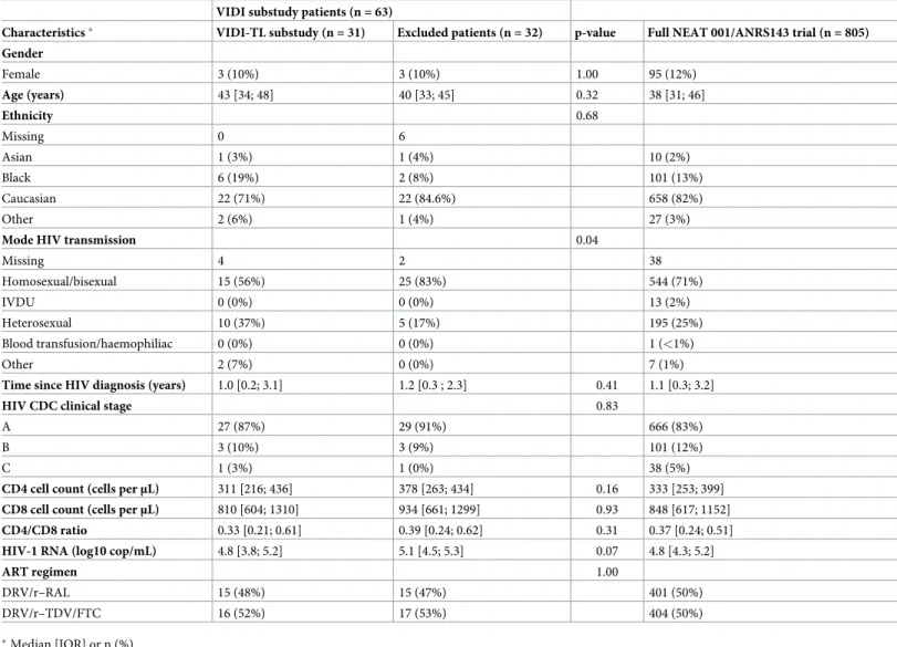
Thirty-one participants from the NEAT-VIDI trial had TL measurement at treatment initia-tion and at week 96 and at least one T cell marker value at treatment initiainitia-tion and at week 96. Baseline characteristics of the participants are described inTable 1. Mode of HIV transmission was significantly different between included participants and participants excluded due to inappropriate blood samples or flow cytometry was not performed were frequently. No other differences were found between included and excluded participants. In both groups, no partic-ipants were co-infected by hepatitis C virus. At week 96, 28 (90.3%) of the particpartic-ipants had achieved a plasma HIV-RNA viral load of less than 50 copies/mL. Mean change in CD4+T cell count was 158 per mm3and mean change in CD4+/CD8+ratio was +0.38.
The mean blood TL between week 0 and week 96 had a statistically significant increase of 0.04 (T/S Ratio) (SD 0.10. raw p-value = 0.03). Adjusted for telomere length at week 0, treat-ment with DRV/r + RAL was associated with an increase of telomere length at week 96 of 0.05 higher than participants treated with DRV/r + TDV/FTC, this difference was not significant (p = 0.1186). In the CD4+T cell population, the change in TL between treatment initiation and week 96 was significantly correlated with the change in the percentage of total CD4+T cell in the same time period. For an increase of 100 percent of CD4+T cell, TL increases by 0.46 (T/S Ratio) (p = 0.03, FDR-adjusted p-value: 0.76) (Fig 1A). On the contrary, the change in the TL was negatively correlated with the change in the percentage of CD4+EM cells. Indeed, for an increase of 100 percent of CD4+EM cells, the TL decreases by 0.55 (T/S Ratio) (p = 0.01, FDR-adjusted p-value: 0.76) (Fig 1B). Trends for a positive correlation between the change in the TL and the change in the percentage of CD4+TEMRA cells (p = 0.05, FDR-adjusted p-value: 0.76) and for a negative correlation with the change in the percentage of CD4+E cells (p = 0.06, FDR-adjusted p-value: 0.76) were also found. No statistically significant association was found among other T cells markers from the CD4+population and the telomere length evolution.
For the CD8+population, a significant negative correlation with the TL change was found only with the change in the number of CD38+molecules on CD8+CM cells. For a decrease of 10,000 CD38+molecules on CD8+CM cells the TL increased by 0.09 (T/S Ratio) between treatment initiation and week 96 (p = 0.05, FDR-adjusted p-value: 0.76) (Fig 1C). We also found trends toward negative correlations between the change in the TL and the changes in the percentages of total CD8+T cells (p = 0.10, FDR-adjusted p-value: 0.77), of CD38+CD8+T
cells (p = 0.08, FDR-adjusted p-value: 0.77), of HLADR+-CD38+CD8+Naïve T cells (p = 0.06, FDR-adjusted p-value: 0.76), of CD38+CCR5+HLADR+CD8+CM cells (p = 0.07, FDR-adjusted p-value: 0.76), the number of CD38+molecules on CD8+E cells (p = 0.06, FDR-adjusted p-value: 0.76) and on CD8+TM cells (p = 0.08, FDR-adjusted p-value: 0.77). After controlling for FDR, no statistically significant association between TL changes and T-cells markers changes were found.
Table 2shows correlations among T cells markers and TL stratified by treatment group. For this analysis we have selected markers that in the overall population were significantly associated with TL change or were close to significance. For the CD8+population, a significant negative correlation (p = 0.04) with the TL change was found only with the change in total CD8+T cells in participants receiving DRV/r-RAL.
Sensitivity analyzes were realised to take into account age as a confounding factor in the association between T cell markers change and telomere length change between W0 and W96 (Supplementary material) and results were very closed to those without adjustment on age at baseline.
Table 1. Characteristics at baseline of the participants from the NEAT-VIDI substudy (n = 31) and NEAT 001/ANRS 143 trial (n = 805). VIDI substudy patients (n = 63)
Characteristics� VIDI-TL substudy (n = 31) Excluded patients (n = 32) p-value Full NEAT 001/ANRS143 trial (n = 805)
Gender Female 3 (10%) 3 (10%) 1.00 95 (12%) Age (years) 43 [34; 48] 40 [33; 45] 0.32 38 [31; 46] Ethnicity 0.68 Missing 0 6 Asian 1 (3%) 1 (4%) 10 (2%) Black 6 (19%) 2 (8%) 101 (13%) Caucasian 22 (71%) 22 (84.6%) 658 (82%) Other 2 (6%) 1 (4%) 27 (3%)
Mode HIV transmission 0.04
Missing 4 2 38 Homosexual/bisexual 15 (56%) 25 (83%) 544 (71%) IVDU 0 (0%) 0 (0%) 13 (2%) Heterosexual 10 (37%) 5 (17%) 195 (25%) Blood transfusion/haemophiliac 0 (0%) 0 (0%) 1 (<1%) Other 2 (7%) 0 (0%) 7 (1%)
Time since HIV diagnosis (years) 1.0 [0.2; 3.1] 1.2 [0.3 ; 2.3] 0.41 1.1 [0.3; 3.2]
HIV CDC clinical stage 0.83
A 27 (87%) 29 (91%) 666 (83%)
B 3 (10%) 3 (9%) 101 (12%)
C 1 (3%) 1 (0%) 38 (5%)
CD4 cell count (cells perμL) 311 [216; 436] 378 [263; 434] 0.16 333 [253; 399]
CD8 cell count (cells perμL) 810 [604; 1310] 934 [661; 1299] 0.93 848 [617; 1152]
CD4/CD8 ratio 0.33 [0.21; 0.61] 0.39 [0.24; 0.62] 0.31 0.37 [0.24; 0.51]
HIV-1 RNA (log10 cop/mL) 4.8 [3.8; 5.2] 5.1 [4.5; 5.3] 0.07 4.8 [4.3; 5.2]
ART regimen 1.00
DRV/r–RAL 15 (48%) 15 (47%) 401 (50%) DRV/r–TDV/FTC 16 (52%) 17 (53%) 404 (50%)
�Median [IQR] or n (%)
https://doi.org/10.1371/journal.pone.0230772.t001
Discussion
In this VIDI substudy of the NEAT 001/ANRS 143 clinical trial we have found that changes in blood TL occurring 96 weeks after initiation of antiretroviral therapy (ART) were correlated positively with the change in the percentage of CD4+on lymphocytes and negatively with the change in the percentage of CD4+EM cells. This is the first time that the correlation between
Fig 1. Correlation between baseline and 96 weeks after antiretroviral treatment initiation change in telomere length and change of the percentage of T cells subsets in participants from the VIDI substudy of NEAT 001 / ANRS 143 trial. Percentage of the CD4+T cells (A), percentage of CD4+effector memory (EM) cells (B) and the number of the CD38+PE molecules on CD8+central memory (CM) cells (C).
https://doi.org/10.1371/journal.pone.0230772.g001
Table 2. Correlation between change of the CD4 and CD8 population markers and change in telomere length between W00 and W96 according to treatment group, n = 31.
DRV/r-RAL (n = 15) DRV/r-TDF/FTC (n = 16) T cell population TL change for an increase of 100 units of the marker�[95% confidence
interval] p-value interaction CD4+(%) Total CD4+ +0.47 [-0.12 ; 1.05] +0.46 [-0.25 ; 1.16] 0.72 E cells -0.49 [-1.01 ; 0.04] -0.18 [-0.67 ; 0.30] 0.35 EM cells -0.53 [-1.16 ; 0.10] -0.95 [-2.15 ; 0.25] 0.85 TEMRA cells +0.35 [-0.56 ; 1.26] +0.18 [-0.12 ; 0.47] 0.70 CD8+(%) Total CD8+ -0.60 [-1.17 ; 0.02] -0.23 [-1.15 ; 0.69] 0.35 CD38+ -0.20 [-0.54 ; 0.14] -0.23 [-0.63 ; 0.17] 0.39
HLA-DR+CD38+Naïve cells -1.29 [-2.88 ; 0.31] -2.07 [-7.36 ; 3.21] 0.77 CD38+PE molecules on CM cells -0.001 [-0.002 ; 0.001] -0.002 [-0.005 ; 0.001] 0.38
CD38+CCR5+ HLA-DR+CM cells -0.38 [-0.81 ; 0.04] -0.69 [-3.13 ; 1.76] 0.69
CD38+PE molecules on E cells -0.001 [-0.004; 0.02] -0.003 [-0.007; 0.001] 0.83
CD38+PE molecules on TM cells -0.001 [-0.005; 0.002] -0.008 [-0.018; 0.001] 0.77
�Adjusted by telomere length at week 0
blood TL and T cell subpopulations has been evaluated in a clinical trial of ART naïve partici-pants using contemporary ART. Compared to baseline, mean blood TL increased significantly at week 96 in the participants included in the VIDI substudy. In two other different datasets we have found progressive increases of blood TL in HIV infected adults receiving effective ART [4,8]. First, in the NEAT 001/ ANRS 143 TL substudy we have found a statistically signifi-cant increase in mean blood TL up to 96 weeks after ART initiation in the 201 participants included [4]. Second, in a cohort of virologically suppressed HIV infected participants we have also found that mean blood TL increased significantly after two years of follow-up [8]. Two older studies [9,10] had also found an increase in T cell TL 24 and 48 weeks after ART. These increases in blood TL after initiation of ART are contrary to studies performed in the general population, in which mean blood TL measured by qPCR analysis shows annual decrease [11].
Our results support the hypothesis that the increase in blood TL occurring in HIV infected persons receiving efficacious ART is a marker of immune reconstitution [4]. We found that increases in blood TL correlated positively with the change in the percentage of total CD4+T cells and negatively with the percentage of CD4+EM cells. After ART initiation there is a decrease of antigenic stimulation secondary to rapid control of HIV replication and a shift towards T-cell subpopulations with longer TL (naïve and central memory). The positive corre-lation between blood TL and change in the percentage of total CD4+T cell might be the reflec-tion of new circulareflec-tion of undifferentiated CD4+cells with long telomeres. The negative correlation between blood TL and the percentage of CD4+ EM cells is also in agreement with a decrease in antigenic stimulation that drives lymphocyte differentiation towards phenotypes with short telomeres such as EM cells [12]. The other statistically significant correlation was an inverse correlation between blood TL and the number of CD38 molecules on CM cells. CD38 is a marker of cell activation than in other studies [13] has also been negatively associated with shorter blood TL.
The main limitations of our study are the small sample size that decreases power to find associations and the multiplicity of comparisons performed that increases the risk of finding false significant results. Another limitation is the lack of measurement of TL within each cell subset which would allow to know if the blood TL is weighted significantly by a specific cell type. Also, we only performed assessments at baseline and W96, which does not allow to deter-mine relation between dynamics of immune recovery and/or control of immune activation and TL changes.
In summary, we found that first-line ART increased blood TL and that this observation is probably a combined consequence of CD4 immune reconstitution and a concomitant decreased immune CD8 cell activation.
Supporting information
S1 Appendix. Correlation between change of the CD4 and CD8 population markers and change in telomere length between W00 and W96 adjusted by telomere length at week 0 and age at baseline, n = 31.
(DOCX)
S2 Appendix. NEAT 001/ANRS 143 Study Group.
(PDF)
Acknowledgments
We are grateful to all study participants, their partners, families, and caregivers, local investiga-tors and study coordinainvestiga-tors from all the centres taking part in the trial. NEAT 001/ANRS 143
Study Group. We thank the HIV CTU, INSERM U1219 Coordinating Unit, Bordeaux, France; MRC Clinical Trials Unit at UCL, London, UK; CHIP Coordinating Unit, Copenhagen, Den-mark; Academic Medical Center Coordinating Unit at the Amsterdam Institute for Global Health and Development, Amsterdam, The Netherlands; ISS, Rome, Italy; Local CTUs: GESIDA, Madrid, Spain, University of Athens Medical School, Greece. We also want to spe-cially thank Dr. Antonio Buño and his laboratory staff at the biochemistry department of Hos-pital Universitario La Paz for performance of adipokines and inflammatory biomarkers.
NEAT 001/ANRS 143 Study Group (SeeS2 AppendixStudy group).
Lead author of NEAT 001/ANRS 143 Study Group: Prof. Francois Raffi. francois.raffi@-chu-nantes.fr
Author Contributions
Conceptualization: Franc¸ois Raffi, Jose Ramon Arribas. Formal analysis: Assia Samri, Brigitte Autran.
Funding acquisition: Franc¸ois Raffi, Jose Ramon Arribas.
Investigation: Mathieu Chalouni, Javier Rodriguez-Centeno, Assia Samri, Natalia
Stella-Ascariz, Franc¸ois Raffi, Jose Ramon Arribas.
Methodology: Javier Rodriguez-Centeno, Franc¸ois Raffi, Jose Ramon Arribas. Project administration: Franc¸ois Raffi, Jose Ramon Arribas.
Supervision: Julian Blanco, Franc¸ois Raffi, Jose Ramon Arribas.
Writing – original draft: Mathieu Chalouni, Javier Rodriguez-Centeno, Jose Ramon Arribas. Writing – review & editing: Mathieu Chalouni, Javier Rodriguez-Centeno, Assia Samri, Julian
Blanco, Natalia Stella-Ascariz, Cedrick Wallet, Hernando Knobel, David Zucman, Belen Alejos Ferreras, Brigitte Autran, Rodolphe Thiebaut, Franc¸ois Raffi, Jose Ramon Arribas.
References
1. Deeks SG, Verdin E, McCune JM. Immunosenescence and HIV. Curr Opin Immunol. 2012 Aug 01; 24 (4):501–6.https://doi.org/10.1016/j.coi.2012.05.004PMID:22658763
2. Liu JCY, Leung JM, Ngan DA, et al. Absolute leukocyte telomere length in HIV-infected and uninfected individuals: evidence of accelerated cell senescence in HIV-associated chronic obstructive pulmonary disease. PloS One. 2015 Apr 17; 10(4):e0124426.https://doi.org/10.1371/journal.pone.0124426PMID: 25885433
3. Raffi F, Babiker AG, Richert L, et al. Ritonavir-boosted darunavir combined with raltegravir or tenofovir-emtricitabine in antiretroviral-naive adults infected with HIV-1: 96 week results from the NEAT 001/ ANRS143 randomised non-inferiority trial. Lancet Lond Engl. 2014 Nov 29; 384(9958):1942–51.
4. Stella-Ascariz N, Montejano R, Rodriguez-Centeno J, et al. Blood Telomere Length Changes After Rito-navir-Boosted Darunavir Combined With Raltegravir or Tenofovir-Emtricitabine in Antiretroviral-Naive Adults Infected With HIV-1. J Infect Dis. 2018 Oct 5; 218(10):1523–1530.https://doi.org/10.1093/infdis/ jiy399PMID:29982509
5. Cawthon RM. Telomere length measurement by a novel monochrome multiplex quantitative PCR method. Nucleic Acids Res. 2009 Feb; 37(3):e21.https://doi.org/10.1093/nar/gkn1027PMID:19129229
6. Iyer SB, Hultin LE, Zawadzki JA, et al. Quantitation of CD38 expression using QuantiBRITE beads. Cytometry 1998; 33(2):206–212.https://doi.org/10.1002/(sici)1097-0320(19981001)33:2< 206::aid-cyto15>3.0.co;2-yPMID:9773881
7. Benjamini Y, Hochberg Y. Controlling the False Discovery Rate: A Practical and Powerful Approach to Multiple Testing. J R Stat Soc Ser B Methodol. 1995 Nov; 57: 289–300
8. Montejano R, Stella-Ascariz N, Monge S, et al. Impact of Nucleos(t)ide Reverse Transcriptase Inhibitors on Blood Telomere Length Changes in a Prospective Cohort of Aviremic HIV-Infected Adults. J Infect Dis. 2018 Oct 5; 218(10):1531–1540.https://doi.org/10.1093/infdis/jiy364PMID:29912427
9. Kaushal S, Landay AL, Lederman MM, et al. Increases in T cell telomere length in HIV infection after antiretroviral combination therapy for HIV-1 infection implicate distinct population dynamics in CD4+ and CD8+ T cells. Clin Immunol Orlando Fla. 1999 Jul; 92(1):14–24.
10. Aladdin H, Katzenstein T, Dreves A-M, et al. T-cell receptor excisional circles, telomere length, prolifera-tion and apoptosis in peripheral blood mononuclear cells of human immunodeficiency virus-infected individuals after 18 months of treatment induced viral suppression. Scand J Immunol. 2003 May; 57 (5):485–92.https://doi.org/10.1046/j.1365-3083.2003.01258.xPMID:12753506
11. Muezzinler A, Zaineddin AK, Brenner H. A systematic review of leukocyte telomere length and age in adults. Ageing Res Rev. 2013 Mar; 12(2):509–19.https://doi.org/10.1016/j.arr.2013.01.003PMID: 23333817
12. Serrano-Villar S, Sainz T, Lee SA, et al. HIV-infected individuals with low CD4/CD8 ratio despite effec-tive antiretroviral therapy exhibit altered T cell subsets, heightened CD8+ T cell activation, and increased risk of non-AIDS morbidity and mortality. Plos Pathog. 2014 May 15; 10(5):e1004078.https:// doi.org/10.1371/journal.ppat.1004078PMID:24831517
13. Gianesin K, Noguera-Julian A, Zanchetta M, Del Bianco P, Petrara MR, Freguja R, et al. Premature aging and immune senescence in HIV-infected children. AIDS. 2016 Jun 1; 30(9):1363–73https://doi. org/10.1097/QAD.0000000000001093PMID:26990630