Evaluation of disease incidence and severity and yield loss
of finger millet varieties and mycelial growth inhibition of
Pyricularia grisea isolates using biological antagonists and
fungicides in vitro condition.
Getachew Gashaw1, Tesfaye Alemu1* and Kassahun Tesfaye1
Department of Microbial, Cellular and Molecular Biology, College of Natural Sciences, Addis Ababa University, P.O.Box. 1176, Addis Ababa, Ethiopia.
* Corresponding author email: talemu2000@yahoo.com
Original submitted in on 12th August 2013 Published online at www.m.elewa.org on 31st January 2014.
ABSTRACT
Objective: The aim of this study was to conduct a survey on the disease incidence and severity at 5 agro-ecological zones of Ethiopia. Moreover, the study was also designed to carry out pathogenicity test, estimate yield losses caused by test pathogen and in vitro evaluation of fungicides and biocontrol agents against finger millet blast isolates.
Methodology and Results: The incidence of finger millet blast was assessed as the percentage of plants with visible symptoms in a field and greenhouse. Blast severity was also evaluated as the percentage of leaf area with symptoms. From the surveyed areas, maximum disease incidence and severity were recorded in west Wollega zone with 63.03 and 34.60%, and lowest disease incidence and severity was recorded in Awi zone with 46.7 and 15.7%, respectively. A total of 42 isolates of P.grisea were collected and isolated from infected finger millet plants and wild relatives from five agro-ecological zones of Ethiopia. The pathogenicity test conducted in greenhouse on three finger millet varieties also indicated that among P.grisea isolates, Pg.11, Pg.41 and Pg.40 showed the highest disease incidence on all the three varieties with 74.8, 69.5 and 66.5%, respectively. Moreover, the highest disease severity with 27.7 and 27.8% were observed by isolates Pg.11 and Pg.41, respectively. In vitro evaluation and testing of Trichoderma viride have showed maximum mycelial growth inhibition with 77.1% and 74.1% by isolates Pg.41 and Pg.22, respectively; while Pseudomonas fluorescens showed maximum mycelial growth inhibition by isolates Pg.40 (57.2%), followed by Pg.26 (56.1%). The efficacy tests of four fungicides evaluated for their antifungal activity showed Sancozeb (85.50- 88.40%) as the most effective fungicide to inhibit mycelial growth of P. grisea.
Conclusion and application of findings: The highest percent of mycelial growth inhibition of P. grisea isolates was observed by T.harzianum and T. viride and followed by Pseudomonas fluorescens. Sancozeb was the most effective fungicide and also showed the highest mycelial growth inhibition on the isolate of P. grisea and followed by ridomil, bayleton, and curzate. From in vitro evaluation of the effectiveness of biological agents and fungicides against the mycelia growth of P.grisea isolates, fungicides were most effective for the control of blast disease of finger millet than biological agents.
Keywords: Biocontrol, Blast disease, Eleusine coracana, Fungicide, Pyricularia grisea.
Journal of Applied Biosciences 73:5883– 5901
INTRODUCTION
Finger millet is one of the most important cereal crops supporting the lives of millions of people across the globe and particularly in the developing world (Dida et al., 2007). It is adapted to a wide range of environments and is known for withstanding harsh environmental conditions including high temperature, moisture deficit and water stagnation but it is susceptible to blast (Lenne et al., 2007). Nutritionally, the grains are equal or superior to other staple cereals as they are a good source of quality protein and various minerals. In marginal and medium agricultural zones, finger millets are high priority staples in Ethiopia, Sudan and Uganda (Mendelsohn et al., 2000; Seyfu Ketema, 2008). The grain is used to make a variety of food products, among these only Injera, spongy fermented flat bread that serves as the staple food for most Ethiopians can be prepared by using finger millet flour. Its straw is highly valued and used as feed for animals (Andualem and Tadesse, 2011). Finger millet production in the world including Ethiopia is subjected to many abiotic and biotic factors that seriously compromise the final yields. Among the menacing biotic factors, finger millet blast caused by P.grisea is a worldwide disease capable of devastating the unprotected finger millet which result reduction of physiological maturity, biomass and yield of the crop (Lenne et al., 2007). The disease affects the crop at all growth stages from seedling stage, causing lesions and premature drying of young leaves, to affecting the panicle causing neck and/or finger blast. The typical symptoms appear on leaves, leaf sheath, rachis, nodes and even the glumes are attacked. Blast in finger millet is caused by a heterothallic filamentous fungus pathogenic to almost 50 plant species in 30 genera of Poaceae including Eleusine (Rossman et al., 1990). The fungus appears to overwinter as mycelia in the infected living leaves or dead plant debris in the soil (Uddin, 2000). High temperature, high relative humidity and leaf wetness are critical environmental factors that influence disease development (Ruiz, 2003). Pazoutova and Bogo (2001) have showed in Uganda and Kenya that the pathogen persists in crop debris, on weed and wild grasses such as E. indica, E.africana, Digitaria spp., Setaria spp. and Doctylocterium spp. These
hosts could serves as potential inoculum reservoirs. Exclusion of the pathogen through plant quarantine is the first line of defense (Agrios, 2005) and other solutions include the elimination of the pathogen’s inoculum and development by good cultural practice; intercropping and rotation; the judicious use of fungicides, understanding and combating virulence mechanisms of the pathogen, biological and chemical control, post harvest protection and improving plant performance through biotechnological approaches (Richard, 2005). Blast has been identified as one of the most important disease infecting finger millet in East African countries including Ethiopia, Kenya and Uganda. However, most of the studies in Africa and particularly in Ethiopia fall in short of providing a quantitative measurement of finger millet blast incidence and severity. Assessment of the incidence and severity of plant disease is important to determine the geographic distribution and status of the disease throughout a region in order to prioritize research. In Ethiopia, no studies have been conducted for the isolation and characterization of the test pathogen and little is known of the biology and potential disease sources of Pyricularia species on finger millet in spite of its nutritional importance. Hence, due to importance of this disease attention needs to be given to the causal agents with special emphasis on its physiological and biochemical requirements for the growth and development of the pathogen, which could serve as an input for efficiently, manage the disease. Moreover, isolation and characterization of blast P. grisea isolates from finger millet and wild relative is important to design better management options. Therefore, this study was undertaken with the objectives of isolation characterization and pathogenicity test of P.grisea isolates. Moreover, blast disease management through biological control agents and the application of fungicides at in vitro condition, in vivo evaluation and estimate yield losses that were caused by P. grisea isolates, were among the objective of the study. The output of this study can be used to design finger millet improved strategy and varietal selection procedure against blast for different agro-ecological zones of the country.
MATERIALS AND METHODS
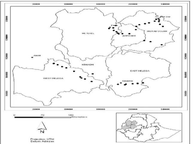
Survey area and collection of blast diseased specimens: Finger millet plants showing typical symptoms of blast were collected from five different ecological zones (East Wollega, West Wollega, Metekel, Awi and West Gojam) areas of finger millet growing parts of Ethiopia using paper bags and envelopes. The surveyed areas were covered the most important finger millet producing regions of Ethiopia with frequent stopping at different intervals depending on variability of finger millet fields in terms of altitude, cropping system and finger millet varieties growing areas of the country. The size of the district, availability and accessibility of finger millet fields were also given due consideration in deciding where to stop on the surveyed route. Surveyed areas
include a wide range of administrative districts and agro-ecological zones, which lie between 9010’ and 11016’ North latitudes and between 34059’ and 37029’East longitudes. Field incidence and severity data were collected from East Wollega, West Wollega, Metekel, Awi and West Gojam zones of the country. The areas also vary in terms of weather conditions and altitude (1033 to 2322 masl). The surveyed route followed major roads to towns and localities in different districts with closer sampling sites separated by 10-12 km from each other. The diseased plants were tagged and kept in refrigerator at 4oC for further studies (leaf, neck, finger and seed blast specimens).
Figure 1: Finger millet blast disease survey sites in West and North West Ethiopia. Dark points and dark star indicate collection sites and major towns in the regions.
Isolation of Pyricularia grisea: The cultures of Pyricularia grisea isolates used throughout the investigation were isolated from blast infected plant parts (Diseased leaves, necks, sheath and seeds) of different finger millet plant, weed and wild species collected from
the farmer’s field of different agro-climatic regions of Ethiopia and also from Research Centers (Bako, Pawe and Adet) during 2011 main cropping seasons. The pathogen was isolated by following standard tissue isolation procedure (Tuite, 1969). Of these plant materials
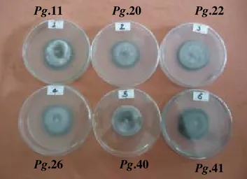
leaves, necks, sheath and seeds were taken from the margin of infected regions and split open longitudinally with the help of a sterilized knife and washed in tap water for two minutes in separate plates in order to minimize surface contaminants. Subsequently, dipped in 70% ethanol for one minute to sterilize the surface and rinsed three times in sterile distilled water to remove the remaining contaminants from the diseased materials (Dhingra and Sinclair, 1993; Aneja, 2005). After complete sterilization, samples were directly transferred in to growth medium containing potato dextrose agar (PDA) amended with 60 mg/l Chloramphenicole or Neuromycine sulphate (Tredway et al., 2003) to suppress the growth and contamination of bacteria. They were incubated in a dark incubator at 27±1 oC for 10 to 15 days and allowed to grow until the emergence of mycelia and sporulation of P.grisea isolates. The pure colony from each isolate was obtained and identified as P.grisea based on its morphological growth pattern, spore shape, conidia and septation. Among 42 isolates, six isolates were selected and designated as Pg.11, Pg.20, Pg.22, Pg.26, Pg.40 and Pg.41, which employed throughout the present studies for detailed morphological, cultural, physiological and finger millet seedling pathogenicity test studies.
In vivo evaluation of finger millet blast (P.grisea) under greenhouse condition
Pathogenicity test: The pathogenicity test in vivo evaluation of susceptibility of finger millet varieties were conducted under greenhouse condition, at the Department of Microbial, Cellular and Molecular Biology, College of Natural Sciences, Addis Ababa University. In the greenhouse, disinfected viable seeds of three finger millet varieties (Boneya, Bako Local Check and Tadesse) which were obtained from Bako Agricultural Research Center (BARC) were sown in 21cm plastic pots filled with 5kg of autoclaved soil. Before sowing, the seeds were surface sterilized with 2% sodium hypochlorite solution for 5 min, and then soaked three times in sterilized distilled water (SDW) to get off from any contamination of microorganisms (Duggar and Davis, 1989). In every three days, each pot was watered with 200 ml of tap water early in the morning. The crop was thinned down to 15 stands per pot after 3 weeks of growth. When the seedlings were six weeks old the leaves were thoroughly cleaned with SDW and the leaves were predisposed to nearly 95 per cent humidity for 24 hours (Sreenivasaprasad et al., 2005). The spore suspensions of 15 days old culture P. grisea isolate cultures with SDW and adjusting the concentration of spore suspension to be (105spore/ml) for
all P.grisea isolates (Takan et al., 2004). The spore suspension contained a mixture of micro and macro conidia. Two hundred (200 ml) spore suspension was prepared for each tested isolates. Thereafter, they were inoculated by spraying on leaves by using hand sprayer (Han et al., 2003). Similarly control plants were sprayed with SDW for comparison. After application of spore suspensions of P.grisea isolates the leaves of the inoculated seedlings were covered by plastic bag and were kept for six days to create conducive condition with high relative humidity near to 95% and to ensure successful penetration and establishment of the test pathogen on the leaf and tissues of seedlings (Sreenivasaprasad et al., 2005). The plastic bags were removed after six days and plants were kept under greenhouse conditions and periodical observations were made regularly for the first appearance and development of symptom on the leaves. Every day the moisture of the soil was maintained with sterilized water. All treatments were replicated three times in Complete Randomized Design (CRD) arranged randomly in the greenhouse. The mean minimum and maximum temperature in the greenhouse during the study period were 12oC and 30o C, respectively. After 30 days of inoculation plants were assessed and recorded for the percentage of disease incidence and severity of leaf infection. As soon as symptoms were developed on the finger millet leaves and stems the diseased plant parts were taken to the laboratory. Subsequently, small cut of diseased parts were made in order to re- isolate P.grisea isolates. The diseased plant parts were surface sterilized using 70% ethanol, 1% sodium hypochlorite (1% NaOCl) for one minute to disinfect the surface, rinsed three times, and put on sterile filter papers to dry off the moisture. The sterilized pieces were then mounted on PDA medium and incubated at, 27OC±1 until the growth of the test pathogen isolates appeared on the PDA medium. After the appearance of the mycelial growth, subsequent sub-culturing was employed to purify the fungal spores and to obtain the pure culture of the test pathogen. Simultaneously, the spores were transferred to PDA and continuous observation was made on the sporulation, pigmentation, conidial structures and other parameters and compared with the original cultures of P.grisea isolates and thus Koch’s postulates were proved. Identification of isolates was done according to the methods employed by Barnett (1960).
Table1: Finger millet varieties used for pathogenicity test on seedlings. Finger millet
varieties
Date of
Susceptibility Sowing Inoculation Data collection
Boneya (KEN#411) Moderate susceptible 29-3-2012 10-5-2012 10-6-2012, 29-11-2012 Bako Local check Susceptible 29-3-2012 10-5-2012 10-6-2012, 29-11-2012 Tadesse (KEN#1098) Moderate resistant 29-3-2012 10-5-2012 10-6-2012, 29-11-2012
Laboratory based germination test were carried out on damp filter paper in Petri dishes to ensure that they had a sufficiently high level of germination, that allow the establishment of adequate number of seedling.
Preparation of inoculum of P. grisea isolates: The inoculums of each isolate of P. grisea were prepared from old culture grown on Oat Meal Agar medium at, 27±1OC in incubator. Conidia were harvested by scraping the surfaces of the colonies with a spatula by washing the
medium with sterilize distilled water and filtered them through nylon mesh cloth. Spore suspensions of different P.grisea isolates were then adjusted to be (105) spores /ml for the isolates with sterile distilled water as described in Takan et al. (2004). The spore suspension counting was done by using Haemocytometer according to the method employed by Summerell et al. (2006) for inoculation of P.grisea spores on finger millet seedlings
.
Figure 2: Microphotograph showing conidial mass of P. grisea with different shapes and size (45 and 100X). Inoculation and pathogenicity study of P. grisea
isolates
Inoculation on finger millet seedlings under greenhouse condition: In the nursery at the greenhouse of College of Natural Sciences, pathogenicity test of each P.grisea isolates was tested on finger millet seedlings that were planted in 21cm plastic pots. Subsequently inoculation each pot was done on leaf with the test pathogen isolate spores suspensions of 1x10⁵ spore/ml. Inoculation experiment was done after 42 days of planting on May 10, 2012. Three non-symptomatic seedlings per each treatment /isolate were prepared for inoculation.
Additional three seedlings were included as controls. The arrangement of varieties was randomized within each pot to ensure experimental integrity. Each pot [3 varieties (15 seedlings of each) x 1 P. grisea isolate] was replicated three times. Three pots were considered as one replicate. Each treatment was labeled and lesion/ symptoms developments were evaluated after 30 days of inoculation. Virulence was determined based on finger millet seedling mortality as well as foliage symptoms and disease development, which subsequently led to disease incidence and severity of the six isolates of P.grisea.
Table 2: Pyricularia grisea isolates used for the finger millet seedling inoculation and their collection sites/locations. No. P.grisea
isolates
Isolate type District/locality Zone Altitude (masl)
Longitude (E)
Latitude (N)
1 Pg.11 Neck Leta Bobina West
Wollega
1622 0350 39’29.8” 090 16’ 19.3” 2 Pg.20 Leaf Abat Bules Metekel 1060 0360 21’15.8” 110 11’ 59.4” 3 Pg.22 Seed Medhin Metekel 1033 0360 21’27.5” 110 13’ 15.4” 4 Pg.26 Leaf Bizra Keni Awi 1679 0360 27’02.9” 110 01’ 26.1” 5 Pg.40 Weed leaf Bikiltu Dila West
Wollega
1893 0350 34’26.0” 090 26’ 08.3”
6 Pg.41 Wild neck Sebat Amit West
Gojam
1786 0370 23’53.0” 110 32’ 12.7”
Finger millet blast disease assessment and scoring: The plants were rated for disease incidence (DI) and disease severity (DS), DI as the presence or absence of disease (percentage of infected leaves on the plant) and DS as the severity percentage of disease damage and yield loss on the three finger millet varieties. Severity of symptoms on individual plants was rated on a scale from 0 to 4 according to percentage of foliage with yellowing or necrosis in acropetal progression: 0 = 0%, 1 = 1 to 33%, 2 = 34 to 66%, 3 = 67 to 100%, and 4 = dead plant, as used previously (Trapero-Casas and Jimenez Diaz, 1985). Incidence (I) and severity data (S) were used to calculate
disease intensity index (DII), DII= (I× S)/4. Blast disease severity and incidence was assessed at, 30 days after inoculating the plant under natural infection by using the following formula (Waller et al., 2002).
Disease Severity (%) = nxv/4N x100;
Where,(n)= Number of plants in each category, (v) = Numerical values of symptoms category.
(N)= Total number of plants, (4) = Maximum numerical value of symptom category.
Disease Incidence (%) = Number of infected plant units X 100
Total number of units assessed
Assessment of yield loss: The assessment of yield loss was carried out mainly based on yield comparisons between infected and healthy plants or between plants with different disease severities using field plots, micro plots (hill plots), single plants or tillers; between resistant and susceptible varieties; between infected plants and
plants treated with fungicides; or between healthy plants and plants where disease damage has been simulated by the removal of essential plant organs, such as the flag leaf on a cereal plant (Cooke, 2006). Percent yield loss (%YL) in terms of grain weight was calculated as follows (Mousanejad et al., 2010).
% YL = Yield in intensive protected plot - Yield in particular treatment X100 Yield in intensive protected plot
In vitro evaluation of antagonistic activity of Trichoderma and Pseudomonas species against finger millet blast (P. grisea) isolates: Two species of Trichoderma [T. harzianum; (AUT1) and T. viride; (AUT2)] and one species of Pseudomonas (P. fluorescens) from culture collection of the Mycology and Applied Microbiology laboratories, Addis Ababa University were used to evaluate the antagonistic activities against the test pathogen (P.grisea) isolates. Dual culture method (Rao,
2003) was employed to evaluate the antagonistic activities of T. harzianum and T. viride. Four mm diameter mycelial disc from the periphery of 10-day-old culture of bioagent was placed on the Petri plate at four locations, approximately 3 cm from the center. Similarly, Pseudomonas fluorescens isolate was assessed for potential antagonistic activity against P.grisea isolates on PDA using dual culture technique (Rangajaran et al., 2003). Four mm diameter mycelial disc was cut from an
actively growing P.grisea culture and placed on the surface of fresh PDA medium at the center of the Petri plates. A loopful of actively growing Pseudomonas fluorescens isolate was placed opposite to the P.grisea disc and streaking the Pseudomonas fluorescens isolate on the plate at four locations, approximately 3 cm from the center. Plates inoculated with P.grisea isolates and without Pseudomonas fluorescens and Trichoderma species alone were used as control. All in vitro tests of antagonism were performed three times. All plates were incubated at, 27oC±1 for 7 days. Visual observations of growth inhibition were recorded every two days and the final measurements were recorded at the 7th days for incubation. Degree of antagonism was determined by measuring the radial growth of pathogen (radial mycelial growth reduction) of P.grisea isolates by two species of Trichoderma and Pseudomonas fluorescens in dual culture in relation to growth of the control and percentage of inhibition was calculated by using the following equation (Riungu et al., 2008).
[I% =(C-T)/C] x100; Where,
I = percentage inhibition of pathogen by antagonists C= radial growth measurement of the pathogen in the control plates and
T= radial growth of the pathogen in the experimental plates
In vitro evaluation and testing of fungicides against P.grisea isolates: To test the effect of fungicides, the method followed by Nene and Thapliyal (1993) was employed. In this technique, the growth medium poisoned with fungicides (fungal toxicants) was used to test the ability of the isolates to grow on the medium. The fungicides were used Bayleton ®, Curzate®, Ridomil® and Sancozeb ®. All fungicides were obtained from Chem Tax PLC Office, Addis Ababa. The fungicide concentration were prepared as follows, if the formulated product (fungicide) has, say 50% active ingredient, for 1 ppm solution 2 mg of the formulated product should be dissolved in a liter solvent (Nine and Thapliyal, 1993).
Stock concentrations of the chemicals used were: bayleton ® 50%(a.i) WP, curzate ® WP (Cymoxanil 4.2%, cupper oxychloride 39.75% (43.95% a.i) and rest inert), ridomil Gold MZ (metenoxam 4%, mancozeb 64% (68% a.i) and inert 32%) and sancozeb 80% WP (mancozeb 80% a.i and inert 20%). The fungicides were added to the autoclaved PDA medium cooled to 45°C, then after obtained required concentrations were obtained. Duplicate culture plates, each containing 20 ml of the test medium, were used to test each isolate at each concentration. PDA plates without fungicide were used as control. Pyricularia grisea isolates were grown on PDA medium for 10 days at, 27±1° C. in an incubator. Mycelia plugs, 4 mm in diameter, were cut from actively growing margins of the culture by sterile cork borer and transferred aseptically in to the center of the Petri dishes contained the PDA medium with different concentrations ranges from 200-1000PPM. Inoculated plates were incubated at, 27±1° C for 7 days. Growth of P.grisea isolates at each concentration was determined by measuring colony diameters in two perpendicular directions on each culture plate in darkness at, 27±1° C. The relative growth reduction for each rate of fungicide was calculated by Riungu et al. (2008).
I = [(C – T)/C] x 100; Where, I= inhibition of radial mycelial growth;
C= radial growth measurement of the pathogen in control; T= radial growth of the pathogen in the presence of the fungicides.
Statistical Analysis: The statistical analysis of growth characteristics of P.grisea isolates at different medium, temperature, and pH and mean comparisons of isolates based on different parameters were conducted using one-way ANOVA procedures of SPSS statistical analysis software (SPSS institute Inc., Cary, NC 2006) version 15. Mean comparisons of each treatments were performed by using Duncan’s multiple range test (α= 0.05). The effect of P.grisea isolates on finger millet yield was also determined by analysis of variance.
RESULTS AND DISCUSSION
Disease Incidence and Severity across Geographic Regions: A set of 42 different isolates of P. grisea obtained from different parts of the crop (diseased leaf, neck and finger/seed) and from weed and wild relative species were collected from different growing areas of east Wollega (7 samples), west Wollega (16 samples),
Metekel (6 samples), Awi (6 samples) and west Gojam (7 samples) and the test isolates were designed as Pg.1-42 (Pyricularia grisea isolates 1-42). For intensive analysis at the later experimental stage, only six isolates were selected based on their aggressiveness and growth characteristics (Table 2). The survey route covered a wide
range of finger millet growing areas located in different parts of Ethiopia. A total of five geographic zones in western and north western Ethiopia (East Wollega, West Wollega, Metekel, Awi, and West Gojam zones) were included in the survey and finger millet blast disease was prevalent in all the surveyed zones but with varying intensity. Incidence of blast in the different zones varied from 46.7 to 63.3%, with the greatest incidence in the west Wollega and West Gojam zones with 63.03% and 57.9%, respectively (Table 3 and Fig.3). Over all, finger millet blast was most severe in the West Wollega zone (34.6%) and least severe in Awi zone (15.7%). Mean disease incidence for the area surveyed was 56.2 % with a range of 29.4 to 76.02% and the mean severity was 26.30% with a range of 10.1 to 41.36% (Table 3). Similarly, Sreenivasaprasad, (2004) has observed that the disease
incidence and severity of finger millet blast were higher at the station compared to farmers’ fields in Busia and Teso districts, in Kenya with incidence range of 2-9 (mean 3.5) and severity range of 10-90%.The survey revealed that the disease incidence and severity were varied from locality to locality due to cultivation of different varieties in different geographical and environmental conditions prevailing in each locality. The highest incidence and severity of the disease in west Wollega and west Gojam may be associated with the presence shad tree and shrubs in the finger millet fields. These conditions reduce light intensity and increase in humidity that favors the distribution of the pathogen. On the contrary the sampling site of Awi zone is characterized by higher altitude, low temperature, and low humidity that did not favor the spread of the pathogen (Srivastava and Tewari, 2002). Table 3: Average disease incidences and severities and their ranges in different geographical regions of Ethiopia. Zone/Region Disease incidence (range) (%) Average disease incidence (% ) ±SD Disease severity (range) (%) Average disease severity (%)±SD Altitude Mean ±SD
East Wollega 35-68.3 54.1±10.30a 18-35.8 28.6±6.7ab 1862.1±165.9ab
West Wollega 45-76.7 63.03±11.04a 22.5-50 34.6±8.4a 1726.2±129.7b
Metekel 10-75 50.8±26.5a 0-25 22.3±23.1ab 1152±153.8c
Awi 20-65 46.7±15.00a 5-27 15.7±8.1b 1913.3±277.9ab
West Gojam 37-95 57.9±16.5a 5-69 23.7±16.3ab 1990.4±185.9a
Mean total 29.4-76.02 56.2±16.04 10.1-41.36 26.30±14.4 1775.1±317.4
Values in the same letters are not significantly different (P<0.05)
0 10 20 30 40 50 60 70 54.1 63.3 50.8 46.7 58.1 28.6 34.6 22.3 15.7 23.6 D is ea se in ci d en ce a n d s ev er it y (% ) Zones
Mean Disease Incidence (%) Mean Disease Severity (%)
Pathogenicity test: The results of pathogenicity test showed that the disease symptoms and development of P.grisea isolates increased up to 60 days of after inoculation with the inoculum of the test pathogen. The lesions were observed on all of the finger millet seedlings. The typical symptoms of dark brown were developed on the leaves after the 7th days of inoculation (Fig. 4). When these infected leaves were removed from the plant and re-isolated the pathogen, a group of conidiophores and conidia of the fungus were observed which were similar to the original culture isolated from the field. The re-isolated P.grisea isolates from inoculated seedlings exactly matched with the characteristics of original isolates (Fig.5). This indicted that all isolates were the causative agents for blast disease of finger millet. Pathogenicity test revealed that all P. grisea isolates were able to infect all the varieties of finger millet. It has also been observed that the two finger millet varieties (Boneya and Tadesse) were more susceptible to blast disease. The variation in aggressiveness and virulence characters of these isolates that were infected on both varieties observed differently. Among the six isolates of P.grisea, three isolates (Pg.11, Pg.41 and Pg.40 ) were showed the highest disease score on the three finger millet varieties, four of P.grisea isolates (Pg.11, Pg.41.Pg.40 and Pg.22) showed the highest disease score on Boneya (KEN#411) and Tadesse (KEN#1098) (Table 4). Similarly, Adipala, (1989) has studied the host range, morphology and pathogenicity of the Genus Pyricularia on finger millet in Uganda. There was significant difference amongst varieties in their susceptibility to the test pathogen and among the tested isolates of P.grisea. Disease incidence and severity of the six P.grisea isolates on the different varieties are indicated in Table 4. The highest mean disease incidence was on
variety Boneya (62.5%) and followed by Tadesse (57.3%), where as the lowest was displayed on Local check (53.9%). It is evidenced that the highest mean percentage disease incidence and severity was observed by isolate Pg.41 (74.8% and 21.4%, respectively) which was significantly different from the remaining isolates. The lowest mean percentage disease incidence and severity was observed by Pg.26 (37.6% and 4.4% respectively) on the three finger millet varieties (data not shown). The highest disease incidence was recorded on Boneya variety by isolates Pg.11, Pg.41 and Pg.40 with 79.7, 76.0 and 74.3%, respectively. Lowest disease severity of 3.2% was recorded by Pg.20, which was followed by Pg.22 (3.8%) on Local check and by Pg.26 (4%) on Tadesse. This study indicated that all the P.grisea isolates were pathogenic to all tested finger millet varieties. Disease incidence recorded in all finger millet varieties clearly revealed that Local check was immune response, while Boneya and Tadesse showed susceptibility to leaf blast for all the six P.grisea isolates. Pyricularia grisea isolates from weeds and wild species were also pathogenic to finger millet, with some isolates being as aggressive as some of the isolates from finger millet. Takan et al. (2004) have observed that the molecular analysis and suggested that the pathogen harbored by the weed and wild millet can serve as an inoculum source. Difference in disease incidence among the three finger millet varieties inoculated at this stage indicated that there was difference in varietal reaction to the blast pathogen and also aggressiveness of the pathogens as well. Similarly, Adipala (1989), in Uganda has indicated that some isolates of blast from weeds will also infect finger millet, which implies the need to removal alternate host such as weedy and wild relative for better disease management. Table 4: Percentage of disease incidence and severity on three finger millet varieties inoculated with six P.grisea isolates under greenhouse condition.
Isolates
Finger millet varieties
Boneya (KEN#411) Local Check Tadesse (KEN#1098)
Incidence (%)±SD Severity (%)±SD Incidence (%)±SD Severity (%)±SD Incidence (%)±SD Severity (%)±SD Pg.11 79.7±2.8a 27.7±1.1a 57.4±1.0b 11.3±1.1b 71.4±3.1ab 20.4±1.4a Pg.20 48.2±0.9d 9.7±1.2c 37.3±1.0f 3.2±0.4d 47.4±3.4c 12.9±1.9c Pg.22 68.4±0.9c 10.4±0.6c 48.6±0.7e 3.8±1.0cd 48.1±3.2c 12.3±0.9c Pg.26 28.1±1.1e 4.4±0.6d 51.2±1.2d 4.9±0.4c 33.5±0.6d 4.0±0.1d Pg.40 74.3±0.9b 25.6±1.5b 55.3±0.6c 10.4±0.5b 69.3±1.6b 17.6±2.0b Pg.41 76.0±0.9b 27.8±0.9a 74.1±0.9a 14.4±0.5a 74.2±1.5a 21.9±1.5a Mean 62.5±19.0a 17.6±10.0a 53.9±11.4c 7.9±4.4c 57.3±15.7b 14.9±6.3b Data followed by the same letter are not significantly different (p<0.05), according to Duncan’s multiple range test
Boneya (KEN#411)
Pg.11 Pg.20 Pg.22 Pg.26 Pg.40 Pg.41
Tadesse (KEN#1098)
Pg 11 Pg.20 Pg.22 Pg.28 Pg.40
Figure 4: Reactions of three finger millet varieties to six different P.grisea isolates originating from various sources and symptoms on leaves.
Inoculation was done after 42 days of planting and the disease symptoms development of P.grisea isolates were evaluated after 30 days of inoculation (Table1).
Figure 5: Re-isolated P.grisea isolates
Assessment of finger millet yield loss caused by P.grisea isolates: There was significant difference amongst finger millet varieties in their percentage yield
loss to the pathogen and among the grain weight infected by the isolates of P. grisea (Table 5). The highest percent of yield losses was recorded on the variety Boneya by isolate Pg.11 (71.9%) and followed by isolate Pg. 41 (62.5%). Similarly, higher yield losses were observed on Tadesse and Local varieties by isolate Pg. 11 and Pg. 41 with 55.9% and 40.9%, respectively. It has been observed that from overall mean value of yield losses, Boneya variety was susceptible to all isolates of P. grisea. The lowest percent of yield loses was recorded by isolate Pg.20 (15.2%), which was followed by Pg.22 (22.1 %) on Local check variety and by Pg.26 (30.1%) on Tadesse variety (Table 5). It has been observed that the highest disease incidence and severity led to the more percent of the yield reduction by different isolates of P. grisea and was also associated with small seeds size; poor seed size (shrink, empty seeds) and quality. Similar result was obtained by Takan et al. (2002), in Kenya and Uganda on which yield losses of 10% to 80% was reported on improved varieties. Similarly Ahmad et al. (2011) have reported that losses due to blast disease may range up to 90% depending up on the component of the plant infected. Overall, there was a trend for seed yield to decrease as the incidence was increased from early leaf infection to
Pg.26 Pg.40 Pg.41
Pg.11 Pg.20 Pg.22
Bako Local Check
Pg.11 Pg.20 Pg.22
late finger development. In this study, it has been observed that indicated that all the isolates were
pathogenic to all tested finger millet varieties with some level of variation in severity and yield loss.
Table 5: Yield losses caused by P.grisea isolates on the three finger millet varieties under greenhouse condition
Is
o
la
te
s
Seed yield (g/pot) Finger millet varieties
Boneya Local check Tadesse
% Y L M ea n ± S D
Infected YL (%) Infected YL (%) Infected YL (%)
Pg.11 0.9±0.6c 71.9±22.8a 8.6±2.2a 40.7±16.2ab 4.1±0.9a 55.9±20.6 a 56.2±15.7a Pg.20 2.0±0.4ab 37.5±2.6bc 12.3±4.7a 15.2±8.4b 5.5±3.4a 40.9±8.7 a 31.2±14.8ab Pg.22 1.6±0.3abc 50.0±0.6abc 11.3±3.1a 22.1±13.4b 5.0±2.3a 46.2±19.3 a 39.4±16.6ab Pg.26 2.2±0.7a 31.3±10.4c 10.1±3.0a 30.3±6.8ab 6.5±3.2a 30.1±11.2 a 30.6±0.8b Pg.40 1.4±0.2bc 56.3±3.1ab 8.7±1.8a 40.0±17.4ab 4.5±1.6a 51.6±10.5 a 49.3±10.3ab Pg.41 1.2±0.2bc 62.5±13.9a 7.3±2.0a 49.7±11.0a 4.9±3.4a 47.3±24.8 a 53.2±6.8a Mean±SD 1.6±0.6b 50.2±16.9a 9.7±3.0a 30.5±15.7b 5.1±2.4a 44.8±16.2a 41.8±10.2 Control 3.2±0.5b 14.5±5.3a 9.3±4.6a
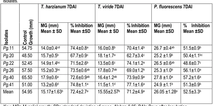
In vitro evaluation of antagonistic activity of Trichoderma species and P. fluorescence against the test pathogen: In dual culture technique the mycelium of both the cultures comes in contact with each other after 4 days of incubation at 27±1oC. After seven days of incubation the hyphal growth of P.grisea isolates was found to be inhibited by the hyphae of Trichoderma species. Further, Trichoderma species almost inhibited the mycelia growth of P.grisea after 10 days of incubation. The advancing hyphae of Trichoderma species covered the entire medium in the Petri plates, suppressing the growth of P.grisea isolates. Both Trichoderma species tested exhibited inhibition of mycelial growth of P.grisea isolates. The results of this study showed that differences in inhibition of the mycelial growth rate of the test pathogen. T. harzianum has rapid growth as compared to all the test isolates of P.grisea and it did not show any clear inhibition zone. Similarly, Tesfaye Alemu and Kapoor (2004 and 2010) studied that Trichoderma and Gliocladium species were effective in reduction of botrytis corm rot (Botrytis Gliocladium) of Gladiolus. Trichoderma
viride has rapid growth, in the form of powdery widespread throughout the Petri plates. As that of T. harzianum, it failed to develop an inhibition zone, since it grew in the form of widespread powders, occupied all the spaces of the medium (Fig.6). The interaction was due to the competition for spaces and nutrients rather than forming inhibition zone. T. viride showed maximum mycelial growth inhibition (77.1% and 74.1%) on P.grisea isolates (Pg.41 and Pg.22), respectively. The minimum percent of mycelial growth inhibition (62.7%) of T. viride was observed on Pg.20. T. harzianum showed maximum mycelial growth inhibition (74.8%) on Pg.41 and minimum mycelial growth inhibition (67.7%) on Pg.20 (Table 6). The effect of the fungal species might be due to the metabolite or exudates that diffused through the medium and reached to the pathogen surface, competition for space, nutrients and intermingling of mycelia rather than forming clear inhibition zones. Similarly, Harish et al. (2007) have indicated that Trichoderma species have an inhibitory effect on Pyricularia species. Similarly, the results of in vitro test of antagonistic activities of P. fluorescens against
the isolates of P.grisea were indicated in Table 6 and Fig.6. Inhibition was clearly showed by limited growth of fungal mycelium in the inhibition zone surrounded by P. fluorescens. The antagonistic effects of P.fluorescens against P.grisea isolates were in the range of 48.6-57.2%. Pseudomonas fluorescens have showed maximum mycelial growth inhibition on isolates Pg.40 (57.2%), followed by Pg.26 (56.1%), Pg.11 (51.5%) and Pg.41 (51.3%). The least percent of mycelial growth inhibition of P. fluorescens was observed on isolate Pg.22 (48.6%). Significance difference was observed in the percentage of mycelial growth inhibition between the isolates of P.grisea. The control plates without P. fluorescens were grown and the Petri plates were covered by the P.grisea isolates. The
mean inhibitory effects of the biological control agents showed significant difference among themselves as well as against the different isolates of the test pathogen (Table 6). The two fungal biocontrol agents were found to be more effective than bacterial antagonist with higher percent of inhibition 72.4% by Trichoderma harzianum and 71.2% by Trichoderma viride compared with P. fluorescens (52.5%) Similar results were reported by Rosales et al. (1995) on which Pseudomonas fluorescens was shown to effectively inhibit Rhizoctonia solani and Pyricularia oryzae by agar plate method. These might be due to producing secondary metabolites, which inhibited the mycelial growth of P.grisea isolates.
Table 6: In vitro evaluation and testing of T. harzianum, T. viride and P. fluorescens against mycelial growth of P.grisea isolates. Is o la te s C o n tr o l G ro w th ( m m )
T. harzianum 7DAI T. viride 7DAI P. fluorescens 7DAI
MG (mm) Mean ± SD % Inhibition Mean ±SD MG (mm) Mean ± SD % Inhibition Mean ±SD MG (mm) Mean ±SD % Inhibition Mean ±SD Pg.11 54.75 14.0±0.4cd 74.4±0.8a 16.0±0.8b 70.4±1.4b 26.7 ±0.4ab 51.5±0.9b Pg.20 48.50 15.7±0.5b 67.7±0.9c 18.1±1.7a 62.7±3.4c 25.2 ±1.9b 50.4±1.1bc Pg.22 52.45 14.9±1.4bc 71.5±2.6b 13.5±0.6c 74.1±1.2a 26.5 ±0.6ab 48.6±0.7c Pg.26 57.50 15.2±0.3bc 73.5±0.6ab 17.8±0.7ab 69.0±1.2b 25.3 ±1.0b 56.1±1.0a Pg.40 65.50 17.9±0.6a 72.6±0.9ab 16.4±1.2ab 73.9±0.9a 27.8 ±1.0a 57.2±1.6a Pg.41 51.00 13.2±0.6d 74.8±1.1a 11.5±1.1d 77.1±1.6a 24.9 ±1.1b 51.3±0.9b Mean 54.95 15.17±1.63b 72.4±2.7a 15.55±2.57b 71.2±4.9a 26.05 ±1.28a 52.5±3.3b Key: MG= Mycelial growth; SD= standard deviation of mean, Alpha= 0.05; DAI= Days after Incubation
A
Pg.26 Pg.40 Pg.41
A = AUT1; B = AUT2; C = P. fluorescens
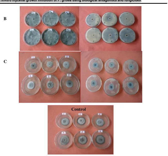
Figure 6: In vitro evaluation and testing of T. harzianum, T. viride and P. fluorescens against mycelial growth of P.grisea isolates.
In vitro evaluation of fungicides against the test pathogen: All the four fungicides showed varied levels of antifungal activity and the highest concentration of fungicides were capable of inhibiting the mycelial growth of the isolates of P.grisea (Table 7). At 1000 PPM, all the fungicides were highly resistant and showed highest percentage inhibition (72.8% to 91.11%) against six isolates of the test pathogen. Pyricularia grisea isolates showed differences in their mycelial growth inhibition at different concentration of Bayleton ® 50% WP. The highest percentage inhibition was observed at the concentration of 1000PPM on isolates Pg.40 (79.72%)
and Pg.11 (79.08%) and the lowest percentage inhibition was observed at the concentration of 200PPM on isolates Pg.22 (61.06%) and Pg.20 (62.33%) (Table7). The highest percentage mycelial growth inhibition by P.grisea isolates were ranges from 78.0-87.60% at 1000PPM and the lowest percentage mycelia growth inhibition were ranges from 53.08-66.36% at 200PPM concentration of Curzate ® WP. The highest percentage mycelial growth inhibition was recorded on isolate Pg.40 (87.60%) at 1000PPM and the lowest percentage mycelial growth inhibition was recorded on isolate Pg.22 (53.06%) at 200PPM. Similarly, Ridomil Gold MZ showed difference percentage of
Control B
mycelial growth inhibition ranges from 70.82-85.04% at difference concentration. The highest percentage mycelial growth inhibition was displayed by isolate Pg.40 (85.04%) at 1000PPM concentration and the lowest percentage mycelial growth inhibition was observed by isolate Pg.41 70.82 at 200PPM concentration. Application of different concentrations of Sancozeb 80% WP on percent mycelial growth inhibition of P.grisea, that ranges from 83.06-91.11% (Table 7 and Fig.7). The maximum percent mycelial growth inhibition was observed on isolates Pg.22 (91.11%), Pg.40 (90.84%) and Pg.20 (90.14%) at 10000PPM and minimum percent mycelial growth inhibition was observed on isolates Pg.11 (83.06%) and Pg.40 (83.35%) at 200PPM concentration. There was significant difference among the concentrations (200-1000 PPM) of all fungicides on mycelial growth inhibition of P.grisea isolates on the growth media (Table 7). However, no significant difference of inhibition was recorded from Sancozeb fungicides with regard to concentration ranges from 200- 1000 PPM in all P.grisea isolates, even maximum inhibition was recorded at 1000PPM. Relatively high percentage of inhibition of 70.82-85.04% and 83.06-91.11% were recorded by 200-1000 PPM of Ridomil and Sancozeb, respectively. Ekkwamu and Mukiibi (1985) have observed that fungicidal control through spraying with mancozeb once at the seedling stage and twice at ear head stage reduces disease incidence. The response
of individual isolates to the different fungicide was shown in Table 8. The highest mean percentage of inhibition of mycelial growth of the isolate was displayed on isolate Pg.26 ranging from 74.9% with Bayleton, 75.30% with Curzate, 82.5% with Ridomil and 88.4% with Sancozeb. Likewise isolate Pg.26 was found to be inhibited to 73.3% by Curzate followed by Bayleton (74.9%), Ridomil (78.3%) and Sancozeb (86.9%). Isolate Pg.11 was found to be inhibited to 69.5% by Curzate followed by Ridomil (72.2%), Bayleton (73.9%) and Sancozeb (85.5%). Generally, isolate Pg.40 seems to be relatively sensitive to all fungicides, where as isolate Pg.11, Pg.22 and Pg.41 was found to be the most resistant. With regard to the efficacy of the fungicides, Sancozeb was found to be the most effective to inhibit the isolates (85.5-88.4%) followed by Ridomil (72.2-82.5%), Bayleton (68.1-74.9%) and Curzate (67.0-75.3%).The fungicide Sancozeb was reported as very effective and used to control fungal blast disease caused by P.grisea on cultivated wild rice and finger millet in Minnesota (Percich et al., 1997). In this study Sancozeb was reduced up to 88.4% inhibition on mycelial growth of P.grisea isolates. Harish et al. (2007) reported that Mancozeb (0.2%), main component of Sancozeb (Agrios, 2005), was observed to be the most effective, which significantly reduced the spore germination of Pyricularia oryzae.
Table 7: Evaluations of different concentration of fungicides on mycelial growth of P.grisea isolates.
Is
o
la
te
s Fungicides Concentration (PPM)
Mycelial growth inhibition (%)
Pg.11 Control 200 500 800 1000 Bayleton ® 50% WP 54.75 67.92±0.8b 74.04±1.4a 74.47±1.9ab 79.08±0.5ab Curzate ® WP 54.75 55.15±0.5cd 61.86±1.0de 76.30±0.6c 84.70±1.0b Ridomil 54.75 71.46±0.3bc 71.69±0.7e 72.68±1.8e 72.83±0.3e Sancozeb 80% WP 54.75 83.06±0.7a 85.09±0.4a 85.98±0.9a 87.98±1.9b Pg.20 Bayleton ® 50% WP 48.50 62.33±0.6d 68.63±1.4cd 72.94±0.4bc 78.98±0.6ab Curzate ® WP 48.50 56.97±0.4bc 64.96±0.8c 77.61±1.1bc 78.00±0.2c Ridomil 48.50 76.29±2.9a 80.17±1.1b 82.20±0.3ab 83.16±1.1b
Sancozeb 80% WP 48.50 85.23±0.5a 86.61±3.4a 87.13±4.6a 90.14±0.6ab Pg.22 Bayleton ® 50% WP 52.45 61.06±0.3d 65.99±0.3d 67.65±0.8d 77.81±0.9b Curzate ® WP 52.45 53.08±0.4e 61.20±1.0e 74.01±0.3d 79.70±0.9c Ridomil 52.45 75.22±2.7ab 79.01±1.4bc 81.20±0.4b 82.08±0.9bc Sancozeb 80% WP 52.45 87.77±2.5a 86.98±0.5a 87.89±0.9a 91.11±1.8a Pg.26 Bayleton ® 50% WP 57.50 70.95±0.1a 74.00±2.3a 76.17±0.9a 78.41±0.6ab Curzate ® WP 57.50 57.86±0.6b 71.96±0.8a 80.84±0.6a 82.66±0.7b Ridomil 57.50 77.18±0.3a 77.39±1.2c 78.70±0.6c 79.85±0.4d Sancozeb 80% WP 57.50 84.64±1.2a 87.04±0.3a 87.62±0.5a 88.13±0.4ab Pg.40 Bayleton ® 50% WP 65.50 65.86±1.5c 72.52±0.7ab 76.16±0.6a 79.72±0.6a Curzate ® WP 65.50 66.36±1.5a 68.16±1.3b 78.89±0.2b 87.60±0.8a Ridomil 65.50 77.57±1.0a 83.01±0.3a 84.35±0.5a 85.04±1.0a Sancozeb 80% WP 65.50 83.35±4.9a 88.62±1.8a 89.33±0.7a 90.84±0.7ab Pg.41 Bayleton ® 50% WP 51.00 63.00±0.8d 70.16±1.3bc 71.24±0.5c 75.98±0.7c Curzate ® WP 51.00 54.89±0.7de 63.97±1.0cd 76.10±0.2c 83.38±1.3b Ridomil 51.00 70.82±0.4c 74.76±0.3d 75.98±0.7d 81.13±o.3cd Sancozeb 80% WP 51.00 85.31±0.5a 85.63±1.9a 87.09±0.7a 88.41±0.7ab * Values with the same letter are not significantly different
Table 8: Mean percent of mycelial growth inhibition of fungicides against P.grisea isolates on PDA medium after 7 days of incubation at 27 ±1oC.
Isolates Mean inhibition percentage by*
Bayleton Curzate Ridomil Sancozeb Mean
Pg.11 73.9±4.6a 69.5±13.4a 72.2±0.7c 85.5±2.0a 75.3±9.0a Pg.20 70.7±7.0a 69.4±10.3a 80.5±3.0a 87.3±2.1a 77.0±9.5a Pg.22 68.1±7.0a 67.0±12.1a 79.4±3.1ab 88.4±1.8a 75.7±11.1a Pg.26 74.9±3.2a 73.3±11.3a 78.3±1.2ab 86.9±1.5a 78.3±7.6a Pg.40 73.6±5.9a 75.3±9.9a 82.5±3.4a 88.0±3.3a 79.8±8.2a Pg.41 70.1±5.4a 69.6±12.7a 75.7±4.3bc 86.6±1.4a 75.5±9.6a Mean ±SD 71.9±5.6c 70.7 ±10.7c 78.1±4.3b 87.1±2.1a 76.9±9.2
Values with the same letter are not significantly different, Alpha=0.05, CV=12
Figure 7: Percentage mycelial growth inhibition of P.grisea isolates by Sancozeb ® 80%WP (A = 200, B= 500, C= 800 and D= 1000PPM).
CONCLUSIONS
The basic data on finger millet blast pathogen, diversity, distribution, disease incidence/severity and blast disease management options has been studied. Moreover, in-depth study was carried out on identification of P.grisea isolates, its biocontrol and fungicide agents that could provides a basis for eco-friendly and sustainable finger millet blast management strategy in Ethiopia. Finger millet blast was found to prevail in most finger millet growing regions of Ethiopia. However, both disease incidence and severity varied significantly across survey districts, altitude groups and climate zones. Over all; West Wollega zone had the highest blast level and followed by West Gojam zones of the country. The pathogenicity test authenticated that the P.grisea isolates are caustic agents of blast disease of finger millet in Ethiopia. All the isolates of P.grisea were able to infect the three finger millet varieties and out of the six isolates Pg.41, Pg.11 and Pg.40 were
the most virulent isolates. Varietal susceptibility to the pathogen has been identified. The most virulent isolates were showed highest finger millet yield reduction. Isolates Pg.11, Pg.41 and Pg.40 were showed maximum yield loss from the data that was by in vivo test under green house condition. In vitro evaluation of the effectiveness of biological agents on the mycelia growth of the isolates, in general, showed that the Pseudomonas fluorescence was less effective than the two Trichoderma species. The fungicides were found to reduce the growth of the different isolates ranging from 67.00% up to 88.40%. The most effective fungicide was found to be Sancozeb followed by Ridomil, Bayleton, and Curzate. The interspecific relative susceptibility of P. grisea isolates showed a pattern of Pg.11 > Pg.22 > Pg.41> Pg.20> Pg.26> Pg.40 on all fungicides.
ACKNOWLEDGMENTS
The authors are greatly acknowledged the Departments of Microbial, Cellular and Molecular Biology, College of Natural Sciences of the Addis Ababa University and BioInnovate Africa Sorghum and Millet consortium for funding, and providing the laboratory facilities and supplying the required consumables and equipments
during the whole period of this study. The authors also acknowledged Office members of Bako Agricultural Research Center, especially Mr. Teshome Bogale and Mr. Dagnachew Lule for providing and supplying finger millet varieties.
B
C
D
REFERENCES
Adipala E, 1989. Host range, morphology, and pathogenicity of the Genus Pyricularia in Uganda. J. East Afr. Agric. 54: 101-105. Agrios GN, 2005. Plant Pathology. 5th edn. Elsevier, USA.
Pp. 948.
Aneja KR, 2005. Experiments in Microbiology Plant Pathology and Biotechnology. 4th edn. New Age International Publishers, New Delhi. Pp. 607. Ahmad SG, Garg VK, Pandit AK, Ali Anwar, Aijaz S,
2011. Disease incidence of Paddy seedlings in relation to environmental factors under temperate agro-climatic conditions of Kashmir Valley. Journal of Research and Development 11: 29-38.
Andualem Wolie and Tadesse Dessalegn, 2011. Correlation and path coefficient analyses of some yield related traits in finger millet (Eleusine coracana (L.) Gaertn.) germplasms in northwest Ethiopia. Afr. J. Agric. Res. 6: 5099-5105. Barnett HL, 1960. Illustrated Genera of Imperfect Fungi.
2ndedn. Burgess publishing company, Morgantown, West Virginia. Pp. 71.
Cooke BM, 2006. Disease assessment and yield loss. In: The Epidemiology of Plant Diseases, pp.43-75, (Cooke, B.M., Jones, D.G. and Kaye, B., eds). Springer, the Netherlands.
Dhingra OD, and Sinclair JB, 1993. Basic Plant Pathology Methods. CRC Press, Inc. of Boca Raton, Florida. Pp. 335.
Dida M, Srinivasachary M, Ramakrishnan S, Bennetzen JL, Gale MD, Devos KM, 2007. The genetic map of finger millet, Eleusine coracana. Theor. Appl. Genet. 114: 321-32.
Duggar BM and Davis AW, 1989. Seed disinfection for pure culture work: the use of hypochlorites. Annals of the Missouri Botanical Garden 6: 159-170.
Han Y, Bonos SA, Clarke BB, Meyer WA, 2003. Inoculation techniques for selection of gray leaf spot resistance in perennial ryegrass. USGA Turfgrass and Environmental Research Online 2: 1-9.
Harish S, Saravanakumar D, Kamalakannan A, Vivekananthan R, Ebenezar EG, Seetharaman K, 2007. Phylloplane microorganisms as a potential biocontrol agent against Helminthosporium oryzae Breda de Hann, the incitant of rice brown spot, Arch. Phytopathol. and Plant Prot. 40: 148 -157.
Lenne, JM, Takan JP, Mgonja, MA, Manyasa EO, Kaloki P, Wanyera N, 2007. Finger millet blast management: A key entry point for fighting malnutrition and poverty in East Africa. Outlook on Agriculture 36: 101–108.
Mendelsohn R, Dinar A, Dalfelt A, 2000. Climate Change Impacts on African Agriculture. A World Bank publication. World Bank. Pp. 278.
Mousanejad S, Alizadeh A, Safaie N, 2010. Assessment of yield loss due to rice blast disease in Iran. J. Agr. Sci. Tech. 12: 357-364.
Nene YL and Thapliyal PN, 1993. Fungicides in Plant Disease Control. Oxford and IBH Publishing CO.PVT.LTD. New Delhi, India. Pp.579. Pazoutova S and Bogo A, 2001. Rediscovery of Claviceps
sorghi (Ascomycotina: Clavicepitaceae) in India. Mycopathologia 153: 99-101.
Percich JA, Nyvall RF, Malvick DK, Kohls CL, 1997. Interaction of temperature and moisture on infection of wild rice by Bipolaris oryzae in the growth chamber. Plant Dis. 81: 1193-1195. Rangajaran S, Saleena LM, Vasudevan P, Nair S, 2003.
Biological suppression of rice diseases by Pseudomonas spp. under saline soil conditions. Plant Soil 251: 73–82.
Rao NSS, 2003. Methods used in soil Microbiological studies. Soil Microbiology. 4th edn. Oxford and IBH Publishing Co.Pvt.Ltd. New Delhi. Pp: 61-72.
Richard NS, 2005. Plant disease: A threat to global food security. Annu. Rev. Phytopathol. 43: 83–116. Riungu GM, Muthorni JW, Narla RD, Wagacha JM,
Gathumbi JK, 2008. Management of Fusarium head blight of wheat and deoxynivalenol accumulation using antagonistic microorganisms. Plant Pathol. J. 7: 13-19. Rosales AM, Thomashow L, Cook RJ, Mew TW, 1995.
Isolation and identification of antifungal metabolites produced by rice associated antagonistic Pseudomonas spp. Phytopathol. 85: 1029-1032.
Rossman AY, Howard RJ, Valent B, 1990. Pyricularia grisea, the correct name of the rice blast disease fungus. Mycologia 82: 509–512.
Ruiz CP, 2003. A new means of control for Pyricularia oryzae, Rhizoctonia solani, and other important rice-disease pathogens in Colombia. Pflanzenschutz-Nachrichten Bayer 56: 399-416. Seyfu Ketema, 2008. Strategic choices and research priorities for the ASARECA sub-region: Food
crops, Livestock, natural resources management, Policy and information. pp.760. Sreenivasaprasad S, 2004. Pathogen diversity and
management of finger millet blast in East Africa: A summary of project activities and outputs. pp.14. International Sorghum and Millets. Newsletter 45: 66-69.
Sreenivasaprasad S, Takan JP, Mgonja MA., Manyasa EO, Kaloki P, Wanyera NM, Okwadi J, Muthumeenakshi S, Brown AE, Lenné JM, 2005. Enhancing finger millet production and utilization in East Africa through improved blast management and stakeholder connectivity. In: Pathways out Of Poverty, Aspects of Applied Biology 75, pp. 11-22 (Harris, D., Richards, J.I., Siverside, P., Ward, A.F. and Witcombe, J.R., eds). UK: Association of Applied Biologists. Srivastava LD and Tewari AK, 2002. Fungal diseases of
wheat and barley. In: Diseases of Field Crops, pp. 58-78, (Gupta, S.C. and Paul, D., eds). Indus Publishing, New Delhi, India.
Summerell BA, Gunn LV, Bullock S, Tesoriero LT, Burgess LW, 2006. Vascular wilt of basin in Australia. Australasian Plant Pathol. 35: 65-67. Takan JP, Akello B, Esele P, Manyasa EO, Obilana AB,
Audi PO, 2004. Finger millet blast pathogen diversity and management in East Africa: A summary of project activities and outputs. Int. Sorghum and Millets Newsletter 45: 66–69. Takan JP, Muthumeenakshi S, Sreenivasaprasad S,
Akello B, Obilana A, Bandyopadhyay R, Coll R, Brown AE, Talbot NJ, 2002. Characterizations of
finger millet blast pathogen populations in East Africa and strategies for disease management. Plant Pathology and Global Food Security Meeting, 8-10 July, 2002, Imperial College, London UK.
Tesfaye Alemu and Kapoor IJ, 2004. In Vitro evaluation of Trichoderma and Gliocladium spp. against Botrytis corm rot (Botrytis gladiolorum) of Gladiolus. Pest Mgt .J. Ethiopia 8: 97-103. Tesfaye Alemu and Kapoor IJ, 2010. Evaluations of
Funginil (Trichoderma formulation) for the control of Botrytis corm rot (Botrytis gladiolorum) of gladiolus varieties under pot culture and field experiment. SINET: Ethiop. J. Sci. 33: 125-130. Trapero-Casas A and Jimenez-Diaz RM, 1985. Fungal wilt and root rot diseases of chickpea in southern Spain. Phytopathology 75: 1146-1151. Tredway LP, Stevenson KL, Burpee LL, 2003.
Components of resistance to Magnaporthe grisea in ‘Coyote’ and ‘Coronado’ tall fescue. Plant Dis. 87:906-912.
Tuite J, 1969. Plant Pathological Methods, Fungi and Bacteria. Burges Publishing Company, New York. pp. 239.
Uddin W, 2000. Gray leaf spot comes on strong. [Online] available: http://groundsmag. Com/ar/grounds_maintenance_gray_leaf_spot/ (09 Oct. 2008).
Waller JM, Lenne JM, Waller SJ, 2002. Plant Pathologist’s Pocketbook. 3rd edn. CABI Publishing, New York. pp. 27.