RT:PCR-ELISA
for
Detection
of
Apple
stem
grooving virus
in
Apple
Trees
Vera Lricia
A.
lVlarinhor,
Julio
Daniels2, Jean Kummert3,
Anne
Chandeliets
&
Philippe
Lepoivre3
rEMBRAPARecursos Genéticos e Biotecnologia, Cx. Postal 02372,CBP70770-900 Brasilia, DF, Brazil, e-mail: vmarinho@cenargen.embrapa.br; 2EMBRAPAClimaTemperado, Cx. Postai 403, CEP 96001-970 Pelotas, RS, Brazil; 3Unité dePhytopathologie, Faculté Universitaire des SciencesAgronomiques, 13,Avenue Maréchal Juin,5030 Gembloux, Belgium
(Accepted
forpublication
on09/04/2003)
Corresponding author: Vera Marinho
MARINHO,
VL.A.,
DANIELS, J., KUMMERT, J. CHANDELIER,A.
&
LEPOIVRE, P. RT-PCR-ELISA for detection of Apple stentgrooving vints în apple trees. Fitopatologia Brasileira 28:374-319.2003.
ABSTRACT
extracts ofplant tissue, as little as 400 fg ofthe virus rvas detected
with
this method. The assay rvith ASGV4F-ASGV4R primers specifically detected the virus in ASGV-infected apple trees from different origins, rvhereas no signal was obsen'ed with ampli{ication products obtained rvith primers targeting the coat protein regionof
the ASGV genome or with primers specific for Apple chlorotic leaf spot virurs(ACLSV)
and Apple stempitting
virrs
(ASPV).The technique combines the porver of PCR to increase the number ofcopies ofthe targeted gene, the specificiry oIDNA hybridization, and the ease
of
colorimetric detection and sample handling in microplates.Additional keyrvords: colorimetric detection ofASGY, P nnus atmeniaca.
RESUNIO
sensibilizada com estreptavidina, e uma sonda de revelaçâo marcada
com digoxigenina. O complexo
foi
detectado utilizando-se um conjugado fosfatase alcalina marcada com anti-DIG Foi avaliada a sensibilidade desse método utilizando-se uma série de diluiçôes devirus purificado, tendo sido possivel detectar até 400 fg de virus. A
especificidade
do
método
foi
demonstrada usando-se osoligonucleotideos iniciadores ASCV4F-ASGV4R e macieiras de
diferentes origens inletadas com ASGV. Nào foram reveladas colorimetricamente as amplificaçôes observadas em gel de agarose,
quando essas mesmas plantas
foram
testadas utilizando-seoligonucleotideos iniciadores para o ASGV escolhidos na regiâo que
codifica para capa protéica e amplificaçôes provenientes de material infetado com o Apple chlorotic leaf spot viras (ACLSV) e Apple
srent pitting vlrus (ASPV). Esta técnica combina o desempenho da
PCR, a especificidade da hibridizaçâo molecular e as facilidades do teste de ELISA.
A method to detect Apple stem grooving vinrs (ASGV) based
on reverse transcription polymerase chain reaction (RI-PCR) was
developed using primers ASGV4F-ASGV4R targeting the viral
replicase gene, follorved by a sandrvich hybridisation, in microtiter plates, for colorimetric detection of the PCR products. The
RT-PCR was performed with the TitanrM RT-PCR system, using
AMV
and diluted crude extracts ofapple (Malus dontestica) lealor barklor the first strand synthesis and a mixture of Taq and PWO DNA
polymerase for the PCR step. The RT-PCR products is hybridised rvith both a biotin-labelled capture probe linked to a
streptavidin-coated microtiter plate and a digoxigenin (DIG)-labelled detection
probe. The complex was detected with an anti-DIG conjugate labelled
with
alkaline phosphatase. When purihed ASGV rvas added toRT-PCR-ELISA para a detecçâo do Apple stem grooving virus
em macieira
Um método foi desenvolvido para a detecçâo do Apple stent
grooving viras (ASGV), baseado na transcriçâo reversa do RNA
viral e na polimerizaçâo em cadeia do DNA (RT-PCR), usando os
oligonucleotideos iniciadores ASGV4F-ASGV4R, localizados na
regiâo
do
gene daRNA polinerase,
sendo os produtos deamplificaçâo detectados por hibridizaçâo "sandwich", em placas de
poliestireno, através de reaçâo colorimétrica. A RT-PCR foi realizada
com a "Titanrr!1 RT-PCR System", complexo enzimâtico formado
pela AMV, responsâvel pela transcriçâo do RNA viral em cDNA e
as DNA polimerases PWO e Taq, usadas para a etapa de PCR,
utilizando-se para isso extrato bruto de lb1has e casca do caule de macieir a (|v[al trs d om es t I c a). Adetecçâo dos produtos de ampl ifi caçào
RT-PCR foi realizada hibridizando esses produtos com uma sonda
de captura marcada com biotina, ligada à placa de ELISA previamente
NTRODUCTION
,
Apple stern
groovingvims (ASGV)
is a member of the genus Capillovirus. This virus infects apple (lufalus domesticaBorkh) trees, usually without causing obvious symptoms, and
is distributed worldrvide.
It
also infects
apricot
(Prunus arnreniacaL.),
cherry
(P. serotinaEhrh.)
and pear (P1'ruscornmunis
L.)
trees(Lister,
1970; Sawamuraet
al.,
1988;374
Takahashi et
al.,
1990;Kinard
etal.,
1996).Control
of
the disease is by planting virus-free stock. The standard detection technique for ASGV relies on graft bioassays, which are time-consuming,although
the useof
thenewly
selected Malttsclones (Howell et a\.,1996) allows detection within two months. Serological
(ELISA)
detection is reliable during a short periodin
the year,but
it
does notwork
well
rvith
dormant woodytissues
(Kinard
et a\.,1996). The use ofreverse transcriptionRT-PCR-ELISA for detection of Apple sterm grooving virus... polymerase chain reaction (RI-PCR) for the detection
ofASGV
has been described (Kinard et al. , I 996; Macken zie et a1., 1997 ;
Kummert et a|.,7998) using an amplification step follorved by
electrophoresis and the detection of the amplicons by ethidium bromide staining. The amplified products cannot be
reliably
detected using ethidium bromide when the
virus titer
is low.Moreover, the specificity of that identification protocol relies on the assessment of the size of amplified products, and false
positive results are possible.
Recently, sensitive assays
for
the detectionof
viral
infections have been based oncolorimetric
(Lassner, 1995; Poljak&
Seme, 1995;A dams et a\.,1996;Lage et aL.,1996) or chemiluminescent (Zammatteo et a\.,1995; Schoen etal.,1996)
detection
of
hybridized PCR
products. These approachescombine the
specificity
of DNA
probe hybridization,
thesensitivity of colorimetric or
luminescent detection, and theease of sample handling in microplates.
\\'e earlier reported a RT-PCR method for diagnosis
ol
ASGV using primers specific for the putative RNA polymerase region of the virus (Marinho et
al.,
l998a,b). The detectionof
the
arrplified
products rvas basedon
UV
fluorescence onetlridium bromide staining
of
agarose gels(Kummert
et al., 1998) or on a colorimetric technique based on direct labellingof
PCR productswith
digoxigenin (Daniels et
al.,
1998).Although
sensitive, thelatter
system can easily giverise
to false positives since any amplified product is labelled.This
paper describesa fast, sensitive and
specific colorimetric assay for detecting amplified ASGV products.It
includes
hybridisation
of
the amplicons,first with
aDIG-labelled detection probe and then rvith a biotin-labe1led caplLrre
probe that is bound to a streptavidin-coated
microtiter
plate.MATERL{I,S ANr:D N IETHODS
Virus
isolates and host plantsTheASGV isolates 1031 I and 10771 were obtained from 're Gorsem E,xperimental Station,
Belgium,
transferredby
mechanical inoculation and multiplie dtn Nicotiana gltttinosa
L.andChenopodiuntquinoa Willd.
Isolates 10291 and 10392r.vere maintained
in
apple trees in thefield.
Isolates of Applechlorotic leafspot vints
(ACLS$
(91300) an d Apple stempiuing
vinrs (ASPV) (91325), maintained in C. quinoa and in apple trees
in the field, respectively, rvere occasionally used as control.
Template preparation for RT:PCR
Clarified
plant extracts:
The extraction protocol of Marinhoet al. (1998a) was used.
Fifty
mg of apple leaves or bark wereground in a
i.5
ml polypropylene tube containing 1ml
of TE buffer (50 mM Tris, pH 8.0,l0
mN4 EDTA). The homogenate was vortexed, centrifuged at 10,000 gfor
10ninat4
oC, andi
;.rl of supernatant was transferred to the RT-PCR mixrure.
Partially purified virions:
The purification method of Sequeira& Lister ( I 969) was used with minor modifications. Frozen leaves
of C. quinoa
infectedwithASGV(isolate
10711) weregroundin three volumes of 0.01 M Tris buffer, pH 8.0, containing 0.20%
Fitopatol. bras.28(4),jul - ago 2003
of
3,3'
diaminodipropylamineand20
mM
iodoacetic acid. Bentonite (40 mg/ml) was added atl0
my'g of leaf tissue afterfiltration of
the homogenate. The mixture was shakenfor
10min, stored at4oC
for
10 min, and centrifuged at 1,500 gfor
10min.
The supematant rvasfiltered
through cheesecloth and then submitted to centrifugation on a 20olo sucrose cushion at 80,000 g for 2 h. Pellets were resuspendedin
1 ml 0.05M
Tris,pH
8.0, containing 0.005M
MgClr.After
a second cycleof
clarification
(3,000 gfor
10 min) and centrifugation on a20Yo sucrose cushion ( 1 00,000 g for 90 min), the final peliets wereresuspended
in
the samebuffer
and the suspensionclarified
as before. The
virions
concentration was estimatedby
light
absorbance at 260 nm using an extinction
coefficient of
2.5cm2mg-r. An aliquot
of
1.0 pl of purified virusorpurified
virusdiluted
in
extracts
of
healthy
Malus
leaveswas
directly
transfened to the RT:-PCR mixture.Reverse Transcription Polymerase Chain Reaction (RT:PCR)
Specific
primers were designedfrom
the nucleotidesequence o f ASGV (P-209) published by Yo shikaw a e t a l. (1 99 2) and from partial sequences of four European
ASGV
isolates. The upstream primerASGV4F (5'-GTT CACTGAGGCAAA
AGC
TGT C-3') site is located between nucleotides 3918 and 3940, and the dorvnstream primerASGV4R (5'-CTT CCG TAC CTCTTC CACAGGAC-3') site is complementaryto nucleotides4 49
|
to 4 4 69, tn the putative viral RNA polymerase-encodingregion (Marinho et
al.,
1998a) and about300
nucleotidesdownstream from the variable region described by Magome el
al. (1997). The primers give an amplification product of 574 bp. The primers designed by Kinard et al. ( I 996) and those reported
by
MacKenzie etal.
(1997) target the putative coat protein gene ofASGV, giving rise to amplicons oî 420 and 524bp, respectively.
The
TitanrNr one hrbe RT-PCR System (Boehringer Mannheim, Germany) was used according to manufacturer'sprotocols. Thermal cycling
was
performed
in
a
TRIO-thermoblock cycler (Biometra, Gôttingen, Germany) for 30 minat 50
'C
for the reverse transcription step, and with thefollowing
parameters for PCR: initial template denahrration at94 "C for 2
min; 35 cycles, of 94 oC for 30 s (denaturation),62 "C
for
1 min (annealing), and 72"C
for 2 min(DNA
synthesis); andfinal
elongation of
72"C for
15 min.Detection of the PCR products
Electrophoresis: Amplified
products (10pl)
rvere separatedby electrophoresis in a 10% agarose gel in TAE buffer (40
mM
Tris, 20 mM acetic acid, 2 mM EDTA). The PCRproducts were stainedwith
0.1% ethidium bromide and photographedwith
Polaroid films.
Southern
blotting:
For the preparationof
the digoxigenin-labelled probe, the amplified products obtained fromASGV
isolate 1 03 I 1 using primers ASGV I F-ASGV I R (Kummert et al., 1998) were recovered using a QIAEX gel extraction kit (Qiagen
GmbH, Germany) and cloned into the pCRrNIil vector using
the TA Cloning
Kit
(Invitrogen, Netherlands). The recombinantV.L.A. Marinho el a/ plasrnids rvere used for PCR synthesis of the
DNA
probe anddirect incorporation of digoxigenin through the pCR DIG probe
Synthesis
Kit
(Boehringer Mannheim, Germany). The probe was 560 bp long, corresponding to nucleotides 3925to
44gSof
ASGV.RNA.
For the detection, PCR products were electrophoresed
in
l0lo agarose and transfened to a positively-charged nylonmembrane (Boehringer Mannheim, Germany). The digoxigenin_ labeled probe (100 ng/ml) was hybridized to rhe membrane and
detected
rvith
the PCR
DIG
Luminescent Detection
Kit
(Boehringer Mannheim) according
to
the
manufacturer's instructions.Colorimetric
detection inmicrotiter
plates: The capture anddetection probes were designed according
to
the sequencesof
fourASGV
isolates. The absence of an unsuitable secondarystructure
in
the
sequenceof
the probewas confirmed by
computer analysis (Oligo 4.0 Soft primers, Biosciences, Inc., Plymouth, USA). The capture probe (BIO-probe), referred to as ASGV-5CI
(Biotine-TTC ACT GAGGCAAAA
GCT GGTCAAACC
TT), was prepared by labelling an extendedASGV4Fprimer sequence
with biotin
at its5'
terminus. The detection probe (DlG-probe). relerred to asASGV-SR2(AGAAGAAù{A
GGG
GAAAAA
GCAcCAAGA
C-DIG),
was prepared bylabelling
a selected nucleotide sequence, homologousto
anintemal fragment in the amplicon, located befween nucleotides
4419 and4147, with digoxigenin at its 3, end. The colorimetric detection
of
the PCR productsis
schematicallyoutlined
in Figure 1. An aliquotof
l0 pl
of pCR product r.vere added to 40irl
of
the
denaturation
soiution
(Boeringher Mannheim,
Germany) to denature the double-stranded DNA and the mixture incubated at room temperature
for
10 min. The denatured pCRproduct (50
pl)
rvas then addedto
450ltl
of
hybridization solution containing 100 ng/ ml of the DlG-labelled detection probe (ASGV-SR2), and rhe hybridizarion was canied our ar 52oC
for 3 h
"vith
shaking (stepA). In
stepB,
the Bio-probe (ASGV-5C1) at 100 nglml
in PBS-Trveen rvas added to eachwellof
amicrotiter
plate (MTPs) (200 1tl perwell)
that was precoatedlr,ith
streptavidin. The plates rvere then incubatedfor
th
at 37 "C. The MTPs were then washed four timeswith
250
plof
PBS-Trveen, and 200plofthe
hybridized pCRproductsrvas added to each of trvo wells. The hybridization rvas carried
out at 37 oC
for
1.5 h r.vith shaking (step C). TheMTps
were then rvashedwith
PBS-Tween, and 200pl
of conjugate buffer(PBS-Trveen plus 0.5% blocking reagent) containing
l:3,000
dilution
ofAnti-Digoxigenin-AP
Fab fragments (BoehringerMannheim, Germany) was added to each well. The MTps were incubated
at3l
"C for 30 min rvith shaking, washed as before, and200 1tl of substrate buffer ( l0 mM dieth44olamine-HCl, pH9.5) containing 0.7 mg/ml of enzyme substlât. fu-nitrophenyl
phosphate-PNPP) was added (step
D).
The optical
density (OD) was measured at 405 nm in a Multiskan plus colorimeterafter incubation of the MTPs
for I
tol6
h at room temperature. The PCR reaction mixture containing water in place of template(blank) was used as a negative control in each assay. The
cut-off
value
of
scoring a positive
samplewas
two
standard376
deviations above the means for the blank PCR mixtures (sample
without
DNA).
Reagents for denaturation and hybridizationsteps and MTPs w'ere purchased from Boehringer Mannheim,
Germany.
RESL'LfS
Sensitivity
of
the colorimetric hybridization
assayfor
detectionofASGV
amplification productsThe TitanrM one tube RT-PCR system and the primers ASGV4F-ASGV4R were used to ampli$TASGV viral sequences
directly from diluted plant
sapof
either ASGV-infected
herbaceous hosts (isolates 1031
I
and 10771) and from leavesor bark
of
apple
trees(isolates
i0291 and
10392).
The amplification products were then visually detected in ethidiumbromide stained gels, by Southem blotting or in the colorimetric
assay.
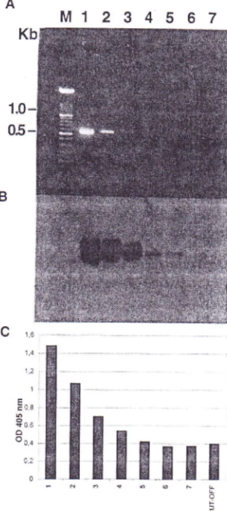
When successive dilutions
of
1:5 through 1;500,000the RTPCR anrplification products rvere tested rvith the differenr systems, the virus rvas detected in dilutions of l:50,000 (Figure2
B-C) using the colorin.retric assay and Southern
blotting,
andin dilutions of about l:500 in the ethidium bromide stained gels
(Figure 2-A).
The sensitivity
of
the assayto
detectvinrs
in
crude extracts rvas also estimated using a serial, 20-folddilution
of
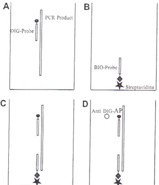
FIG
1 - Steps of colorimetric RT-PCR assay for Apple stem groovingvirrs
(ASGV) detection.A.
Denaturationof
the amplicon andhybridization to the digoxigenin (DIG)-labelled detection probe; B.
Biotin-labelled capture probe (BIO-probe) bound on plate coated
with streptavidin; C. Hybridization
of
the amplicon/-digoxigenin labelled probe complex with the biotin labelled capfure probe; D.DIG detection by anti-DIG alkaline phosphatase (AP) conjugate, reaction of AP with its substrate (PNPP) and colour development.
A
,,",.[il
PCR Pr'duc'lc
A}
il
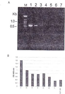
purified virus (starting with a virus concentration of 0.g
pgl
pl)
in crude
jtrice
ofhealthy Malrrs sp. leaves (dilutedl:
200 in TEbuffer, pH 8.0). The detection
limit
of the amplified product in agarose gels was about 40 pg of thepurified
virus (Figure3-A), whereas the colorimetric hybridization assay (Figure 3-B)
and Southern
blotting
(results not shown) detected aslittle
as400 fg
ofpurified
virus.Specificity of the colorimetric hybridization
assayfor
detectionofASGV
In order to determine the specificity of the colorimetric
assay for the detection of the ASGV amplified product, the test was performed on crude extracts from leaves or bark tissues
of
plants infected with different isolatesofASG!
on healthyplantsand on
RNA
preparationsfrom
plants infectedwith ASGV
(isolate 10392),ACLSV
orASPV. The plant extracts orRNA
preparations were submittedto
theTitan
one tube RT_pCRnplification in the presence of the specihc primers ASGV4F_
ASGV4R or primers for detection ofACLSV (isolate 9 I 300) and
ASPV
(isolate 91325). The RT-pCR products obtained fromextracts of ASGV-infected plants amplified
with
the primers targeting the coat protein gene (Kummert e t at., l99g;Sa,uvamuraet al., 1988) were used as checks for the colorimetric detection assay.
No significant absorbance rvas measured (ODro, values
< 0. 1) with the processing of extracts of healthy plants fsample
l0)
orplants infectedrvithACLSV orASpV
(samples g and 9).AIso,
no
significant
absorbance was seenwith
theASGV
amplicons using the primers targeting the coat protein gene (samples3,4, 5,6),
even though these pCR products showed the correct band size when analyzedby
ge1 electrophoresis(Figure
4).
In
contrast, theamplification
products obtainedwith the specific primers ASGV4F-ASGV4R resulted in ODoo.
values of
>
1.0 for all of the testedASGV-infected samples.DTSCUSSION
Validation of diagnostic tests for qualify control
ofplant
material is a classic statistical problem of hypothesis testing.Tr,vo types of mistakes may occur:
(i)
a false positive occurs when a healthy plant is claimed to be infected, and(i
i)
a false negative occurs when the test concludes that the pathogen isabsent in an infected plant.
Any
quality control program must assure the grower that the probability of either mistakewill
beconfined to a small and predetermined range. This objective
of
"qualiry
assurance" can be achieved by a proper sample size and by an assay technique exhibiting adequate sensitivity andspecificity. Moreover, the diagnostic
testmust
alsobe
of
practical
usefor
routine application on
a large numberof
samples.
f
The aim
of
PCR is to produce detectable amountsof
the target
DNA
sequence. Therefore, thesensitivify
and thespecificity
of the whole procedure rest on the detection steps andon the amplification itself, which
is
dependenton
thet€mplate, primers, and the parameters of thermocycling.
We
shorvedthat the specific primers ASGV4F
andFitopatol. bras.
28(4),jul
- ago 2003RT-PCR-ELISA for detection of Apple sternt grooving vinrs...
C 1,6 1.4 1,2 1 i o.e i 0.6 ô o o.o 0
FIG
2 - Detection of successive dilutions (from l:5 to l:500,000)of
RT-PCR amplified products obtained from Chenopodiwn quinoaleaves infected withApple stem groovingvirus
(ASGp
isolatel03l
1by
agarose gel electrophoresis(A),
Southern blotting(B),
and colorimetric hybridization assay (C). LanesI
to 6 correspond to thedilutions of RI-PCR products; lane 7, blank (no template added);
Lane
M,
size of markers in Kb. The cut-off value of the colorimetric assay was defined as two standard deviations above the mean for theblank PCR mixtures.
ASGV4R (M arinho et al., I 998a) designed to amplify a 57 4 bp
fragment located
in
the putativeRNA
polymerase geneof
ASGV can be used in a one step RI-pCR procedure with crude extracts
of
apple trees. However, the detectionof
theASGV
amplification products was based on ethidium bromide staining in agarose gels (Kumme rt et al., I 998). This diagnosis is based on the size of the amplicon and does nottotally
discount thepossibility of false positives due to unspecific amplification
of
non-target sequences.
Moreover,
this
detection
method is much less sensitive than Southem blotting and time-consumingfor processing large numbers of the samples.
Novel strategies for detection of amplified products
in
routine tests have been published recently. Schoen et al.
(1996)
have shown that the fluorescence of a probe (Taq ManrMprobe)
that hybridizes specifically to the target pCR product results
in
a sensitive and specific detectionof
amplicons. Ho."veverthat technique requires a luminescence spectrometer. On the
V.L.A. Marinho eral.
5
4
3
FIG
3-
Detectionof
the amplified producrsof
a serial. 20-fold dilutions of the prtrifi,edApple stern groovingvirus (ASGV) by agarose gel electrophoresis (A) and colorimetric hybridization assay (B). Lane 1 to 6, serial, 2O-fold dilutions ofpurified
ASGV
isolate 10771(starting virus concentration 0 .8 pgl pl) prepared in crude extracts
of
healthy leaves of Malus sp. Lane 7, blank (no template added). LaneM, size of markers in Kb. The cut-off value was defined as trvo standard
deviations above the mean for the blank PCR mixtures,
other hand,
colorimetric
detectionof amplification
productsusing
PCR-ELISA only
requires equipment rvidely usedfor
conventional
enzyme
immunoassaysin
routine ciinical
microbiology
laboratories and,
therefore,
represents anattractive alternative.
In the simplest PCR-ELISAprorocol, reported byAdams
et
al.
(1996)
for
the detection and
typing
of
Huntan
papillomavinrs (FIPV), two primers are labelled with biotin anddigoxigenin, respectively. Although sensitive for detection
of
HPV, this method can easily result in false positives since any amplified product rvill be labelled. The capture of the amplicons by a specific probe bound to the micro titer plate introduces an
additional level of specificily for colorimetric detection.
Two possible means to detect captured
amplification
products have been described. In thefirst,
the amplicons canbe directly labelled during PCRby incorporation ofbiorin dUTp
or digoxigenin dUTP, or by using 5'-labelled primer (poljak
&
Seme, 1995; Adams et
al.,
1996; Daniels?r a/., 1998). In the second, an internally-labelled detection probe hybridises to aspecific region of the target amplicon (Lage et
al.,
1996).Thefirst
alternative is not as specific, as non-target ampliconswill
also be labelled. Such a false positive was observedwith
theprimer pair
ASGVIF-ASGVIR
when aDIG
dUTP-labelledamplification product and the forward primer labelled with biotin
were used for detection ofASGV. In this case, apositive signal
378
Kb
1.0
-0.5-gBx
FIG
4 - Specificity of the colorimetric hybridization assay for the detection of Apple stem groo,-irtg virus (ASGV) -amplified products. Lanes 1 and 3, crude extracts fromNicotiana glutinosaleaves infectedwith isolate 1 03 1 I ; lanes 2 and 4, crude extracts from bark tissues
of
Maltts sp infected rvith isolate 10392;lane 5, total RNA preparation from Chenopodium quinoa leaves infected with isolate 1077 1 ; lanes 6 and 9, total RNA preparation from Malus sp leaves infected with isolate 10392; lanes 7 and 8, total RNA preparations from Malus
leaves infected with ACLSV and ASPV isolates, respectively; lane 10, crude extracts from C.quinoa healthy leaves; lane 1 1, blank (no
template added). For samples 1,2,9, l0 and I 1, ASGV4F-ASGV4R
primers were used for M-PCR. For samples 3, 4, 5 and 6, the primers
used amplified the coat protein gene. For samples 7 and 8, ACLSV
and ASPV specific primers were used, respectively. Lane M, size
c'
markers in Kb. The cut-off value was defined as two standard deviatiL above the mean for the blank PCR mixtures.
$.as observed for a sample
ofACLSV-infected
plant material(Daniels et al.,1998).
In order to improve the specificity of the detection step,
we adopted the second strategy using a DIG-labelled detection probe corresponding to a region ofthe amplicon that is specific
to the ASGV genome. Although the ASGV4F-ASGV4R primers and amplification conditions used in the RT-PCR offer a high
degree of specificity, our colorimetric hybridization protocol
provides the additional
assurance neededfor
a
routine
diagnostic
test. The specificity
of
this
system
wasdemonstrated
by
using amplification products obtainedwith
primers targeting another region of the ASGV genome or
with
specificprimers
forACLSV
and ASPV,which did not
givecolorimetric
signals, althoughamplification
products were observed in gels using ethidium bromide staining.Three systems
to
detect theM-PCR
products were Kb1.0-
0.5-25B
,.u 12 3oa o O o5 0.2 0t
lllllrrr
Ë E l,so-,*,$'***
o È o 6RT-PCR-ELISA for detection of Apple sterm grooving virus... compared
for
sensitivity. Thedilution
thresholdlimits
foundfor colorimetric hybridization and gel electrophoresis follorved
by Southern blotting were 100 times lower than that found
for
ethidium bromide staining of agarose gels. Similar results were reported by Lage et al. (1996) forthe colorimetric hybridization
assay in the detection of amplified
llelicobacter pr'lori DNA.
In
conclusion,the colorimetric hybridization
assaydescribed
in
this
paper
is
a
very
sensitive
assayfor
thedetection
of ASGV
in
apple trees. The assay combines thespecificity of
DNA
hybridizationwith
the ease and speedof
colorimetric detection. Partial automation is possible, and nearly 100 samples can be processed simultaneousiyin
one micro titer plate. Thus the protocol is well suited to the routine testingofplanting
material for a quality control program.ACKNO\\LEDGN,A\TS
This
research was supportedby
the Federal IVlinistry,fAgriculture,
DGVI,
Belgium. We thank P. Simon, Gorsem,Belgium, for providing
ASGV
isolates. Thefirst
tu'o authors also thankEMBRAPA (Brazilian
Enterprise forAgricultural
Research), which provided financial support for her PhD and his post-doctoral fellowships, respectively.
LITERATTIRE CITED
ADAlvlS, V.,
MOLL, C.,
SCHIMID,
M.,
RODRIGUES, C., IVIOOS,R.
&
BRINER,
J.
Detection andtyping
of
human papillomavirus in biopsy and cytological specimens by polymerase chain reaction and restriction enzyme analysis: A method suitable for semi-automation. Joumal of lvledical Virology 48:161- 170. 1996DANIELS, J., MARINHO,
VL.A.,
KUMMERT, J. & LEPOIVRE, P. Development of colorimetric RI-PCR tests for ASGV detection in apple trees. Acta Horticulturae 472:105-111,1998.HOWEL, W.E.,
lv{INK,
G.1., HURTT, S.S., FOSTER,J.A. &
POSTIVIAN, J.D. Select Maltrs clones for rapid detection of apple stem grooving virus. Plant Disease 80:1200-1202. 1996.
:INARD, GR., SCOTT, S.W.
&
BARNETT, O.W. Detectionof
apple chlorotic leaf spot virus and apple stem grooving viruses using RT-PCR. Plant Disease 80:616-624.1996.
KU]V{MERT, J., MARINHO. V.L.A., RUFFLARD, G, COLINET, D. & LEPOIVRE, P. Sensitive detection of apple stem grooving and
apple stem pitting viruses from inlected apple trees by RT-PCR.
Acta Horticulturae 472:97 -104. 1 998.
LAGE, A.P, FAUCONNIER, A., BURETTE, A., GLUPCZ\}ISKI,
\', BOLLEN, A. & GODFROID, E. Rapid colorimetric hybridization
assay for detecting amplified Helicobacter pylori DNA in gastric biopsy specimens. Journal of Clinical Microbiology 31:530-533.
r996.
LASSNER, D. Quantitation of mRNA by the ELOSA technique using external standards. Quantitation of mRNA by polymerase chain
reaction. In: Quantitation ofmRNA bypolymerase chain reaction: non
radioactive PCR methods. Springer Germany. 1995. pp.117-123.
LISTER, R.M. Apple stem grooving virus. CMI/AAB Descriptions of Plant Viruses n.31. 1970.
MACKENZIE, D.J., MACLEAN, M.A., MUKERJI, S. & GREEN,
M. Improved RNA extraction from woody plants for the detection
of viral pathogens by reverse transcription-polymerase chain reaction.
Plant Disease 8l :222-226. 1997 .
NIAGOME, H., YOSHIKAWA, N., TAKAHASHI, T., ITO, T.
&
MIYAKAWA, T.
Molecular
variability
of
the
genomesof
capilloviruses from apple, Japanese pear, European pear, and citrustrees. Phytopathology 87 :389-396. \.997.
MARINHO, V.L.A.,
KUMMEM,
J., RUFFLARD, G., COLTIIET,D. & LEPOIVRE, P. Detection of apple stem grooving virus in dormant
apple trees rvith crude extracts as templates for one-step RT-PCR. Plant Disease 82:785-790. 1998a.
IVIARINHO,
VL.A.
Caractérization et diagnostic moléculaires duvirus du bois rayé du pommier ("apple stem grooving virus", ASGV)
(Ph.D. Thesis). Université des SciencesAgronomiques de Gembloux,
Belgica. 1998 b.
POLJAK, N,1.
&
SElvIE, K. Rapid detection and typing of human papillomavirusesby
Consensus polymerase chain reaction andenzyme-linked immunosorbent assay. Joumal of Virological Methods 56:231-238.1995.
SAWAMURA, K., \'AMASHITA, K.
&
ARAI, K. An apple stemgrooving virus strain isolated lrom European pear (Pyrus cornmLtnis).
Bulletin of Facculty of Agriculrure, Hirosaki University 50:22-26.
1 988.
SCHOEN, C.D., KNORR, D.
&
LEONE, G. Detection of potatoleafroll virus
in
dormant potato tubers by immunocapture and a fluorogenic 5' nuclease RT-PCR assays. Phytopathology 86993-999.1996.
SEQUEIRA, O.
&
LISTER, R.M. Purification and relationshipsof
sonre filamentous viruses from apple. Phytopathology 59:1740-1749.
1969.
TAKAHASHI, T.,.SAITO, N., GOTO,
M.
&
KAWAI, M. APPIC stem grooving virus isolated from Japanese apricot (Pnrnus ntunte)imported from China. Research Bulletin of Plant Protection Sen'ice 26:15-21.1990.
YOSHIKAWA, N., SASAKI, E., KATO, M.
&
TAKAHASHI, T, The nucleotide sequence ofapple stem grooving capillovirus genome.Virology 191 :98-105. 1992.
ZAMMATTEO,
N.,
MORIS, P., ALEXANDRE,I.,
VAIRA, D.,PIETTE,
J.
&
REMARCLE,i.
DNA
probehybridization
in microwells using a new bioluminescent system for the detectionof
PCR-amplified HIV-i proviral DNA. Journal of Virological Methods 55:185-197.1995.02040