C1q as a unique player in angiogenesis with
therapeutic implication in wound healing
Fleur Bossia,1, Claudio Tripodob, Lucia Rizzia, Roberta Bullaa, Chiara Agostinisc, Carla Guarnottab, Carine Munautd, Gustavo Baldassarree, Giovanni Papaf, Sonia Zorzeta, Berhane Ghebrehiwetg, Guang Sheng Lingh, Marina Bottoh,2, and Francesco Tedescoa,2
aDepartment of Life Sciences, University of Trieste, 34127 Trieste, Italy;bDepartment of Human Pathology, University of Palermo, 90127 Palermo, Italy; cInstitute for Maternal and Child Health, Istituto di Ricovero e Cura a Carattere Scientifico Burlo Garofolo, 34137 Trieste, Italy;dLaboratory of Tumor and Development Biology, Centre Hospitalier Universitaire, Groupe Interdisciplinaire de Génoprotéomique Appliqué-Cancer, University of Liège, B-4000 Liège, Belgium;eCentro di Riferimento Oncologico, Division of Experimental Oncology 2, National Cancer Institute, 33081 Aviano, Italy;fPlastic Surgery Unit, University Hospital of Cattinara, 34142 Trieste, Italy;gDepartment of Medicine, Stony Brook University, Stony Brook, NY 11794; andhCentre for Complement and Inflammation Research, Department of Medicine, Imperial College, London W12 0NN, United Kingdom
Edited by Napoleone Ferrara, University of California, San Diego, La Jolla, CA, and approved February 4, 2014 (received for review June 26, 2013) We have previously shown that C1q is expressed on endothelial
cells (ECs) of newly formed decidual tissue. Here we demonstrate that C1q is deposited in wound-healing skin in the absence of C4 and C3 and that C1q mRNA is locally expressed as revealed by real-time PCR and in situ hybridization. C1q was found to induce permeability of the EC monolayer, to stimulate EC proliferation and migration, and to promote tube formation and sprouting of new vessels in a rat aortic ring assay. Using a murine model of wound healing we observed that vessel formation was defective inC1qa−/−mice and was restored to normal after local application of C1q. The mean vessel density of wound-healing tissue and the healed wound area were significantly increased in C1q-treated rats. On the basis of these results we suggest that C1q may rep-resent a valuable therapeutic agent that can be used to treat chronic ulcers or other pathological conditions in which angiogen-esis is impaired, such as myocardial ischemia.
complement
|
vasculogenesis|
animal modelsA
ngiogenesis is a complex process consisting of the growth of new capillaries from preexisting blood vessels and is critical for supplying nutrients, oxygen, and defense factors to newly formed tissues. Endothelial and mural cells are key players in this phenomenon, which starts with the disruption of the vas-culature followed by migration and proliferation of endothelial cells (ECs) and ends with the organization of a network of tube-like structures that eventually become new blood vessels (1–3). The process is very active during embryonic development, con-tributing to organ growth, but it becomes relatively quiescent in the postnatal period, except for certain conditions such as for-mation of maternal decidua in pregnant women, tumor growth, inflammatory processes, and tissue repair (4).In physiologic conditions such as wound healing, the angio-genic process proceeds in an orderly fashion under a balanced control of proangiogenic and antiangiogenic factors. Conversely, in pathologic conditions such as malignant tumor growth, rheu-matoid synovitis, and several other disorders, the persistence of stimuli can tilt the balance toward sustained angiogenesis, result-ing in excessive formation of blood vessels. Other diseases in-cluding ischemic heart disease, preeclampsia, and chronic ulcer are characterized by reduced angiogenesis that prevents revas-cularization and tissue regeneration (4).
Several angiogenic factors promote angiogenesis acting on different vascular cells. VEGF plays a pivotal role in the initia-tion and progression of blood vessel formainitia-tion (5, 6) with the contribution of platelet-derived growth factor, transforming growth factor-β, fibroblast growth factor, and angiopoietins (7, 8). The complement (C) system has been shown to establish multiple in-teractions with the endothelium of blood vessels, leading to ex-pression of adhesion molecules, leukocyte mobilization, secretion of proinflammatory cytokines and chemokines, and increased
vascular permeability (9). The finding of C3a, C5a, and mem-brane attack complex (MAC) deposited in drusen in patients with age-related macular degeneration raised the possibility that C may play an important role in the development of choroidal neovascularisation (CNV), a typical feature of this disease (10, 11). Failure of C3- and C5-deficient mice to develop laser-induced CNV supports the hypothesis that C contributes to angiogenesis (12, 13). Consistent with this, the degree of CNV is decreased in C3aR- and C5aR-deficient mice or in animals treated with C3aR and C5aR antagonists (11) or with recombinant CD59a-Fc, which prevents the assembly of MAC (14). However, Langer et al. (15) have recently reported an inhibitory role of C on angiogenesis in a mouse model of retinopathy of prematurity. They observed a defective neovascularization in WT animals compared with C3-and C5aR-deficient mice C3-and found that C5a was responsible for the inhibition of angiogenesis by stimulating macrophages to se-crete the antiangiogenic soluble VEGF receptor-1. Importantly, all these conflicting reports on the effects of the C system on angiogenesis depend on C activation and are associated with an inflammatory process.
Herein we report an alternative way by which the C system can promote angiogenesis through the action of C1q via an activation-independent mechanism. C1q is well known to activate the classical pathway and to act as a defense molecule favoring the removal of infectious agents, apoptotic cells, and immune complexes (16). In
Significance
C1q is a well-known initiator of the complement classical path-way and interacts with several immune and nonimmune cells inducing complement activation-independent functions. Endo-thelial cells represent one of the potential targets of C1q that binds to cell surface-expressed receptors and stimulates in-flammation. Here we report a unique and hitherto unrecognized function of C1q to promote angiogenesis acting through the globular heads. The angiogenic activity of C1q was supported by its ability to induce new vessel formation in in vitro and in vivo models of wound healing. These findings have important impli-cations for the treatment of clinical diseases associated with im-paired angiogenesis such as chronic skin ulcers in diabetic patients.
Author contributions: F.B. designed research; L.R., C.A., C.G., G.P., S.Z., and G.S.L. per-formed research; G.B., B.G., and M.B. contributed new reagents/analytic tools; C.T., R.B., and C.M. analyzed data; and M.B. and F.T. wrote the paper.
The authors declare no conflict of interest. This article is a PNAS Direct Submission.
Freely available online through the PNAS open access option. 1To whom correspondence should be addressed. E-mail: fbossi@units.it. 2M.B. and F.T. contributed equally to this work.
This article contains supporting information online atwww.pnas.org/lookup/suppl/doi:10. 1073/pnas.1311968111/-/DCSupplemental.
IMMUNOLO
recent years, a wealth of data has been collected indicating that C1q can modulate the function of different cells of both innate and adaptive immunity (17). For example, C1q can target ECs by binding to the receptors for the collagen-like tail (cC1qR) and the globular head of the molecule (gC1qR) as well as to other ligands such as heparan sulfate. As a result of these interactions, C1q in-duces adhesion and spreading of ECs and stimulates expression of adhesion molecules (18) and release of the chemotactic factors IL-8 and monocyte chemoattractant protein-1 and IL-6 (19). C1q has also been reported to recognize apoptotic ECs (20) and to stimulate expression of genes in neurons that are associated with neuro-protection-enhancing neurite outgrowth and limiting neuronal stress and inflammation (21).
In this paper we will present in vitro and in vivo data dem-onstrating that C1q exerts a proangiogenic effect and may be used as a unique therapeutic tool to induce vessel formation in wound healing.
Results
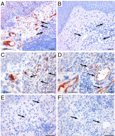
In Vivo Localization of C1q on Newly Formed Vessels in Granulation Tissue.Our previous findings that C1q is deposited on the ECs of vessels in maternal decidua but is absent in other vascular dis-tricts (22) raised the question of whether C1q may be associated with newly formed vessels. To this end, we examined the pres-ence and distribution of C1q in wound healing, a tissue charac-terized by active angiogenesis. Immunohistochemical analysis showed that C1q was deposited in the lesional area but was undetectable in the derma of intact skin (Fig. 1A and B). The protein was mainly localized on the vessel endothelium, in the surrounding stroma, and in scattered inflammatory cells. To as-sess the ability of C1q to activate the C system through the classical pathway, the granulation tissue was examined for the presence and distribution of C1r and C1s using specific anti-bodies (Fig. S1). Unlike C1q, C1r was absent on endothelium and was only detected in tissue-infiltrating cells with hystiocytic morphology (Fig. 1C). C1s showed a distribution similar to that of C1q and were mainly observed in infiltrating inflammatory cells and vessels (Fig. 1D), whereas both C4 and C3 were un-detectable (Fig. 1E and F).
To investigate the source of C1q, we analyzed wound-healing tissue for the expression of mRNA for the three chains of C1q by quantitative RT-PCR and the results were compared with those obtained with intact skin. As shown in Fig. 2A, the expression level of the three mRNAs in granulation tissue was significantly higher than that in normal skin. In situ hybridization revealed the presence of mRNA for the C chain of C1q in stromal cells and vascular endothelium of the lesional area, whereas it was un-detectable in the cells of intact skin (Fig. 2B–E). We have in-vestigated specifically the C1qC mRNA expression because we have previously shown that ECs isolated from normal skin ex-press only the mRNA for A and B chains of C1q (22).
C1q Induces an Angiogenic Phenotype in Cultured ECs.The detection of C1q on endothelium and other cells of a tissue characterized by active angiogenesis led us to examine the role of C1q in angiogen-esis, analyzing its contribution to vascular permeability, proliferation, and migration of ECs.
The concentration of C1q used in all experiments (10μg/mL) was selected as the maximal amount that binds to ECs in a dose– response curve (Fig. S2). The endothelial permeability induced by C1q was evaluated measuring the amount of fluorescein-labeled BSA leaked through a monolayer of ECs and using bradykinin as Fig. 1. Immunohistochemical analysis of human dermal granulation tissue and intact skin. Complement deposition was analyzed using polyclonal antibodies to various complement components and revealed by the strep-tavidin–biotin–peroxidase complex method using aminoethyl carbazole as chromogenic substrate (red signal). Note the localization of C1q on the endothelium and stroma of the granulation tissue (A), whereas C1q was undetectable in intact skin (B). Note also that C1r is present only in tissue-infiltrating cells with histiocytic morphology (C), whereas C1s distribution resembles that of C1q and is expressed on vessels and infiltrating flammatory cells (D). Both C4 (E) and C3 (F) were undetectable. Arrows in-dicate blood vessels. (Scale bars, 30μm.)
Fig. 2. Local expression of C1q mRNA in granula-tion tissue of wound-healing and intact skin. (A) Quantitative PCR analysis of RNA extracted from wound-healing and intact skin. The relative amount of the three chains of C1q was normalized with 18S expression and RNA extracted from blood-derived macrophages was used as calibrator. Data are pre-sented as mean of three independent experiments± SD; *P< 0.05, **P < 0.01 vs. intact skin. (B–E) In situ hybridization of wound-healing (B and C) and intact skin (D and E) sections for the C1q C chain mRNA expression using digoxigenin-labeled antisense (B and
D) and sense (C and E) RNA probes followed by incubation with alkaline phosphatase-conjugated antidigoxigenin antibody and staining with Nitro blue tetra-zolium and 5-Bromo-4-chloro 3-indolyl phosphate. Arrows indicate blood vessels. (Scale bars, 30μm.)
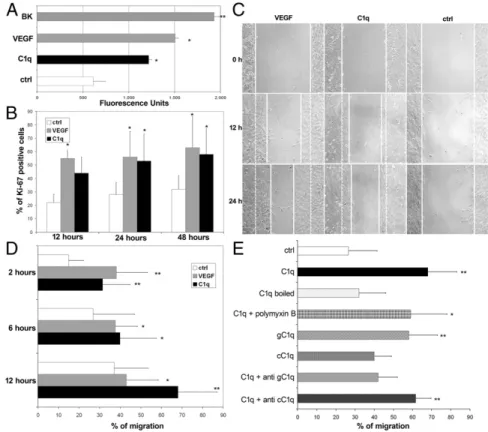
a positive control. As shown in Fig. 3A, C1q exerted a significant permeabilizing effect on ECs.
C1q was also tested in a proliferation assay incubating ECs with C1q for different time intervals followed by single-cell analysis of expression of the proliferation marker Ki-67. The data presented in Fig. 3B show that the percentage of ECs ex-pressing Ki-67 increased in the presence of C1q to almost double that of the control.
The effect of C1q on cell migration was analyzed using a scratch assay and monitoring the mobilization of ECs toward the scratched area for 24 h. As shown in Fig. 3C, ECs stimulated by C1q started to enter the scratched area by 12 h and migrated farther than cells exposed to medium alone after 24 h. The promigratory activity of C1q on ECs was also investigated using a Transwell model system with the membrane insert covered with fibronectin. ECs were added to the upper chamber and the number of cells that migrated into the lower chamber containing either C1q or VEGF or medium was counted at different time intervals. The results of a dose–response curve established with increasing amounts of C1q showed a promigratory effect of this C component that peaked at 5μg/mL and plateaued thereafter (Fig. S3). C1q used at a concentration of 10μg/mL to allow its maximal binding to ECs stimulated migration of∼30% of the cells after 2 h and mobilized more than 70% of the cells at 12 h (Fig. 3D). In this as well as in all previous assays, VEGF was used as a positive control and the combination of the two molecules induced mi-gration of a higher number of cells than each of them tested separately, suggesting an additive effect (Fig. S4). The possibility that endotoxin contaminating C1q was responsible for its biologic effects on cell migration was excluded by boiling C1q for 10 min, a process that destroys C1q but not LPS, and by blocking LPS with polymyxin B (50μg/mL) (Fig. 3E). To identify the portion of C1q molecule responsible for the promigratory effect, the globular head (gC1q) and the collagen tail (cC1q) of C1q were examined separately in the assay and the activity was found to reside mainly
in gC1q (Fig. 3E). The major contribution of gC1q was also sup-ported by the neutralizing effect of a specific antibody against gC1qR, whereas an antibody against cC1qR did not exert a sta-tistically significant inhibition (Fig. 3E). The biologic effect of the commercial C1q was confirmed using our own preparation of C1q that was found to be equally active at similar concentration.
To investigate the ability of C1q to activate signaling mole-cules in human umbilical vein endothelial cells (HUVECs), cell monolayers were incubated with C1q (10 μg/mL) or VEGF (20 ng/mL), used as a positive control, for different lengths of time at 37 °C, and the phosphorylation status of ERK1/2 on total cell lysates was evaluated by immunoblotting. As shown inFig. S5, C1q stimulated the activation of ERK1/2, which was clearly evident at 15 min and decreased to the baseline at 30 min. In contrast, the activation induced by VEGF persisted up to 60 min. Analysis of C1q- and VEGF-mediated activation of p38 revealed a similar but slightly delayed kinetic expression pattern com-pared with that of ERK1/2, although the effect of C1q did not reach statistical significance.
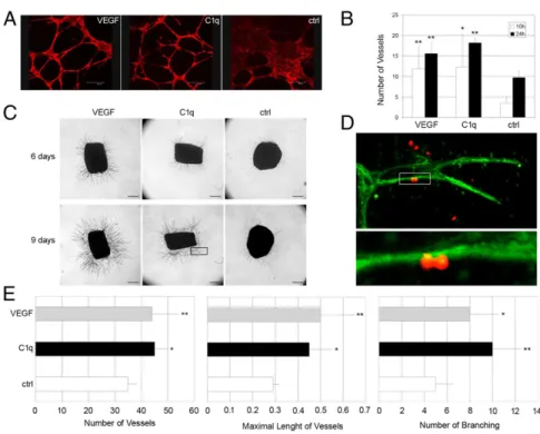
In Vitro and ex Vivo Proangiogenic Effect of C1q.Having established that C1q stimulates EC proliferation and migration, we next evaluated the ability of C1q to induce vessel formation using two different experimental approaches. First, C1q was tested in the in vitro tube formation assay, growing ECs in Matrigel and staining the cells for actin to visualize tube formation. The cells incubated in medium tended to cluster together, forming large aggregates in the absence of tubular organization. Conversely, C1q induced marked changes in the cell pattern, with the formation of tubules assembled by elongation and joining of ECs, and a similar pattern was observed with VEGF (Fig. 4A). Quantitative analysis of the data showed that the effect of C1q was clearly seen at 10 h and became more evident after 24 h of incubation (Fig. 4B).
The ex vivo model of rat aortic ring assay was used as a second approach to analyze the effect of C1q on vessel sprouting from
Fig. 3. In vitro effects of C1q on endothelial cells. (A) The permeabilizing activity of C1q (10μg /mL), VEGF (20 ng/mL), or bradikinin (BK, 10−6M) added to the upper chamber of the Transwell was evalu-ated measuring the amount of FITC-labeled BSA leaked through a monolayer of endothelial cells into the lower chamber. (B) Proliferation of endo-thelial cells was investigated counting the number of cells expressing the proliferation marker Ki-67 at various time intervals after addition of C1q (10 μg/mL) or VEGF (20 ng/mL). (C) Migration of endo-thelial cells using the scratch assay. A confluent monolayer of HUVEC was stripped in the middle with a pipette tip and the scratched area covered by cells that had migrated after exposure to C1q (10μg/mL) or VEGF (20 ng/mL) or medium alone up to 24 h was measured. The continuous line in the scratched area represents the limit of cell migration. The dotted line indicates the border of the scratched area. (D and E) Migration of endothelial cells from the upper to the lower chamber of a Transwell system. C1q (10μg/mL), VEGF (20 ng/mL), cC1q (0.5 μg/mL), or gC1q (0.5μg/mL) was added to the lower chamber and serum-free medium was used as a control. Boiled C1q and C1q mixed with polymyxin B (50μg/mL) were also analyzed to exclude the presence of LPS. Inhi-bition experiments were performed incubating the cells with F(ab)2anti-cC1qR or anti-gC1qR (40μg/mL) for 30 min at 37 °C before addition of C1q. The cells were allowed to migrate up to 12 h. The data are expressed as mean of five independent experiments run in triplicate± SD; *P < 0.05, **P < 0.01 vs. control.
IMMUNOLO
aortic rings. As shown in Fig. 4C, C1q stimulated microvessel formation, which was already visible after 6 d of incubation and became more apparent after 9 d. Because apposition of mural cells around ECs is required for the assembly of newly formed vessel, the microvessel sprouting from the aortic ring was exam-ined by confocal microscopy and found to be made up of a con-tinuous layer of ECs (green) surrounded by pericytes (red) (Fig. 4D). Quantitative analysis of the proangiogenic activity of C1q showed that the number of vessels induced by C1q, their maxi-mal length, and the number of branchings were significantly in-creased compared with the control values (Fig. 4E).
In Vivo Proangiogenic Activity.The data collected using in vitro and ex vitro models suggested that C1q has the features of an angiogenic factor but did not clarify whether it also exhibits an-giogenic activity in vivo. To address this issue, an animal model of wound healing was established using WT andC1qa−/−mice. The animals were killed 14 d after surgery and skin samples were collected and analyzed for the presence of blood vessels. As shown in Fig. 5A, a limited number of small vessels was observed in the wound healing of C1q-deficient animals, in contrast to the overt angiogenesis observed in WT and C3−/− animals. Interestingly, local application of C1q to the wound ofC1qa−/−mice restored vessel formation to the level found in WT mice. Evaluation of mean vascular density (MVD) in wound healing confirmed the visual observation of tissue sections. In particular, MVD values were found to be significantly lower in C1qa−/−mice compared with bothC3−/−and WT animals and increased to levels compa-rable to those observed in C-sufficient mice following treatment of C1q-deficient animals with C1q (Fig. 5B). The increased number of vascular vessels induced by C1q was associated with a signifi-cantly enhanced healing of wounds compared with untreated wounds (Fig. 5C).
To investigate the potential therapeutic use of C1q as an an-giogenic factor, the effect of topical application of C1q was evaluated in a rat model of wound healing. Full-thickness round wounds were produced on the dorsum of rats and treated with either C1q or VEGF or saline as positive and negative controls, respectively. After 14 d the animals were killed and the skin samples were analyzed for the presence of microvessels. Several
small vessels were observed in both VEGF- and C1q-treated skin with an MVD value significantly higher than that of the saline control (Fig. 6A and B). The animals receiving C1q showed also a significant increase in the healed wound area, although less marked than the growing number of blood vessels obtained with C1q or VEGF (Fig. 6C).
To further explore the mechanism of the angiogenic activity of C1q, we evaluated the tissue expression of gC1qR using an an-tibody against this receptor that was found to effectively inhibit C1q-mediated migration of ECs in the initial in vitro experi-ments.Fig. S6shows vessels stained for gC1qR in a tissue section of wound healing whereas the surrounding intact skin revealed negligible expression of gC1qR.
Discussion
Understanding the mechanisms by which angiogenesis is regulated in pathophysiologic conditions is critical to devise successful strat-egies to control this process in various diseases characterized by abnormal vessel formation. C has been implicated in the regulation of angiogenesis, with the main contribution of the activation prod-ucts of the late components found to exert both a proangiogenic and antiangiogenic effect in models of choroidal neovascularisation and retinopathy of prematurity, respectively. The data provided in this study support a definite role for C1q in promoting angiogenesis by a direct effect on ECs.
Skin wound healing provided an invaluable tool to investigate the contribution of C1q to new vessel formation in tissues characterized by active angiogenesis. This analysis was prompted by our previous finding that C1q is deposited on ECs of decidua, serving the important function of promoting adhesion of endo-vascular trophoblasts and their migration through the inter-endothelial cell junctions of spiral arteries to partially replace ECs (22). Because decidua is a newly formed tissue at an embryo implantation site, we hypothesized that cell-bound C1q may also play a role in promoting angiogenesis in other situations. The detection of C1q on ECs of the lesional areas in wound healing and its absence from the dermal endothelium of intact skin are in line with our working hypothesis. Although C1s showed a distri-bution similar to that of C1q in granulation tissue, failure to reveal colocalization of C1r and C1q and to detect C4 and C3 on Fig. 4. In vitro and ex vivo angiogenesis induced by C1q. (A) Capillary-like tubules were allowed to form from endothelial cells exposed to C1q (10μg/mL) or VEGF (20 ng/mL) in Matrigel. The tubules were vi-sualized after staining with Phalloidin–Alexa Fluor 546. (Scale bars, 300μm.) (B) Vessel number was counted using LAS software connected to Leica microscope. (C) Photomicrographs showing vessel sproutings formed after 6 and 9 d of incubation of aortic rings with C1q (10μg/mL) or VEGF (20 ng/mL). (Scale bars, 500μm.) (D) Photomicrograph at two magnifications (40× and 100×) of a microvessel sprout (indicated by the rectangular area in C) formed after 9 d of incubation with C1q showing endothelial cells stained in green with isolectin-B4 and pericytes stained in red with rabbit IgG anti-NG2. (E) Quantifi-cation of number and maximal length (millimeters) of vessels and number of branchings formed after in-cubation of aortic rings with C1q for 9 d. The data are expressed as mean of five independent experiments run in duplicate± SD; *P < 0.05, **P < 0.01 vs. control.
the endothelium suggests that the expression of C1q in the granulation tissue was not associated with complement activa-tion through the classical pathway despite extensive tissue remodeling and inflammation. This is not surprising, because a similar situation is encountered in the decidua that is also char-acterized by remodeling and active angiogenesis and by endothelial
C1q deposition in the absence of C4 and C3. Plasma is a possible source of C1q interacting with the membrane of ECs on lesional areas. However, the involvement of circulating C1q, although po-tentially possible, is unlikely because binding of plasma C1q to target cells usually leads to complement activation through the classical pathway, which apparently does not occur in wound healing, as suggested by the lack of C4 and C3 deposition and the in vivo findings with the C3-deficient mice. An alternative possibility supported by the in situ hybridization data is that C1q is produced by ECs and interacts with specific receptors and other binding sites on the cell surface, as already shown for decidual ECs (22).
C1q is one of the most pleiotropic components of the C system and can elicit a wide variety of biologic activities in different cells of both innate and acquired immunity. The data presented in this paper reveal a yet-unrecognized function of C1q to promote angiogenesis acting on ECs as primary target. C1q exhibits all of the features of an angiogenic factor inasmuch as it induces an in-crease in vascular leakage as a result of loosening of interendo-thelial cell junctions and stimulates cell proliferation and migration. The increase in EC permeability induced by C1q and VEGF (23) allows the extravasation of plasma proteins and formation of ex-tracellular matrix favorable to endothelial and stromal cell migra-tion (24). C1q also shares with VEGF the ability to induce the formation of vascular structure evaluated using both the in vitro Matrigel assay and the ex vivo aortic ring assay.
The C system has been implicated in the promotion of angio-genesis in laser-induced CNV through C5a and MAC, which pri-marily stimulate the retinal pigment epithelium to release VEGF and other angiogenic factors (11, 12). A direct stimulating effect of C5a on the proliferation and migration of ECs and on their ability to form ring-shaped structures has more recently been reported by Kurihara et al. (25). However, the in vivo relevance of this ob-servation is unclear because Langer et al. (15) provided compel-ling evidence that C5a exerts an antiangiogenic effect in a murine model of retinopathy of prematurity, extending previous findings that C5a triggers the release of antiangiogenic factors associated with placental dysfunction and poor pregnancy outcomes (26).
As an angiogenic factor, C1q differs significantly from other components or activation products of the C system for several reasons. First, its action is independent of C activation and seems to be related to a direct stimulating effect on ECs. This con-clusion is supported by the finding that the vessel density in the wound healing of C3-deficient mice is not different from that observed in WT animals. Second, the selective expression in skin undergoing wound healing, but not in intact skin, as revealed by in situ hybridization, is compatible with a physiologic role played by locally released C1q in vessel formation of regenerating Fig. 5. Angiogenesis in a wound-healing model in C1qa−/−and WT mice.
Sections of paraffin-embedded skin of wound-healing samples were ob-tained from WT, C3−/−, C1qa−/−, and C1qa−/−mice treated with a topical application of C1q (5μg) 14 d after surgery and stained for the expression of CD34. Three animals for each group were used. The MVD was quantified on five different 200× microscopic fields of skin sections immunostained for CD34. (A) Representative pictures of CD34 immunostained sections showing the MVD in WT, C3−/−, C1qa−/−, and C1qa−/−mice treated with C1q. Arrows indicate blood vessels present in the tissue. (Scale bars, 30μm.) (B) Quanti-fication of MVD in five fields of skin sections from WT, C3−/−, C1qa−/−, and C1q-treated C1qa−/−mice. (C) Evaluation of wound closure expressed as per-centage of the initial punch biopsy area. The data are presented as mean± SD; *P< 0.05 vs. C1qa−/−.
Fig. 6. Angiogenesis in a rat wound-healing model. (A) Representative pictures of paraffin-embedded skin sections of wounds treated with local application of either C1q (5μg) or VEGF (1 ng) or saline 14 d after surgery. The sections were stained with anti-CD34. Arrows indicate blood vessels present in the tissue (Scale bars, 30μm.) (B) Sections obtained from five rats were also analyzed blindly for MVD in five different fields at 200× magnification. (C) Evaluation of wound closure expressed as percentage of the initial punch biopsy area. The data are presented as mean± SD; *P< 0.05, **P < 0.01 vs. saline.
IMMUNOLO
tissues. Finally, the finding that angiogenesis is impaired in the wound bed ofC1qa−/−mice and can be rescued to levels similar to those of WT mice following topical application of purified C1q further supports its important contribution to the angiogenic process during wound healing.
The observation that C1q is very effective in inducing new vessel formation in wound healing both inC1qa−/−mice and in WT rats raises the issue of the therapeutic use of C1q to promote this process in pathologic conditions characterized by slow or impaired angio-genesis. Chronic wounds associated with burns, venous and arterial insufficiency, diabetes, and peripheral neuropathy are potential indications for topical administration of C1q. Because the globular head of C1q interacting with gC1qR seems to be implicated in the proangiogenic effect of C1q, the fact that gC1qR expression is mainly restricted to the skin ulcer and negligible in normal tissue may fa-cilitate the local therapeutic effect of C1q. This prevalent expression in wound healing would avoid undesired effects that may derive from stimulation of angiogenesis in normal skin as a result of repeated administrations when the receptor is constitutively expressed, as is the case of VEGF receptor. Data from Lee et al. (27) suggest that un-controlled expression of VEGF may lead to the formation of disor-ganized vessels and hemangiomas.
In conclusion, this study provides evidence indicating that C1q is selectively localized on the vessel endothelium and in the stroma of wound healing in the absence of C4 and C3 and acts as a unique angiogenic factor. Based on these findings, we suggest that C1q is involved in new vessel formation in tissues un-dergoing regeneration and may be used as a therapeutic tool to promote angiogenesis in chronic wound healing and also in ischemic conditions.
Materials and Methods
Detailed information on materials and methods used in this study is provided inSI Materials and Methods.
Human Tissue. Dermal granulation tissue and skin biopsy specimens obtained from patients with skin ulcers were examined for immunohistochemical analysis, quantitative real-time PCR and in situ hybridization. The primers used for qPCR and in situ hybridization are reported inTable S1.
Animals. WT mice and rats were purchased from Harlan Laboratories, and C1qa−/−mice were generated by M.B. and used for the in vivo model of wound healing.
Reagents. A full list of antibodies and chemicals is provided in detail inSI Materials and Methods.
Angiogenic Assays. In vitro tests include cell proliferation and migration, EC permeability, tube formation, and rat aortic ring assay. Wound healing in mice and rats was used as an in vivo model of angiogenesis.
ACKNOWLEDGMENTS. We thank L. Spazzapan and A. Delpin for assistance in animal experiments; D. Delapierre, E. Konradowsky, and S. Berton for technical assistance in vitro experiments; and G. Baj for bioinformatic support with confocal microscopy analysis. This work was supported by grants from European Network of Excellence Embryo Implantation Control within FP6 (Contract LSHNCT-2004-512040), the Italian Association for Cancer Research, Italian Ministry of University and Research (Progetti di ricerca di rilevante interesse nazionale MFXE7L), Region Friuli-Venezia Giulia (Network for Lym-phoproliferative diseases and Approccio integrato trattamento tumori art. 23 legge regionale 26/2005), Fondazione Casali-Trieste (to F.T.), Wellcome Trust Grant 088517 (to M.B.), and National Institutes of Health Grants R01 AI 060866 and R01 AI-084178 (to B.G.). C.M. is a Research Associate from the F.N.R.S.-Fonds National de la Recherche Scientifique (Belgium).
1. Folkman J (2003) Fundamental concepts of the angiogenic process. Curr Mol Med 3(7):643–651.
2. Sato Y (2012) The vasohibin family: Novel regulators of angiogenesis. Vascul Phar-macol 56(5–6):262–266.
3. Raza A, Franklin MJ, Dudek AZ (2010) Pericytes and vessel maturation during tumor angiogenesis and metastasis. Am J Hematol 85(8):593–598.
4. Carmeliet P (2005) Angiogenesis in life, disease and medicine. Nature 438(7070): 932–936.
5. Dvorak HF, Brown LF, Detmar M, Dvorak AM (1995) Vascular permeability factor/ vascular endothelial growth factor, microvascular hyperpermeability, and angiogen-esis. Am J Pathol 146(5):1029–1039.
6. Chung AS, Ferrara N (2011) Developmental and pathological angiogenesis. Annu Rev Cell Dev Biol 27:563–584.
7. Carmeliet P, Jain RK (2011) Molecular mechanisms and clinical applications of an-giogenesis. Nature 473(7347):298–307.
8. Semenza GL (2007) Vasculogenesis, angiogenesis, and arteriogenesis: Mechanisms of blood vessel formation and remodeling. J Cell Biochem 102(4):840–847.
9. Fischetti F, Tedesco F (2006) Cross-talk between the complement system and endothelial cells in physiologic conditions and in vascular diseases. Autoimmunity 39(5):417–428. 10. Johnson LV, Leitner WP, Staples MK, Anderson DH (2001) Complement activation and
inflammatory processes in Drusen formation and age related macular degeneration. Exp Eye Res 73(6):887–896.
11. Nozaki M, et al. (2006) Drusen complement components C3a and C5a promote cho-roidal neovascularization. Proc Natl Acad Sci USA 103(7):2328–2333.
12. Bora PS, et al. (2005) Role of complement and complement membrane attack com-plex in laser-induced choroidal neovascularization. J Immunol 174(1):491–497. 13. Bora NS, et al. (2006) Complement activation via alternative pathway is critical in the
development of laser-induced choroidal neovascularization: Role of factor B and factor H. J Immunol 177(3):1872–1878.
14. Bora NS, et al. (2007) CD59, a complement regulatory protein, controls choroidal neovascularization in a mouse model of wet-type age-related macular degeneration. J Immunol 178(3):1783–1790.
15. Langer HF, et al. (2010) Complement-mediated inhibition of neovascularization re-veals a point of convergence between innate immunity and angiogenesis. Blood 116(22):4395–4403.
16. Nayak A, Pednekar L, Reid KB, Kishore U (2012) Complement and non-complement activating functions of C1q: A prototypical innate immune molecule. Innate Immun 18(2):350–363.
17. Nayak A, Ferluga J, Tsolaki AG, Kishore U (2010) The non-classical functions of the classical complement pathway recognition subcomponent C1q. Immunol Lett 131(2): 139–150.
18. Feng X, Tonnesen MG, Peerschke EI, Ghebrehiwet B (2002) Cooperation of C1q re-ceptors and integrins in C1q-mediated endothelial cell adhesion and spreading. J Immunol 168(5):2441–2448.
19. van den Berg RH, Faber-Krol MC, Sim RB, Daha MR (1998) The first subcomponent of complement, C1q, triggers the production of IL-8, IL-6, and monocyte chemoattractant peptide-1 by human umbilical vein endothelial cells. J Immunol 161(12):6924–6930. 20. Navratil JS, Watkins SC, Wisnieski JJ, Ahearn JM (2001) The globular heads of C1q
specifically recognize surface blebs of apoptotic vascular endothelial cells. J Immunol 166(5):3231–3239.
21. Benoit ME, Tenner AJ (2011) Complement protein C1q-mediated neuroprotection is correlated with regulation of neuronal gene and microRNA expression. J Neurosci 31(9):3459–3469.
22. Bulla R, et al. (2008) Decidual endothelial cells express surface-bound C1q as a mo-lecular bridge between endovascular trophoblast and decidual endothelium. Mol Immunol 45(9):2629–2640.
23. Kevil CG, Payne DK, Mire E, Alexander JS (1998) Vascular permeability factor/vascular endothelial cell growth factor-mediated permeability occurs through disorganization of endothelial junctional proteins. J Biol Chem 273(24):15099–15103.
24. Dvorak HF (1986) Tumors: Wounds that do not heal. Similarities between tumor stroma generation and wound healing. N Engl J Med 315(26):1650–1659. 25. Kurihara R, et al. (2010) C5a promotes migration, proliferation, and vessel formation
in endothelial cells. Inflamm Res 59(8):659–666.
26. Girardi G, Yarilin D, Thurman JM, Holers VM, Salmon JE (2006) Complement activa-tion induces dysregulaactiva-tion of angiogenic factors and causes fetal rejecactiva-tion and growth restriction. J Exp Med 203(9):2165–2175.
27. Lee RJ, et al. (2000) VEGF gene delivery to myocardium: Deleterious effects of unregulated expression. Circulation 102(8):898–901.