HAL Id: in2p3-00660910
http://hal.in2p3.fr/in2p3-00660910
Submitted on 18 Jan 2012HAL is a multi-disciplinary open access archive for the deposit and dissemination of sci-entific research documents, whether they are pub-lished or not. The documents may come from teaching and research institutions in France or abroad, or from public or private research centers.
L’archive ouverte pluridisciplinaire HAL, est destinée au dépôt et à la diffusion de documents scientifiques de niveau recherche, publiés ou non, émanant des établissements d’enseignement et de recherche français ou étrangers, des laboratoires publics ou privés.
Sorption Speciation of Nickel(II) onto
Ca-Montmorillonite: Batch, EXAFS Techniques and
Modeling
X. Tan, J. Hu, Gilles Montavon, X. Wang
To cite this version:
X. Tan, J. Hu, Gilles Montavon, X. Wang. Sorption Speciation of Nickel(II) onto Ca-Montmorillonite: Batch, EXAFS Techniques and Modeling. Dalton Transactions, Royal Society of Chemistry, 2011, 40, pp.10953-10960. �10.1039/c1dt10740b�. �in2p3-00660910�
Sorption Speciation
of Nickel(II) onto Ca-Montmorillonite: Batch, EXAFS
Techniques and Modeling
XiaoLi Tan,†* Jun Hu,† Gilles Montavon‡, XiangKe Wang†*
Key Laboratory of Novel Thin Film Solar Cells, Institute of Plasma Physics, Chinese Academy of Sciences, P.O. Box 1126, Hefei, 230031, P.R. China; and Laboratory SUBATECH, Groupe de Radiochimie, UMR Ecole des Mines/CNRS/Université, 4 rue A. Kastler, BP 20722, 44307 Nantes cedex
03, France
The sorption speciation of Ni(II) on Ca-montmorillonite was evaluated using a combination of batch experiments, extended X-ray absorption fine structure (EXAFS) spectroscopy and modeling. The pH and temperature at the aqueous-montmorillonite interface affects both the extent of Ni(II) sorption as well as the local atomic structure of the adsorbed Ni(II) ions. At 0.001 mol/L Ca(NO3)2 and low pH, the
study reveals that the majority of Ni(II) is adsorbed in the interlayers of Ca-montmorillonite coordinated by six water molecules in an octahedron as an outer-sphere complex. At higher pH, inner-sphere surface complexes are formed. The Ni-Si/Al distances (RNi-Al=3.00 Å, RNi-Si1=3.10 Å and RNi-Si2=3.26 Å)
determined by EXAFS confirm the formation of mononuclear complexes located at the edges of Ca-montmorillonite platelets at pH 7.5 and 8.5. At pH 10.0, the Ni-Ni/Si distances (RNi-Ni=3.07 Å and
RNi-Si =3.26Å) indicates the formation of Ni-phyllosilicate precipitates. A rise in temperature promotes
inner-sphere complexation, which in turn leads to an increase in Ni(II) sorption on Ca-montmorillonite. Sorption edges are fitted excellently by surface complexation model (SCM) with the aid of surface species determined from EXAFS spectroscopy.
*
: Corresponding author. Tel: +86-551-5592788; Fax: +86-551-5591310. Email: tanxl@ipp.ac.cn (X.L. Tan),
1. Introduction
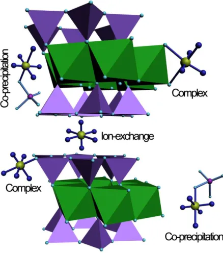
Availability of Ni(II) for bio-uptake and transport in the environment is controlled by sorption processes and speciation of Ni(II). The final form of metal sorption products influences the mobility, bioavailability, and ultimately toxicity in the environment.1,2 Thus, it is critical to understand Ni(II) sorption speciation in soils.3-8 However, accurately determining Ni(II) speciation in whole soils is challenging because soils are heterogeneous with respect to mineralogy and composition. This difficulty can be overcome by studying more controlled systems in which major interactions between Ni(II) and specific soil constituents can be isolated. Montmorillonite is present in most soils and aquatic systems, which has been well characterized in detail.8 Montmorillonite presents several types of sorption sites (such as exchange sites, amphoteric edge sites). Ni(II) sorption mechanisms on this mineral are expected to be complex (Fig. 1). The determination of Ni(II) species on montmorillonite allows one to clearly identify the adsorbed Ni(II) species on clay minerals, and to evaluate the interaction of Ni(II) on soils.
Understanding metal speciation and sorption complexes can also help to select appropriate management and remediation methods for contaminated environments.
Sorption of Ni(II) on clay minerals has been extensively studied by sorption experiments.3-12 Many mechanisms have been postulated for Ni(II) sorption, including ion exchange, surface complexation (inner-sphere and outer-sphere), precipitation/coprecipitation, and diffusion into particle micropores.3-12 However, these approaches did not yield any direct structural information on the adsorbed ions. Recently, extended X-ray absorption fine structure (EXAFS) studies have been a powerful tool for the investigation of Ni(II) speciation on mineral surfaces at atomic level over short time scales at pH levels undersaturated with respect to pure metal hydroxide solubility, and at metal surface coverage below theoretical monolayer coverage.13-17 Utilizing this method, Scheidegger et al.14 observed the presence of
a mixed Ni-Al hydroxide phase at low surface loading and at reaction conditions undersaturated with respect to the formation of Ni(OH)2(s). Dähn et al.17 demonstrated that the uptake of Ni(II) on
montmorillonite resulted in the neoformation of a phyllosilicate. And they found that the reaction time may determine the structure of the adsorbed Ni. Often the sorption speciation of metal ions is contingent upon the existent physical and chemical conditions of the system, including solution pH and temperature fluctuation, which can affect the fate and transport of heavy metals in the environment. However, few works were carried out to give structural information of the adsorbed Ni complexes under variable conditions.
Surface complexation modeling (SCM) has successfully described the sorption of metal ions onto adsorbents. However, the species are usually determined in the absence of spectroscopic data that may provide more detailed structural information on surface complexes formed.10-12 To make the sorption mechanisms onto montmorillonite system more reliable, SCM simulation basing on spectroscopic data is important. However, to date, only a few reports involve the combination of SCM with spectroscopic investigation, although this may offer a fundamental level investigation as far as we know.18
In this paper, Ni(II) sorption onto montmorillonite utilizing EXAFS spectroscopy (allow one to investigate the sorption mechanisms at molecular scale and thus to identify the transition between cation exchange and inner-sphere binding to edge sites at different pH and temperature) was investigated. And more importantly, basing on the EXAFS data, the SCM simulation of the Ni(II) sorption onto montmorillonite was performed.
2. Experimental Details
2.1 Materials. All solutions were prepared with Milli-Q water. Commercially available
Ca-montmorillonite was rinsed with Ca(NO3)2 solution and filtration. Then the sample was washed with
methanol and dried at 80oC and used in the experiments. The N2-BET surface area of
Ca-montmorillonite is 64.4 m2/g. The cation exchange capacity (CEC) is 110 mmol/100g by using the ammonium acetate method,19 and the point of zero charge (pHpzc~5) is determined by using
potentiometric titration method (Fig. SI-1 and Table SI-1). All chemicals used in the experiments were purchased in analytical purity. Milli-Q water was used in the experiments.
2.2 Sorption Experiments. All the experiments were carried out using batch technique. The
Ca-montmorillonite suspension, Ca(NO3)2, Ni(NO3)2 stock solution, HNO3 or buffer were added in the
polyethylene test tubes to achieve the desired background electrolyte concentrations and pH of the aqueous solutions. The pH values were adjusted with negligible amount of HNO3 (0.1 mol/L or 0.01
mol/L) or buffer (4 mmol/L Tris(hydroxymethyl aminomethane)). The pH values were determined using pH meters by temperature compensation to eliminate the error of the measurements. The test tubes were shaken for 2 weeks and then centrifuged at 9000 rpm for 40 min to separate the solid from liquid phases. The amount of Ni2+ adsorbed on Ca-montmorillonite was calculated from the difference between the initial concentration and the equilibrium one. It is necessary to note that the solid-liquid separation by filter for the removal of colloid was not carried out to evaluate the effect of colloid on the sorption of Ni(II) to Ca-montmorillonite. The colloid is important to influence the physicochemical behavior of metal ions. Ni-63 (99.99% purity) was used as radiotracer in the experiments. The carrier free Ni-63 was dissolved in HNO3 to achieve radioactive Ni(NO3)2 solution, which was same to Ni(NO3)2 stock
solution used in the experiments. The concentration of Ni-63 was analyzed by liquid scintillation counting using a Packard 3100 TR/AB Liquid Scintillation analyzer (PerkinElmer). The scintillation cocktail was ULTIMA GOLD AB (Packard). The separated liquid phases were also analyzed for Si and
Al concentrations by optical emission spectroscopy (ICP-OES). The amount of Ni(II) adsorbed on Ca-montmorillonite was calculated from the difference between the initial concentration and the equilibrium one.
The removal of Ni(II) by Ca-montmorillonite was calculated by the following equation:
% 100 C C C % Sorption 0 e 0 (1)
where C0 is the initial concentration of Ni(II), and Ce is the equilibrium one in supernatant after
centrifugation.
The amount of metal ions adsorbed on per weight unit of solid after equilibrium (Cs) (mol·g-1) is
calculated by the following equation:
m V ) C C ( Cs 0 e (2)
The distribution coefficient (Kd) was calculated from the initial concentration of Ni(II) in suspension
(C0) and the final concentration of Ni(II) in supernatant (Ce) according to the following equation:
m V C ) C C ( K e e 0 d (3) where V is the volume of the suspension and m is the mass of Ca-montmorillonite.
Detailed sample preparation for EXAFS analysis is shown in the Supporting Information (SI).
2.3 EXAFS Data Collection and Analysis. Nickel K-edge X-ray absorption spectra at 8333 eV were
recorded at room temperature at the national synchrotron radiation laboratory (NSRL, USTC, China) in fluorescence (for Ni2+(aq), Ni/montmorillonite samples) and in transmission (for β-Ni(OH)2) modes.
The electron beam energy was 0.8 GeV and the mean stored current was 100 mA. A superconductor wiggler with a maximum magnetic field B0 of 6 T inserted in the straight section of the storage ring was
Ionization chambers with N2 atmosphere were used to collect the Ni K-edge spectra in transmission
mode at room temperature. A multi-element pixel high purity Ge solid-state detector (Canberra) was used to collect the fluorescence signal. IFEFFIT and their graphical interfaces ATHENA and ARTEMIS for background subtraction and fitting.20 The input parameter to ATHENA that determines the maximum frequency of the background, Rbkg, was set to 1.1 Å.21 The data range used for Fourier
transforming the EXAFS v(k) data was 3.2-12 Å-1 with a Kaizer-Bessel window function and a dk value of 0.5 Å-1. The amplitude reduction factor S02 was set to 0.85. The theoretical scattering phases and
amplitudes used in data analysis were calculated with the scattering code FEFF722 using the crystal structure of Ni(II)(aq), β-Ni(OH)2 and Ni-Talc.23 Errors given were estimated on the basis of the fitting
results. The R values were accurate to ± 0.02 Å, and the N values were accurate to ± 20% for the first shell and ± 40% for the second shell.
3. Results and Discussion
3.1 Effect of Solution pH and Temperature on the Ni2+ Ion Sorption to Ca-montmorillonite: The
sorption of Ni(II) on Ca-montmorillonite as a function of pH was measured in the presence of 0.001 mol/L Ca(NO3)2. The variations of Si and Al concentrations in solution during the experiment were also
monitored. The concentration of Si amounted to 315 µM and up to 394 µM at different pH. The Al concentration in solution was too low (4 µM) to produce reliable ICP-OES measurements. The Ni sorption results (Fig. 2(A)-(C)) were consistent with previous research which distinguished divalent metal ion sorption on edge and interlayer sites based on the background electrolyte concentration and pH dependence of sorption. The effect of pH can be explained in terms of pHpzc of Ca-montmorillonite.
At pH<pHpzc, the surface charge is positive, and the ion exchange between Ni2+ and H+/Ca2+ on
different ionic strength also deduce that cation exchange is the main mechanism for Ni(II) sorption on Ca-montmorillonite, especially at low pH values (Fig. SI-2). An increase in pH above pHpzc shows an
increase in sorption in which the surface of the adsorbent is negatively charged, thereby it is easy for the positive charged Ni(II) to be adsorbed on the negatively charged Ca-montmorillonite surface. Another reason is that the aluminol and silanol groups of montmorillonite are less protonated with pH increasing. Hence, these groups are more available to retain Ni(II) ions. While at pH up to 10.0, the precipitation of Ni(II) may contribute to the sorption, and the precipitation have been confirmed in the following EXAFS study. In general, the effect of pH on the sorption of Ni(II) on Ca-montmorillonite is the result of the combination of the mentioned factors above. It is also necessary to note that the sorption edges spread over three pH units is often relevant of presence of many surface complexes.3
It can be seen from Fig. 2(A)-(C) that the sorption increases with increasing temperature in the whole pH range. The increase reflects that sorption of Ni(II) on Ca-montmorillonite is an endothermic process. Due to the endothermic ion-exchange reactions of Ni(II) ion with Ca-montmorillonite, it is reasonable that the sorption of Ni(II) increases with increasing temperature at low pH. The state and speciation of Ca-montmorillonite can also be modified with increasing temperature, which makes the direct comparison of the temperature effects for the same pH value a little difficult.24 Tertre et al.25 studied the sorption of Ni(II) on Ca-montmorillonite and found that the sorption was clearly pH dependent with a net increase of Kd (distribution coefficient) with increasing pH, which was consistent with the results of
this study. Increasing reaction temperature promotes the sorption of Ni(II) on Ca-montmorillonite. To evaluate the sorption ability of Ca-montmorillonite, sorption isotherms obtained at pH 6.4 are shown in Fig. 2(D). With increasing Ni(II) concentration, the sorption first increases rapidly and then increases slowly. The sorption isotherm achieves the highest at 60oC and the lowest at 20oC. These
results indicate that high temperature favors the sorption, which agrees with the endothermic process. In order to gain a better understanding of the mechanism and to quantify the sorption data, the thermodynamic parameters (the standard enthalpy changes ΔH, the Gibbs free energy change ΔG, and the entropy change ΔS) of Ni(II) sorption on Ca-montmorillonite were determined from sorption isotherms at different temperatures. The free energy changes (ΔG) are calculated by the following equation: 0 lnKd RT G (4)
where R is the ideal gas constant (8.314 J·mol-1·K-1), T (K) is the temperature in Kelvin, and Kd0 is the
sorption equilibrium constant. Values of lnKd0 are achieved by plotting lnKd vs. Ce and extrapolating Ce
to zero, and the value of intercept is the value of lnKd0. From the value of lnKd0, the value of ΔG can be
calculated from Eq. (1). The standard entropy change (ΔS) can be calculated using the following equation: T G S (5)
The average standard enthalpy change (ΔH) is then calculated by using the values of ΔG and ΔS:
S T G
H
(6) The values obtained from Eqs. (1)-(3) are listed in Table 1.
As can be seen from Table 1, the standard enthalpy changes (ΔH) are positive, which indicates that Ni(II) sorption on Ca-montmorillonite is an endothermic process. Furthermore, the observed enthalpy change is greater than that for typical ion-exchange reactions (typically less than 8.4 kJ·mol-1),26 suggesting that specific interactions and not only outer-sphere electrostatic interactions are operative in the Ni(II)/montmorillonite system. The Gibbs free energy change (ΔG) is negative as expected for a
spontaneous process. The value of ΔG at T = 60o
C is more negative than those at T = 20oC and 40oC, which indicates the more efficient sorption at higher temperature. Cations are readily desolvated at higher temperature and hence their sorption becomes more favorable. The positive values of entropy change (ΔS) reflects the affinity of Ca-montmorillonite toward Ni(II) in aqueous solutions and
suggesting structure changes in the adsorbents.27 A positive entropy change for Ni(II) sorption on Ca-montmorillonite is due to the fixation of ions on the exchange sites of the randomly distributed species on solid surfaces. This is consistent with the results that Ni(II) sorption at pH 6.4 is dominated by cation exchange and surface complexation.
At pH 6.4, Ni(II) presents in the form of octahedral hydrous Ni(II) ions ([Ni(H2O)6]2+). In order to
adsorb on solid surface, the hydration shell of Ni2+ is partially stripped. The dehydration process of Ni2+ requires energy, and the energy of dehydration exceeds the exothermicity of Ni2+ attaching to the surface. The removal of water molecules from ions is essentially an endothermic process, and it appears that the endothermicity of the desolvation process exceeds that of the enthalpy of sorption by a considerable extent.27 This implies that structure changes in Ni(II) ions and Ca-montmorillonite units occur during the sorption process. As a result of sorption, the number of the water molecules surrounding Ni(II) decreases and thus the degree of the freedom of the water molecules increases.26 The interpretation of the temperature-dependent sorption is further investigated at molecular level, such as the local atomic structures of Ni(II) adsorbed on montmorillonite, the transition between cation exchange and inner-sphere binding to edge sites, which are discussed in EXAFS analysis.
3.2 EXAFS Study: EXAFS is a technique to determine the local atomic structures including
interatomic distances, coordination number, and type of near-neighbors surrounding surface-adsorbed metal ions and can be used to probe the mechanism of metal ion sorption to oxides and natural minerals
at the molecular level. The energy and features of the main absorption edge are consistent with octahedrally coordinated Ni(II) ions (Fig. SI-3).13-17
In the EXAFS analysis of references (Fig. 3), the k3χ(k) spectrum of aqueous Ni2+ ions is dominated by the backscattering from oxygen atoms, resulting in one shell in the pseudo radial distribution functions (RDFs) and consistent with [Ni(OH2)6]2+ octahedra. The solutions of [Ni(OH2)6]2+ are
immediately generated upon dissolution of simple Ni(II) salts in water containing non- or weakly-coordinating counter-anions.28 Spectrum of the β-Ni(OH)2(s) reference reveals backscattering
from heavier (Ni) atoms, which represents a second shell in the RDFs. The two-shell fitting for
β-Ni(OH)2(s) reference sample results in the first coordination shell of 5.4 O atoms at the interatomic
distances (R) of ~2.05 Å and the second-neighbor shell of 6.08 Ni atoms at the distances of ~3.11 Å (Table 2).
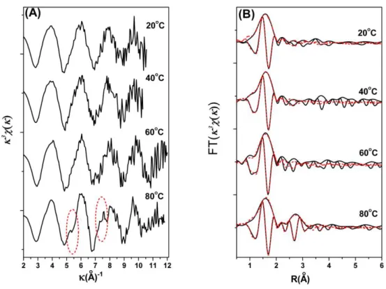
EXAFS data were collected for pH 5.0, 6.4, 7.5, 8.5 and 10.0 sorption samples. The k3-weighted functions of sorption samples show distinct changes as pH increases from 5.0 to 10.0 (Fig. 3A). At low pH, the EXAFS spectral noise is more pronounced due to the low Ni loading on the solid surface. The spectra are dominated by O shell backscattering resembling the Ni2+(aq), as expected based on the spectrum discussed above. As pH increases, pronounced features appear in the higher k range at k=5 Å-1 and k=8 Å-1, which is consistent with the presence of heavy backscattering in the local coordination environment of adsorbed Ni(II).29-31 These features indicate more than one ordered neighboring shells around adsorbed Ni atoms. Therefore, outer-sphere complexation is not the predominant sorption mode of Ni(II) on montmorillonite at high pH values.
The resulting RDFs are shown in Fig. 3B. A broad first-shell O peak is observed for the low pH samples along with a small first-neighbor shell, and reasonable fits were obtained by fitting Ni-O shell.
The EXAFS coordination number of the first shell (NNi-O) is about 6.0 (Table 2). The interatomic
distance (2.03~2.04 Å) is typical of sixfold coordinated Ni. For all RNi-O distances fall in the 2.03~2.04
Å, they cannot explain the formation of hydrated outer-sphere surface complexes on Ca-montmorillonite as we discussed in the batch experiments. Between pH 7.5 and 10.0, a feature appearing at about 3.0~3.3 Å can be explained by the formation of surface or even polynuclear Ni complexes. The different Ni-M (M represents metal element) shell contributes to the samples prepared at high pH as determined from the fitting procedure. For the both pH 7.5 and 8.5 sorption samples, it is
appropriate to fit the experimental data using a combination of Ni-Al and Ni-Si pairs, and a good fit with one Ni-Al and two Ni-Si (Ni-Si1 and Ni-Si2) pairs was obtained. Spectral analysis leads to the identification of three nearest cationic subshells containing Al at 3.00 Å, Si at 3.10 Å and 3.26 Å. The fitting data in this way gives a conclusion that inner-sphere complexes exist in our sorption samples at these conditions. These interatomic distances determined from EXAFS fitting are the characteristic of edge-sharing linkages between Al octahedra and Ni octahedral, and of corner-sharing linkages between Ni octahedra and Si tetrahedra (i.e., the Ni-Si and Ni-Al interatomic distances support the formation of monodentate complexes on the Si tetrahedral and bidentate complexes on the Al octahedra), as in clay structures.29
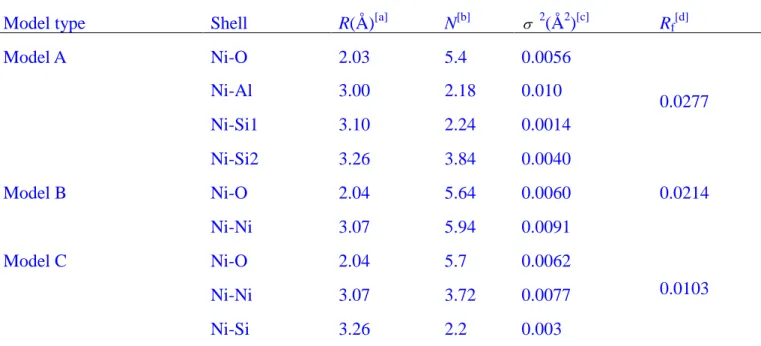
At pH 10.0, the precipitation of Ni(II) may contribute to the sorption. The second RDFs peaks, however, increase in amplitude with pH increasing, but their positions remain constant. Ni(II) complexes or precipitation (Ni(OH)2(s) and Ni-phyllosilicate phase) are assumed to contribute to the
sorption products. In order to get accurate EXAFS fitting, the RDFs peaks in the sample at pH 10 were fitted with three models (model A: fit with O, Al, Si1 and Si2; model B: fit with O and Ni; and model C: fit with O, Ni and Si).32-34 It can be seen from Table 3 that model A and model B have the poor fit with
the reduced error Rf>0.02. The samples were best fitted with ~6 Ni atoms at 3.07 Å and 4~5 Si atoms at
3.26 Å in the second coordination shell. The simultaneous appearance of the short Ni-O and Ni-Ni/Si bonds, similar to that of Ni-phyllosilicate (RNi-Ni=3.05 to 3.08 Å, RNi-Si=3.26 to 3.27 Å), was interpreted
by the nucleation of a Ni phase having a Ni-phyllosilicate-like local atomic structure. The formation of Ni complexes or Ni(OH)2(s) may not be dismissed, while we can conclude that the majority of Ni(II)
sorption is through co-precipitate. The sorption product does not require the addition of Si in solution, and Si dissociate from montmorillonite facilitates the formation of Ni phyllosilicate in our montmorillonite sorption system. Dähn et al.16,17 used polarized XAFS spectral analysis to distinguish whether α-Ni-hydroxide or Ni-phyllosilicate phase was formed on montmorillonite. Their results indicated the formation of Ni-phyllosilicate in the montmorillonite, which supported our results.
The effect of temperature on Ni2+ sorption was also selected for EXAFS measurements. The spectra of the sorption samples at pH 6.4 and the four temperature levels (20, 40, 60 and 80oC) appear comparable (Fig. 4), but differ from either of the references, Ni2+(aq) or -Ni(OH)2(s). The peak at
about 2.03~2.04 Å dominates the RDFs, which has no significant difference for the four samples. EXAFS data do not suggest that the surface complexes at temperatures of 20 and 40oC are significantly different. The second shell is not very pronounced in the RDFs except the sample prepared at 80 oC, and the heavy metal backscattering in the sample at 80oC is more readily observed in the k3-weighted structure. Fig. 4A shows the differences between the k3χ(k) values of the samples at the four temperatures. The spectra have a beat pattern at about k=5 Å-1 and a multifrenquence wave shape at 80oC. These features reflect the second coordination sphere, which is indicative part of inner-sphere complexes forming on Ca-montmorillonite surfaces. EXAFS results reveal that a rise in temperature results in a gain in the inner-sphere complexation, which in turn leads to an increase in Ni(II) sorption
on Ca-montmorillonite. The sorption of Ni(II) ions at the surface at elevated temperatures is likely increased due to electrostatic effects at the surface owing to a lowering of the pHpzc on montmorillonite
as a function of elevated temperature.24,35 The sixfold coordination ([Ni(H2O)6]2+) should also be
favored entropically because of the release of H2O to form inner-sphere complexes.27 At pH 7.5, as can
be seen from the corresponding RSFs of Ni(II) sorbed on montmorillonite for the three different temperatures (i.e., 20, 40 and 60oC) (Fig. SI-4), the first (Ni-O) peak at R 2.03~2.04 Å remains relatively constant in amplitude with increasing temperature. The second (Ni-Al/Si) peak in the RSFs increases with increasing temperature, demonstrating the growth of the Ni(II) inner-sphere complexes with increasing temperature.
3.3 Surface Complexation Modeling: A multisite surface complexation model was considered in this
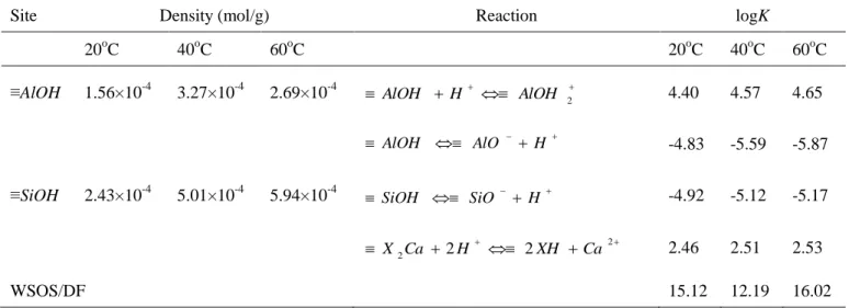
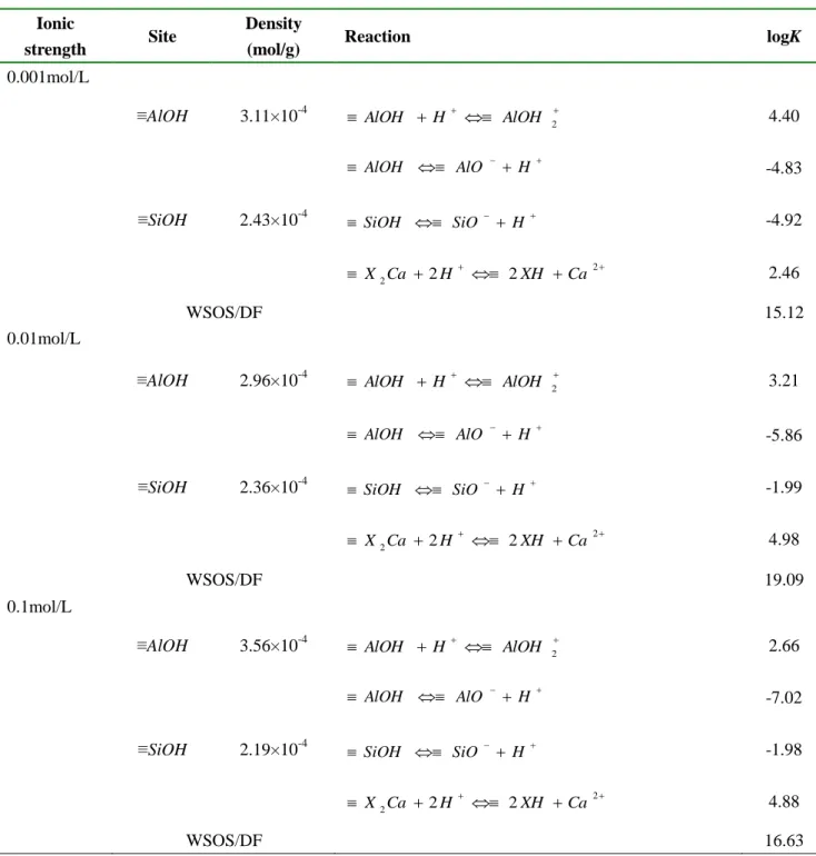
study. The sorption data modeling was completed according to the above structural investigation. Potentiometric titrations were completed on the Ca-montmorillonite used in this study (Fig. SI-1). The characteristics of the amphoteric behavior of the montmorillonite were described in Table 4 as well as the concentrations of each type of sites:1.1×10⁻³ mol/g for exchange sites and for the edge sites, (1.56~3.27)×10-4 and (2.43~5.94)×10-4 mol/g for aluminol and silanol sites, respectively. 10,24,36
The sorption data modeling was completed according to EXAFS structural investigation, which showed that Ni(II) was adsorbed on montmorillonite substrates as monodentate and bidentate surface complexes. The hydrolysis and aqueous complexation constants used in this work to account for nickel chemistry were those reported by Tertre et al.25 We had no information regarding the number of proton released during the sorption process and the choice of the sorption equilibria were based on the goodness of the modeling for this point. The best fits were obtained considering the following sorption equilibria:
(≡X)2Ca + Ni2+ ↔ (≡X)2Ni + Ca2+ (7)
≡Al(OH)2 + Ni2+ ↔≡Al(OH)2Ni2+ (8)
≡SiOH + Ni2+
↔ ≡SiONi+ + H+ (9)
The surface species repartition diagrams (Fig. 2(A)-(C)) are closely linked with the EXAFS structural results, with mainly three species for pH values ranging from 3.0 to 10.0. The surface speciation diagrams show that the ion exchange species (≡X)2Ni is the dominant species at low pH
values, ≡Al(OH)2Ni2+ becomes the major species with increasing pH and ≡SiONi+ is the most abundant
surface species at higher pH. The adsorbed species are same, some differences can be underlined in the surface species repartition depending mainly on pH and temperature. The global surface charge evolution of Ca-montmorillonite versus pH and temperature has to be taken into account to explain this behavior. The model predicts the ion exchange species (≡X)2Ni increase when the temperature elevate
at low pH, with pH increasing, ≡Al(OH)2Ni2+ and ≡SiONi+ species contribute greatly to the increased
sorption as observed at macroscopic and molecular level. EXAFS shows a surface precipitate at pH up to 10.0. However, surface complexation models cannot provid an estimate of the precipitate processes, and the surface species modeling are not corresponding to the EXAFS results.
4. Conclusion
There are several different surface species that Ni(II) can be adsorbed on Ca-montmorillonite, depending on Ca-montmorillonite properties, pH, and ionic strength. At low pH, sorption is via ion exchange with hydrogen and calcium ions that saturate the exchange sites. Electrostatically held outer-sphere Ni(II) commonly adsorbs in the interlayers of Ca-montmorillonite. At high pH, possible inner-sphere complexes for Ni(II) sorption on Ca-montmorillonite that have been observed, which include inner-sphere surface complexes and co-precipitation. The sorption of Ni(II) on
Ca-montmorillonite is the result of the combination of these factors, and the elevated temperatures lead to an increase in the inner-sphere complexation. The results are quite relevant to understand and to evaluate the physicochemical behavior of Ni(II) in the natural environment.
In the broader context of predicting Ni(II) ion mobility, bioavailability, and transport in different aqueous geochemical environments, improvements in geochemical models await further studies establishing the structural basis for Ni(II) ion sorption. The findings in this study are an important step toward a molecular-level description of Ni(II) uptake onto minerals and will help to improve our understanding of Ni(II) ion sorption processes at the water-mineral interface. More work is needed to address sorption reversibility as a function of coverage, pH and aging time etc. before such generalizations, if any, can or should be made in future.
Acknowledgements. Financial support from National Natural Science Foundation of China (20907055;
20971126) and 973 projects (2007CB936602; 2011CB933700) from MOST of China and Anhui Province Technology Fund for Outstanding Youths (10040606Y34) are acknowledged. The authors gratefully acknowledge Dr. Bo He and Dr. Zhi Xie of NSRL, USTC for helpful technical assistance of EXAFS experiments. We also express our thanks to Prof. B. Grambow (SUBATECH Laboratory, France) for favorable discussions.
References
1 R. Mandal, N. M. Hassan, J. Murimboh, C. Chakrabarti, M. H. Back, Environ Sci Technol. 2002, 36, 1477-1484.
3 P. Chang, X. Wang, S. Yu, W. Wu, Colloid Surf. A 2007, 302, 75-81.
4 X. L. Tan, J. Hu, X. Zhou, S. M. Yu, X. K. Wang, Radiochim. Acta 2008, 96, 487-495. 5 C. O. Ijagemi, M. H. Baek, D. S. Kim, J. Hazard. Mater. 2009, 166, 538-546.
6 S. S. Gupta, K. G. Bhattacharyya, J. Colloid Interf. Sci. 2006, 295, 21-32. 7 M. E. Argun, J. Hazard. Mater. 2008, 150, 587-595.
8 K. G. Bhattacharyya, S. S. Gupta, Adv. Colloid Interface Sci. 2008, 140, 114-131. 9 M. H. Bradbury, B. Baeyens, Geochim. Cosmochim. Acta 2009, 73, 990-1003.
10 H. Marcussen, P. E. Holm, B. Strobel, H. C. B. Hansen, Environ. Sci. Technol. 2009, 43, 1122-1127. 11 M. H. Bradbury, B. Baeyens, Geochim. Cosmochim. Acta 1999, 63, 325-336.
12 B. Baeyens, M. H. Bradbury, J. Contam. Hydrol. 1997, 27, 199-222. 13 A. Voegelin, R. Kretzschmar, Environ. Sci. Technol. 2005, 39, 5311-5318.
14 A. M. Scheideger, G. M. Lamble, D. L. Spark, Environ. Sci. Technol. 1996, 30, 548-554. 15 L. Charlet, A. Manceau, Geochim. Cosmochim. Acta 1994, 58, 2577-2582.
16 R. Dähn, A. Scheidegger, A. Manceau, M. Schlegel, B. Baeyens, M. H. Bradbury, J. Synchrotron
Rad. 2001, 8, 533-535.
17 R. Dähn, A. M. Scheidegger, A. Manceau, M. L. Schlegel, B. Baeyens, M. H. Bradbury, M. Morales,
Geochim. Cosmochim. Acta 2002, 66, 2335-2347.
18 C. L. Peacock, D. M. Sherman, Geochim. Cosmochim. Acta 2004, 68, 2623-2637.
19 E. Ferrange, C. Tournassat, E. Rinnert, B. Lanson, Geochim. Cosmochim. Acta 2005, 69, 2797-2812. 20 B. Ravel, M. Newville, J. Synchrotr. Radiat. 2005, 12, 537-541.
21 M. Newville, P. Līviņš, Y. Yacoby, J. J. Rehr, E. A. Stern, Phys. Rev. B 1993, 47, 14126-14131. 22 A. L. Ankudinov, J. J. Rehr, Phys. Rev. B. 1997, 56, 1712-1715.
23 B. Perdikatsis, H. Burzlaff, Z. Kristallogr. 1981, 156, 177-186.
24 E. Tertre, S. Castet, Z. G. Berger, M. Loubet, E. Giffaut, Geochim. Cosmochim. Acta 2006, 70, 4579-4599.
25 E. Tertre, G. Berger, S. Castet, M. Loubet, E. Giffaut, Geochim. Cosmochim. Acta 2005, 21, 4937-4948.
26 W. Li, G. Pan, M. Zhang, D. Zhao, Y. Yang, H. Chen, G. He, J. Colloid Interf. Sci. 2008, 319, 385-391.
27 Q. Fan, D. Shao, Y. Lu, W. Wu, X. Wang, Chem. Eng. J. 2009, 150, 188-195.
28 Y. Xu, L. Axe, T. Boonfueng, T. A. Tyson, P. Trivedi, K. Pandya, J. Colloid Interf. Sci. 2007, 314, 10-17.
29 R. Dähn, A. M. Scheidegger, A. Manceau, M. L. Schlegel, B. Baeyens, M. H. Bradbury, D. Chateigner, Geochim. Cosmochim. Acta 2003, 67, 1-15.
30 A. M. Scheidegger, E. Wieland, A. C. Scheinost, R. Dähn, P. Spieler, Environ. Sci. Technol. 2000,
34, 4545-4548.
31 D. R. Roberts, A. Scheidegger, D. Sparks, Environ. Sci. Technol. 1999, 33, 3749-3754.
32 K. Fahmy, M. Merroun, K. Pollmann, J. Raff, O. Savchuk, C. Hennig, Selenska-Pobell, Biophys. J. 2006, 91, 996-1007.
33 M. L. Merroun, J. Raff, A. Rossberg, C. Hennig, T. Reich, S. Selenska-Pobell, Appl. Environ.
Microbiol. 2005, 71, 5532-5543.
34 B. K. Teo, EXAFS: basic principles and data analysis. Springer-Verlag Press. 1996.
35 M. Rozalén, P. Brady, F. J. Huertas, J. Colloid Interf. Sci. 2009, 333, 474-484.
Table 1 Thermodynamic data of Ni(II) sorption on Ca-montmorillonite at different temperatures
Temperature ΔG (kJ/mol) ΔH (kJ/mol) ΔS (kJ/(mol·K)) 20oC -19.54 31.81
0.18 40oC -22.90 31.94
Table 2 Structure parameters derived from EXAFS analysis of reference samples and sorption samples
at Ni K-edge
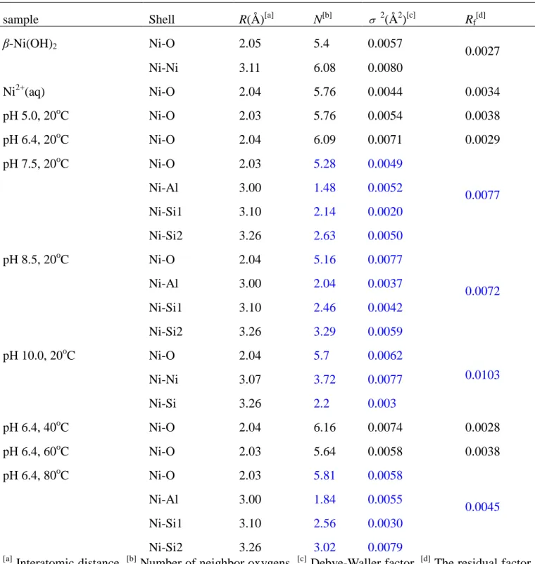
sample Shell R(Å)[a] N[b] 2(Å2)[c] Rf[d]
β-Ni(OH)2 Ni-O 2.05 5.4 0.0057 0.0027 Ni-Ni 3.11 6.08 0.0080 Ni2+(aq) Ni-O 2.04 5.76 0.0044 0.0034 pH 5.0, 20oC Ni-O 2.03 5.76 0.0054 0.0038 pH 6.4, 20oC Ni-O 2.04 6.09 0.0071 0.0029 pH 7.5, 20oC Ni-O 2.03 5.28 0.0049 0.0077 Ni-Al 3.00 1.48 0.0052 Ni-Si1 3.10 2.14 0.0020 Ni-Si2 3.26 2.63 0.0050 pH 8.5, 20oC Ni-O 2.04 5.16 0.0077 0.0072 Ni-Al 3.00 2.04 0.0037 Ni-Si1 3.10 2.46 0.0042 Ni-Si2 3.26 3.29 0.0059 pH 10.0, 20oC Ni-O 2.04 5.7 0.0062 0.0103 Ni-Ni 3.07 3.72 0.0077 Ni-Si 3.26 2.2 0.003 pH 6.4, 40oC Ni-O 2.04 6.16 0.0074 0.0028 pH 6.4, 60oC Ni-O 2.03 5.64 0.0058 0.0038 pH 6.4, 80oC Ni-O 2.03 5.81 0.0058 0.0045 Ni-Al 3.00 1.84 0.0055 Ni-Si1 3.10 2.56 0.0030 Ni-Si2 3.26 3.02 0.0079 [a]
Interatomic distance, [b] Number of neighbor oxygens, [c] Debye-Waller factor, [d] The residual factor.
β-Ni(OH)2 and Ni2+(aq) are named as reference samples, whereas the other samples of
Table 3 Structure parameters derived from EXAFS analysis using different fit approach of sample at pH
10.0
Model type Shell R(Å)[a] N[b] 2(Å2)[c] Rf[d]
Model A Ni-O 2.03 5.4 0.0056 0.0277 Ni-Al 3.00 2.18 0.010 Ni-Si1 3.10 2.24 0.0014 Ni-Si2 3.26 3.84 0.0040 Model B Ni-O 2.04 5.64 0.0060 0.0214 Ni-Ni 3.07 5.94 0.0091 Model C Ni-O 2.04 5.7 0.0062 0.0103 Ni-Ni 3.07 3.72 0.0077 Ni-Si 3.26 2.2 0.003 [a]
Table 4 Surface site concentration and intrinsic surface complexation constants of Ca-montmorillonite calculated by FITEQL 3.2. CSolid=5.0 g/L, BET=64.4 m2/g, NX2Ca=110 mmol/100g, I=0.001mol/L
Ca(NO3)2.
Site Density (mol/g) Reaction logK
20oC 40oC 60oC 20oC 40oC 60oC
≡AlOH 1.56×10⁻⁴ 3.27×10⁻⁴ 2.69×10⁻⁴ AlOH H AlOH 2 4.40 4.57 4.65
AlOH AlO H -4.83 -5.59 -5.87
≡SiOH 2.43×10⁻⁴ 5.01×10⁻⁴ 5.94×10⁻⁴ SiOH SiO H -4.92 -5.12 -5.17
2 2Ca 2H 2XH Ca X 2.46 2.51 2.53 WSOS/DF 15.12 12.19 16.02
Figure Captions
Fig. 1 Illustration of sorption sites for Ni on Ca-montmorillonite.
Fig. 2 Sorption percent of Ni(II) on Ca-montmorillonite and corresponding surface species repartition
diagrams at temperature 20oC (A), 40oC (B), 60oC (C). Sorption isotherms of Ni(II) on Ca-montmorillonite at three temperatures (D). m/V=0.5 g/L, I=0.001 mol/L Ca(NO3)2. (A-C):
CNi(II)initial=10 mg/L; (D): pH=6.4.
Fig. 3 Raw k3-weighted χ(k) spectra of references and experimental samples (A) and their corresponding pseudo radial distribution functions (RDFs) (B) at different pH. m/V=0.5 g/L, CNi(II)initial=10 mg/L,
pH=5.0, 6.4, 7.5, 8.5 and 10.0, T=20oC, I=0.001 mol/L Ca(NO3)2.
Fig. 4 Raw k3-weighted χ(k) spectra of experimental samples (A) and their corresponding pseudo radial distribution functions (RDFs) (B) at three different temperatures. m/V=0.5 g/L, CNi(II)initial=10 mg/L,
Fig. 2 Sorption percent of Ni(II) on Ca-montmorillonite and corresponding surface species repartition
diagrams at temperature 20oC (A), 40oC (B), 60oC (C). Sorption isotherms of Ni(II) on Ca-montmorillonite at three temperatures (D). m/V=0.5 g/L, I=0.001 mol/L Ca(NO3)2. (A-C):
CNi(II)initial=10 mg/L; (D): pH=6.4.
Fig. 3 Raw k3-weighted χ(k) spectra of references and experimental samples (A) and their corresponding pseudo radial distribution functions (RDFs) (B) at different pH. m/V=0.5 g/L,
Fig. 4 Raw k3-weighted χ(k) spectra of experimental samples (A) and their corresponding pseudo radial distribution functions (RDFs) (B) at three different temperatures. m/V=0.5 g/L, CNi(II)initial=10
Table of contents:
Supporting Information on
Sorption Speciation of Nickel(II) onto Ca-Montmorillonite: Batch, EXAFS
Techniques and Modeling
XiaoLi Tan†*, Jun Hu†, Gilles Montavon‡, XiangKe Wang†*
Key Laboratory of Novel Thin Film Solar Cells, Institute of Plasma Physics, Chinese Academy of Sciences, P.O. Box 1126, Hefei, 230031, P.R. China; and Laboratory SUBATECH, Groupe de Radiochimie, UMR Ecole des Mines/CNRS/Université, 4 rue A. Kastler, BP 20722, 44307 Nantes
cedex 03, France Xiaoli Tan: tanxl@ipp.ac.cn
Jun Hu: jhu@ipp.ac.cn
Gilles Montavon: montavon@subatech.in2p3.fr Xiangke Wang: xkwang@ipp.ac.cn
*: Corresponding author. Tel: +86-551-5592788; Fax: +86-551-5591310. Email: tanxl@ipp.ac.cn (X.L. Tan), xkwang@ipp.ac.cn (X.K. Wang). †: Institute of Plasma Physics; ‡: Laboratory
Figure Captions
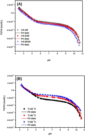
Fig. SI-1 Acid-base titrations of Ca-montmorillonite (5.0 g/L) as a function of pH in 0.001, 0.01 and
0.1 mol/L Ca(NO3)2 at T = 20 oC (A) and at three different temperatures in 0.001 mol/L Ca(NO3)2
(B). The lines are calculated using FITEQL 3.2.
Fig. SI-2 Sorption edges of Ni(II) at different ionic strengths. m/V = 0.5 g/L, CNi(II)initial = 10 mg/L, T
= 20 oC and I = 0.001, 0.01 and 0.1 mol/L Ca(NO3)2.
Fig. SI-3 XANES spectra of the reference and sorption samples at different pH (A) and temperature
(B).
Fig. SI-4 Raw k3-weighted χ(k) spectra of experimental samples (A) and their corresponding pseudo radial distribution functions (RDFs) (B) at three different temperatures. m/V = 0.5 g/L, CNi(II)initial =
Acid-base Titration of Ca-montmorillonite. Potentiometric titration of Ca-montmorillonite
suspensions (5.0 g/L) were performed over a wide range of pH (3.2-10.6) to measure proton sorption/desorption as a function of pH and ionic strength (0.1, 0.01, and 0.001 mol/L Ca(NO3)2) at
20 oC with a Mettler-Toledo DL 50 Automatic Titrator. Before the titration, the suspensions were titrated up to pH ~3 quickly with 1 mol/L HNO3 and purged with argon gas for about 1 h. Then the
titration was carried out from pH ~3 to pH ~10.5 by using 0.0911 mol/L NaOH.
3 4 5 6 7 8 9 10 11 -4.0x10⁻³ -3.0x10⁻³ -2.0x10⁻³ -1.0x10⁻³ 0.0 1.0x10⁻³ I=0.1M Fit data I=0.01M Fit data I=0.001M Fit data T O T H ( m o l/ L ) pH (A) 3 4 5 6 7 8 9 10 11 -5.0x10⁻³ -4.0x10⁻³ -3.0x10⁻³ -2.0x10⁻³ -1.0x10⁻³ 0.0 1.0x10⁻³ 2.0x10⁻³ (B) T=20 oC Fit data T=40 oC Fit data T=60 o C Fit data T O T H ( m o l/ L ) pH
Fig. SI-1 Acid-base titrations of Ca-montmorillonite (5.0 g/L) as a function of pH in 0.001, 0.01 and
0.1 mol/L Ca(NO3)2 at T = 20 oC (A) and at three different temperatures in 0.001 mol/L Ca(NO3)2
Table SI-1 Surface site concentration and intrinsic surface complexation constants of
Ca-montmorillonite calculated by FITEQL 3.2. CSolid = 5.0 g/L, BET= 64.4 m2/g, NX2Ca = 110
mmol/100g, T = 20 oC.
Ionic
strength Site
Density
(mol/g) Reaction logK
0.001mol/L
≡AlOH 3.11×10⁻⁴ AlOH H AlOH 2 4.40
AlOH AlO H -4.83
≡SiOH 2.43×10⁻⁴ SiOH SiO H -4.92
2 2Ca 2H 2XH Ca X 2.46 WSOS/DF 15.12 0.01mol/L
≡AlOH 2.96×10⁻⁴ AlOH H AlOH 2 3.21
AlOH AlO H -5.86
≡SiOH 2.36×10⁻⁴ SiOH SiO H -1.99
2 2Ca 2H 2XH Ca X 4.98 WSOS/DF 19.09 0.1mol/L
≡AlOH 3.56×10⁻⁴ AlOH H AlOH 2 2.66
AlOH AlO H -7.02
≡SiOH 2.19×10⁻⁴ SiOH SiO H -1.98
2 2Ca 2H 2XH Ca X 4.88 WSOS/DF 16.63
3 4 5 6 7 8 9 10 0 20 40 60 80 100 0.001 mol/L Ca(NO 3)2 0.01 mol/L Ca(NO 3)2 0.1 mol/L Ca(NO3)2 pH A d s o rp ti o n %
Fig. SI-2 Sorption edges of Ni(II) at different ionic strengths. m/V = 0.5 g/L, CNi(II)initial = 10 mg/L, T
= 20 oC and I = 0.001, 0.01 and 0.1 mol/L Ca(NO3)2.
From the ionic strength dependence, one can deduce that cation exchange is the main mechanism for Ni(II) sorption on Ca-montmorillonite at pH < 7, which is also supported by the very slow increase of Ni(II) sorption at this pH range. This is consistent with increased sorption on the permanent charge sites with decreasing Ca2+ concentration. The ionic strength-dependent sorption suggests that Ni2+ exchange reaction on permanent charge sites is the predominant sorption process at the lowest Na+ concentration. The results of Ni(II) sorption are similar to that found and described previously for Cu(II) sorption as a function of pH and Na+ concentrations.1 It was reported that an increase in Na+ concentration results in a displacement of metal ions from the interlayer sites and a shift in metal ions sorption from the interlayer to the edge sites.
Sample Preparation for EXAFS Analysis. Ni(II) sorption samples were prepared by adding
Ni2+ (in the form of Ni(NO3)2 solution) to freshly prepared Ca-montmorillonite suspensions. The
initial Ni(II) concentration (10 mg/L) and the reaction pH were chosen to achieve high Ni(II) loading on Ca-montmorillonite while avoiding that the bulk solutions were undersaturated with
respect to crystalline Ni(OH)2(s).2,3 The samples were shaken end-over-end for 2 weeks. Samples
for EXAFS measurements were prepared from the residual wet pastes obtained after centrifugation of the suspensions. Samples preparation and equilibration steps were carried out in a glove box under N2 atmosphere (CO2 < 5 ppm, and O2 < 5 ppm).
8200 8400 8600 8800 9000 0 2 4 6 8 10 pH10.0 pH8.5 pH7.5 pH6.4 pH5.0 Ni2+ Ni(OH)2 N o rm a li z e d A b s o e b a n c e Energy(eV) (A) 8200 8400 8600 8800 9000 0.0 0.5 1.0 1.5 2.0 2.5 3.0 3.5 4.0 4.5 5.0 5.5 6.0 80oC 60oC 40oC 20oC (B) N o rm a li z e d A b s o e b a n c e Energy(eV)
Fig. SI-3 XANES spectra of the reference and sorption samples at different pH (A) and temperature
2 3 4 5 6 7 8 9 10 11 12 (A)-1 60oC 40oC 20oC (A) o 1 2 3 4 5 6 60oC 40oC 20oC (B) FT ( ) R(A)o
Fig. SI-4 Raw k3-weighted χ(k) spectra of experimental samples (A) and their corresponding pseudo radial distribution functions (RDFs) (B) at three different temperatures. m/V = 0.5 g/L, CNi(II)initial =
10 mg/L, pH = 7.5, T = 20, 40 and 60 oC, I = 0.001 mol/L.
Literature Cited
1 Morton, J.D.; Semrau, J.D.; Hayes, K.F. An X-ray absorption spectroscopy study of the structure and reversibility of copper adsorbed to montmorillonite clay. Geochim. Cosmochim.
Acta 2001, 65, 2709-2722.
2 Dähn, R.; Scheidegger, A.M.; Manceau, A.; Schlegel, M.L.; Baeyens, B.; Bradbury, M.H.; Chateigner, D. Structural evidence for the sorption of Ni(II) atoms on the edges of montmorillonite clay minerals: a polarized X-ray absorption fine structure study. Geochim.
Cosmochim. Acta 2003, 67, 1-15.
3 Dähn, R.; Scheidegger, A.M.; Manceau, A.; Schlegel, M.L.; Baeyens, B.; Bradbury, M.H.; Morales, M. Neoformation of Ni phyllosilicate upon Ni uptake on montmorillonite: a kinetics
study by powder and polarized extended X-ray absorption fine structure spectroscopy. Geochim.