Université de Montréal
Understanding the Role of the Matricellular Protein SMOC-2 in Renal Cell Carcinoma
Par
Daniel Feng
Département de Pharmacologie et Physiologie Faculté de Médecine
Thèse présenté en vue de l’obtention du grade de M. Sc en Pharmacologie
Août, 2019
Université de Montréal
Faculté des études supérieures et postdoctorales
Cette thèse intitulée
Understanding the Role of the Matricellular Protein SMOC-2 in Renal Cell Carcinoma
Présentée par
Daniel Feng
A été évaluée par un jury composé des personnes suivantes
Dr. Christian Beauséjour Président-rapporteur Dr. Casimiro Gerarduzzi Directeur de recherche Dr. Sylvain Meloche Membre du jury
Résumé
Les proteins matricellulaires (MPs) sont des macromolécules non structurales de la matrice extracellulaire (ECM) qui sont induites de façon transitoire lors du développement, de la réparation et du remodelage tissulaire et lors de l’inflammation. L’expression des MPs peut être déclenchée par des dommages tissulaires aigus, et leur expression à long terme peut contribuer à certaines maladies chroniques. Les MPs agissent principalement pour médier les événements du remodelage tissulaire en facilitant les interactions et les signaux à partir de l’ECM vers l’environnement cellulaire avoisinant.
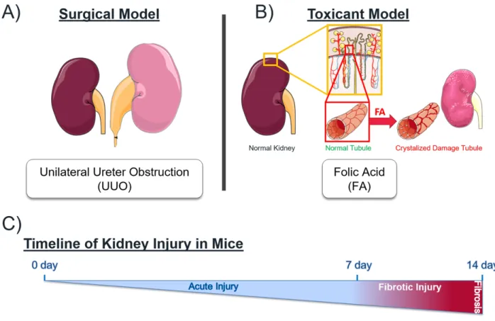
En utilisant des données de RNA-seq provenant de deux modèles distincts de dommages rénaux, soit l’Acide Folique (FA) ou l’Obstruction Urétérale Unilatérale (UUO), nous avons analysé les profils d’expressions de plusieurs familles bien connues de MPs lors des blessures aigues et chroniques. Nous révélons de nouvelles MPs impliquées dans les dommages rénaux et présentons de nouveaux réseaux entre les membres de chaque famille de MPs, en utilisant des outils bioinformatiques. L’expression de l’ARNm de certaines MPs a été confirmée par immuno-buvardage de type Western (WB).
Afin d’approfondir notre connaissance des mécanismes de réparation tissulaire et de remodelage de la matrice, nous avons choisi SMOC-2 comme MP modèle dans l’étude des carcinomes cellulaires rénaux (RCC), cancers qui présentent de fortes tendances métastatiques. Nous avons démontré que la surexpression de SMOC-2 ainsi que le traitement avec la protéine recombinante de lignées cellulaires RCC (786-O, et ACHN) induisent un profil métastatique de transition épithélio-mésenchymateuse (EMT) par WB et des tests fonctionnels. Nous avons également démontré que l’inhibition de SMOC-2 par siRNA donne les résultats opposés.
L’ensemble de nos travaux utilise la compréhension des patrons d’expressions temporels des MPs pour améliorer notre compréhension des mécanismes et conditions qui supportent une activation persistante dans des états pathologiques chroniques. Globalement, notre étude sur SMOC-2 offre une perspective ainsi qu’un modèle intéressant pour l’étude et la caractérisation de nouvelles MPs dans des maladies impliquant le remodelage et la réparation de la matrice.
Mot Clès: protéine matricellulaire, transition épithéliale-mésenchymateuse, acide folique, obstruction unilatérale de l'uretère, matrice extracellulaire, carcinome à cellules rénales, métastase du cancer.
Abstract
Matricellular proteins (MPs) are non-structural ECM macromolecules induced transiently during development, tissue repair and remodeling, and inflammation. Expression of MPs can be triggered by acute tissue injury and their sustained expression can contribute to chronic disease. MPs primarily act to mediate tissue remodeling events by facilitating interactions and signals from the ECM to the surrounding cellular niche.
Using published RNA-seq data from two distinct models of kidney injury, Folic Acid (FA) and Unilateral Ureteral Obstruction (UUO), we analyzed the expression profile of various members of well-known MP families during the acute and fibrotic injury phases. We reveal novel MPs implicated in renal injury and present informative networks between members of each MP family using bioinformatic tools. mRNA expression of select candidate MPs were confirmed by Western blot.
To extend our understanding of translatable mechanisms in repair and matrix remodeling, we chose SMOC-2 as our MP model to study in Renal Cell Carcinoma (RCC) which has strong metastatic tendencies. SMOC-2 overexpression and recombinant protein treatment of RCC cell lines (786-O, ACHN) were shown to induce a metastatic EMT profile by Western blot analysis, supported by functional assays (proliferation, migration). Silencing SMOC-2 by siRNA showed the contrary results.
Taken together, our work utilizes the understanding of temporal expression patterns of MPs to gain insight into mechanisms and conditions that support persistent activation in chronic injury states. Overall, our work with SMOC-2 provides a valuable perspective and template to approach studying and characterizing novel MPs in diseases involving pathological matrix remodeling and repair.
Key Words: matricellular protein, epithelial-to-mesenchymal transition, folic acid, unilateral ureteral obstruction, extracellular matrix, renal cell carcinoma, cancer metastasis.
Table of Contents
List of Thesis Figures ...7
List of Manuscript Figures ...8
List of Thesis Tables ...9
List of Manuscript Tables ...9
List of Abbreviations ...10
Acknowledgements ...11
1.0 Introduction 1.1. Extracellular Matrix Composition & Function ...12
1.2. Matricellular Protein Function and Integrin Signaling ...15
1.3. Matricellular Protein Families ...18
1.3.1. SMOC-2 as a Model Matricellular Protein ...22
1.4. Tissue Repair Processes Overview ………..24
1.4.1. Fibrosis – Deregulated Repair ...26
1.4.2. The Kidney as a Model Organ to Study Injury and Maladaptive Repair 28 1.5. Development Overview ………...29
1.5.1. Cancer – Deregulated Development ...30
1.6. Epithelial-to-Mesenchymal Transition ...33
1.6.1. Epithelial-to-Mesenchymal Transition in Cancer Metastasis ...35
1.7. Rationale & Research Objectives ...37
2.0 Manuscript - Characterization of Matricellular Protein Expression Signatures in Mechanistically Diverse Mouse Models of Kidney Injury 2.1 Preface to Manuscript ...38
2.2 Manuscript ...39
2.3 Discussion of Manuscript - SMOC-2 as a Candidate MP ...72
3.0 Understanding the Role of SMOC-2 in Renal Cell Carcinoma 3.1 Preface ...74
3.2 Manuscript in Preparation - SMOC-2 Promotes Epithelial Mesenchymal Transition and
a pro-Metastatic Phenotype in Epithelial Cells of Renal Cell Carcinoma Origin ...75
3.3 Introduction - Renal Cell Carcinoma Background ...76
3.4 Materials & Methods ...78
3.4.1 Cell Culture and Media ...78
3.4.2 Histology & Immunostaining ...79
3.4.3 SMOC-2 Transfection and Recombinant Protein Treatment ...79
3.4.4 Western Blotting ...81
3.4.5 Functional Assays ...82
3.5 Results ...83
3.5.1 Histological Confirmation of Tumorigenic Tissue in RCC Patient Biopsy Samples ...83
3.5.2 SMOC-2 is Highly Expressed in RCC Patient Biopsy Samples with Co-localization of Vimentin ...84
3.5.3 Endogenous SMOC-2 Expression in RCC Cell Lines ...85
3.5.4 Activation of EMT Markers by SMOC-2 Overexpression ...86
3.5.5 Activation of EMT Markers by SMOC-2 Recombinant Protein ...89
3.5.6 SMOC-2 Silencing in Stimulated RCC cells Downregulate EMT Marker ...92
3.5.7 SMOC-2 Overexpression & Silencing Affects Cell Proliferation ...94
3.5.8 SMOC-2 Modulation Regulates Cell Migration ...97
3.6 Discussion ...102
4.0 Overall Discussion ...105
5.0 Future Perspective ...108
6.0 Conclusion ...110
List of Thesis Figures
Figure 1. Integrin pathways activated by ECM protein interactions ...17
Figure 2. SMOC-2 protein domains ...22
Figure 3. MPs in metastatic cancer development ...32
Figure 4: H&E staining of RCC patient biopsy samples ...83
Figure 5: Immunofluorescence staining for patient biopsy samples ...84
Figure 6: SMOC-2 endogenous levels detected by Western blot ...85
Figure 7: SMOC-2 overexpression induces EMT makers ...88
Figure 8: Recombinant SMOC-2 Protein Treatment induces EMT markers ...91
Figure 9: SMOC-2 silencing downregulates EMT markers ...93
Figure 10: SMOC-2 overexpression promotes cell proliferation by MTT assay ...95
Figure 11: SMOC-2 silencing reduces cell proliferation by MTT assay ...96 Figure 12: rSMOC-2 promotes cell migration ... 98-99 Figure 13: SMOC-2 silencing inhibits cell migration ... 100-101
List of Manuscript Figures
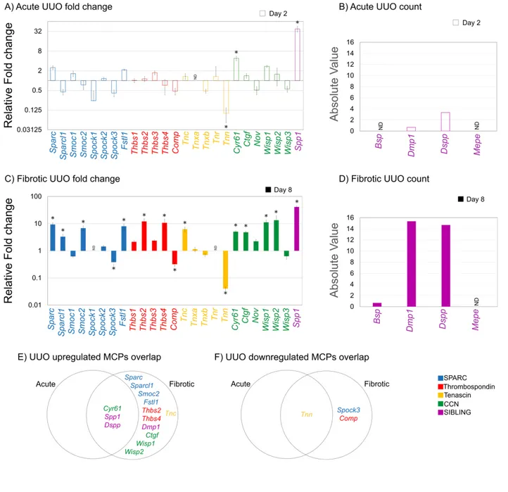
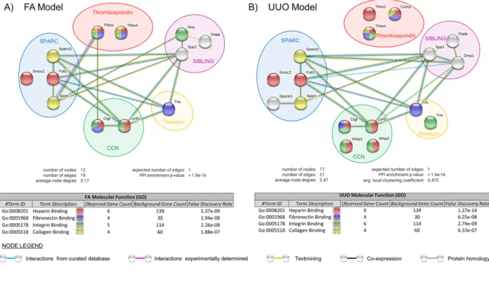
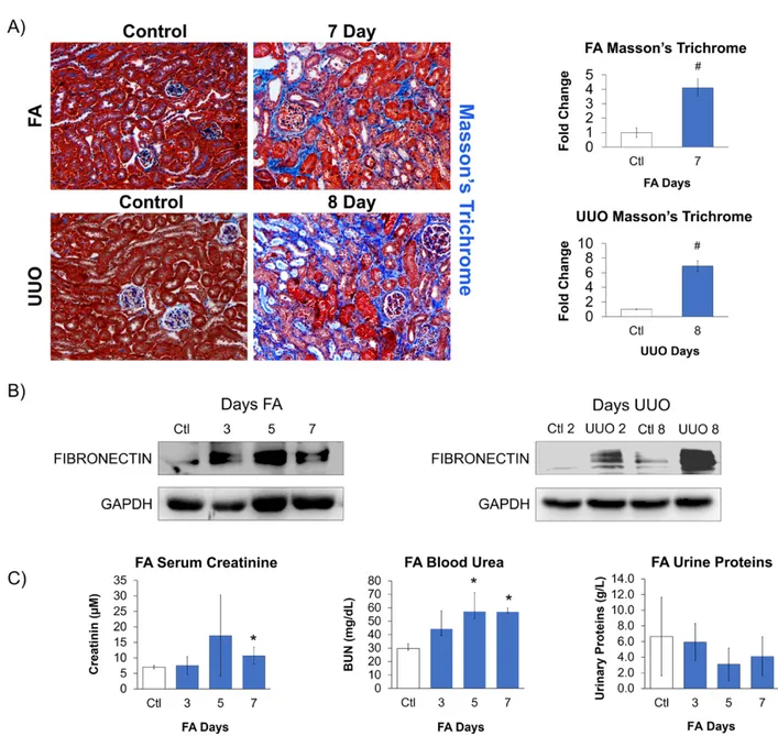
Figure 1. Mechanistically distinct mouse models of kidney injury ...61 Figure 2. MP expression in mouse kidney at Acute and Fibrotic time points after FA treatment ...62 Figure 3. MP expression in mouse kidney at Acute and Fibrotic time points after UUO treatment ...63 Figure 4. Differential expression of MP mRNA at different time-points between FA- and UUO-induced injuries ...64 Figure 5. Interaction analysis of differentially expressed MP genes within the fibrotic phase of kidney injury using the database STRING ...65 Figure 6. Confirmation of kidney injury within mouse models ...66 Figure 7. Validating RNA-Seq data of selected MPs from kidney injury models ...68 Supplementary Figure 1. Functional networks of significantly regulated MCP and ECM genes within the Fibrotic phase of kidney injury using the STRING database ...71
List of Thesis Tables
Table 1. Functional Roles and Characteristics of Matricellular Protein Families ...21
List of Manuscript Tables
Table 1. Listing of candidate MP genes based on published fibrotic relevance within in vitro and/or in vivo models ...60
List of Abbreviations
AKI – acute kidney injury
a-SMA – a-alpha smooth muscle actin BUN – blood urea nitrogen
CKD – chronic kidney diease DPC – days post coitum DE – differentially expressed ECM – extracellular matrix EGF – epidermal growth factor
EMT – epithelial-to-mesenchymal transition FA – folic acid
FBS – fetal bovine serum
GEO – gene expression omnibus GAG – glycosaminoglycan GO – gene ontology
i.p – intraperitoneal injection
KUPKB – kidney and urinary pathway knowledge base LOX – lysyl oxidase
MP – matricellular protein MMP – matrix metalloprotease
PDGF – platelet derived growth factor RCC – renal cell carcinoma
SPARC – secreted protein acidic and rich in cysteine
SIBLING – small integrin-binding ligand, N-linked glycoprotein SLRP - Small leucine-rich proteoglycan
SMOC-2 – secreted modular calcium binding protein-2
STRING – search tool for the retrieval of interacting genes/proteins THBS - thrombospondin
TN - tenascin
UUO – unilateral ureteral obstruction VEGF – vascular endothelial growth factor
Acknowledgements
I would like to first extend my greatest gratitude to both my professors, Dr. Vincent Pichette and Dr. Casimiro Gerarduzzi. Thank you Dr. Pichette for taking me on as a student and giving me the opportunity to pursue scientific research and opening your lab space to us.
Thank you to Dr. Gerarduzzi for taking me on as his first student and being my friend, mentor and fellow Raptors fan throughout this journey. Thank you for all the opportunities and challenges you gave me and for always pushing me to see my potential.
Thank you to Nathalie Henley, the super research assistant. Thank you especially for helping me navigate the complexities and intricacies of HMR. I am most grateful for all the guidance and support you provided and always having answers to my constant “Hey
Nathalie….”. Thank you for teaching me to appreciate the little details in experiments.
Thank to you to all my lab mates over the course of time. Each of you brought your own perspectives and personalities, and it was great to have more company in the lab.
And lastly, thank you to the research community and staff of HMR. I am sorry I cannot list everyone but it has been a pleasure meeting and working with everyone. I hope our paths cross again someday.
1.0 Introduction
The idea of matricellular proteins is credited to the group of Paul Bornstein and their 1991 manuscript titled “Extracellular Proteins That Modulate Cell-Matrix Interactions”1. The proteins
described by Bornstein have come to be known today as matricellular proteins (MPs) and have been appreciated and recognized as important contributors to the plethora of ways cells
communicate and regulate the surrounding extracellular environment. The term ‘matricellular’ is used to describe a particular group of extracellular matrix (ECM) proteins that do not contribute directly to the formation of structural elements, but instead serve to modulate cell–matrix interactions and cell function2. MPs work in an orchestrated manner with major structural
proteins (ie. collagens, fibronectin, proteoglycans) to provide the heterogenous ECM scaffold for cellular functions. The primary cell functions governed by MPs include migration and
chemotaxis, matrix (dis)-assembly, signal transduction of diffusible small proteins (ie. cytokines and growth factors) and supporting a state of intermediate cell adhesion to allow rapid response to external stimuli requiring increased adhesion or de-adhesion2.
1.1 Extracellular Matrix Composition & Function
The extracellular matrix (ECM) is a non-cellular component present in all tissues and organs that provides a physical framework for cellular constituents and acts in a reciprocal manner towards the surroundings of cell populations by mediating biochemical and
biomechanical cues. The ECM is not simply a static structure but is better understood as a highly dynamic environment that reflects the needs and demands of the local cellular environment or in some cases the whole tissue. Broadly, the ECM is made of water, proteins and polysaccharides, but each tissue has a unique heterogenous composition and topology generated during
development3. Owing to the various demands of each unique organ, mechanical parameters such
as compressive strength, elasticity and density are affected by the nearby cells as the delicate balance of ECM components adapts to the changing demands of the tissue.
The two major classes of macromolecules in the ECM are proteoglycans and fibrous proteins3. Proteoglycans are proteins that have been post-translationally modified by
glycosylation by covalent linkage of glycosaminoglycan (GAG) chains to a peptide core and fill the majority of the interstitial space by forming linked matrices to provide resistance towards compressive forces3. Three main families of proteoglycan proteins in the ECM are small
leucine-rich proteoglycans (SLRPs), modular proteoglycans and cell surface proteoglycans3. In addition
to being structural proteins, proteoglycans also serve as mediators of ECM signal transduction as they are upstream of intracellular signaling cascades owing to their localization3, 4. Fibrous
proteins of the ECM are predominantly collagen, fibronectin, elastin and laminin which compose the main structural proteins that provide tensile strength to tissues to enable a physical response to mechanical forces3, 5. Collagens are a ubiquitous protein in vertebrae secreted primarily by
local fibroblasts and are the most abundant protein in the ECM. Fibrous type collagens type I, II, III are the major forms in the ECM and form a rod like triple helix structure made of three a-chains in a hetero-or homotrimeric fashion with context dependent variable length3, 6. Covalent
cross-linking of collagen fibers mediated by lysyl oxidases and association with the other fibrous proteins in the ECM allow tissues like the skin, heart and bones to resist sheer, tensile and
pressure forces to maintain function. The delicate balance of collagen production and
degradation in the ECM is often perturbed in pathologies like organ fibrosis with excess collagen deposition causing tissue stiffening or Ehlers-Danlos syndrome in which genetic collagen
mutations lead to frequent joint dislocations and hypermobility.
Fibronectin is an abundant modular glycoprotein of the ECM that assembles into dimers to create a branched mesh network that extends and connects adjacent cells and can facilitate binding to collagen, tethering to cell surface integrins and other fibronectin proteins through its various modular domains7. Its own dimeric assembly is a cell mediated process and begins as
thin fibrils which eventually cluster together to form more robust and thicker bundle structures from the multimerization of individual dimers7. These mature fibronectin fibrils have remarkable
tensile strength as they can be stretched several times beyond its resting length by cell traction forces, which has also been shown to expose hidden integrin binding domains for adaptive cell signaling3, 7.
The capacity of the ECM to induce intracellular pathways and signaling cascades is mediated primarily by binding to different heterodimeric integrins on the cell surface. Integrin binding domains on matrix proteins can recognize unique integrin combinations associated with the desired response or induce ligand occupied integrin translocation. For example, avb3
integrins were found to predominate at the cell periphery for formation of focal adhesions and stress fibers, whereas a5b1 integrins were observed to associate with intracellular actin bundles to
transmit cell generated tension forces7, 8. The fibronectin network itself can also act as a
mechano-regulator and indicator of tissue stiffness as conformational changes can expose cryptic integrin binding sites. Processes like cell spreading and adhesion can adapt to different levels of stiffness as seen with experiments showing predominant a5b1 interactions in maintaining cell
adhesion and resisting detachment but preferential avb3 binding on stiffer substrates3, 9.
Elastin’s are another type of fibrous protein found in the ECM and have important functions in allowing tissues to recoil from stretching forces, as their name suggests. Formation of elastic fibers occur by lysyl oxidase mediated crosslinking of the precursor, tropoelastin and are modified with fibrillin’s to maintain structural integrity3, 10.
Laminins are large heterotrimeric multimodal proteins, perhaps best known for their role in basement membrane formation but also act to reinforce the ECM through association with other matrix macromolecules11. Laminins, like the other major fibril proteins of the ECM,
connect to the surrounding cells via integrins to fulfill their mechanical anchoring properties but to also mediate cell migration and adhesion. Of the three subunits, α, β, and γ, the a-chain plays a significant role in mediating biological processes, especially organogenesis during embryo development12.
Lastly, within the ECM are also non-structural proteins, known as MPs that are secreted specifically into the pericellular matrix, a sub-compartment of the ECM directly adjacent to the cell membrane2. MPs are present in a context dependent manner typically during development,
tissue injury and repair processes. The presence and expression of MPs in the ECM are widespread throughout many organs and produced by cell types ranging from fibroblasts to
macrophages due to their multimodal functions and have been shown to have contradicting properties depending on the context.
1.2 Matricellular Protein Function and Integrin Signaling
In contrast to the common structural ECM proteins, MPs are not constitutively expressed. Instead, MPs are both spatially and temporally expressed at specific sites during times of active remodeling such as in development, tissue repair or in response to an injury or chronic disease states13. From a therapeutic aspect, the context specific expression can be exploited to target MPs
during aberrant repair or chronic disease states in which MPs are most active, with hopes that resolution of the complication will also signal their innate downregulation and thus leading to fewer side effects.
The main functions of MPs are primarily to mediate ECM remodeling processes such as synthesis, contraction or proteolytic degradation as well as trigger intracellular events to
stimulate proliferation, cell migration and invasion14. Like classical ECM proteins, MPs can also
interact with matrix components by their distinct functional domains that allow them to anchor and contribute to the intricate network of the ECM15.
Integrin signaling is the primary mechanism by which MPs exert their function on the surrounding cells to communicate extracellular signals to initiate intracellular responses, allowing cells to quickly adapt to the changing external environment16, 17. Integrins are
categorized as transmembrane cell surface receptors but also provide mechanical anchoring to both the ECM and cytoskeleton16. Moreover, they influence a broad range of cellular functions
from proliferation, migration, differentiation and importantly confers mechano-sensing capability to cells18. As such, deregulation of integrin signaling is often observed in the development of
pathological processes where it is adapted to instead sustain pathologies like chronic inflammation and cancer18.
Integrin receptors are composed of two subunits, a and b, that non-covalently interact to form a functional heterodimeric receptor to bridge the ECM and intracellular cytoskeleton
through a helical transmembrane domain, extracellular domain and a short cytoplasmic tail14, 19.
In vertebrates, there exists 18 a and 8 b subunits that assemble into 24 unique ab heterodimer structures18, 19. Integrins are ubiquitously expressed by all cells but typically only express a
subset of the many ab heterodimer combinations. The integrin profile is relevant to the cell type but can also be influenced by the current surrounding ECM composition19, 20. The most
abundantly produced integrin subunits have been found to be av and b1, due to their ability to
adapt and pair with several other a- and b- subunits18. For example, the b1 subunit has been
shown to be essential in fibroblasts to regulate wound repair events such as wound closure and de-novo ECM secretion and organization21. Major processes such as cell migration rely on large
ECM proteins such as fibronectin that can also serve as ligands to integrin receptors (ie. avb3,
a5b1, , a4b1). Moreover, MPs also function primarily through integrin mediated signaling by
direct physical interactions at specific binding sites, which has been exploited for therapeutic benefits22.
Integrins also exhibit bidirectional signaling, a unique feature among transmembrane receptors that allow them to bind both extracellular and intracellular proteins and signaling molecules to exert their function16, 18. The intracellular to extracellular signaling cascade, or
so-called inside-out activation of integrin by cytoskeletal proteins induces conformational changes that promotes high affinity ligand binding and integrin clustering by oligomerization of various a and b subunits17. Similarly, outside-in signaling begins with ligand binding in the extracellular
domains to cluster with other bound integrins on the plasma membrane to form structures known as focal adhesions. Focal adhesions are connected to the cytoskeleton and high local
concentrations is one way integrins can activate downstream intracellular responses and influence the cell cytoskeleton17. Important pathways initiated by integrin mediated signaling
include the Focal adhesion kinases (FAK), ERK, JNK, Akt and PI3K23. These extensive and
broad acting pathways are the drivers of many major cell processes like proliferation, survival, motility, and matrix remodeling (Figure 1).
Figure 1: Integrin pathways activated by ECM protein interactions. Gene expression changes that influence proliferation, survival, cell motility and matrix remodeling triggered by ECM protein-integrin interaction. Figure sourced from Hastings et al23.
As stated, both ECM structural proteins like fibronectin and MPs can interact with integrins to trigger intracellular downstream signaling. For example, CCN1 is a well
characterized MP expressed in diverse cell types such as fibroblasts, smooth muscle cells, and monocytes24. Recognition and binding of CCN1 occurs by different integrin receptors dependent
not only on the cell type but the intended cellular response, as MPs are multimodal. For instance, in fibroblasts, CCN1 engages the integrins a6b1, avb5, and avb3 for cell adhesion, migration and
DNA synthesis/proliferation respectively24. Therefore, expression of cell-specific integrin
receptors is one mechanism by which cells can discriminate between the multifunctional role of MPs.
1.3 Matricellular Protein Families
Within the last two decades, many MPs have been discovered and placed within distinct families based on shared structural similarities and/or functional domains25. The major MP
families are Secreted Protein Acidic and Rich in Cysteine (SPARC), CCN, Thrombospondin (THBS), Small Integrin-Binding Ligand, N-linked Glycoprotein (SIBLING) and Tenascin (TN). MPs have been well-studied for their important function in regulating tissue repair mechanisms but emerging roles for MPs in pathologies involving dysregulated remodeling are becoming increasingly common2, 26. MPs have been implicated in a variety of pathologies
ranging from skeletal development, fibrosis, cancer and metastasis with disparate and context dependent roles of MPs within the same family for added complexity25, 27, 28.
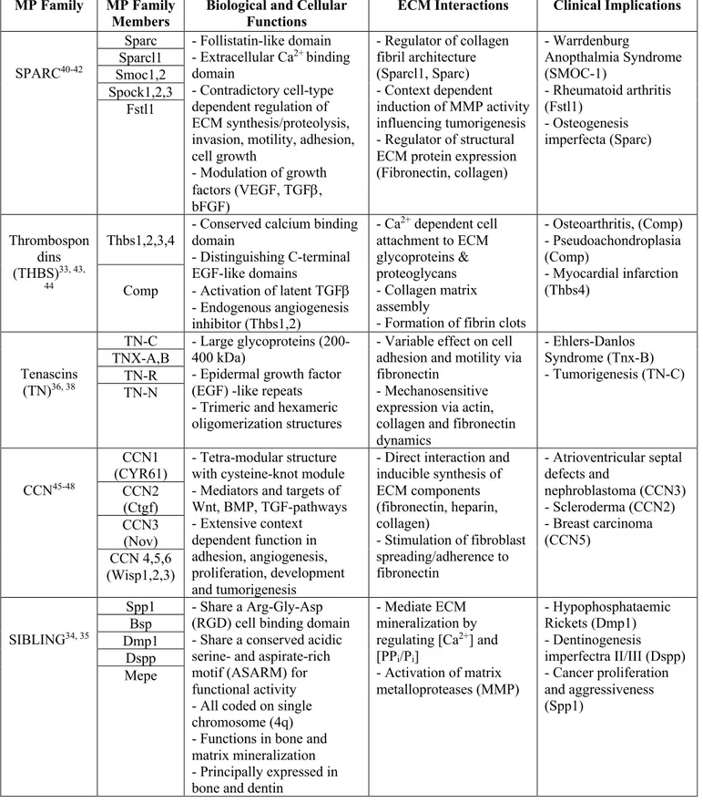
Most notably and best described is the SPARC family with eight members (SPARC, SPARCL1, SMOC1-2, SPOCK1-3, FSTL1) that all share a follistatin-like domain and an extracellular calcium binding E-F hand domain29. Among one of original MPs first described,
SPARC or otherwise referred to as Osteonectin or BM-40 served as the prototype for understanding the counter-intuitive functions of MPs in the ECM as it showed de-adhesive activity, but later re-defined as a state of “intermediate adhesion”30, 31.
The CCN family encompasses six proteins designated CCN1-6, though many alternative names exist for each member, and are unified by a cysteine-knot module (apart from CCN5) composed of a series of 38 cysteine residues with strict conservation in position and number32.
Moreover, CCN proteins have a tetra-modular structure, allowing for up to four independent and unique interactions working in a synergistic or antagonistic manner, hence the mosaic structure of CCN proeins32.
Thrombospondins (THBS), specifically THBS-1, was also amongst the first described MPs, which has now expanded to five total members, namely THBS1-4 and COMP. The hallmark of all THBS proteins lies in the highly conserved globular C-terminal domain of each peptide that consists of a calcium binding domain and a variable number of repeating epidermal growth factor (EGF)-like domains that serve as the THBS signature33. Variability between THBS
proteins come from the N-terminal domain which confer functional differences between family members, such as the anti-angiogenic properties of THBS1-2 attributed to their properdin domain33.
The SIBLING family consist of 5 defining proteins (OPN/SPP1, BSP, DMP1, DSPP and MEPE) with OPN being the most characterized of the set. All SIBLING proteins contain an integrin binding Arg-Gly-Asp (RGD) motif important for cell adhesion and its overall
hydrophilic structure34. Most SIBLING proteins also activate specific matrix metalloproteases
(MMPs) that play important roles in ECM degradation35. However, SIBLING proteins remain
understudied in the context of cancer, with the exception of OPN and BSP which have been associated with tumor aggressiveness in processes of invasion, metastasis and angiogenesis34,
and in development of mineralized tissue such as bone and dentin34.
Tenascin-C (TN-C) is the founding member of the TN family and was the third original MP first described by the Bornstein group. The TN family has now expanded to five proteins, TN-C, TNX-A, TNX-B, TN-R and TN-N, that all share a N-terminal heptad repeats, EGF-like repeats and fibronectin type-III repeats36. Tenascins can assemble into large oligomeric proteins,
primarily forming trimers via the highly conserved heptad repeats but formation of large symmetrical hexamers from two trimers is seen in the case of TN-C36. Less is known about the
other members, but functional capabilities of TN-C includes its ability to bind directly to fibronectin to modulate cell adhesion properties and as a integrin-binding ligand to trigger intracellular pathways affecting cell spreading36, 37. Moreover, de-novo expression of TN-C in
wound healing, chronic inflammation and cancer exemplifies its wide ranging capabilities from its initial discovery36, 38.
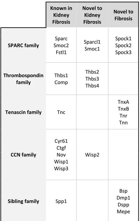
Active research in the field of MPs has shed light on their diverse functions and continues to show novel contexts in which MPs have not yet been considered (Table 1). Though our
knowledge of familiar MPs is constantly expanding into new research domains, there still
remains much to be explored for lesser known proteins. For example, considering splice variants of MPs is something that may be important for understanding contradictory roles of MPs in different contexts and differences between family members39. With increasing appreciation for
MPs, emphasis on finding links to disease by adapting screening studies to focus on MPs has also helped to expand the growing body of knowledge.
Table 1: Functional Roles and Characteristics of Matricellular Protein Families
MP Family MP Family
Members Biological and Cellular Functions ECM Interactions Clinical Implications
SPARC40-42
Sparc - Follistatin-like domain - Extracellular Ca2+ binding
domain
- Contradictory cell-type dependent regulation of ECM synthesis/proteolysis, invasion, motility, adhesion, cell growth - Modulation of growth factors (VEGF, TGFb, bFGF) - Regulator of collagen fibril architecture (Sparcl1, Sparc) - Context dependent induction of MMP activity influencing tumorigenesis - Regulator of structural ECM protein expression (Fibronectin, collagen) - Warrdenburg Anopthalmia Syndrome (SMOC-1) - Rheumatoid arthritis (Fstl1) - Osteogenesis imperfecta (Sparc) Sparcl1 Smoc1,2 Spock1,2,3 Fstl1 Thrombospon dins (THBS)33, 43, 44 Thbs1,2,3,4
- Conserved calcium binding domain - Distinguishing C-terminal EGF-like domains - Activation of latent TGFb - Endogenous angiogenesis inhibitor (Thbs1,2) - Ca2+ dependent cell attachment to ECM glycoproteins & proteoglycans - Collagen matrix assembly
- Formation of fibrin clots
- Osteoarthritis, (Comp) - Pseudoachondroplasia (Comp) - Myocardial infarction (Thbs4) Comp Tenascins (TN)36, 38 TN-C - Large glycoproteins (200-400 kDa)
- Epidermal growth factor (EGF) -like repeats - Trimeric and hexameric oligomerization structures
- Variable effect on cell adhesion and motility via fibronectin
- Mechanosensitive expression via actin, collagen and fibronectin dynamics - Ehlers-Danlos Syndrome (Tnx-B) - Tumorigenesis (TN-C) TNX-A,B TN-R TN-N CCN45-48 CCN1
(CYR61) - Tetra-modular structure with cysteine-knot module - Mediators and targets of Wnt, BMP, TGF-pathways - Extensive context dependent function in adhesion, angiogenesis, proliferation, development and tumorigenesis
- Direct interaction and inducible synthesis of ECM components (fibronectin, heparin, collagen) - Stimulation of fibroblast spreading/adherence to fibronectin - Atrioventricular septal defects and nephroblastoma (CCN3) - Scleroderma (CCN2) - Breast carcinoma (CCN5) CCN2 (Ctgf) CCN3 (Nov) CCN 4,5,6 (Wisp1,2,3) SIBLING34, 35 Spp1 - Share a Arg-Gly-Asp
(RGD) cell binding domain - Share a conserved acidic serine- and aspirate-rich motif (ASARM) for functional activity - All coded on single chromosome (4q) - Functions in bone and matrix mineralization - Principally expressed in bone and dentin
- Mediate ECM mineralization by regulating [Ca2+] and
[PPi/Pi] - Activation of matrix metalloproteases (MMP) - Hypophosphataemic Rickets (Dmp1) - Dentinogenesis imperfectra II/III (Dspp) - Cancer proliferation and aggressiveness (Spp1) Bsp Dmp1 Dspp Mepe
1.3.1 SMOC-2 as a Model MP
Secreted Modular Calcium binding protein-2 (SMOC-2) was first described by the Hartmann lab in 2003 and is part of the SPARC family of matricellular proteins49. It is encoded
on human chromosome 6q27 and has highly conserved intron and exon structure to the murine equivalent on chromosome 1749. SMOC-2 contains a homologous extracellular calcium binding
and follistatin-like domain shared among all SPARC family members, two thyroglobulin-like domains and a SMOC-protein unique domain49. Moreover, an N-terminal signal consensus
sequence and peptidase cleavage site along with the absence of a transmembrane hydrophobic domain allows it to be secreted into the ECM like other members of the SPARC family49 (Figure
2).
Figure 2: SMOC-protein domains. Image sourced from Hartmann et al49
By Northern blotting and RT-PCR, SMOC-2 was found to be expressed widely in ovary, testis, heart, spleen, thymus, lung, liver, kidney, skin, colon, pancreas and skeletal muscle49, 50.
Expression of SMOC-2 has been shown to be detected in mice as early as 8.5 dpc (days post-coitum)51 and mRNA detectable at 12.5 dpc in the development of facial and cranial features and
limbs51, 52. Moreover, SMOC-2 mutations have been shown to be linked to human dental
developmental defects despite no other observed complication at birth50. Beyond developmental
processes, SMOC-2 expression has also been found to be highly upregulated in murine models of renal fibrosis53 and metastatic colorectal cancer54. However, conflicting observations have
been made showing its downregulation in tumor development in ovarian55, pancreatic56, breast57
and thyroid58 cancer by microarray studies. The contradicting roles of SMOC-2 simply speak to
its highly context dependent function and our only partial understanding of its role in various diseases.
The molecular functions of SMOC-2 range from mediating cell cycle progression59 and
growth factor signals to regulating mitogenic and angiogenic processes60. It is also shown to
influence cell growth, migration and attachment51, 61. The broad functions and involvement in
various biological processes highlight not only the breadth of SMOC-2, but importance and capability of matricellular proteins on a broader scale as both mediators and main drivers to trigger intracellular changes from the ECM.
Supporting cellular attachment and anchoring to the ECM is an important function of matricellular proteins. SMOC-2 was shown to promote adhesion of keratinocytes exclusively by its EC domain51. The EC domain is important for extracellular Ca+2 binding to enhance affinity
for binding ECM ligands/proteins and as such, removal of soluble Ca+2 by EDTA chelation
completely abolishes cell attachment51. SMOC-2 overexpression was also shown to stimulate
keratinocyte migration in the presence of fibronectin but not BSA in a modified Boyden chamber assay51. SMOC-2 overexpressing endothelial cells also showed increased migration with bFGF60
used as a chemoattractant, showing an enhanced pro-angiogenic and migratory phenotype when coupled with potent growth factors.
To regulate cell processes like cell attachment and migration, SMOC-2 signals from the ECM are translated by integrin receptors at the cell surface. Specifically, the avb1
integrin-subunit complex is important in promoting cell attachment, while inhibition of only the b1
subunit is sufficient to drastically reduce adhesive capability of keratinocytes51. Moreover, avb1
has been shown to be highly expressed on the surface of activated fibroblasts to bind TGFb-162
to trigger intracellular cascades like the Smad pathway62, 63, Snail64, 65 and Rho-like GTPases65
which all can promote cell migration and attachment by either directly influencing gene expression.
SMOC-2 has also been shown to have pro-angiogenic effects in endothelial cells (HUVEC) overexpressing a Myc-SMOC-2 fusion protein using a adenoviral vector (Ad-Myc-SMOC-2), as indicated by increased projections in Matrigel coated wells60. Complementary to
by [3H]thymidine incorporation in a dose dependent manner with VEGF and bFGF (basic
Fibroblast Growth Factor) in Ad-SMOC-2 transduced cells compared to the Ad-GFP control60.
Overall, SMOC-2 remains a understudied MP and our understanding of its functions pales in comparison to other members in the SPARC family. Emerging research in the field of MPs will hopefully spark interest in studying the lesser known proteins.
1.4 Tissue Repair Process Overview
In all mammalian organ systems, the wound repair process occurs by an overlapping three step process, beginning with inflammation, followed by a proliferative phase and concludes with matrix remodeling events66. However, the timing of these events are also tightly regulated
to ensure prompt repair but also distinct completion of each phase to avoid prolonged states of inflammation or matrix remodeling which is the underlying cause of many other pathologies.
The first responders to the wound site include recruitment of, but are not limited to, platelets, fibrin and fibronectin that act as a hemostatic plug and provide the first basis of a provisional matrix for incoming inflammatory cells to further build upon67. Local release of
growth factors such as platelet derived growth factor (PDGF), epidermal growth factor (EGF), TGFb-1, pro-inflammatory cytokines like IL-1b, IL668, 69 act as stimulants to transform resident
monocytes to macrophages, recruit neutrophils to the wound site and initiate the development of granulation tissue using the provisional matrix as a foundation. As the primary purpose of the inflammation phase is to clear damaged cellular debris and prevent infection, pro-inflammatory signals must be properly sequestered once they served their purpose to transition into the next phase and avoid a state of chronic inflammation.
Following inflammation, the next major theme in the repair process is the proliferative phase which is important to regenerate the wound site by replacing the provisional matrix with granulation tissue that forms a new foundation. Local concentration of aforementioned wound
repair signals trigger migration and increased proliferation of cells such as, keratinocytes, fibroblasts, granulocytes and surviving epithelial cells to help fill the wound gap and build upon the granulation tissue69. Moreover, anti-inflammatory M2 macrophages can secrete factors such
as EGF to promote re-epithelization and nitric oxide for wound contraction69. Migration is aided
by the loss of physical tension and cell-cell contact during injury and stops once this contact is re-established and new adhesion structures restored. This wave of migratory cells work in essence to replace the provisional matrix laid down during hemostasis to make way for deposition of structural ECM proteins like collagen and fibronectin that is secreted by
fibroblasts, which also releases degradative proteases such as MMPs69, 70. The presence of these
two major ECM structures distinguish the temporary provisional matrix from the lasting granulation tissue71, 72 and provide a basic scaffold for further cell adhesion, migration, growth
and differentiation to occur through the subsequent phases of wound repair. Another important aspect during the proliferative phase is angiogenesis, which is the process of forming new blood vessels to support the growing tissue and is initiated by growth factors like PDGF and vascular endothelial growth factor (VEGF). The local concentration of these factors work to attract endothelial cells of nearby vessels to migrate and proliferate towards this source of angiogenic stimuli69.
The last phase to conclude the repair process is a compilation of matrix remodeling events with the goal of restoring functionality to the injured tissue73. The injured parenchymal
tissue orchestrates the repair process by promoting both epithelial and fibroblast transformation to their effector forms to mediate remodeling. A major event during this phase include restoring the tensile forces that hold the tissue together. This primarily occurs by TGFb-1 driven fibroblast differentiation into myofibroblasts that express a-smooth muscle actin (a-SMA) which is
incorporated into de-novo contractile bundles of actin of the myofibroblast, coined stress fibers69, 74. These stress fibers provide an intracellular link to the ECM via transmembrane integrin
receptors to form large structures known as focal adhesion complexes74, 75. Moreover, the rapidly
deposited collagen backbone, composed of predominately collagen IV is converted to collagen I which provides greater tensile strength after cross-linking66, 69, 74, 76. Re-organization and
alignment of the present ECM materials to restore normal tissue architecture can still be actively occurring from 6 months – 1 year post injury, depending on the severity of the insult66, 73 and can
only regain up to 80% of the tensile strength in the case of dermal wounds77. The conclusion of
the remodeling phase must also signal the shutdown of inflammatory, proliferative, regenerative and matrix protein secretory pathways as their prolonged activation can cause scar formation, overly stiffened/inflexible tissue and long term pathologies associated with dysregulated or imperfect repair such as organ fibrosis69, 73.
MPs are involved during all three stages of the repair process and targeted disruption of MP expression or function in murine models report altered inflammation responses, reduced angiogenesis at the wound site and impaired matrix deposition78. For example, THBS-2 null
mice were able to recover from a full thickness excisional wound but with abnormal
neovascularization within the wound bed and a disorganized ECM in the dermal layer compared to wildtype. Though interestingly, although the repair process occurred faster in THBS-2 null mice, the end result appeared highly irregular78. Another example is the absence of OPN, which
significantly impaired the synthesis and organization of collagen I protein in cardiac remodeling after myocardial infarction that resulted in reduced strength of the repaired ECM79. Collectively,
these studies highlight the implications of MP absence during wound repair at varying stages that result in sub-optimal restoration of the injured tissue.
1.4.1 Fibrosis – Deregulated Repair
Fibrosis is defined as the stiffening or scarring of tissue that leads to the destruction of the organ architecture and impairment of function associated with excessive deposition of ECM components80, 81. Fibrosis affects nearly every tissue in the body including but not limited to,
bone marrow, kidney, heart, intestine, lung and skin80. Existing therapies are limited to only
helping manage or slow the progression of fibrosis as there are currently no treatments to stop or completely reverse fibrotic tissue in any organ82. One note of positivity is the histological
similarities between different organs after fibrotic onset, providing hope for potentially
elucidating and targeting common mechanism(s) to be effective across many organs. However, organ specific triggers and pathological impact on overall health still vary depending on the severity and progression in afflicted tissues.
As the ECM is a dynamic structure, normal up-and downregulated production of ECM materials is crucial for cells to adapt quickly to subtle and transient changes in its surroundings83.
There is consensus in the literature that the overproduction of these ECM materials come from activated fibroblasts (myofibroblasts) that secrete primarily collagen type I and IV and
fibronectin in an unregulated manner3, 82. Without proper management or degradation, this
deposition and accumulation over time causes elevated mechanical stress, tension and reduced elasticity to the entire ECM network. The differentiation of originally reparative fibroblasts, resident epithelial cells and even circulating bone marrow derived mesenchymal stem cells3, 84
into myofibroblasts from this dysregulated system creates a destructive feedback loop of ECM stiffening and cellular differentiation. However, it is worth noting that these processes normally described occur during wound repair with strict feedback mechanisms allowing for restoration of tissue function without onset of fibrosis3.
Within the dysregulated matrix during fibrotic onset, MPs have also been shown to be strongly upregulated and contribute to sustaining fibrotic mechanisms. CCN2 is a
well-characterized pro-fibrotic MP that works by supporting the TGFb-1 induced fibrosis mechanism in part by activation of Smad1 and Erk1/2 signaling for sustained ECM deposition and
remodeling in human fibroblasts in-vitro85. Osteopontin (OPN) is another pro-fibrotic MP which
has been shown to be upregulated in the tubules of obstructed kidneys after unilateral ureteral obstruction (UUO) in mice. Mice subjected to ureteral ligation developed hydronephrosis, tubular atrophy and interstitial inflammation and fibrosis, compared to unobstructed control animals86. From the tenascin family, TN-C levels are found to be significantly upregulated in
fibrotic lungs in patients with idiopathic pulmonary fibrosis, compared to healthy patients87.
Primary fibroblasts cultured from diseased lungs was found to constitutively release higher levels of TN-C, and more sensitive to TGFβ stimulation87.
Though a large majority of MPs are pro-fibrotic, it is worth noting that there are anti-fibrotic MPs, even members amongst same family, that have been reported to attenuate the fibrotic phenotype. For example, CCN1 overexpression in a model of liver fibrosis induces ER stress and reactive oxygen specific formation to induce apoptosis of activated hepatic stellate
cells that are major producers of excessive ECM88. Overall, fibrosis is a powerful stimuli and
context for inducing MP expression beyond the normal tissue repair and regeneration timeline.
1.4.2 The Kidney as a Model Organ to Study Injury and Maladaptive Repair
Acute kidney injury (AKI) is a pervasive clinical syndrome and is a major burden on global health, with increasing incidence rates that is directly associated with short and long term morbidity89. Repeated insults such as ischemia, chemical toxins, inflammation, hypoxia and
physical injuries are common causes of AKI which causes cellular injury and can lead to
development of chronic kidney disease (CKD)89, 90. Internal damage to biological structures such
as the tubules, nephrons, glomeruli and blood vessels obstruct homeostatic function and triggers the pathophysiological mechanisms that underlie many clinical renal pathologies. Maladaptive repair after AKI can lead to accelerated kidney damage due to poor adaptation to the inefficient function. One possible end-stage renal disease outcome is fibrosis, which is a common cause of organ failure and outcome in chronic tissue injuries82, 90.
The kidney is a robust model organ to study the connected mechanisms of dysregulated repair/remodeling and pathogenesis because of the extensive animal models available,
convenient internal control with two kidneys, informative functional readouts through urine analysis, extensive cell lines and there still remains an large unmet need for therapeutic options to treat renal pathologies82, 91, 92. Moreover, as we are interested in studying MPs whose
expression is context dependent, the kidney provides a safer alternative to other organs for inducible injury to follow the progression of repair and disease. Therefore, the kidney represents an all-encompassing model for our purposes in search of translatable mechanisms with MPs as the focus of our research.
1.5 Development Overview
During development, the ECM has dually important functions as both the traditional concept as a passive support scaffold for tissue growth, and as a actively remodeling structure that imparts important cues to incoming cells to direct development of tissue architecture93.
On a macroscopic level, the ECM can act as a physical barrier between cells, tissue structures and work as a protective cushion that secure and hold organs intact94. The composition
and delicate ratio of ECM structural proteins is a determinant for the strength, elasticity/stiffness and overall biomechanical properties and is remodeled accordingly during development to be tissue specific. For example, collagen cross-linking is a key process in ECM development in both a regulated and un-regulated manner and is crucial for stiffening of the cardiovascular system94.
Knockout of the collagen cross-linking enzyme lysl oxidase (LOX) result in mortality at birth due to inefficient cardiac output by a weak cardiovascular system and a fragile diaphragm in murine models94.
In the context of development, MPs also have important roles and are upregulated to participate in active remodeling. MPs within the ECM are also important in the development of bone and cartilage, though the specifics are beyond our scope. Interestingly, SMOC-2 has been reported to be responsible in the development of cranial and facial features of dogs95. Using
genome-wide association studies, a mutation in canine SMOC-2 is linked to the development of canine brachycephaly, which causes the shortened head, nose and flat face best exemplified by breeds such as Pugs, Shih Tzus and Pekingese dogs95.
The importance for maintaining tight regulation of the ECM and MPs during
development is best highlighted by the consequences of dysregulation when this delicate balance is perturbed. To do so, we look to cases of de-regulated development where we can study its consequences.
1.5.1 Cancer – Deregulated Development
Malignant tumors are a complex structure of numerous cell types in a pool of oncogenic secretory factors all being structurally supported by a dynamically adaptive ECM. Interactions with the ECM can set off a cascade of downstream molecular signalling events that adapt the tumor microenvironment for immune suppression, communication, rapid growth and metastatic dissemination27. To understand the tumor stroma, comparisons have been drawn to the
dysregulated ECM of chronically injured wounds that fail to heal3, 96 but proliferating cancer
cells interacting with their microenvironment can be considered a unique entity or separate tissue entirely with functional disorder3, 97.
The biology of a developing tumorigenic ECM is strikingly similar to the phenotypes observed in fibrosis. Excessive ECM deposition of fibronectin, type I collagen and increased secretion of MPs all contribute to accelerating tumor growth94. Buildup of excessive matrix
components can support oncogenic proliferation by interfering with cell-cell and cell-matrix interactions to amplify growth factor signals and enhance integrin signaling98. Though seemingly
disorganized, the developing tumor ECM can undergo remodeling processes to facilitate
invasion as seen with organization and alignment of collagen fibrils that act as tracks for outward movement98.
The developing tumor ECM can also be distinguished from non-malignant ECM by the abnormalities in its biomechanical properties. ECM plasticity influences how cells perceive the external environment and subsequently how they respond99. Compromised tensile homeostasis in
tumorigenic tissue is one of the key characteristics, and is used as a parameter when screening for breast tumors as they tend to be stiffer than the surrounding tissue100, 101. Within the tumor
microenvironment, these elevated contractile forces can positively influence cell growth and invasion by promoting maturation of focal adhesions and matrix remodeling for rearrangement of collagen fibrils to push the invasive front of tumors outwards needed for spreading100, 102, 103.
In contrast to tensile stress, compressive stress by the expanding tumor matrix onto surrounding tissue extracellular matrix and vasculature, effectively shrink the total interstitial space, aiding in tumor invasion into the parenchyma100. Moreover, changes in the interstitial pressure and
progressive increasing density of solid tumors adds yet another layer of complexity towards delivery and penetration of anti-tumor therapeutics100, 104.
Deregulation of MP expression in the developing ECM is a hallmark of cancer as they are implicated in many pertinent processes required to sustain the malignancy. In an analogous fashion to wound healing and fibrosis, the actively transforming oncogenic ECM provides the necessary context to stimulate MP expression. For example, OPN is the best characterized member of the SIBLING family and its expression is found to be elevated in tumor ECM of different histotypes, which all report a positive correlation with tumor progression and
metastasis25. Within the developing ECM, OPN has also been showed to induce endothelial cell
migration which is important for vascularization of the growing tumor niche to supply oxygen and nutrients to proliferating malignant cells25. From the thrombospondin family, THBS-1
expression has also been widely used as a prognostic marker of many cancer types including, melanoma, ovarian and pancreatic carcinomas105-107. At sites of human primary melanoma,
increased levels of THBS-1 was detected by microarray analysis compared to benign nevi and associated with melanoma cell motility105. Though interestingly, it was reported that THBS-1
was negatively correlated with SLUG expression, a master regulator of cell motility suggesting an alternative independent pathway105. Another well characterized MP is connective tissue
growth factor (CTGF), or otherwise CCN2. First associated with breast and pancreatic cancer, aberrant CCN2 expression has been linked to many human cancers since its discovery108. Its
divergent expression, whether abnormally up- or downregulated compared to normal cell
counterparts is often associated with a poor clinical outcome108. In breast cancer, malignant cells
overexpressing CCN2 gain enhanced migratory ability through activation of the ERK1/2 pathway via integrin mediated signaling109. Importantly, many MPs also work to transform the
local matrix environment and contribute to tumor growth by influencing cell processes like invasion, migration and proliferation. In colorectal cancer, follistatin-like protein 1 (FSTL-1) from the SPARC family is reported to be significantly upregulated in cancerous tissue compared to normal and promoted invasive behavior of colorectal cancer cell lines110. Higher FSTL-1
expression lead to a more aggressive phenotype by activating focal adhesions for cytoskeletal rearrangement and inducing TGFb-Smad2/3 signaling to increase cell migration110. Metastatic
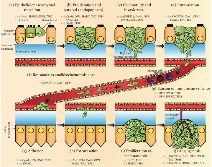
compared to control animals110. Figure 3 illustrates the major milestones of a primary tumor in
its progression to a metastasizing cancer. Supportive or inhibitory roles of known matricellular proteins during each stage along with various stromal cells is also highlighted.
Figure 3: MPs in metastatic cancer development. Different MPs are implicated at each stage of cancer development. Action of MPs, can be pro- or anti-tumorigenic, depending on the processes involved. In the early stages of tumor initiation (a,b), MPs induce cellular and biochemical changes in vulnerable cells and promotes dysregulated survival and growth. Cell motility is aided through ECM degradation by MPs and allows tumor cells to invade into circulation (c-e) and provide protection during circulation to the metastatic site (f). Interaction with MPs to establish at the metastatic site and promote adhesion and proliferation of invaded cells (g-j). Figure is sourced from Chong et al27.
1.6 Epithelial-to-Mesenchymal Transition
Epithelial-to-mesenchymal transition (EMT) is a reversible biological process by which polarized epithelial cells undergo biochemical changes to transform and acquire mesenchymal, fibroblast-like properties27, 111. Transformed cells lose their apical-basal polarity, cuboidal
morphology and cell-cell junctions in favor of a mesenchymal phenotype with fibroblast-like appearance, enhanced migratory potential, loose organization in the ECM and ability to catalyze ECM and cytoskeletal rearrangements for invasive growth111-113. EMT is a crucial process that
has been well described and associated with embryonic development and wound healing/tissue regeneration, but our understanding of its contribution in various human pathologies remains an area of active research111.
Considered a defining event in EMT progression is the repression and loss of E-cadherin, which in a sense opens the gateway for cells to exhibit features of a mesenchymal phenotype. E-cadherin is a tight junction protein whose function is to keep epithelial cells bound tightly to maintain their polarity and the separation between the apical epithelial layer and underlying stroma114. Loss of normal tissue architecture and tight junctions allows cells to move both
horizontally and vertically beyond their normal imposed constraints and receive autocrine signals from diverse cell types to reinforce the EMT phenotype114. EMT-inducing transcription factors
such as Snail, Slug, and noncoding microRNAs can act to repress E-cadherin expression while simultaneously activating Wnt/b-catenin, TGFb and TWIST pathways to promote mesenchymal transformation by augmenting cell polarity, cell-cell contact and attachment.
Concomitant changes in the ECM and cell cytoskeleton also support the loss of E-cadherin and provide the necessary environment and tools for mesenchymal cell migration and invasion. Two important cytoskeletal markers of EMT that appear in mesenchymal cells are vimentin and a-SMA115. Upregulation of extracellular proteins like fibronectin and
non-structural proteins such as MPs help remodel the ECM to complement and support the intracellular changes115.
Vimentin is an intermediate filament protein expressed by various cell types including fibroblasts, endothelial cells, mesenchymal originating cells and epithelial cells undergoing EMT116, 117. It has functions in maintaining cell integrity, migration and invasion and when
overexpressed in cancers, vimentin has been shown to drive EMT and have positive correlation tumor invasiveness and metastasis and serves as a marker of mesenchymal cells115. Although the
exact molecular mechanisms of vimentin function remain to be elucidated, overwhelming evidence shows that its upregulation in the context of transforming epithelial or transformed mesenchymal cells have potent pro-EMT effects. Vimentin has been shown to be important in recycling endocytosed integrin receptors back to the plasma membrane to promote cell motility, which was severely attenuated with vimentin inhibition116, 118. Moreover, vimentin filaments are
shown to be transiently concentrated and upregulated at the leading edge of sub-confluent metastatic breast cancer cells using a scratch assay to stimulate migration by generating a artificial wound gap for cells to fill in116. Co-operativity between actin filaments and vimentin
have also been shown to regulate formation of lamellipodia and filopodia membrane protrusions that are required to propel the cell on the ECM substrate for migration119. Another pro-EMT
function of vimentin is its ability to influence the expression of other EMT-linked genes and proteins such as receptor tyrosine kinase Axl and integrin subunit-b4, both of which have roles in
cell proliferation and migration117.
a-SMA is a type of actin filament commonly used to identify the myofibroblast cell type but is also expressed by cancer-associated fibroblasts and mesenchymal cells in the tumor microenvironment120, 121. In many epithelial derived cancers, TGFb induced EMT can trigger
a-SMA expression by promoting trans-differentiation of stromal mesenchymal cells into
myofibroblasts which become the effector cells121. Beyond just a cell marker, a-SMA is shown
to be important in the formation of focal adhesions which are integrin receptors and associated protein complexes that provide a link between the cytoplasmic actin network and the ECM important for coordinating cell migration122.
Fibronectin is a major component of the ECM and its aberrant expression has been associated with increased metastasis in numerous cancer types115. Fibronectin and its associated
signaling cascades to promote cell proliferation and invasion. Increased fibronectin is also able to stimulate formation of focal adhesion at the leading edge of metastatic lung cancer cells, which also upregulated matrix metalloprotease mediated matrix degradation to facilitate invasion and ultimately metastasis123, 124.
1.6.1 Epithelial-to-Mesenchymal Transition in Cancer Metastasis
From a disease perspective, EMT has been regarded as an important driver of tumor progression and metastasis because it provides tumor cells with the necessary molecular mechanisms and cellular components to migrate out of the primary site and invade secondary tissues112. EMT has been linked, but not limited to, cancers of the breast, neck, lung, colon,
kidney and ovaries112. As is the case with all cancers, re-purposing and deregulation of normal
cellular machinery and pathways turn EMT from a regenerative process to benefit and enable the expansion and growth of the tumor niche and propagate cancer associated cells beyond the primary lesion.
To explain the contribution of EMT in cancer metastasis, two current hypothesis exist125.
Firstly, cancer progenitor cells do not undergo simultaneous EMT but can undergo
differentiation at different times to create heterogeneity among the tumor cell population that represent the various advanced stages of the cancer125, 126. The cancerous cell population
therefore exists at different stages of EMT which can be accelerated by other factors in the developing tumor. A second hypothesis proposes that some cancer progenitor cells initially undergo EMT to metastasize after clonal expansion, which suggests that the metastatic tumor cell population shares a cellular signature with the original transformed cancer progenitor cells125, 126.
In human epithelial derived carcinomas, cells undergoing EMT have been shown to concentrate at the invasive front of the primary tumor and thus suggests an influence from the surrounding activated matrix environment127, 128. The combined loss of epithelial cell polarity
tissue carcinomas129. The well characterized epithelial adherent junction protein, E-cadherin, has
been linked to cancer suppression mechanisms and as such, its impaired and eventual loss of function during the early stages of EMT is pivotal step in the transformation process. As mentioned previously, many of the EMT associated factors can be classified by their direct or indirect repressive actions on E-cadherin expression and un-surprisingly also have pro-oncogenic roles. Thus, combined tumorigenic stresses such as physical constraints, inflammation and metabolic stress can all lead to dysregulation in TGFb, Wnt and TWIST pathways and create favorable conditions to trigger EMT.
Within the activated matrix environment are also MPs, which can promote EMT and initiate downstream signaling cascades that lead to invasion, migration, proliferation to promote dissemination of tumor cells to other organs by metastasis27. For example, the founding member
of the Tenascin family, TN-C, is a well-known mediator of cell motility and invasion in various carcinomas. TN-C knockdown was shown to reduce migration of metastatic breast cancer cells measured by a Transwell assay and significantly decreased metastasis in a lung colonization assay with the same cells after inoculation into immunodeficient mice130. In another murine
model of osteosarcoma, TN-C was shown to be crucial for driving lung metastasis that is dependent its associated integrin receptor, a9b1131. In the progression of colon cancer, secreted
modular calcium-binding protein-2 (SMOC-2) has been reported to promote EMT of colorectal cells. SMOC-2 overexpressing cells adopted a more elongated mesenchymal-esque morphology and did not form tight compact colonies at high cell densities compared to control cells54. This
was coupled with a downregulation of E-cadherin, a marker of epithelial cells that is lost during EMT111, 125. Colorectal cancer cells overexpressing SMOC-2 also exhibited greater motility,
1.7 Rationale & Research Objectives
Aim 1: To identify and analyze significant expression of MP families and their members using two mechanistically different models of murine kidney injury.
Aim 2: Analyze the role of SMOC-2 in mediating EMT induced metastasis of tubular epithelial cells in the context of Renal Cell Carcinoma.
Overall Research Objective: To study MPs in the context of tissue remodeling processes in order to understand de-regulated processes of repair in fibrosis and development of metastatic cancers.
2.0 Manuscript - Characterization of Matricellular Protein Expression Signatures in Mechanistically Diverse Mouse Models of Kidney Injury
2.1 Preface to Manuscript
The following manuscript is presented as part of my overall project completed during my training. The manuscript is published in the journal of Scientific Reports, with reference:
Feng, D., Ngov, C., Henley, N. et al. Characterization of Matricellular Protein Expression Signatures in Mechanistically Diverse Mouse Models of Kidney Injury. Sci Rep 9, 16736 (2019) doi:10.1038/s41598-019-52961-5.
2.2 Manuscript
Characterization of Matricellular Protein Expression Signatures in
Mechanistically Diverse Mouse Models of Kidney Injury
Daniel Feng1,2#, Cindy Ngov3#, Nathalie Henley2, Nadia Boufaied4 and Casimiro Gerarduzzi1,2,5* 1Département de Pharmacologie et Physiologie, Faculté de Médecine, Université de Montréal,
Montréal, Québec, Canada
2Centre de recherche de l'Hôpital Maisonneuve-Rosemont, Faculté de Médecine, Centre affilié à
l'Université de Montréal, Montréal, Québec, Canada
3Department of Microbiology and Immunology, McGill University Health Centre Research
Institute, Montréal, Québec, Canada
4Division of Urology and Cancer Research Program, McGill University Health Centre Research
Institute, Montréal, Québec, Canada
5Département de Médecine, Faculté de Médecine, Université de Montréal, Montréal, Québec,
Canada
#These authors contributed equally to this work. *Corresponding author
Running title: Characterizing Matricellular Protein Expression during Kidney Injury Key words: Chronic Kidney Disease, Acute Injury, Fibrosis, Animal Models, Folic Acid, Unilateral Ureteral Obstruction (UUO), RNA-Seq
*Corresponding author: Casimiro Gerarduzzi
Division of Nephrology, Maisonneuve-Rosemont Hospital CIUSSS de l'Est-de-l'Île-de-Montréal 5345, boul. de l'Assomption Montreal, QC, Canada H1T 2M4 Tel: 514-252-3400 ext:2813 casimiro.gerarduzzi@umontreal.ca
Fibrosis is the most common pathophysiological manifestation of chronic Kidney Disease (cKD). It is defined as excessive deposition of extracellular matrix (ECM) proteins. Embedded within the ECM are a family of proteins called Matricellular Proteins (MCPs), which are typically expressed during chronic pathologies for ECM processing. As such, identifying potential MCPs in the pathological secretome of a damaged kidney could serve as
diagnostic/therapeutic targets of fibrosis. Using published RNA-Seq data from two kidney injury mouse models of different etiologies, Folic Acid (FA) and Unilateral Ureteral Obstruction (UUO), we compared and contrasted the expression profile of various members from well- known MCP families during the Acute and Fibrotic injury phases. As a result, we identified common and distinct MCP expression signatures between both injury models. Bioinformatic analysis of their differentially expressed MCP genes revealed similar top annotation clusters from Molecular Function and Biological Process networks, which are those commonly involved in fibrosis. Using kidney lysates from FA- and UUO-injured mice, we selected MP genes from our candidate list to confirm their mRNA expression by Western blot, which correlated with injury progression. Understanding the expressions of MCPs will provide important insight into the processes of kidney repair, and may validate MCPs as biomarkers and/or therapeutic targets of CKD.