Pulsed Laser Manipulation of Cells Decorated by Plasmonic Nanoparticles
MENGJIAO QI Institut de génie biomédical
Mémoire présenté en vue de l’obtention du diplôme de Maîtrise ès sciences appliquées Génie biomédical
Avril 2019
Ce mémoire intitulé :
Pulsed Laser Manipulation of Cells Decorated by Plasmonic Nanoparticles
présenté par Mengjiao QI
en vue de l’obtention du diplôme de Maîtrise ès sciences appliquées a été dûment accepté par le jury d’examen constitué de :
Frédéric LESAGE, président
Michel MEUNIER, membre et directeur de recherche Marc LAVERTU, membre
DEDICATION
This thesis is dedicated to my parents. As I had gone through super difficult time studying abroad, they offered me help and suggestions to support me to go on. I gained a lot during my study, both scientific knowledge and experience to tackle problems. They are good examples for me to be independent and strong in any situation.
ACKNOWLEDGEMENTS
I should thank my supervisor Professor Michel Meunier for offering me opportunity to finish the study abroad. He offered help when I was in hard situations. He cares a lot about his students. I appreciate all the guidance, help and support from my supervisor.
I would also thank the members of jury for my thesis, professor Frédéric Lesage and professor Marc Lavertu, for their precious time on reviewing my thesis and taking time for my thesis defense. Many thanks to the research associate Dr. Sergiy Patskovsky in the group, he spent much time with me to tackle the problems in my experiment, initiated me with some ideas those are quite important for me. I appreciated so much for his efforts and help.
Thanks to my colleagues in the group. Their kindness and support helped me to solve many specific problems in the lab.
RÉSUMÉ
Au cours des dernières décennies, la thérapie cellulaire a suscité un intérêt considérable dans les domaines de la recherche biomédicale et théranostique. Une large gamme d’applications biomédicales a obtenu des avantages fructueux par une manipulation directe de cellules (cellules seuls ou groupes de cellules individuelles) en contrôlant la concentration de nanoparticules plasmoniques fonctionnelles et de lasers à impulsions. Cependant, tous ces protocoles de transfection, accompagnés d’une efficacité et d’une toxicité de transfection cellulaire variées, sont applicables pour une condition spécifique. Ainsi, l’optoporation doit être explorée, optimisée et généralisée pour un large éventail de thérapies cellulaires délicates et complexes par le laser.
Un suivi en temps réel des cellules optoporées révèle les mécanismes impliqués dans une transfection réussie sans induire de cytotoxicité. Dans cette thèse, le laser pulsé nanoseconde (532 nm) utilisé pour optoporer une seule cellule cancéreuse humaine du sein (MDA-MB-231) montre clairement les effets des fluences du laser pulsé. L’optoporation intériorise les molécules exogènes (Iodure de Propidium) dans l’intervalle fonctionnel compris entre 0.3 et 0.7 J/cm2 sans provoquer
d’effet secondaire (confirmé par Calcein AcetoxyMethyl). La position du faisceau détermine clairement dans quels compartiments subcellulaires les molécules exogènes à intérioriser avec précision. Le faisceau laser focalisé près du noyau dirige intensément l’Iodure de Propidium (PI) pour qu’il réagisse avec les nucléotides cellulaires, alors que pour le faisceau focalisé loin du noyau, le PI se déverse à peine dans le site d’action. Ce protocole d’optoporation, indiquant le rôle critique des nanoparticules plasmoniques liées, nécessite un faisceau laser dirigé vers la position de la nanoparticule plasmonique sur la membrane cellulaire. L’éclairage latéral par LED développé ici visualise simplement la nanoparticule plasmonique liée sur la membrane cellulaire dans une grande zone.
Par la suite, un laser femtoseconde dans le proche infrarouge (800 nm) est également utilisé pour optoporer des cellules de lymphocyte T humaines supersensibles (Jurkat) avec un taux de survie négligeable après une exposition de courte durée à une fluence relativement élevée (252 mJ/cm2).
En explorant plusieurs fluences laser et durées d’irradiation, nous obtenons donc une gamme applicable de fluences laser (63 à 71 mJ/cm2) et de temps d’irradiation (10 ms) pour l’optoporation
de cellules de Jurkat liées à des nanoparticules plasmoniques sans réduire leur viabilité cellulaire. Ces résultats fondamentaux indiquent comment effectuer une optoporation réussie en ajustant les
paramètres du laser (fluence, durée d’irradiation, position, etc.) sur différentes lignées cellulaires afin d’atteindre une haute transfection pour une large gamme d’applications biomédicales.
ABSTRACT
In the last decades, cell therapy has attracted tremendous interests in biomedical and theranostic research fields. A wide range of biomedical applications has gained fruitful benefits by a direct manipulation of cells (whether single cell or bunch of individual cells) by controlling the concentration of functional plasmonic nanoparticles and pulsed lasers. However, all these transfection protocols, accompanied by a varied cellular transfection efficiency and toxicity, are applicable for a specific condition. Thus, the optoporation needs to be explored, optimized, and generalized for a broad range of delicate and complex laser mediated cell therapies.
A real-time monitoring of the optoporated cells reveals mechanisms involved in a successful transfection without inducing cytotoxicity. In this thesis, the nanosecond pulsed laser (532 nm) used to optoporate a single adherent human breast cancer cell (MDA-MB-231) clearly shows effects of the pulsed laser fluences. The optoporation internalizes the exogenous molecules (Propidium Iodide) at the functional range between 0.3 to 0.7 J/cm2 without causing a side effect
(confirmed by Calcein AcetoxyMethyl). The beam pointing location clearly determines in which subcellular compartments the exogenous molecules to be internalized precisely. The laser beam localized close to the nucleus intensively directs the Propidium Iodide there to react with cellular nucleotides, whereas the beam far away from the nucleus barely fluxes into the site of action. This optoporation protocol, indicates the critical role of the bound plasmonic nanoparticles, is required a laser beam to be directed to the position of the plasmonic nanoparticle on the cellular membrane. The lateral LED illumination developed here simply visualizes the bound plasmonic nanoparticle on the cell membrane in a large area.
Afterward, near-infrared femtosecond laser (800 nm) is also employed to optoporate supersensitive human T lymphocyte cells (Jurkat) with a negligible survival rate after a short time exposure to a relatively high fluence (252 mJ/cm2). Exploring several laser fluences and irradiation durations,
we therefore obtain an applicable range of laser fluences (63 to 71 mJ/cm2) and irradiation time
(10 ms) for the optoporation of plasmonic nanoparticles bound Jurkat cells without reducing their cell viability. These fundamental results indicate how to perform a successful optoporation by adjusting laser parameters (i.e., fluence, irradiation duration, position, etc.) on different cell lines in order to achieve high transfection for a wide range of biomedical applications.
TABLE OF CONTENTS
DEDICATION ... III ACKNOWLEDGEMENTS ... IV RÉSUMÉ ... V ABSTRACT ...VII TABLE OF CONTENTS ... VIII LIST OF TABLES ... XI LIST OF FIGURES ... XIII LIST OF SYMBOLS AND ABBREVIATIONS... XVIII LIST OF APPENDICES ... XX
CHAPTER 1 INTRODUCTION ... 1
1.1 Problems and objectives ... 3
CHAPTER 2 LITERATURE REVIEW ... 6
2.1 Nanosecond pulsed laser perforation on cells ... 9
2.1.1 Mechanism of nanosecond pulsed laser-induced cell perforation ... 9
2.1.2 Nanosecond optoporation of adherent cell ... 10
2.2 Femtosecond pulsed laser perforation on cells ... 11
2.2.1 Mechanism of femtosecond pulsed laser-induced cell perforation ... 12
2.2.2 Femtosecond optoporation of adherent cell ... 13
2.2.3 Femtosecond optoporation of suspension cell ... 14
2.3 Summary ... 15
CHAPTER 3 REAL-TIME MONITORING OF PLASMONIC NANOPARTICLE-MEDIATED PULSED LASER SINGLE-CELL OPTOPORATION ... 16
3.1.1 Research question ... 16
3.1.2 Objective ... 17
3.2 The proposed approach ... 17
3.3 Methodology ... 18
3.3.1 Cell culture, PI transfection, and live cell assay ... 18
3.3.2 Preparation of optical setup for optoporation and visualization ... 19
3.4Results and discussions ... 21
3.4.1 RLM imaging of NPs in a cellular environment ... 21
3.4.2 NP-laser alignment ... 22
3.4.3 Analysis of perforation dynamics ... 22
3.4.4 Effects of irradiation areas ... 26
CHAPTER 4 ARTICLE 1: COST-EFFECTIVE SIDE-ILLUMINATION DARKFIELD NANOPLASMONIC MARKERS MICROSCOPY ... 29
4.1 Authors ... 29 4.2 Abstract ... 29 4.3 Introduction ... 30 4.4 Experimental ... 31 4.4.1 Cell culture ... 31 4.4.2 Plasmonic nanoparticles ... 32
4.4.3 Side-illumination microscopy adaptor. ... 32
4.5 Results and discussion ... 34
4.5.1 Side-illumination darkfield multispectral plasmonic NP microscopy. ... 34
4.5.2 Multispectral plasmonic NP imaging in homogeneous medium ... 36
4.5.3 Plasmonic NP imaging in cell-NP complex. ... 37
4.7 Acknowledgement ... 41
CHAPTER 5 PLASMONIC NANOPARTICLE MEDIATED FEMTOSECOND LASER JURKAT CELL OPTOPORATION ... 42
5.1 The proposed approach ... 42
5.2 Methodology ... 43
5.2.1 Cell sample preparation ... 43
5.2.2 Optical setup ... 44
5.3 Results and discussions ... 45
5.3.1 NPs attachment on the cell membrane ... 45
5.3.2 Femtosecond laser treatment of Poly-L-Lysine attached Jurkat cells ... 48
5.3.3 Femtosecond laser treatment of floating Jurkat cells without NPs ... 49
5.3.4 Femtosecond laser treatment of floating Jurkat cells decorated with NPs ... 50
CHAPTER 6 GENERAL DISCUSSION ... 55
6.1 Real-time monitoring of plasmonic nanoparticle-mediated pulsed laser single-cell optoporation ... 55
6.2 Visualization of plasmonic nanoparticles bound cell membrane ... 56
6.3Plasmonic nanoparticle-assisted femtosecond laser Jurkat cells optoporation ... 58
CHAPTER 7 CONCLUSION AND RECOMMENDATION S... 60
7.1 Main contributions of the thesis ... 60
7.2 Recommendations and future work. ... 60
BIBLIOGRAPHY ... 62
LIST OF TABLES
Table 2.1 Characteristic example of cell membrane optoporation and transfection application by the pulsed laser, referring to Etienne Boulais et al.[45]. Various cell types were optoporated or transfected by nanosecond laser or femtosecond laser under specific conditions. λ is wavelength, τp is pulse duration, F is single laser pulse fluence, E is the pulse exposure. T is
the targeted cell. ... 7 Table 3.1 Schematic of the cellular post-irradiation responses to different laser fluences. There is
no fluorescence intensity detetction or cellular response before the laser irradiation (Region 1). In region 2, the dramatic increase of fluorescence intensity is related to the effect of the initial excitation and internalization of PI molecules into the locally perforated membrane. The nucleotides in the cytoplasm react with the internalized PI molecules, and quickly activate fluorescent responses (the initial intensity peak). This trend is te same for all four curves. In region 3 and 4, there is an intensity drop followed by another increase which is related to different mechanisms for curve I, II and III. For the intensity drop after the initial irradiation, 1) the membrane reparation stopped the internalization and 2) photobleaching of the PI molecules caused by the illumination. On the other hand, the intensity rise might be caused by a slow diffusion of big PI molecule-nucleic acid complexes. The free internalized PI molecules also enter into the nucleus and bind with nucleotides (second intensity peak). The fluorescent intensity mainly depends on the perforation of the membrane. At the low laser fluence the perforated membrane reseals by itself, the PI intensity gradually decreases due to the photobleaching (curve I in region 5). The saturation effect on curve II is also observed in region 5 due to the existed excretion of nucleotides from the damaged cell [88] during optoporation which activates PI in the solution (Figure 3.5.III.B). Higher laser fluence would cause cell damage easily by necrosis [89] and many PI molecules cound enter into the nucleus, causing cell death and giving bright fluorescence as curve III and IV in region 5. ... 25 Table 5.1 Cell viability results from the fluorescence of Jurkat cells without NPs decoration with
different laser parameters. ... 50 Table 5.2 Cell viability of the NPs decorated suspension Jurkat cells after the laser irradiation with
Table A.1 Cell viability stains fluorescence information is tested by comparing the side-illumination device (Blue LED and Green LED) with the corresponding emission filters to the standard XCite fluorescence setup. ... 73
LIST OF FIGURES
Figure 1.1 Schematic of traditional transfection methods. Reprinted from W. Ding et al. [14] ... 2 Figure 1.2 The NPs decorated cells have higher throughput optoporation with D) weak focused
laser than A) the tightly focused laser beam. B) and E): the irradiated NPs provide spatial localization of the cell membrane rupture. C) and F): the permeabilized cell membrane takes the extracellular molecules into the cytoplasm. ... 3 Figure 2.1 A) Scattering, absorption and extinction cross sections for 100 nm AuNP in water,
showing the on-resonance wavelength around 570 nm and off-resonance wavelength in infrared. B) Electric field enhancement around the NP surface with low absorption and near field enhancement. Adapted from Ref. [45]. ... 9 Figure 2.2 Schematic of the experimental procedure. After incubating cells with AuNPs and with
the molecular agents. By applying different laser energies, cell membrane integrity is ruptured by vaporized bubbles (high laser energy) or heating of AuNPs (low laser energy). The treated cell membrane allows exogenous molecules to enter the cells. Referring to[57]. ... 10 Figure 2.3 A) SEM of fixed HeLa CCL-2 cells on the thermoplasmonic substrate, referring to [58].
B) Illustration of the cell optoporation selectivity according to the black pixels of Albert Einstein drawing. The green fluorescence FITC-dextran with 10 kDa is successfully delivered into the optoporated cells. Scale bar is 1000 µm. Adapted from Ref. [59]. ... 11 Figure 2.4 Schematics of different NPs phenomena with increased laser irradiation classified in the
term of laser intensity. Adapted from Ref. [45]. ... 12 Figure 2.5 A) demonstrates a side view of the laser irradiation treated cell samples with a loosely
focused femtosecond laser. B) shows the laser irradiation on NPs decorated cell samples and the permeabilized membrane allows the exogenous nucleic acid to enter. C) programmed laser irradiation scanning pattern for treating cells in large quantity. Adapted from Ref. [69]. ... 13 Figure 3.1 Schematic of the experimental setup for the single-cell optoporation. AuNP serves as
Figure 3.2 Cell-NP images taken with I) 60X objective and II) 100X objective. A) Images acquired in pDF imaging of cells; B) RLM imaging of NPs on the cell surface; C) Combination of pDF and RLM imaging. The scale bar is 10 µm; III) RLM imaging of NP distribution on a single cell at -2 μm, B) 0 μm (at the bottom of petri dish substrate) and C) 10 μm position (NPs on top surface of the cell membrane). The scale bar is 5 µm. ... 21 Figure 3.3 Illustration of the 3D energy distribution of the laser focus when NA equals to A) 1.3
and B) effective diaphragm is 1.8 mm. C) shows the Point Spread Function (PSF) normalization of energy distributions of two NAs. ... 22 Figure 3.4 Typical dynamics of PI uptake and PI-nucleic acid binding diffusion in a cell after
nanoparticle-mediated optoporation. A) shows the normalized fluorescence intensity to dynamic tendencies and relative intensity changes; B) shows a log transformation of the absolute fluorescence intensity values without the normalization. I-IV demonstrates responses with different laser fluences. ... 23 Figure 3.5 I) Combined imaging of cell transmission imaging and PI fluorescence imaging after
laser irradiation. A) Laser fluence is between 200-300 mJ/cm2; B) Laser fluence is between
300-700 mJ/cm2; C) Laser fluence higher than 1.5 J/cm2. Colored squares demonstrate the
perforated cells. II) Cellular response with laser fluence lower than 300 mJ/cm2. A) shows the
transmission imaging of a single cell; B) Cal-AM fluorescence of the cells 2 hours post-irradiation; C) and D) demonstrate PI fluorescence imaging of the cell before and after laser irradiation. The yellow square indicates the irradiated area. III) Fluorescence imaging of a single cell with laser irradiation fluence around 700 mJ/cm2. A) Cal-AM fluorescence of the
cell; B) RLM of NP and PI fluorescence of the cell with excretion. ... 25 Figure 3.7 demonstrates the laser targeted cell on different locations. I.A) The schematic of
different irradiation areas on the cell; B) pDF imaging of the cell and RLM imaging of NPs with visible nucleus. II) and III) are the laser irradiation are far from and close to the nucleus respectively: A) RLM imaging of NPs on cell membrane; B) Cal-AM fluorescence imaging 2 h post-irradiation; C) and D) PI fluorescence imaging before and after the laser irradiation. The yellow square is the irradiated area. ... 28
Figure 4.1 A) Principle of the side-illumination darkfield technique. B) Schematics and C) experimental prototypes of the darkfield microscopy with side LED illumination designed for inverted and D) upright microscopes. ... 33 Figure 4.2 Resonance peaks of NPs are selected to fit the spectral emission of the color LED
used for lateral illumination and to the imaging camera detector filters. A) CL246 LED spectral emission properties. B) Scattering properties of 70 nm Ag NPs, 80 nm Au NPs and 40x80 nm Au NRs in PBS (solid line) and in Vectashield mounting medium (dashed line). C) Transmission spectral profiles of CMOS camera Bayer filters ... 34 Figure 4.3 Comparative conventional darkfield microscopy (DFM) with Nikon darkfield
condenser (left column) and side-illumination microscopy (SIM) (right column) detection of Au NPs samples with different sizes (80 nm, 60 nm, 50 nm). NPs in the PBS buffer between conventional microscopy slide and thin coverslip. Objective 60x, 0.7 NA. ... 37 Figure 4.4 A) Example of NP mixture on a coverslip visualization with SIM microscopy. B)
Magnified Ag NPs imaging with only blue LED, C) Ag and Au NPs imaging with blue and green LEDs, D) Ag, Au, Au NR mixture imaging with all LEDs. Objective 60x, 0.7 NA. ... 39 Figure 4.5 Cell-NP complex imaging by DFM A), C) and SIM B), D). Adherent cell line
MDA-MB-231 A), B) and suspension cell line Y79 C), D). Insets show a magnified view of the selected area ... 40 Figure 4.6 A) SIM image of cells-NP complex taken with objective 60x, 0.95 NA. B)
corresponding 3D intensity patterns show high contrast of NPs placed on the MDA-MB-231 cell membrane. ... 41 Figure 5.1 Demonstration of the cell sample preparation. Dashed arrows mean the alternative
treatment for the sample preparation depending on different experiments (cell sorting and cell optoporation). ... 44 Figure 5.2 Schematic of the optical setup for femtosecond laser irradiation of the NP-cell
Figure 5.3 A) Spectrum of the NPs in the NP-Jurkat cell samples after three individual centrifugations. B) Spectrum of the NPs in the NP-Jurkat cell samples before the centrifugation and the NPs concentration in the supernatant after two individual centrifugations with and without the agitation. All tests were performed in the 96-well plate with the same volume of medium (200 µl). The spectrum of deionized (DI) water was given as a reference. The spectrum of the commercial NPs showed the plasmonic peak characteristic of the uniform NP distribution. ... 46 Figure 5.6 Suspending Jurkat cells floating in a Petri dish are irradiated with the femtosecond laser
at a fluence of A) 78 mJ/cm2 and B) 252 mJ/cm2 respectively, introducing a hole on the cell
membrane with a diameter size of 1.3 µm and cutting the cell into half. The solid white vertical line comes from the damage pixels of the camera. ... 51 Figure 6.1 NPs decorated cell membrane optomechanical phenomena after local perforation. A.1)
NP is driven away; A.2) NP is sinking into the cell membrane; A.3) NP is destroyed and the local membrane is ruptured; B.4) NP near the cell nucleus is destroyed and both the cell and the nucleic membrane are ruptured; B.5) NP afar from the cell nucleus is destroyed and the cell bleb is induced by the pressure wave. ... 56 Figure 6.2 A) Traditional darkfield imaging of the NPs in a cell sample, NA matching between the
objective and the darkfield condenser is necessary; B) The wide-field hyperspectral backscattering imaging of the NPs in a cell sample, high NA objective is needed for the NPs visualization. Adapted from Ref. [84, 124]. ... 57 Figure 6.3 Side-illumination imaging of the NPs in a cellular environment with A) 60x and B) 20x
objective respectively. Both 60x and 20x objectives provide enough NPs-cell complex contrast. 60x objective with more NPs details and 20x objective with a larger field of view. Scale bar is 10 µm. ... 57 Figure 6.4 Flow chart of laser treatments on the suspension Jurkat cell with different laser
parameters. The NPs decorated suspension Jurkat cells have an ideal laser fluence range from 56 mJ/cm2 to 71 mJ/cm2 for the optoporation. For the specific NPs decorated Jurkat cells
sorting from the co-culture, the ideal laser fluence range is from 63 mJ/cm2 to 79 mJ/cm2. 59
Figure A.1 A) Side-illumination device offers the cell viability stains Cal-AM and PI compared with B) the fluorescence information from the standard XCite illumination. The LED arrays
can excite the cell stains to some degree, providing similar fluorescence details of the cell viability. ... 73 Figure A.2 Cell viability stains fluorescence information comparison: A) and D) by using the side-illumination (Blue LED and Green LED) with the corresponding emission filters; B) and E) by using the standard XCite fluorescence setup; The grey images C) and F) are the combination of the brightfield imaging plus the fluorescence imaging. The straight line in the middle is due to the damaged camera pixels. ... 74 Figure B.1 The modified femtosecond laser optical setup for suspension Jurkat optoporation with
high throughput. The NPs decorated Jurkat cells are pumped into the microfluidic channel at a specific speed. The desired reshaped laser line irradiates the cell flow with a uniform energy deposition. The laser treated cells are collected in a container for the further optoporation and viability analysis. ... 76 Figure B.2 NPs visualization in Jurkat cellular environment within a microfluidic channel by the
side-illumination device. The pinkish bright spots are the cell membrane attached NPs, 40x objective is used. ... 77 Figure B.3 A) Modifications on reshaping the laser in the laser optical path; B) The proposed
reshaped laser line is to replace the laser focal spot scanning; C) The laser reshaping is realized by applying a cylindrical lens before the objective. ... 78 Figure B.4 Laser focus characterization before reshaping. The laser focal point diameter is around
16 µm. ... 78 Figure B.5 Laser focus characterization after reshaping. The focal point (~16 µm) is reshaped to a
crescent-shaped line with a size of 9 µm x 0.3 mm. The reshaped line could be applied to irradiate the suspension cells in the microfluidic channels. ... 79
LIST OF SYMBOLS AND ABBREVIATIONS
Abs Antibodies
AgNPs Silver Nanoparticles
AuNPs Gold Nanoparticles
AuNRs Gold Nanorods
BE Beam Splitter
bp base pair
Cal-AM Calcein AcetoxyMethyl
DFM Dark Field Microscopy
DI Deionized
DMEM Dulbecco’s Modified Eagle’s Medium
FBS Fetal Bovine Serum
IHC Immunohistochemistry
LEDs Light Emitting Diodes
NA Numerical Aperture
NPs Nanoparticles
PBS Phosphate Buffered Saline
pDF partial Dark Field
PDs Polymer Dots
PS Penicillin and Streptomycin
PSF Point Spread Function
QDs Quantum Dots
RI Refractive Index
RLM Reflected Light Microscopy
ROI Region Of Interest
SIM Side-Illumination Microscopy
LIST OF APPENDICES
Appendix A – Side-illumination for fluorescence information ... 72 Appendix B – Modifications of femtosecond laser setup for the optoporation of jurkat cells ... 76
CHAPTER 1
INTRODUCTION
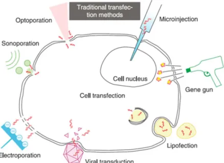
Cell membrane as a biological barrier always keeps separated intracellular and extracellular compartments. Delivery of exogenous biomolecules into the intracellular compartments is highly interesting and functional for such applications including gene therapy. The small molecules in the cell medium like ions can easily penetrate the cell membrane by the passive diffusion through the ion channel [1]. However, large molecules such as DNA plasmids cannot penetrate in the same way due to a limited membrane permeability [2]. There are many methods, which enhance the internalization of large molecules into the cells (Figure 1.1). Virus-mediated transduction employs viral vectors, which contain functional nucleotides to be delivered into the cells [3-5]. However, this technique is not favored due to all inflammation and toxicity concerns and limited package load, even though the transduced cells earn a stable expression. The chemical engineered polymer or particles incorporating nucleotides do not have stable behaviors such as the destabilization of the structure due to the different environments of the treated cells [6, 7]. Mechanical methods such as microinjection and gene gun have high selectivity; however, these strategies show certain drawbacks such as low throughput of the cell treatment and high demand for skilled technicians[8, 9]. The physical methods, including I- electroporation (depending on charging electricity to increase cell membrane permeability), II- sonoporation (using ultrasound generated microbubbles to rupture cell membrane) and III- optoporation (applying laser energy to damage cell membranes integrity by heating or vapor bubble formation) provide a controlled gene delivery [10-17]. Electroporation has high cell throughput in one shot (quick treatment strategy) but it is accompanied by reducing cell viability due to the high voltage. Sonoporation make an uneven ultrasound pressure on targeted cells, which causes cell death. Optoporation uses a laser to produce transient pores on the cell membrane and the exogenous molecules either passively diffuse into the cell or are actively pushed into the cells by the generated vapor pressure. Laser manipulation of the cells has no contact to cells to induce toxicity and offers high selectivity and precision. The laser mediated cell therapy provides a manipulation at a single cell level even at the subcellular level [18].
Figure 1.1 Schematic of traditional transfection methods. Reprinted from W. Ding et al. [19] With different cell therapies mediated by lasers, the involved mechanisms for a membrane permeabilization depend on different laser types and irradiation parameters. Plasmonic nanoparticles (NPs) due to heating properties and near field enhancement effects play an important role in a laser-mediated cell perforation and significantly reduce the laser fluence threshold for minimizing cell damages [20-22]. The plasmonic NPs have variable optical and biological properties depending on different sizes, shapes and composites [23]. The NPs are able to offer spatial and temporal information by a laser irradiation with tunable parameters such as wavelength, laser energy, pulse duration. Moreover, the surface of plasmonic NPs can be chemically modified to bind to specific cells for a target-oriented laser irradiation. Among plasmonic NPs, the gold nanoparticles (AuNPs) are the most popular applied in the biochemical and biomedical therapeutic fields [24].
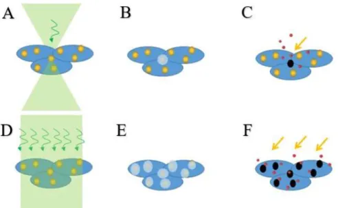
The AuNPs decorated cell optoporation mediated by the laser is a growing research field. In general, the permeabilization of the cell membrane is tuned by the heating and nanobubbles caused by the laser energy absorption of plasmonic NPs (Figure 1.2). By tightly focusing the laser on a single NP bound on the cell membrane, the permeability is increased, and the exogenous materials can locally flow into the cytoplasm. Whereas the higher throughput of the cell treatment is achieved by a loosely focused laser irradiating on NPs bound cells, and the treated NPs can rupture the integrity of the cell membrane to internalize extracellular materials.
Figure 1.2 The NPs decorated cells have higher throughput optoporation with D) weak focused laser than A) the tightly focused laser beam. B) and E): the irradiated NPs provide spatial localization of the cell membrane rupture. C) and F): the permeabilized cell membrane takes the extracellular molecules into the cytoplasm.
1.1 Problems and objectives
Laser parameters (i.e., wavelength, pulse duration, laser fluence) are adjusted to focus on a single cell or target on a large quantity of cells for optoporation studies. Aiming at high transfection efficiency and cell viability through optoporation, most research focused on verifying cell optoporation efficiency and viability after hours of the laser treatment. Such a long waiting period makes it much tricky to precisely analyze these effective parameters and disregards all real-time changes. The study on the real-time responses of the laser treated cells is necessary for the optimized laser manipulation on the cells. As the adherent cells are investigated more for the laser manipulation studies because they are easy to operate, optoporation of the suspension cells with high efficiency is difficult to achieve due to the random and uneven hitting of the cell membrane by the laser irradiation [25]. Here, this thesis suggests studying the cellular real-time responses to the laser irradiation to optimize the optoporation parameters. The modifications on the optical setup for the visualization of NPs are also done to simplify the optoporation process. The exploration on the optoporation of NPs bound suspension cells (Jurkat cells) by the femtosecond laser paves the way for the highly efficient optoporation and transfection.
In this thesis, we target on solving certain issues involved in the laser-mediated cell manipulation for transferring exogenous materials into the cells without reducing the cell viability. The thesis is composed of different Chapters summarizing studies in this field through a literature review and indicating new experimental results:
1. Chapter 2 summarizes the related research in this field. Involving the viral-mediated cell transfection research, non-viral chemical-mediated studies such as the incorporated plasmid in the liposome, naked plasmid DNA, small interfering RNA (siRNA), micro RNA related research, DNA vaccine transfection method, physical-mediated methods and the most popular CRISPR involved transfection method in past few years.
2. Chapter 3 covers a real-time monitoring of single cell responses after the localized laser irradiation in order to understand the involved mechanisms. For this study, a nanosecond (ns) laser (532 nm) is used to irradiate the single AuNP (100 nm in diameter) bound MDA-MB-231 cell (an adherent human breast cancer cell). The localized membrane damage caused by the AuNP results in the uptake of the exogenous molecules. For this reason, Propidium Iodide (PI) staining molecules are used to indicate kinetics of the membrane damage by recording the real-time fluorescence signal change. The cell viability is then verified by means of Calcein AcetoxyMethyl (Cal-AM). The cellular responses after the laser irradiation are captured by monitoring intensity changes of the used fluorescent dyes and accordingly interpreted based on different laser parameters.
3. Chapter 4 describes how to develop a simple and elaborated side-illumination imaging device for the detection of plasmonic NPs in a cellular environment. As with the traditional NPs detection methods, the optical setup which has to incorporate laser irradiation optical path, fluorescence illumination path and signal collection path becomes more complicated, limiting the applications of cell manipulation by the laser. The side-illumination device is to solve the problem. A mixture of plasmonic NPs with different spectra can be also individually visualized and distinguished by their unique colors. The side-illumination imaging device is a useful setup for a wide range of biomedical applications such as disease diagnostic in pathology and cytology.
4. Chapter 5 presents an optimized range of effective parameters (i.e., laser fluence) of infrared femtosecond laser-mediated Jurkat cells optoporation. The individual Jurkat cell is
attached to the Petri dish substrate for investigation and optimization of femtosecond laser parameters. Suspension Jurkat cells are optoporated by femtosecond laser without and with decoration of AuNPs to study the influence of cell viability with different laser irradiation parameters. With experience from cellular response after laser irradiation, we use PI and Cal-AM to verify optoporation efficiency and cell viability. Various laser fluences are chosen for different applications, such as Jurkat cell optoporation and specific cell sorting. For further application of high-efficiency constant suspension cell optoporation mediated by femtosecond laser, some works are described in the appendix.
5. Chapter 6 discusses all results obtained earlier in the mentioned Chapters: monitoring of the plasmonic NPs bound single-cell response to ns laser optoporation paves the way to understand the relation of laser fluence and cellular real-time response, indicated by fluorescence signal change; side-illumination device replaces the traditional imaging methods of plasmonic NPs, with easy adaptation and high contrast; the proposed plasmonic NPs bound suspended cell optoporation mediated by femtosecond laser makes it possible for high throughput suspended cell optoporation.
CHAPTER 2
LITERATURE REVIEW
As the transfection is a process that the exogenous nucleic acids are delivered into the cells, many techniques in different categories are widely developed such as the traditional viral vectors-mediated transfection, lipid-based transfection, and the popular physical methods in past decades, for instance, electroporation and optoporation [26]. However, the advantages and disadvantages of each of the most often used transfections methods are discussed: viral vectors can pack nucleic acids and introduce them into the cells with high efficiency, but it is time-consuming and causing the mutation concerns [27, 28]. Using recombinant viral vectors and non-viral carriers such as liposome is developed. Lipid-based transfection involves the lipid-soluble structures to be transferred into the cells incorporating the nucleic acids with minimum toxicity but low transfection efficiency [29, 30]. However, there are transfection methods for naked nucleic acids such as plasmid DNA and siRNA. Liu et al. demonstrated the significant gene expression in the liver of mice using the injection of naked plasmid DNA by simple mechanical massage [31]. The same method has been employed by siRNA (small interfering RNA) and microRNA for the transfection of kidney, liver and renal tubulointerstitial fibrosis cells [32-34]. Ghartye-Tagoe et al. reported the plasmid DNA transfection on differentiated intestinal epithelial monolayers by electroporation, and siRNA gene silencing. It was found that electroporation induced more expression and DNA uptake than lipofection with the reporter plasmids and siRNA resulted in up to 90% inhibition of targeted protein production [35]. DNA vaccines are plasmids that combine sequences required for replication and selection in Escherichia coli (bacterial region) with sequences needed to express an encoded transgene in vertebrate cells (eukaryotic region) [36]. DNA vaccines are inherently safe because the vectors are non-replicating, encode and express only the target antigen, not as with viral vectors. As they are well tolerated and the DNA product is highly stable, DNA vaccines have excellent safety profile in human clinical investigations [37]. The licensure of four animal health DNA vaccine products demonstrates the utility of DNA vaccination in large animals such as horses and pigs [38]. Wang et al. found that the human serum amyloid P binds and inhibits plasmid transfection and DNA vaccine induced adaptive immune responses more strongly than the murine counterpart [39], which raised difficulties for the DNA vaccine efficacy in humans. Moreover, most of developed delivery methods (such as the physical method: electroporation) can transfer the plasmid into the cytoplasm but rarely into the nucleus directly [40]. Thus, the developed and innovated design of the plasmid for the efficient delivery and diffusion to the nucleus need
highlighted exploration. Physical methods increase the cell membrane permeability by the external forces to open holes on the membrane, allowing exogenous nucleic acids to enter. The tunable parameters (electrical pulses for electroporation; laser wavelength, pulse width and laser fluence for optoporation) offer the alternatives for the transfection optimization [41, 42]. The electroporation treats the cells in the conductive suspension medium applying the electrical pulses, the induced charge accumulation opens the holes on the cell membrane. The membrane can repair when the electric field is removed. The electroporation is an easy method with a high quantity of the treated cells within a short time. However, it is required a commercial device and an optimized protocol for specific cell type transfection. A lot of cells are dead after the electrical pulse treatment due to the high voltage which needs the optimization [43-45]. The optoporation has been studied as a non-contact transfection method with spatial and temporal selectivity, and high targeting precision [46]. In the past few years, scientists have been working with a set of molecular scissors CRISPR (an enzyme that cuts DNA), which is the biggest breathtaking story in science as CRISPR makes genome edits cheaper and faster [47-50]. An important step to employ CRISPR on transfection is to deliver it into the cytoplasm or nucleus of the targeted cells. The transfection methods in different categories mentioned above can work with respective merits and drawbacks. As CRISPR has the biggest limitation that it is not a hundred percent efficient, and it has the off-target phenomenon [51-53] which is still under vast investigations. With various attention on the laser-mediated cell transfection, lots of studies are focusing on developing and optimizing the transfection process using the pulsed laser.
The objective of this master thesis is to investigate the pulsed laser optoporation accompanied by the plasmonic NPs, providing a controlled permeability to internalize exogenous materials into the cell. Both nanosecond and femtosecond lasers have been used to optoporate various cells (Table 2.1).
Table 2.1 Characteristic example of cell membrane optoporation and transfection application by the pulsed laser, referring to Etienne Boulais et al.[54]. Various cell types were optoporated or transfected by nanosecond laser or femtosecond laser under specific conditions. λ is wavelength, τp is pulse duration, F is single laser pulse fluence, E is the pulse exposure. T is the targeted cell.
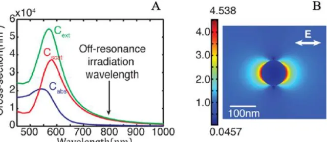
The role of plasmonic AuNPs on the cell membrane is crucial for providing an efficient laser-mediated cell optoporation and subsequent transfection. Plasmonic NPs are able to accumulate the electrons into an oscillatory movement by the irradiating light. When the frequency of the incident light is near the intrinsic plasmon frequency of the plasmonic NP, on-resonance phenomenon is accompanied by strong energy absorption. On the other hand, off-resonance happens when the NP’s plasmon frequency is far from the frequency of the incident light and the energy absorption is no longer dominant to heat up, but to enhance the near field [60]. Boulais et al. [54] showed that the calculated absorption, scattering and extinction cross sections of AuNPs (100 nm in diameter) in water as a function of incident light (Figure 2.1.A). Figure 2.1.B showed the electric field distribution near the AuNP (100 nm in diameter) for low absorption and high near field enhancement. By tuning size, composite, shape of NPs and wavelengths of the incident light, NPs can be manipulated either on-resonance or off-resonance for different biomedical applications.
Application Laser&Target Particles Principle Comments Ref . Cell membrane optoporation and transfection λ=532 nm, τp=20 ns, F=500 mJ/cm2 E : ~100 pulses T : Lymphocytes -20 nm -AuNPs -Conjugated
-Bubbles -Membrane recovers in 2 mins
-Uptake of 10 kDa FITC-D [55] λ=532 nm, τp=6 ns, F=500 mJ/cm2 E : 1-50 pulses T : Hodgkin’s disease and lymphoma cells -15,30 nm -AuNPs -Conjugated -Bubbles -8-68% efficiency [56] λ=532 nm, τp=0.5 ns, F=110-150 mJ/cm2 E : single pulse T : Various
-Size not given -AuNPs clusters
-Bubbles -Transfection of DNA plasmids -~70% efficiency [57] λ=760nm, femtosecond, F=18 mJ/cm2 E : 10 s@80 MHz T : epithelial breast cancer cells -80 nm -AuNPs -Conjugated -Low- density plasma
-Uptake of 10 kDa
FITC-D [58] λ=800 nm, τp=45 fs, F=100 mJ/cm2 E : 10 s@80 MHz T : melanoma cells -100 nm -AuNPs -Unconjugated -Plasma-mediated bubbles -~70% optoporation efficiency -Transfection of YFP-SMAD2 plasmids -~25% transfection efficiency [59]
Figure 2.1 A) Scattering, absorption and extinction cross sections for 100 nm AuNP in water, showing the on-resonance wavelength around 570 nm and off-resonance wavelength in infrared. B) Electric field enhancement around the NP surface with low absorption and near field enhancement. Adapted from Ref. [54].
2.1 Nanosecond pulsed laser perforation on cells
As nanosecond laser is compact and relatively easy to operate, many optoporation studies have been performed with this type of laser [56, 61-63]. For the nanosecond laser cell perforation, the laser with wavelength in visible range (~532 nm) which is around the plasmon peak of 100 nm AuNPs (~570 nm) absorbs laser energy strongly and induces a temperature rise leading to the membrane permeability [64, 65]. The nanosecond laser cell perforation, mainly based on heating mechanisms of plasmonic NPs, causes a fragmentation of NPs and induces cytotoxicity. However, the use of a near-infrared laser (700-1000 nm) is highly desirable for a cell optoporation because the absorption by cellular components in this wavelength range is significantly low and indicates a great potential for a clinical translation.
2.1.1 Mechanism of nanosecond pulsed laser-induced cell perforation
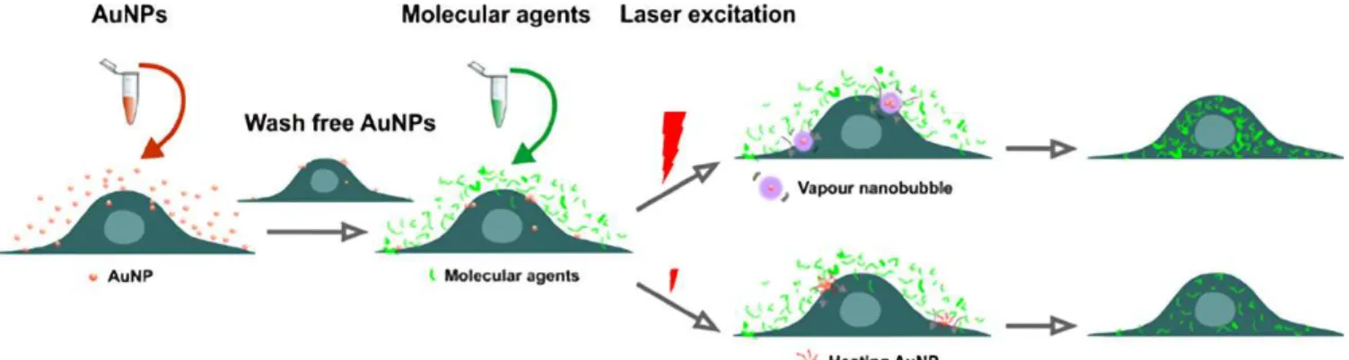
The fluence for a single cell optoporation can be lowered through irradiation of single plasmonic NP binding to the membrane to keep the highest viability without reducing the transfection efficiency. The permeabilization using the plasmonic NP assisted optoporation involves two mechanisms including heating and vaporized nanobubbles (Figure 2.2) [66]. Allowing the fluorescent molecular agents enter the cytoplasm and becoming fluorescent.
Figure 2.2 Schematic of the experimental procedure. After incubating cells with AuNPs and with the molecular agents. By applying different laser energies, cell membrane integrity is ruptured by vaporized bubbles (high laser energy) or heating of AuNPs (low laser energy). The treated cell membrane allows exogenous molecules to enter the cells. Adapted from Ref. [66].
2.1.2 Nanosecond optoporation of adherent cell
Nanosecond laser is a functional and applicable device for almost all cellular optoporation. For example, Krawinkel et al. have employed the Nd: YAG infrared nanosecond laser (1064 nm, 17 ns, 10 Hz) to deliver the PI molecules and GFP inserted plasmids into human breast adenocarcinoma MCF-7 cells. The cells were targeted by the laser and irradiated one by one. The optoporation (indicated by PI with energy density ~4 x 10⁴ J/cm2) and transfection (indicated by
green fluorescent protein encoding plasmid into MCF-7 cells) were confirmed by the fluorescence microscopy [62]. Krawinkel et al. incubated primary human gingival fibroblasts (pHFIB-G) cells with AuNPs prior to the nanosecond laser irradiation (532 nm, 1 ns, 22.5 kHz). The optical setup could irradiate the cell samples by the laser with a large Rayleigh range (16.2 mm) by installing an extra lens (focal length f =250 mm). The laser irradiation resulted in high throughput of pHFIB-G manipulation (up to 85%) with 97% cell viability. The fibroblasts internalized dextran polymers with molecular weights up to 500 kDa to estimate the size of the pores on the membrane [63]. Yao et al. have also shown a cell specific optoporation by using antibody (BerH2: against CD30 and ACT1: against CD25) decorated AuNPs to target cells (Hodgkin’s disease cell line L428 and human lymphoma cell line Karpas 299). The optoporation with 30 nm AuNPs transfected 68% of cells with FITC-Dextran with a cell death (27%) compared to the smaller AuNPs 15 nm [56]. Saklayen et al. cultured the HeLa CCL-2 cells on a thermoplasmonic substrate with an array of pyramids with a gold surface (Figure 2.3.A), developed controllable hotspots generated bubbles at
the underneath of the cell membrane using a nanosecond laser irradiation to internalize FITC-Dextran. The flow cytometry was used to evaluate the delivery efficiency: for the delivered cargos size ranging from 0.6 to 2000 kDa, the delivery efficiency was up to 95% for the smallest molecule and cell viability was up to 98% [67]. Xiong et al. selectively optoporated cells (Figure 2.3.B) using spatially resolved laser scanning manner and the nanoparticle-sensitized photoporation to internalize FITC-Dextran (10 kDa) according to the pre-defined pattern (for a selective location) or image-guided interactive method (a single cell targeting) [68].
Figure 2.3 A) SEM of fixed HeLa CCL-2 cells on the thermoplasmonic substrate, referring to [67]. B) Illustration of the cell optoporation selectivity according to the black pixels of Albert Einstein drawing. The green fluorescence FITC-dextran with 10 kDa is successfully delivered into the optoporated cells. Scale bar is 1000 µm. Adapted from Ref. [68].
However, a real-time monitoring of cellular response of the individual cell receiving nanosecond laser irradiation has not been reported in order to further optimize the optoporation technique and facilitate a clinical translation of the optoporation.
2.2 Femtosecond pulsed laser perforation on cells
Plasmonic NPs-mediated femtosecond laser cell optoporation is quite popular, especially for near-infrared femtosecond laser due to avoiding the fragmentation of NPs and the toxicity [59, 69]. The near field enhancement around plasmonic NPs generated by a multiphoton ionization forms nanocavitation, to permeabilize the cell membrane. The femtosecond laser cell optoporation with high selectivity, efficiency and viability is a hot topic for cell therapy and further clinical translation.
2.2.1 Mechanism of femtosecond pulsed laser-induced cell perforation
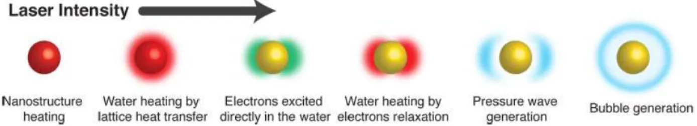
The infrared femtosecond laser (~800 nm) perforating cell membrane has been studied using an objective in the inverted microscope optical setup to precisely target the specific cell and generate spatially confined holes [70-72]. The opened pores allow exogenous biomolecules to diffuse into the cell cytoplasm. However, it is needed an alignment of the femtosecond laser to precisely focus onto the cell surface and target surface bound plasmonic NPs. Thus, the membrane bound NPs address the location of the cell membrane due to their intrinsic scattered light. Locating the cell membrane surface by bound NPs, cells can be permeabilized with femtosecond laser irradiation. The femtosecond laser-mediated AuNPs bound cell membrane permeabilization is based on two fundamental mechanisms including a near field enhancement and particle heating effect [54, 73, 74]. The near field enhancement occurs due to collective oscillations of electrons in AuNPs caused by the incident laser electromagnetic field. The heating effect of the NPs occurs owing to the energy absorption from the incident irradiation. The dominant mechanism for an infrared femtosecond laser irradiation, for instance, on AuNPs (100 nm in diameter) involves an energy transfer and plasma generation in order to induce nanocavitation. The mechanism, corresponding to the energy absorption and light scattering by AuNPs in different laser irradiation intensities, depicted in Figure 2.4. The NPs absorption dominates in the nanostructure and heats the gold electrons when it is in-resonance. In off-resonance, the absorption is relatively low, and the light can be scattered in the near field of NPs to enhance the near field effect. When the laser intensity reaches over the optical breakdown threshold, the electronic plasma is generated near the NPs structure. Continuous laser energy absorption by the plasma finally transfers into the release of the pressure waves, which results in a nanocavitation bubble eventually [54, 75]. These induced effects, in turn, reduce cell membrane permeability, enabling diffusion of exogenous molecules into the cytoplasm.
Figure 2.4 Schematics of different NPs phenomena with increased laser irradiation classified in the term of laser intensity. Adapted from Ref. [54].
2.2.2 Femtosecond optoporation of adherent cell
The femtosecond laser for cell therapy shows a great potential in a wide range of biomedical and biophotonic applications for providing efficient transfection through a non-toxic cell optoporation. The femtosecond laser optoporation has used to transfect the adherent cells, such as CHO cells (represent a mammalian model system for optical transfection study), melanoma cells (human skin cancer model), and MDA-MB-231 cells (transfection of human breast cancer cells) [59, 69, 76, 77]. The adherent cells should be pre-cultured into a petri dish in the suggested cultured medium complex until reaching at confluency more than 90%. On the day of the laser treatment, the cells are incubated with plasmonic NPs at a specific concentration. Depending on specific applications, some bound NPs need to be surface modified by binding specific membrane antigen receptors to attach specific cell types [69]. The pre-programmed laser scanning pattern allows a high throughput of the treated cells [78, 79]. The perforation of cell membrane by femtosecond laser is controllable to generate a wide range of patterns by different laser scanning modes (Figure 2.5).
Figure 2.5 A) demonstrates a side view of the laser irradiation treated cell samples with a loosely focused femtosecond laser. B) shows the laser irradiation on NPs decorated cell samples and the permeabilized membrane allows the exogenous nucleic acid to enter. C) programmed laser irradiation scanning pattern for treating cells in large quantity. Adapted from Ref. [79].
To apply the optimized protocol for a single cell optoporation to a bunch of cells, Georg Breunig et al., achieved the automated CHO cell optoporation by using the infrared femtosecond laser (800 nm, 85 MHz, sub-15 fs). The whole automated setup was designed to find the location of the cells and designate the cells for a laser focal point for irradiation. The software dominant method could target 7-8 cell positions per second for an illumination duration of 100 ms and achieved effective 10,000 cells treated per hour automatically. Employing a uniform laser focal region (i.e., beam shaping to quasi-Bessel beam geometry) can also raise the number of the laser treated cells. The
maximum cell viability after the optoporation was observed around 60-70% [78]. Baumgart et al. used off-resonance femtosecond laser based high throughput virus and fragmentation free transfection method for AuNPs bound melanoma cells. The femtosecond laser was with a center wavelength of 800 nm and a repetition rate of 1 kHz. The laser irradiation optical setup was incorporated with the in-situ spectroscopy to study the fragmentation of NPs and the confocal microscopy for the visualization. A high throughput of laser treated cells was obtained using an optimized speed (40 mm2/min) with a low toxicity (<1%) and high transfection efficiency (~3 fold
higher than the lipofection). They also proposed that the further work on the bioconjugation modified NPs could lead to the promising spatially selective treatment of cells of interest [59]. Bergeron et al. modified the surface of plasmonic NPs and applied these antibody conjugated NPs to target specific cells for a laser treatment. With the conjugated NPs, optoporation of the targeted specific cells in co-culture cell lines (MDA-MB-231 cells positive CD44, and RAW661 cell negative CD44) was achieved using an infrared laser (800 nm) without affecting the non-targeted cells. The threshold to have a selective optoporation was found with 49 pulses at 250 Hz or 97 pulses at 500 Hz [69].
For the numerous investigations on femtosecond laser applied for NPs bound adherent cell optoporation, the cells were on the petri dish substrate and could be controlled by the built-in translation stage in any designed movement. As a comparison, the manipulation of the suspended cells for an efficient transfection using the femtosecond laser optoporation remains challenging because the floating nature of suspended cells, making it hard to manipulate.
2.2.3 Femtosecond optoporation of suspension cell
Transfection of suspended cells has been proven to be quite difficult due to the intrinsic nature of the cell floating character. The common liposome-based non-viral vectors due to a low molecular binding and uptake are not suitable for the transfection of suspended cells [80-82]. However, optoporation is a cell type non-dependent method to perforate the membrane efficiently including the suspended cells. Certain protocols have been developed for a high-efficiency transfection of suspension cells such as chicken eggs coated culture plates to eliminate the nonadherent cell[83] as well as a real time femtosecond laser irradiation in microfluidic system for a spatial confinement of the cell flow [84]. However, cells’ immobilization onto the substrate, requires a pre-treatment
on the petri dish. Moreover, the microfluidic setup with a constant flow needs sub-micron accuracy for positioning of the laser beam on the cell membrane to reach a high-efficient perforation [85]. Marchington et al. have used femtosecond laser (800 nm, 80 MHz, 100 fs) to irradiate individual suspending human embryonic kidney (HEK293) cells in a microfluidic channel. The focused laser beam generated transient pores which allowed uptake of the extracellular molecules in the surrounding fluid into the cytoplasm. The optical setup enabled the rate of cell treatment at 1 cell/s and several thousand cells per hour. The optoporation efficiency (42 ± 8%) and cell viability (28 ± 4%) were tested by PI and Cal-AM cell stains. They also suggested the future development should include the integration of the lab-on-chip technology with the existing setup, incorporating fluorescence spectroscopy [86]. Georg Breunig et al. explored the transfection of the trypsinized CHO cells by using the 800 nm, 85 MHz, sub-15 fs laser, embedded into a microflow system, including the reshaped laser beam focal region into a zero-order quasi-Bessel beam for relative uniform laser energy deposited on the cells. After the transfection (24 h), a small part (~1%) of the bunch of laser treated cells expressed GFP. The lower transfection efficiency compared to the single cell optoporation in petri dish was related to a shielding effect, impedes well-contributed laser fluence in the confined microfluidic channel. The low transfection efficiency might be improved by increasing the cell number and scanning rate of the laser beam [84].
2.3 Summary
Different transfection methods are mentioned with their advantages and disadvantages, transfection by optoporation is chosen for the study due to the variable parameters (i.e., wavelength, pulse width, fluence etc.), controllable spatial and temporal selectivity, and cell type non-dependent.
CHAPTER 3
REAL-TIME MONITORING OF PLASMONIC
NANOPARTICLE-MEDIATED PULSED LASER SINGLE-CELL
OPTOPORATION
In this chapter, a single cell therapy was demonstrated with the aid of plasmonic NP-pulsed laser optoporation. A reflected light microscopy (RLM) with a high numerical aperture (NA) objective is initially developed to provide precise localization of an individual NP bound to the cell membrane and spatially control of the single cell optoporation. All effective parameters including nanosecond laser exposure, laser fluence and optomechanical particle-membrane interaction were then optimized by developing an algorithm for an error-free automatic alignment of laser focus with the bound NP. We proceed to study the single cell optoporation by a systematic analysis of the internalization of PI molecules and perforation kinetics. The internalization rate of PI molecules through a time-dependent perforation indicates a range of suitable fluences (0.3-0.7 J/cm2) without
inducing cell death. The developed NP imaging strategy for a live cell based on RLM, fully compatible with a simultaneous fluorescence microscopy imaging, also provides a real-time observation and systematic analysis of cellular responses. Our results demonstrate that optimized mechanisms and parameters involved in the single-cell optoporation are critical to achieve a successful perforation and cell therapy for a broad range of biomedical applications.
3.1 Introduction
3.1.1 Research question
An elaborated nanomedicine, provides a single cell therapy among a cluster of different cells (i.e., cancer and healthy cells with a broad range of physical and biological features), is now achievable by employing a plasmonic particle-mediated single-cell optoporation [87]. A non-specific optoporation of a cluster of cells by a blind scanning mode consisted of an unequal distribution of Gaussian intensity of laser light (i.e., generation of a wide range laser powers on the cell membrane and reoccurrence of the laser exposure) mainly induce cell death. Therefore, single-cell optoporation, which focuses on a precise laser manipulation of an individual cell targeted by functional plasmonic NPs, controls all effective aspects of laser irradiation, including laser fluence and irradiation duration; however, the blind laser scanning mode affects most of the cells around the laser point and subsequently their normal proliferation and cycle [69]. Moreover, the laser
operation in the highly focused pointing mode, which provides greater selectivity compared to the loosely focused scanning mode for an individual cell optoporation, can act as a microscalpel to generate a local disruption of the cell membrane locally and precisely transfer exogenous biomolecules into intracellular compartments [88].
The position of the laser beam whether to irradiate nuclear-targeted or cytoplasm-targeted plasmonic NPs is critical to obtain suitable cellular responses and improve optoporation efficiency [89, 90]. The irradiation near nuclei provides a narrow laser parameter manipulation range due to inducing cell death; however, irradiation far from nuclei gives a wider laser manipulation range, which is suitable to achieve a high optoporation efficiency (i.e., the threshold to induce cell death when the irradiation is near the nuclei is below the laser fluence of 695 mJ/cm2 but not for the
irradiation afar).
Therefore, plasmonic NPs bound on the cell membrane, acts as an error-free membrane marker, provide a highly spatial localized optoporation using the highly focused pointing mode. Highly focused laser optoporation requires well-developed plasmonic NPs (i.e., target a specific cell), simultaneous visualization of the fluorescence dynamic (i.e., permeabilize the plasma membrane by adjusting the transient pore size and resealing rate), and numerical simulation of the optoporation process (i.e., provide accurate optoporation and transfection for a wide variety of biomedical applications). Thus, a successful development of the single cell optoporation can minimize side effects of the laser irradiation and improve the transfection of the targeted cell at the subcellular level.
3.1.2 Objective
In this chapter, we investigate a real-time single cell response to nanoparticle-mediated pulsed laser optoporation and then develop a spatial and temporal monitoring system to in situ control and optimize in vitro single cell laser optoporation.
3.2 The proposed approach
We here develop a nanosecond laser-based optoporation setup to integrate plasmonic NPs (act as the cellular markers) with a high NA objective (allows a precise localization of NPs), automated laser focus positioning, and longtime measurement of the dynamic of fluorescence. An extensive
study is also aimed to evaluate effective laser parameters (i.e., laser fluence) on biomolecular delivery in situ. In this case, the optimized parameters of the laser for generating an effective optoporation will be confirmed by pursuing cellular targeting (i.e., a backscattering imaging system using a high numerical aperture objective to visualize single NP bound plasma membrane), delivery of exogenous substances such as fluorescent PI molecules with a strong quantum yield [91, 92], and their transport kinetics into the optoporated cell and intracellular compartments. A real-time measurement of the corresponding fluorescence intensity of the exogeneous fluorescent PI molecules, inflows to the cytoplasm (simulated according to Davis’s diffusion model [11]), determines the permeabilization and subsequent recovery processes. Using high-speed shadowgraphic imaging in situ also shows how to follow the status of bubble generation and sizes on the pore formation and recovery.
3.3 Methodology
3.3.1 Cell culture, PI transfection, and live cell assay
MDA-MB-231 cell (Human breast cancer cell line, American Type Culture Collection, Manassas, VA, USA) with a density of 1x10⁵ cells/well were seeded into a glass bottom Petri dish (19.625
cm2, MatTek, Ashland, MA, USA) by adding Dulbecco’s Modified Eagle’s Medium (DMEM)
supplemented with 10% fetal bovine serum (FBS, Invitrogen) and 1% penicillin (100 units/mL, Invitrogen) to reach 80% confluency overnight (37 °C with 5% CO2). After a washing step (using
buffered phosphate saline, PBS, Sigma-Aldrich), the cells were then incubated with 8 μg/ml of AuNPs (50 μg/mL, 100 nm in diameter, A11-100-CIT, Nanopartz, Loveland, CO, USA) in the DMEM for 2 h in the incubator (37 °C with 5% CO2). Afterwards, the treated cells were washed
three times with PBS prior to the laser treatment to remove unbound NPs. In this chapter, AuNPs (100 nm in diameter) were used without further modifications. The dynamic is detected by adding PI (ThermoFisher Scientific) with a final concentration 1.5 μM (diluted in DMEM) to the treated cells30 minutes before the laser irradiation. The cell viability of the treated cells was evaluated by using Calcein Acetoxymethyl (Cal-AM, ThermoFisher Scientific) with a concentration of 1 μM at 2 h post-irradiation.
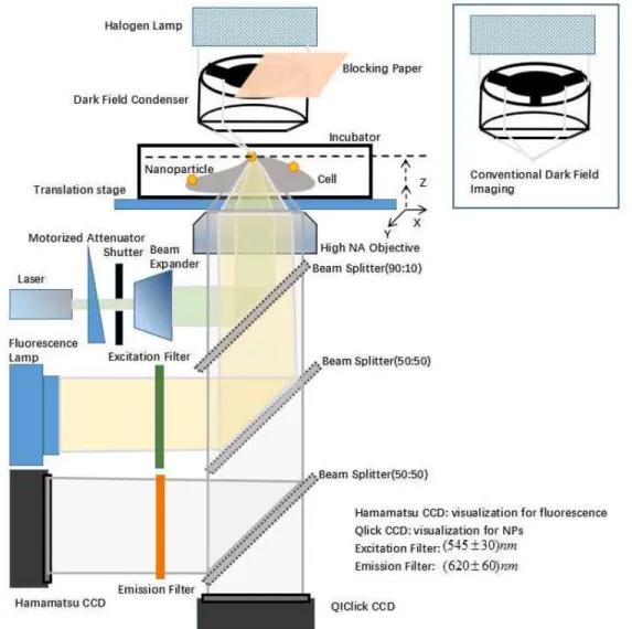
3.3.2 Preparation of optical setup for optoporation and visualization
The experimental setup for a single-cell optoporation (Figure 3.1) consists an inverted Eclipse Ti microscope (Nikon, Mississauga, ON, Canada) equipped with a high NA objective (100X, oil immersion, variable NA:0.5-1.3, Nikon) accompanied by a refractive index (RI) of immersion oil is 1.51 at 23 °C (Olympus Scientific Solutions Americas). The glass-bottom petri-dish was then placed in a humidified CO2 cellular incubator system (Live Cell Instrument, Korea) installed on a
flat top 3D motorized translation stage (H117P1, ProScan, Prior Scientific, Rockland, MA, USA). The laser beam was automatically focused on the targeted NPs by using the z-step scanning system. The focal spot size was roughly calculated as the value of wavelength around 500 nm.
A fluorescence lamp C-HGFI Intensilight (Nikon) with ET-DsRed excitation filter (545 ± 30 nm, Chroma Technology, USA 49005) provided a light for PI fluorophores excitation (~ 535 nm) and cell-NP complex visualization with a QIClick digital CCD camera (QImaging, Surrey, BC, Canada). The detection of real-time living-cell fluorescence dynamic was performed using another CCD camera (Hamamatsu R2) equipped with a corresponding PI fluorophore emission filter (620 ± 60 nm, Chroma Technology, USA 49005). Cal AM was then detected by a filter sets (Ex: 500 ± 20 nm, Em: 535 ±30 nm, Chroma Technology, 49003-ET-EYFP, USA).
The laser source used for cell perforation was an ultra-compact 15 nanosecond laser (QC532, CrystaLaser, USA) working at 100 Hz and 532 nm wavelength. The laser power was controlled by the homemade software (developed function integrated in LabView to control attenuator and shutter for laser irradiation) with the rotation of a half-wavelength waveplate combined with a plate polarizer on the beam path. The irradiation time was automatically controlled by the mechanical shutter (Thorlabs) connected to the software. A beam expander accompanied by a diaphragm was also used to adjust laser focal point size by changing the effective NA. As double decks were placed under the objective from the Nikon microscope to provide two optical pathways: (1) the upper path was conducted to a laser 90:10 beam splitter (Thorlabs, Newton, NJ) from the backport of the microscope to the sample after going through the objective, and (2) the second path separated a wide field NP imaging and fluorescence illumination using a 50:50 beam splitter. Cell perforation was performed by using LabView software to generate a Gaussian fit for PSF of the bound NP, recording the position of the NP, and controlling the translation stage to move the NP’s position to the initial laser focal point position. The NP was precisely selected and well aligned according to
the laser beam path to provide an effective optoporation on a single cell. Note, several automatic iterations of alignment of NP and laser focus processes were employed to achieve the laser focal point due to certain instabilities (i.e., vibration of the optical table).
A standard white light source (Nikon) with 50 W halogen lamp was employed to provide a conventional transmission microscopy and 3D imaging with partial Dark-Field (pDF) microscopy. Partial illumination from Halogen lamp was blocked using an opaque paper before the darkfield condenser (NA=0.9, Nikon) to enhance imaging contrast by creating artificial shadows (Figure 3.1).
Figure 3.1 Schematic of the experimental setup for the single-cell optoporation. AuNP serves as membrane position target and field enhancement factor for localized optoporation.
3.4 Results and discussions
3.4.1 RLM imaging of NPs in a cellular environment
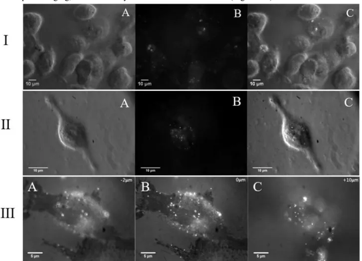
The developed optical setup provides an excitation light for fluorescence imaging following laser irradiation on the chosen region of interest (ROI). Using a high magnification oil immersion objective and darkfield condenser, bound NPs on the cell membrane can be precisely visualized. To achieve a reliable identification and spatial localization of NPs on the cell membrane, the NPs were imaged based on the high backscattering signal detection [93, 94]. RLM, which is compatible with pDF imaging, simultaneously visualize both cell and NPs (Figure 3.2).
Figure 3.2 Cell-NP images taken with I) 60X objective and II) 100X objective. A) Images acquired in pDF imaging of cells; B) RLM imaging of NPs on the cell surface; C) Combination of pDF and RLM imaging. The scale bar is 10 µm; III) RLM imaging of NP distribution on a single cell at -2 μm, B) 0 μm (at the bottom of petri dish substrate) and C) 10 μm position (NPs on top surface of the cell membrane). The scale bar is 5 µm.
The typical experimental images of cells decorated with AuNPs (100 nm in diameter) are illustrated in Figure 3.2.I (60X oil immersion objective with NA:1.4, Nikon) and Figure 3.2.II (100X oil immersion objective with NA:0.5-1.3, Nikon). The images acquired by pDF and RLM and their combination (overlapped images) using 100X oil immersion objective shows an improved contrast to differentiate targeted NPs and cells and analyzed the distribution of bound NPs at different positions of the cell membrane by tuning z-step (Figure 3.2.III).
3.4.2 NP-laser alignment
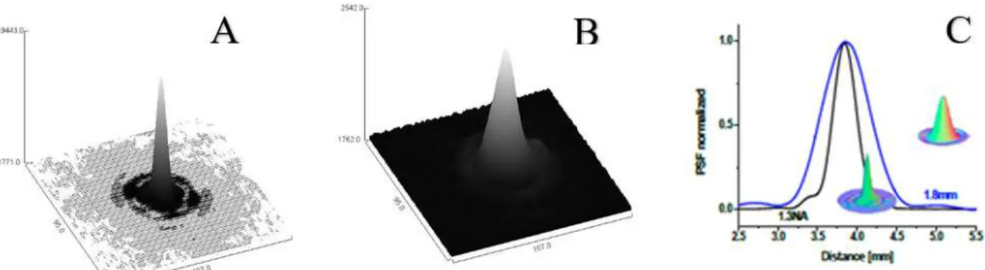
A controlled optoporation of an individual cell depends on the precision of 3D spatial alignment of laser focal point with a bound NP on the cell membrane. Owing to the high NA (0.5-1.3) objective and sharp laser energy distribution, small miss-displacement (in a nanoscale) of the laser focal point from the bound NP induces a dramatic laser energy loss. Therefore, a fine z-scanning (-2 μm to +2 μm, step size: 0.1 µm) remarkably indicates the laser beam 3D intensity distribution (laser Gaussian energy distribution) and focal point on a Si-wafer (full reflection of laser irradiation) (Figure 3.3).
Figure 3.3 Illustration of the 3D energy distribution of the laser focus when NA equals to A) 1.3 and B) effective diaphragm is 1.8 mm. C) shows the Point Spread Function (PSF) normalization of energy distributions of two NAs.
3.4.3 Analysis of perforation dynamics
Accurate delivery of exogenous biomolecules into an individual cell and its subcellular compartment provides a precise functionality and control in cell therapy [95]. The investigation and analysis of cellular responses after the laser irradiation can be tackled down by using the developed microscopic setup (see above section). Therefore, the perforation dynamics can be
![Figure 2.3 A) SEM of fixed HeLa CCL-2 cells on the thermoplasmonic substrate, referring to [67]](https://thumb-eu.123doks.com/thumbv2/123doknet/2344957.34664/31.918.121.812.324.486/figure-sem-fixed-hela-cells-thermoplasmonic-substrate-referring.webp)