HAL Id: tel-01310153
https://tel.archives-ouvertes.fr/tel-01310153 Submitted on 2 May 2016
HAL is a multi-disciplinary open access archive for the deposit and dissemination of sci-entific research documents, whether they are pub-lished or not. The documents may come from teaching and research institutions in France or abroad, or from public or private research centers.
L’archive ouverte pluridisciplinaire HAL, est destinée au dépôt et à la diffusion de documents scientifiques de niveau recherche, publiés ou non, émanant des établissements d’enseignement et de recherche français ou étrangers, des laboratoires publics ou privés.
Role of mTOR kinase activity in skeletal muscle
integrity and physiology
Qing Zhang
To cite this version:
Qing Zhang. Role of mTOR kinase activity in skeletal muscle integrity and physiology. Biochemistry, Molecular Biology. Ecole normale supérieure de lyon - ENS LYON; East China normal university (Shanghai), 2015. English. �NNT : 2015ENSL0987�. �tel-01310153�
THÈSE
en vue de l'obtention du grade de
Docteur de l’Université de Lyon, délivré par l’École Normale Supérieure de Lyon En cotuelle avec East China Normal University (Shanghai)
Discipline : Sciences de la Vie
Laboratoire de Biologie Moléculaire de la Cellule
École Doctorale de Biologie Moléculaire, Intégrative et Cellulaire de Lyon
présentée et soutenue publiquement le 30 Mars 2015
par Monsieur Qing ZHANG
_____________________________________________
Role of mTOR kinase activity in skeletal muscle integrity and physiology ______________________________________________
Directeur de thèse : Monsieur Laurent SCHAEFFER
Co-tuteur de thèse : Monsieur Shuzhe DING
Après l'avis de : Monsieur Shiyi CHEN
Monsieur Xin XU
Devant la commission d'examen formée de :
Monsieur Laurent SCHAEFFER, Ecole Normale Superieure de Lyon, Directeur
Monsieur Shiyi CHEN, Huashan Hospital, Fudan University, Rapporteur
Monsieur Shuzhe DING, East China Normal University, Co-directeur
Monsieur Xin XU, Shanghai University of Sport, Rapporteur
ABSTRACT
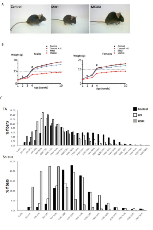
Mammalian target of rapamycin (mTOR) is a key protein kinase that integrates and coordinates diverse signaling information mediated by growth factors, nutrient availability and energy status. It forms at least two functionally distinct signaling complexes, named mTOR Complex 1 (mTORC1) and mTOR Complex 2 (mTORC2). mTOR regulates a great deal of cellular activities such as translation, transcription, autophagy, etc. The knockout of mTOR in mice is embryonic lethal. Mice with skeletal muscle-specific deletion of mTOR suffer from a progressive myopathy that causes premature death, and exhibited muscle atrophy, impaired mitochondrial function and increased glycogen content. My study aims to characterize the role of mTOR kinase activity in muscle integrity and function. To this aim, I have generated a mouse line named mTOR MKOKI which is defective in endogenous muscle mTOR (mTOR MKO), while expressing a FLAG-tagged muscle-specific kinase-inactive allele (Asp2357Glu) of mTOR (FLAGmTOR MKI), thereby mimicking the effects of a potent, bioavailable and selective mTOR catalytic inhibitor in muscle.
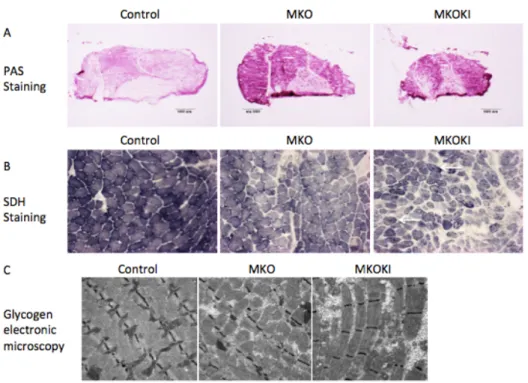
Here I describe the phenotype of MKOKI and compared it with MKO phenotype. I found that: 1) a mTOR kinase dead allele did not rescue any altered parameters associated to the loss of the mTOR protein, but in contrast exacerbates these alterations. 2) Muscle mTOR kinase activity was required for oxidative metabolism maintenance and dystrophin expression. 3) MKOKI muscles show a hyper-accumulation of glycogen that gave rise to myofibrillar disorganization, a feature not observed in MKO. 4) Glycogen hyper-accumulation is due to a stronger Akt hyperactivation that leads to myophosphorylase downregulation. Glycogen degradation inhibition gave rise to myofibrillar disorganization following glycogenosis. Sustained muscle Akt1 and Akt2 activation actually improved glucose uptake and storage as glycogen but also compromised its utilization further leading to severe Glycogen Storage
Disease. 5) Autophagy inhibition in mTOR MKOKI, in contrast to MKO further leaded to pronounced atrophy, dystrophy. 6) mTOR catalytic inhibitors might have severe mutiple effects in postmitotic tissues such as skeletal muscles, which are able to activate the feedback loop on Akt due to mTOR kinase inhibition.
RESUME
Cible de la rapamycine chez les mammifères (mTOR) est une protéine kinase importante, qui intègre et coordonne les informations de multiples voies de signalisation, sert de médiateur des facteurs de croissance, de la quantité de nutriment disponible et du statut énergétique. Il a au moins deux complexes de signalisation, fonctionnellement différents, appelés mTOR complexe 1 (mTORC1) et mTOR complexe 2 (mTORC2). mTOR régule de nombreux processus moléculaires et cellulaires tels que la transcription, la traduction, l’autophagie, et ainsi de suite. Les souris avec l’inactivation du gène mTOR sont mortels dans la période embryonnaire. Les souris qui est knock-out conditionnels de mTOR dans les muscles squelettiques subissent une myopathie progressive, conduisant à une mort prématurée, et présentent une atrophie musculaire, une altération de la fonction mitochondriale et à augmenter le glycogène.
Mes études sont pour caractériser le rôle de l'activité de la kinase mTOR dans l’intégrité musculaire et les fonctions musculaires, pour démontrer des fonctions de mTOR dans des muscles qui demandent ou demandent pas l’activité de la kinase, et donc qui ne sont pas distribuées par mTOR dans le corps. Dans ce but-là, on génère une ligne de souris nommée mTOR MKOKI qui est défectueux dans des muscles endogènes mTOR (mTOR MKO), tout en exprimant une allèle kinase-inactive, muscle-spécifique et drapeau-étiquetable (Asp2357Glu) de mTOR (FLAGmTOR MKI), imitant ainsi les effets dans le muscle d'un inhibiteur catalytique mTOR, puissant, biodisponible et sélective. Ici, je décris le phénotype des souris MKOKI et MKO. Je trouve que 1) un allèle mort de kinase mTOR ne sauve pas des paramètres modifiés associés à la perte de la protéine mTOR, mais au contraire exacerbe ces altérations. 2) l’activité de la kinase mTOR dans les muscles est nécessaire pour l’entretien du métabolisme oxydatif et l'expression de la dystrophine. 3) MKOKI muscles ont
hyper-accumulation de glycogène qui a provoqué la désintégration de myofibrilles et n'est pas observé sur le MKO. 4) l'hyper-accumulation de glycogène est à cause de plus de l'hyper-activation de l’Akt qui provoque une baisse de la phosphorylase musculaire. L’inhibition de la dégradation de glycogène augmente la désorganisation de la myofibrille suite à la glycogénèse. L’activation entretenue des muscles Akt1 et Akt2 effectivement améliore l'absorption et le stockage du glucose comme du glycogène mais également compromet son utilisation entraînant davantage à la maladie plus sévère du stockage du glycogen. 5) l’inhibition de l'autophagie des souris MKOKI davantage conduit à l'atrophie prononcée et la dystrophie par rapport à MKO. 6) mTOR inhibiteurs catalytiques ont peut-être de multiples effets graves dans les tissus postmitotiques comme les muscles squelettiques, qui sont capables d'activer la boucle de réaction sur l’Akt malgré l'inhibition de la kinase mTOR.
INDEX
1. INTRODUCTION ... 1
1.1. Overview of skeletal muscle ... 1
1.1.1. Structure and function of skeletal muscle ... 1
1.1.2. Skeletal muscle fiber type diversity ... 5
1.1.3. Formation of skeletal muscle ... 7
1.1.4. Regulation of skeletal muscle anabolism and mass ... 9
1.1.5. Central role of muscle protein in whole-‐ body metabolism ... 9
1.1.6. Role of muscle in chronic disease ... 10
1.1.7. Skeletal muscle and physical exercise ... 11
1.2. Catching a glimpse of mTOR ... 18
1.2.1. TOR kinase ... 18
1.2.2. mTOR complexes ... 20
1.2.3. Functions of mTOR in metabolic tissues ... 22
1.3. Downstream effects of TOR signaling ... 28
1.3.1. TOR and functions of TOR kinase ... 28
1.3.2. TORC1 mediates repression of autophagy ... 34
1.4. mTOR complex 2 signaling network ... 34
1.4.1. mTORC2 and its function ... 34
1.4.2. mTORC2 and autophagy regulation ... 36
1.5. Upstream regulation of TORC signaling ... 38
1.5.1. TSC complex ... 41
1.5.2. PI3K/Akt ... 42
1.5.3. Amino acids ... 43
1.6. mTOR signaling and muscle ... 45
1.6.1. mTOR and protein synthesis ... 45
1.6.2. Role of mTOR in hypertrophy and atrophy ... 48
1.6.3. mTOR and Ubiquitin/proteasome system ... 54
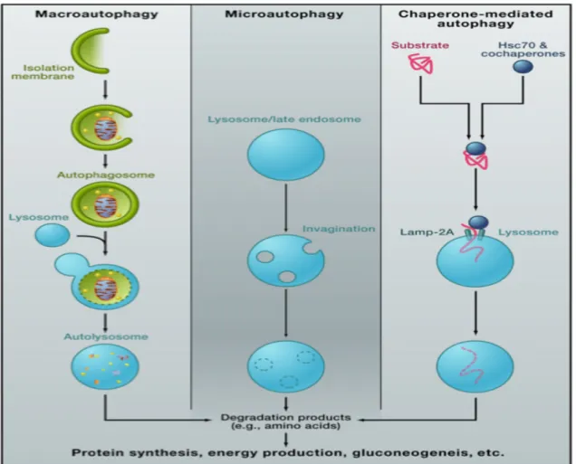
1.7. Autophagy ... 57
1.7.1. Process of macroautophagy ... 61
1.7.3. Autophagy as a regulator of metabolism ... 66
1.7.4. Autophagy in skeletal muscle homeostasis ... 68
1.7.5. Physical exercise and autophagy ... 75
1.7.6. Autophagy in disease ... 79
1.7.7. LC3, p62 and autophagy ... 81
1.7.8. AMPK regulates autophagy via FoxOs ... 90
1.7.9. Akt regulates autophagy via GSK3/NBR1 ... 92
2. EXPERIMENT PART ... 93
2.1. Introduction ... 93
2.2. Results and Discussion ... 98
2.2.1. Generation of mTOR MKOKI mice ... 98
2.2.2. Metabolism in mTOR MKOKI mice ... 100
2.2.3. Autophagy in mTOR MKOKI mice ... 116
2.2.4. Conclusion and perspective ... 118
3. SUPPLEMENTAL MATERIALS AND METHODS ... 122
3.1. Animals ... 122
3.2. Muscle histology, morphometric measurements, and imaging ... 123
3.3. Quantitative real-‐time PCR ... 127
3.4. Protein immunoblot ... 134
4. LIST OF ABBREVIATIONS ... 143
REFERENCES ... 145
1. INTRODUCTION
1.1. Overview of skeletal muscle
1.1.1. Structure and function of skeletal muscle
Skeletal muscle is a form of striated muscle tissue that is controlled by the somatic nervous system. It is responsible for voluntary movements, and also the automatic movements (e.g. standing, holding up head, and breath). Skeletal muscle, cardiac muscle and smooth muscle are the three major muscle types. Taken literally, most skeletal muscles are attached to bones via tendons, which are bundles of collagen fibers. Skeletal muscles function as the motors, brakes and also the shock absorbers. They can be used as heaters when shivering and also being an important store of protein.
Skeletal muscle is made up of large numbers of components known as myocytes, or "muscle cells", sometimes called "muscle fibers" or myofibers, which are long, cylindrical, multinucleated cells. They are formed from the fusion of developmental myoblasts (a type of embryonic progenitor cell) in myogenesis process.
They are arranged in bundles, separated by sheets of connective tissue containing collagen. Each muscle fibre is surrounded by a cell membrane, which allows the contents of the fibres to be quite different from that of the body fluids outside them. The myofibers are composed of myofibrils. The myofibrils are in
a sarcomere, which is the basic functional unit of the myofiber and forms the basic machinery necessary for muscle contraction. Just because of the sarcomere, the skeletal muscle has its striated appearance. The term muscle actually refers to numerous bundles of muscle fibers gathered by connective tissue.
The segment between two neighbouring Z-lines is defined as the sarcomere. With electron micrographs, we noticed the Z-line is a series of dark lines. Surrounding the Z-line, it is the light band region—I-band. After the I-band, it is the dark band—A-band. Then, there is a paler region called the H-band inside the A-band. The names of A- and I-band come from their properties of reflecting the polarized light under a polarization microscope. And within the H-band, it is the middle of the sarcomere, called M-line.
Actin filaments are the main component of the I-band and extend into the A- band. Myosin filaments extend throughout the A-band and overlap in the M-band. The titin (or known as connectin) protein binds to the thin filament system in the Z-line. It extends from the Z-line to the M-band, where it interacts with the thick filaments. In the Z-line and in the M- band of the sarcomere, multiple proteins critical for the stability of the sarcomeric structure are found. Actin filaments and titin protein are cross-linked in the Z-line by the Z-line protein α-actinin. The M-band myosin and the M- proteins build the thick filament bridge system to the M-band part of titin (the stretchy filaments) (Figure 1).
Figure 1. Schematic representation of skeletal muscle structure.
The muscle contraction occurs through an interaction of the actin with the myosin molecules (called crossbridges). Each of the crossbridges can produce about 5 × 10-12 Newtons force, meanwhile,it can pull the thin filament along past the thick filament by about 10 × 10-9 metres. Numerous of these tiny movements and tiny forces will shorten the myofibrils, and thus the whole muscle; therefore, some part of the skeleton is moved by the contraction of
attached muscles which are linked directly or via tendons to the bone.
As the body carries out a movement, events in the brain and the spinal cord produce action potentials in the axons of the motor neurons. Every axon branch sends action potentials to multiple muscle fibres. At the nerve terminals of each axon branch (neuromuscular junction) acetylcholine is released through the coming action potential, and this associates with receptors on the muscle fibre membrane, leading to generate an action potential. This action potential spreads over the whole surface of the fibre and is subsequently conducted into the interior via an extensive network of T-tubules.
Thereby, the permeability of the sarcoplasmic reticulum is changed by action potential, leading the sarcoplasmic reticulum of muscle to release ionic calcium, which then interacts with the regulatory protein troponin. Calcium-bound troponin changes its conformation, facilitating the movement of tropomyosin, thus exposing the myosin-binding sites on actin. This promotes myosin and actin ATP-dependent cross-bridge cycling and shortening of the muscles—the muscle contraction.
As soon as calcium is released from the sarcoplasmic reticulum, the calcium pumps in the membranes of the sarcoplasmic reticulum start to move the calcium back inside, therefore bringing to an end of a muscle twitch. More sustained periods of activity are the amount of movements we make; a series of action potentials are required to be sent to the muscle. The contractions generated in this way are stronger than a single twitch.
The muscle contraction is an ATP- dependent process. Mitochondria, which are located closed to Z- line, provided the energy. Muscle contraction needs energy to drive the crossbridges by means of their cyclic interactions with actin. The myosin molecule does work in moving the thin filament in each cycle. Energy is also utilized for the process of calcium pumping via the sarcoplasmic reticulum. When muscles are used to do external work, the energy consumption is highest. For example, the energy comsumption is high when the body weight has to be lifted in the movement of climbing stairs. However energy is also used when holding up a weight without doing work on it (e.g. isometric contraction). Less energy is need when muscles are used to lower weight, such as descending stairs.
1.1.2. Skeletal muscle fiber type diversity
In mammals, skeletal muscle constitutes about 40% of total body mass, meanwhile, it is responsible for about 30% the resting metabolic rate in human adult [1]. Skeletal muscle has a key role in regulating glycemic and metabolic homeostasis. It is the main (about 80%) site of glucose disposal under insulin- stimulated conditions [2]. Besides, skeletal muscle is the biggest glycogen storage organ. Moreover, exercise increases skeletal muscle glucose uptake by an insulin-independent pathway [3].
Skeletal muscle fibers are classified as slow- or fast-twitch according to the contractile property. This classification accords with histochemical staining for
highest ATPase activity. Type I, slow-twitch muscles are red in appearance. They are specialized for a more continuous activity and a fatigue-resistant phenotype. They mainly develop an oxidative metabolism, having high mitochondrial content and rich blood capillaries. Similarly, type IIa fibers are rich in mitochondria and capillaries, so that they also appear red. Although stain strongly for succinate dehydrogenase (SDH), type IIa fibers are also rich in glycolytic enzymes, thus they are classified as fast-twitch oxidative glycolytic fibers and form fast fatigue-resistant units. Type IIx fibers, also called IId, are characterized by a more glycolytic metabolism; in rat skeletal muscle, Type IIx fibers have strong staining for SDH. They has characteristic of a velocity of shortening intermediate between Type IIa and IIb. However, in humans their SDH staining is the weakest, because that they mostly rely on the glycolytic metabolism. Therefore, Type IIx fibers are the fastest and the most fatigable ones in human. In rodents, Type IIb fibers constitute the most glycolytic and fastest fibers (white, fast- twitch glycolytic fibers); in humans, MHC IIb is not expressed.
Immunohistochemical staining and protein electrophoretic separation are able to determine myosin heavy chain (MHC) isoform protein expression independently. This is another important criterion of fiber type classification. Each isoform has different contractile properties that parallel ATPase activity and twitch characteristics. Uniform fibers containing MHC1, 2A, and 2X presence, while mixture fibers containing 1-2A and 2A-2X isoforms have also been found. Muscle fiber type is genetically determined during development, but the
adaptive transformation of muscle fibers is controversial [4]. Under appropriate training stimulus, the plasticity of muscle allows changes in metabolic potential and morphology [5].
1.1.3. Formation of skeletal muscle
Skeletal muscle is formed in various of species from a pool of muscle precursor cells (myoblasts). Myoblasts proliferate, differentiate and then fuse together to form multinucleated muscle cells—myotubes. These myotubes become larger, innervated, and finally mature into myofibers.
In mammals (e.g. mouse), myofibers are formed through fusion of myoblast during the period of utero embryogenesis. There are big differences between skeletal muscles of the head, limbs, and body, with different sources of precursor cells and expression modes of regulatory genes [6]. The myofiber number is generally fixed by birth and subsequently is the postnatal growth and muscle maturation processes.
In mature mammals, there exist satellite cells on the surface of the mature myofiber (outside the sarcolemma and beneath the basement membrane), which are a small part of quiescent myoblasts. They are often referred to as myogenic stem cells. These satellite cells are activated when the myofiber homeostasis is disturbed, especially in the condition of myofibers necrosis, to generate new muscle to replace the damaged segment [7, 8].
Skeletal myogenesis is initiated in the embryo due to signalling molecules from surrounding tissues that specify the myogenic cell fate. Transgenic analysis of
DNA sequences that regulate Myf5 (myogenic determination factor 5) demonstrates the complexity of elements required to regulate the spatio-temporal expression of this gene. Myf5 and MyoD are essential for the acquisition of myogenic identity. The signalling pathways that influence the onset of myogenesis lead to activation of Myf5 and Myod either directly or indirectly [9].
The myogenic regulatory factors also function during the formation of adult muscle fibres. Myf5 (Myf5- nlacZ) is transcribed in most satellite cells [10], as these cells differentiate, MyoD accumulates. Without MyoD, muscle growth and regeneration are affected [11]. Pax7, the orthologue of Pax3, plays an important role in the formation of adult skeletal muscle [12]. This Pax gene is expressed in satellite cells. In addition, satellite cells are absent in Pax7 mutant mice.
Fetal myoblasts and embryonic satellite cells are maintained in an undifferentiated state through the TGF-β (Transforming growth factor) and BMP (Bone morphogenetic protein) family members. Moreover, FGF (fibroblast growth factor) and PDGF (platelet-derived growth factor) may regulate the proliferation and growth of satellite cells [13]. However, additional signals important for specifying embryonic muscle are still required to be identified, particularly during human development.
1.1.4. Regulation of skeletal muscle anabolism and mass
The mass and integrity of skeletal muscle is very important to the body health. Skeletal muscle plays major roles in whole-body disposal of glucose, amino acids and fatty acids, the accumulation of which can worsen the metabolic state [14-16]. Defects in skeletal metabolism and muscle growth will give rise to or aggravate many diseases, such as diabetes, cancer, and HIV, chronic kidney diseases, congestive heart failure, peripheral artery disease, rheumatoid arthritis [17-20]. In multiple diseases, muscle mass is a key predictor of disease prognosis, length of hospital stay, and treatment outcomes [16, 21].
There are many factors that regulate muscle mass and function. Physical activity and nutrition are the two key factors. The anabolic effects of resistance exercise on muscle protein synthesis and mass are well demonstrated [18]. Among macronutrients, proteins and branched-chain amino acids have remarkable effects in activating muscle protein anabolism [18, 22]. At the molecular level, the nutrient or energy sensor mTORC1 appears to mediate the anabolic effect of amino acids and resistance exercise [23, 24].
1.1.5. Central role of muscle protein in whole- body metabolism
Protein content maintenance of certain tissues and organs is fundamental for survival, such as the skin, brain, heart, and liver. These tissues and organs depend on a stable supply of amino acids through the blood to being as precursors for the protein synthesis to balance the protein breakdown rate in all
tissues in the postabsorptive condition. Moreover, muscle protein functions as the key reservoir to replace blood amino acid taken up by other tissues when there is no nutrient intake [25-27]. In the fasting condition, blood amino acids serve as precursors for both the protein synthesis and hepatic gluconeogenesis [28]. Therefore, the protein content of essential tissues and organs, as well as the concentration of necessary plasma glucose, can be maintained in a relatively homeostasis even that the nutritional intake is absent, provided muscle mass is sufficient to provide the necessary amino acids.
The breakdown of net muscle protein remarkably maintains plasma amino acid concentrations, providing sufficient muscle mass. For example, obese individuals who had increased muscle mass can maintain normal plasma amino acids concentrations after ≥60 days of fasting [29]. By comparison, depletion of muscle mass is detrimental for life. For example, there is firm correlation between the depletion of body muscle mass and the survival time of seriously ill patients with AIDS [30].
1.1.6. Role of muscle in chronic disease
Chronic diseases caused by unhealthy lifestyle account for more than two-thirds of deaths in the world. Studies focused on daily diet and physical activity and measure indexes to predict risk of disease, such as blood lipids, body mass index, and bone biomarkers. However, few studies have assessed muscle mass or physical or metabolic function to make clear the role of muscle. Nonetheless, changings in muscle has a crucial role in the most common conditions and
diseases. In the United Stats, heart disease and cancer are the major chronic diseases [31]. Both cancer and cardiac failure are often implicated with rapid and massive loss of muscle mass, strength, and metabolic function. Under these cachexia conditions, the muscle mass is a key determinant of survival [32, 33]. Sarcopenia is the degenerative loss of skeletal mass, quality, strength and function associated with aging. Sarcopenia is known as a widespread syndrome that has a destructive effect on the life quality and survival. Sarcopenia is a component of the frailty syndrome, and behaves impairment of the ability to perform activities of daily living.
1.1.7. Skeletal muscle and physical exercise
Physical activity is beneficial to human health. It can prevent many preventable death and premature death. About one in three adults and four in five adolescents around the world do not achieve the recommended quantity and quality of daily exercise. Regular exercise and physical activity are the basis in the prevention, management, and treatment of many diseases, including coronary heart disease, hypertension, obesity, type 2 diabetes mellitus (T2DM), and age-related muscle wasting (sarcopenia) [34, 35]. Actually, regular exercise combined with dietary intervention is more effective than pharmacological intervention in the prevention and treatment of T2DM [36] and sarcopenia [37]. 1.1.7.1. Exercise and skeletal muscle mass
denervation, endurance exercise, electrical stimulation), stimuli modifying applied load (resistance exercise, unloading, microgravity) and other environmental factors such as growth factors, nutrient availability, hypoxia, heat, and inflammation mediators. There are 3 potential approaches to maintain or improve muscle mass and function: exercise, and nutrition, hormonal therapy. Here the review focuses on the exercise.
Exercise can improve muscle function and increase muscle mass. Improved function may include both the muscle contractile properties and muscle metabolism. Exercise training improves the insulin sensitivity [38]. Progressive loss of strength [39] and muscle mass [40] happens throughout life, and the rate of loss is accelerated in the middle age and maintained until the old age [41]. Therefore, earlier intervention (e.g. in middle age or younger ages) is imperative to prevent and decrease the deleterious effects of sarcopenia in old age.
According to either increasing muscle endurance or increasing muscle strength, physical exercises modifying the muscle plasticity are generally classified into two categories. Muscle endurance, as the ability of muscle to sustain repeated relatively low intensity affairs for long periods of time, is also linked to the aerobic capacity.
On the other side, muscle strength, the ability of muscles to overcome a resistance, is built up by increasing muscle load rather than by repeating low intensity exercise. This is supported by muscle fiber hypertrophy. It mostly depends on the glycolytic metabolism, thus resistance training only slightly
improves the aerobic capacity and assists only marginally in the maintenance of cardiovascular health [42].
1.1.7.2. The Molecular Basis of Skeletal Muscle Adaptation to Exercise
Skeletal muscle displays remarkable plastic in functional adaptation and remodeling in response to contractile activity. Training-induced adaptations are demonstrated by changes in contractile protein and function [43], metabolic regulation [44], mitochondrial function [45], transcriptional responses [46], and intracellular signaling [47]. The molecular mechanisms that govern the adaptation to exercise training comprise a gradual alteration in protein content and related enzyme activities. These progressive changes show activation and/or repression of specific signaling pathways that regulate the process of transcription and translation, and exercise-responsive gene expression.
Transient postexercise changes in gene transcription include immediate early genes, myogenic regulators, carbohydrate (CHO) genes metabolism, lipid mobilization, mitochondrial metabolism and oxidative phosphorylation, transport and oxidation, and transcriptional regulators of gene expression and mitochondrial biogenesis [46, 48]. A single bout of exercise alters the DNA binding activity of many transcription factors, including HDACs [49], MEF2 [50], and NRFs [51]. Both protein stability and subcellular localization of transcriptional factor complexes within the nucleus and mitochondria are also influenced [52, 53].
Activated Protein Kinase Signaling. AMPK modulates cellular metabolism by the phosphorylation of metabolic enzymes [54] and by transcriptional regulation [55]. AMPK activation is regulated through a cellular energy deficit, which is revealed by increases in the AMP/ATP and Cr/PCr (creatine-phosphocreatine) ratios. Beside intense exercise, cellular stresses that deplete ATP or increase the cellular AMP/ATP ratio (such as glucose deprivation or oxidative stress) also facilitate the activation of AMPK [56]. Given the rate of ATP turnover during muscle contraction, by responding to an altered cellular energy status, AMPK plays the role as a signal transducer for metabolic adaptations. Acute exercise increases the phosphorylation of AMPK and enzymatic activity in an intensity-dependent manner [57], showing intensity-dependent effects of physical exercise on ATP turnover and adenine nucleotide concentrations [58]. AMPK activation acts to conserve ATP by suppressing biosynthetic and anabolic pathways, while stimulating catabolic pathways simultaneously to restore cellular energy [56]. In skeletal muscle, acute AMPK activation inhibits glycogen [54] and protein synthesis [59], but improves glucose transport [60] and lipid metabolism [61]. Chronic AMPK activation changes metabolic related gene expression and induces mitochondrial biogenesis [62], partly through AMPK-induced regulation of the DNA binding activity of transcription factors including HDACs, MEF2, and NRF-1[62].
1.1.7.3. Regulation of Skeletal Muscle Gene Expression and Adaptation
An increasing network of transcription factors and co-regulator proteins has emerged. Through integrating signals from physiological stimuli and
coordinating metabolic adaptation it regulates the skeletal muscle phenotype. This network applies molecular control on contractile, metabolic, and mitochondrial adaptation, clarified by an ability to change the expression of key enzymes in CHO and lipid metabolism, and also the coordination of myogenesis and mitochondrial biogenesis in answer to exercise. The complicated and highly regulated process of mitochondrial biogenesis requires the co-ordination and co-expression of both the nuclear genomes and the mitochondrial genomes for the assembly and expansion of the reticulum, and a dynamic mitochondrial network.
By using transgenic animals and pharmacological manipulation, mechanistic studies have demonstrated the roles of key regulators of skeletal muscle phenotype. Various regulators are sufficient to activate mitochondrial biogenesis, fiber-type transformation, and reprogramming of skeletal muscle metabolism, but many of them are not indispensable for exercise-induced skeletal muscle adaptation. For instance, PGC-1α acts as a transcriptional coactivator through recruitment and co-regulation of numerous transcription factors that regulate skeletal muscle gene NRF-1, NRF-2, ERRα, and Tfam expression [63]. PGC-1α activity is highly controlled by many posttranslational modifications: phosphorylation and deacetylation [55, 64]. Through acute exercise, key upstream kinases and deacetylases regulating these modifications are activated [51, 57, 64], corresponding with changes in protein stability, functional activity, and subcellular localization [52, 53]. Abnormal expression of PGC-1α in muscle
changing the PGC-1α activity induces molecular adaptations that enable the cell to meet the changing energy demands, augmenting cellular respiration rates and substrate utilization [66]. The phenotype of PGC-1α overexpression in rodent skeletal muscle is similar to aerobically trained muscle, characteristic of increased mitochondrial density, ATP synthesis, respiratory capacity, antioxidant defense in type II muscle fibers, and improved exercise performance [66]. Whereas, in muscle-specific PGC-1α KO mice, various of these adaptations are reversed, including that muscle fibers exhibit a more glycolytic phenotype, reduced exercise capacity, impaired mitochondrial respiratory function, and impaired recovery from exercise [67]. Nevertheless, after exercise training, myoglobin, metabolic gene expression, and mitochondrial biogenesis increase, which are similar to wild-type animals, whereas training-induced alterations in mitochondrial enzyme expression are only properly reduced. Remarkably, exercise-induced angiogenesis is reduced in PGC-1α KO mice [68]. Thus, despite strong effects on skeletal muscle phenotype, PGC-1α is not
essential for the majority of adaptive responses to exercise training.
Numerous proteins play roles in the regulation of metabolic gene expression and mitochondrial biogenesis in skeletal muscle. Exercise-induced modulation of these pathways is critical to skeletal muscle adaptation and modulation of the mitochondrial phenotype [69].
In addition, Dysfunctional mitochondria have been involved in sarcopenia [70] and insulin resistance [71], however, long-term aerobic exercise training may
prohibit the age-related declines in health that proceed as a function of mtDNA mutations, as found in mtDNA mutator mice [72]. Therefore, mitochondrial biogenesis and adaptation with regular exercise have implications for a broader range of health issues, not only benefit exercise performance.
1.1.7.4. Regulation of Skeletal Muscle Protein Synthesis by Contraction
The control of protein translation and synthesis produces protein during the exercise-induced hypertrophy, while the activation and incorporation of satellite cells add the newly formed myofibrils to the contractile machinery. The muscle hypertrophy induced by resistance exercise is strongly implicated with the p70S6K phosphorylation [73]. mTOR mediates nutrient and metabolic stimuli to regulate cell growth and proliferation. Contraction-induced p70S6K activation is dependent on mTOR. Activation of this pathway by contraction drives translational processes, and mediates muscle hypertrophy by protein accretion. Activation of mTOR is critical to load-induced muscle growth. This is demonstrated through the attenuation of hypertrophic responses and protein synthesis by rapamycin-the mTOR inhibitor [74]. The mTOR pathway controls mechanisms of protein synthesis at multiple levels including translation capacity, translation efficiency etc. through increases of specific mRNAs translation. mTORC1, which contains raptor and confers rapamycin sensitivity, is need for signaling to p70S6K and 4E-BP1. And meanwhile, mTORC2, which contains rictor and is rapamycin insensitive, is needed for signaling to Akt-FoxO [75]. The function of mTOR activity on downstream regulators of protein synthesis is
Nutrient-dependent regulation of muscle growth is accomplished through insulin- and Akt-dependent activation of the mTOR pathway. These pathways exhibit cooperativity to promote muscle growth in athletes and in disease states. They can be enhanced through adequate nutritional intake such as post exercise carbohydrate (CHO) and amino acid ingestion or increased dietary protein [77]. On the other side, both insulin and the systemic IGF-1 isoforms are involved in mTOR activation in active muscle. Then the activated mTOR upregulates protein synthesis [78]. Both insulin and IGF-1 activate PI3K, which generates PIP3 (phosphatidylinositol-3, 4, 5-triphosphate). Subsequently, PIP3 recruits Akt/PKB to the plasma membrane, where at least two distinct kinases, PDK1 and the TORC2 complex phosphorylate it. Of the three isoforms in mammals, exercise is related with the Akt1 (PKBα) activation, which is upregulated by IGF1 and by still unclear contraction signals. Insulin can activate Akt2. Akt/PKB upregulates protein synthesis through activating the TORC1 pathway and through inhibiting GSK3β (glycogen synthase kinase 3β). The GSK3β downregulates protein synthesis via eIF2B (eukaryotic initiation factor 2B) inhibition [75].
1.2. Catching a glimpse of mTOR 1.2.1. TOR kinase
TOR is a conserved serine/threonine protein kinase that belongs to the phosphoinositide 3-kinase (PI3K)-related kinase family. The discovery of TOR
kinases originate from the study of rapamycin (sirolimus as the trade name), a natural compound first isolated from the Streptomyces Hygroscopicus bacteria. These bacteria were found in Easter Island (called Rapa Nui in the native language) in the South Pacific Ocean. Rapamycin was originally developed as an anti-fungal agent. To date, it is used as an FDA (Food and Drug Administration) -approved immunosuppressant and chemotherapeutic agent. The potent anti-proliferative properties and specificity of rapamycin have also proven to be very useful characteristics for studying cell growth regulation. As early as the beginning of 1990s, genetic screens in budding yeast identified TOR1 and TOR2 as mediators of the toxic effects of rapamycin. Soon after, biochemical approaches in mammals succeeded in purification of mTOR and its discovery as the target of rapamycin[79-81].
In the cell, the dimer that formed by rapamycin interacts with FKBP12 (FK506-binding protein 12 kDa) binds directly to TOR, inhibiting TOR activity [82]. From Drosophila to mammals, being a single gene product, TOR is commonly referred to as dTOR (Drosophila TOR) and mTOR (mammalian TOR). TOR kinase has putative orthologs throughout eukaryotes and possesses a marked conservation of its core cellular functions despite limited sequence similarities.
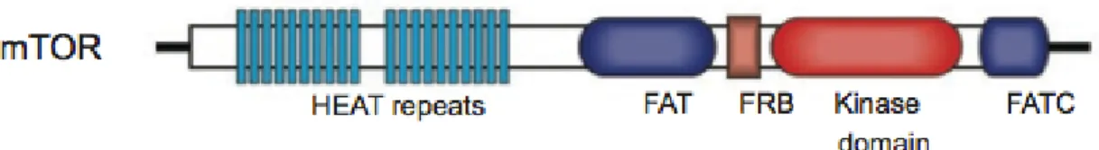
mTOR, with a predicted molecular weight of 289 kDa has a physiological important role of mTOR which is confirmed by the fact that the knockout of mTOR in mice is embryonic lethal [83-85]. mTOR contains 20 tandem HEAT
(a protein-protein interaction structure motif of two anti-parallel α-helices that found in Huntingtin, Elongation factor 3, PR65/A and TOR) repeats at the amino-terminal region, followed by a FAT (FRAP, ATM, and TRRAP) domain (Figure 2) [86]. The HEAT repeats functions to mediate protein-protein interactions. The C-terminal to the FAT domain is the FRB (FKBP12/rapamycin
binding) domain, which offers a docking site for the FKBP12/rapamycin
complex. FATC (FAT C-terminus) domain is at the C-terminus of the protein. The kinase domain is between the FRB and FATC domain. The mechanism that FAT and FATC domains modulate mTOR kinase activity is not clear yet.
Figure 2. The domain structure of mTOR.
HEAT: a protein-protein interaction structure motif of two tandem anti-parallel α-helices structures found in Huntingtin, Elongation factor 3, PR65/A and TOR; FAT: a domain structure shared by FRAP, ATM and TRRAP, all of which are from PIKK family; FRB: FKBP12/rapamycin binding domain; FATC: FAT C-terminus
1.2.2. mTOR complexes
mTOR interacts with multiple of proteins to form two distinct complexes, which are mTORC1 (mTOR complex 1) and mTORC2 (mTOR complex 2). The mTORC1 and mTORC2 complexes have different sensitivities to rapamycin as
well as upstream and downstream outputs (Figure 3, top panel) [87]. mTORC1 and mTORC2 share the catalytic mTOR subunit, mLST8 (mammalian lethal with sec13 protein 8, or G protein beta subunit-like, GβL) [88, 89], DEPTOR (DEP domain containing mTOR-interacting protein) [90], and also the Tti1/Tel2 complex [91]. By comparison, raptor (regulatory-associated protein of mTOR) [92, 93] and PRAS40 (proline-rich Akt substrate 40 kDa) [94-97] are specific to mTORC1, whereas rictor (rapamycin-insensitive companion of mTOR) [88, 98], mSin1 (mammalian stress-activated map kinase-interacting protein 1) [99, 100], and protor1/2 (protein observed with rictor 1 and 2) [96, 101, 102] are only components of mTORC2. The known molecular functions of the mTOR complex are described in Figure 3 (Figure 3, bottom panels) [87].
Figure 3. mTORC1 and mTORC2 Complexes. The mTOR kinase has two protein complexes termed mTORC1 and mTORC2 separately. mTORC1, acutely sensitive to rapamycin, responds to amino acids, growth factors, stress, oxygen, and energy. It facilitates cell growth through inducing and inhibiting anabolic and catabolic processes respectively, and drives cell-cycle progression. mTORC2 is insensitive to acute rapamycin treatment but its structure can be disrupted by chronic exposure to the drug. It responds to growth factors and regulates cell survival, metabolism and the cytoskeleton. The lower panel demonstrates the known functions of the protein components of the mTOR complexes and the bottom panel schematically depicts their interaction sites.
1.2.3. Functions of mTOR in metabolic tissues
mTOR signaling is found in all tissues, however, it is probably particularly significant in metabolic tissues. Metabolic organs including liver, muscle and adipose tissue are sensitive to nutrients, insulin/IGF-1, and energy, the three inputs that regulate mTOR. Liver, muscle and adipose tissue regulate the whole body glucose and lipid homeostasis.
1.2.3.1. mTOR in the liver
In the fasting condition, the liver produces glucose through glycogenolysis (glycogen breakdown) or through gluconeogenesis (glucose synthesis), to prevent hypoglycemia. In the feeding condition, the liver reduces blood glucose levels through various manners including glycolysis (blood glucose consumption), glycogenesis (conversion of glucose to glycogen) and lipogenesis (triglyceride). Mice with mTOR signaling defective in the liver are glucose intolerant, hyperglycemic, hyper- insulinemic and have decreased glycogen content [103-107], demonstrating that mTOR plays a major role in glucose
homeostasis in liver. The above defects are similar to those observed in type 2 diabetes patients, suggesting that defective hepatic mTOR signaling is related with the pathophysiology of type 2 diabetes.
Lipogenesis is activated through the transcription factor SREBP (sterol regulatory element-binding protein) [108-110]. As first demonstrated in mouse embryonic fibroblasts (MEFs) and retinal pigment epithelial cells, mTORC1 mediates SREBP-1 maturation in an S6K1-dependent manner to stimulate de novo synthesis of lipid [111, 112]. However, mTORC1 stimulates SREBP-1 expression in an S6K-independent manner in primary hepatocytes [113]. Above studies shows that mTORC1 activates SREBP expression and maturation, but through two separate effector pathways.
The S6K-independent pathway involves the phosphatidic acid phosphatase lipin-1, which is the mTORC1 substrate and functions as a negative regulator of SREBP-1 activity [114]. mTORC1 directly phosphorylates lipin-1 in response to nutrients and growth factors. This blocks the translocation of lipin-1 into the nucleus, hence allowing SREBP transcriptional activity. Studies demonstrate well that mTORC1 is essential to activate SREBP-1 and lipid synthesis in cultured cells, however, the mTORC1 function in lipogenesis in vivo is still less clear. Liver-specific mTORC1 deficient (raptor knockout) mice present decreased triglyceride content in liver and a reduction in plasma cholesterol levels only in the condition of a high fat diet [114]. Therefore, mTORC1
signaling is likely essential for the accumulation of hepatic triglyceride in vivo only in the pathological conditions.
Study shows that protection against the accumulation of hepatic lipid in LTsc1KO (liver-specific tsc1 knockout) mice is due to the Akt signaling attenuation, as restoration of Akt2 (the main hepatic isoform of Akt) restores the lipogenesis [107]. This demonstrates that mTORC1 and Akt are independently essential for lipogenesis. Decreased Akt signaling in LTsc1KO mice is due to the mTORC1-mediated negative feedback loop [107]. Akt is proposed to prevent expression of Insig2a encoding an SREBP inhibitor [107]. mTORC1 is essential in the activation of SREBP as shown above. Therefore, both Akt and mTORC1 are required for lipogenesis, and the molecular mechanism of selective hepatic insulin resistance still need to be further studied. However, there was data showed that mTORC1 is not necessary for the accumulation of hepatic lipid, because the treatment of rapamycin does not prevent high-fat diet or Pten deletion- induced hepatic steatosis [106].
mTORC2 is also insulin-stimulated and is important for lipid and glucose homeostasis in the liver. LiRiKO (Liver-specific rictor knockout) mice are hypolipidemic and hyperglycemic [103-105], showing total hepatic insulin resistance. This is due to the loss of phosphorylation on Akt Ser473 site [104]. LiRiKO mice, like the LTsc1KO mice, present reduced SREBP-1c activity. Akt signaling restoration suppressed the defects in SREBP-1c activity and de novo lipogenesis [104].
LiRiKO mice shows defects in SREBP-1c activity and hepatic lipogenesis, and mTORC2 but not mTORC1 is impaired. These mean that Akt regulates SREBP-1c at least partly independently of mTORC1. The regulation of Insig2a was not changed in the liver of LiRiKO mice, indicating that the phosphorylation of Akt Ser473 is not necessary for Insig2a inhibition. To sum up, mTORC1, mTORC2, and Akt are required for hepatic lipogenesis.
Hepatic mTORC2 regulates glucose homeostasis through the glycolysis activation and gluconeogenesis inhibition [104]. mTORC2 stimulates glycolysis by the activation of glucokinase and the transcription factor ChREBP. mTORC2 inhibits gluconeogenesis through inhibiting the FoxO1nuclear accumulation. The regulation of glucokinase and FoxO1 are through the Akt Ser473phosphorylation. These studies demonstrate that hepatic mTORC2 tightly regulates Akt to regulate glucose and lipid homeostasis and thus the whole body metabolism. A defect of mTORC2 signaling in the liver may lead to the development of diabetes.
1.2.3.2. mTOR in adipose: Regulation of adipogenesis and lipogenesis
mTOR signaling plays a essential role in adipogenesis [115]. Adipogenesis leads to the adipose tissue formation, the most important storage site of energy in mammals. In vitro, mTORC1 inhibition blocks adipogenesis and impairs the fat cells [116-118]. In contrast, mTORC1 overactivation promotes adipogenesis [119]. There are numerous downstream effectors implicated in the regulation of
transcription factors, regulates the commitment of embryonic stem cell to adipogenic progenitors [120]. The 4E-BPs regulate the terminal differentiation of adipocytes by the translational control of PPAR-g, the master regulator of adipogenesis [120, 121].
Mice with adipose-specific knockout of the mTORC1 are lean and have smaller and fewer adipocytes. The high-fat diet cannot induce obesity of these mice [118]. On the other side, mice with adipose-specific knockout of mTORC2 have normal fat mass but a defect in adipose tissue phosphorylation of Akt that leads to an increase in lipolysis and circulating free fatty acids (FFA) [122].
mTORC1 is greatly active in the tissues of obese and high-fat-fed rodents [123-125]. Elevated circulating insulin levels, nutrients (e.g. branch-chain amino acids and glucose), and proinflammatory cytokines, probably promote the activity of mTORC1 in obese animals. In addition to directly contributing to the expansion of adipose tissue by the activation of adipogenic/lipogenic factors in adipose tissue, mTORC1 promotes insulin resistance by the S6K1-mediated inhibition of insulin signaling [125]. The reduction of insulin action in adipose tissue probably exacerbates systemic insulin resistance through promoting the release of FFA via adipocytes, ectopic fat deposition, and lipotoxicity [126]. Studies show that the high rate of protein synthesis linked with mTORC1 activation probably induces insulin resistance through promoting ER (Endoplasmic reticulum) stress and the UPR (unfolded protein response) [127]. ER stress is prevalent in enlarged adipocytes. ER stress impairs insulin signaling
by the destabilization of IRS1 via JNK (c-Jun N-terminal kinase) [128]. Further studies are required to know what extend the activation of mTORC1 in the adipose tissue of obese individuals promotes the ER stress and insulin resistance.
Rictorad−/− (adipose-specific rictor knockout) mice present an increase in body size because of an increase in lean mass and largely unaffected fat mass [122, 129]. This can be explained through the study that mTORC2 in WAT negatively regulates IGF-1 and the production of insulin via the liver and pancreas, respectively, thus controlling systemic growth and glucose and lipid metabolism [129]. In the adipose tissue, a negative feedback endocrine loop may attribute to the mTORC2-mediated regulation of IGF-1 and insulin, considering that these hormones active mTORC2 itself. Further studies are needed to identify the possible factor(s) that signal from adipose to the liver and pancreas as part of such a loop. And this loop might maintain the hormone homeostasis.
1.2.3.3. mTOR in muscles
mTOR or raptor knockout mice have been generated to study the function of mTORC1 signaling in skeletal and cardiac muscle. Skeletal muscle-specific knockout mice present progressive muscle dystrophy, decreased oxidative capacity and increased glycogen content [130, 131]. Skeletal muscle of S6K1 deficient mice develops atrophic and accumulates glycogen, showing that mTORC1 regulates muscle mass and physiology via at least S6K1 [132, 133].
decreased, implying that mTORC1 may control mitochondrial oxidative capacity by a substrate other than S6K1 [133]. Mice with cardiac-specific mTOR or raptor knockout have dilated cardiomyopathy due to the loss of 4E-BP1 inhibition and subsequently reduced protein synthesis [134, 135].
Studies show that the increased glycogen accumulation in skeletal muscle-specific mTOR or raptor knockout mice is mediated through the Akt hyperactivation due to the loss of the negative feedback loop [130, 131]. Although Akt hyperactivation, muscle-specific raptor knockout mice present slightly glucose intolerant. This is beyond expectation because Akt activates glycolysis and glucose uptake. Reduction in PGC-1α leads to the decrease in mitochondrial oxidative capacity in the raptor knockout mice. The defect is suppressed through the restoration of PGC-1α expression [136].
Although with reduced Akt Ser473 phosphorylation, skeletal muscle-specific rictor knockout mice have no obvious phenotype [131, 137]. The slightly glucose intolerant of them may be due to loss of Akt-medited AS160 phosphorylation. AS160 (Akt substrate of 160 kDa) is a main Akt substrate essential for insulin-stimulated translocation of the glucose transporter GLUT4 (Glucose transporter type 4) to the plasma membrane [137].
1.3. Downstream effects of TOR signaling 1.3.1. TOR and functions of TOR kinase
Amino acids or growth factors can induce mTORC1, activated mTORC1 promotes diverse cellular processes, including ribosome biogenesis, stimulation of mRNA translation, and inhibition of apoptosis [138]. This leads to increased size and number of cells. Those outcomes are caused by the action of mTORC1 on its substrates, especially 4E-BP1 and ribosomal S6 kinase (S6K1) (Figure 4.) [138, 139]. Likewise, inhibition of TORC1 activity leads to a decrease in cell size and a reduction in proliferation. Other substrates of mTORC1 are involved in regulating the gene transcriptions required for the ribosome production.
mTORC1 is also been implicated in regulating miRNA (micro RNA). This class of small RNAs regulates gene expression through binding to the 3′ end of mRNA, thus inhibiting translation and (or) destabilizing the target mRNA [140]. Among these small RNA, miR-1 is a muscle-specific miRNA involved in myogenesis regulation. In myotubes, mTORC1 regulates the transcription of this miRNA, as rapamycin treatment reduces its abundance [141].
The best-characterized functions of mTORC1 in skeletal muscle are those mediated by the actions of the complex on 4E-BP1 (eukaryotic initiation factor 4E-binding protein) and S6K1 [81]. Nutrients and growth factors are the upstream signaling of TORC1. They are transported through TORC1 to the translation machinery by eIF4E (eukaryotic initiation factor 4E) and 4E-BP1. 4E-BP1 and S6K act upon the regulatory complexes bound to the untranslated regions of mRNAs [81]. eIF4E is inhibited by 4E-BP1. This inhibition is relieved by mTORC1 through phosphorylating 4E-BP1, thus promoting
formation of eIF4F complex and mRNA translation [142]. Under nutrient limitation conditions, 4E-BP negatively regulates the assembly of initiation factors through binding and isolating eIF4E, thereby the function of eIF4E to recruit the translation initiation complex to the 5’ mRNA cap structure is suppressed [139, 143].
TORC1 activation also promotes initiation of translation through phosphorylation of S6K [142]. S6K phosphorylates various substrates implicated in translation initiation, including the eIF4B regulatory subunit of RNA helicase (eIF4A), leading to the progression of the small ribosomal subunit towards the start codon (Figure 4) [139, 143].
S6K is also implicated in a negative-feedback loop that acts to decrease the activity of TORC1 (Figure 4); the phosphorylation and inactivation of IRS1 (insulin receptor substrate 1) by S6K results in a decrease in PI3K activity and thus reduces AKT activation and hence the TORC1 signaling [144, 145].
In TSC1−/− or TSC2−/− MEFs with hyperactivation of mTORC1 and S6K1, PI3K-Akt signaling cannot be affected by insulin [146-149]. However, this can be rescued through the prolonged treatment of rapamycin. Similarly, the Akt activity of drosophila TSC1−/− larvae is attenuated, which can be restored via the knockout of dS6K [150].
These studies suggest that active mTOR-S6K1 impinges on Akt signaling. IRS1 (insulin receptor substrate 1) connects insulin receptors with PI3K through direct binding [151, 152]. Interestingly, the expression of IRS1 was reduced in cells
with an elevated S6K1 activity [153]. Further studies indicated that active S6K1 phosphorylates IRS1 on many inhibitory sites and promotes its degradation [144, 145, 154]. Therefore, the activation of mTOR-S6K1 can attenuate Akt signaling [125]. This auto-regulatory pathway is recognized as S6K1-dependent negative feedback inhibition [155]. This may explain why TSC tumors are normally benign, because over-activation of Akt, such as in the condition of PTEN mutations, often gives rise to malignancy. With TSC mutations, mTOR and S6K1 activities are enhanced, leading to overgrowth of tissue; meanwhile, they restrain other pathways responsible for reduced apoptosis and increased proliferation through inhibiting Akt signaling. Besides the modulation on PI3K-Akt signaling, S6K1 also can directly phosphorylate mTOR on a C-terminal site [156]. It is not clear of the phosphorylation consequence. It may increase the intrinsic kinase activity of mTOR. This provides a mechanism for mTORC1 to regulate its function through S6K1.
Moreover, TORC1 has been demonstrated to regulate ribosome biogenesis and tRNA production. In a nutrient-sensitive manner, TORC1-dependent phosphorylation of the key RNA polymerase regulatory subunit TIFIA regulates the outcomes of RNA gene transcripts [157].
Figure 4. Downstream of mTORC signaling. (A) mTORC1 (Mammalian target of rapamycin complex 1) phosphorylation of eukaryotic initiation factor 4E (eIF4E)-binding protein (4E-BP) breaks the interaction of 4E-BP with eIF4E, which releases eIF4A to facilitate the binding of ribosomes to the transcriptional initial site. mTORC1 also activates ribosomal S6K, which activates the eukaryotic translation initiation factor 4B (eIF4B) and the S6 ribosomal protein through direct phosphorylation. mTOR can also function with general transcription factor III C (TFIIIC) and relieve its inhibitor Maf1, and thus lead to increased tRNA production. TORC1 activation also promotes association between the transcription initiation factor 1A (TIF-1A) and polymerase I (PolI), then promoting rRNA synthesis. At last, mTORC1 signaling can phosphorylate Unc-51-like kinase 1 (ULK1) and autophagy related 13 homolog (ATG13), consequently inhibit autophagy. (B) mTORC2 activates AKT and serum/glucocorticoid regulated kinase (SGK) by phosphorylation, and is involved in signaling via protein kinase C (PKC) and Rac/Rho. Overall, these mTORC2-mediated activities promote growth factor signaling and cytoskeletal reorganization. mTORC2 can also interact with ribosomes in response to insulin signaling. Abbreviation: ATG101, autophagy-related protein 101; FIP200, FAK family interacting protein of 200 kDa.
1.3.1.2. Kinase-independent function of mTOR in myofiber formation and growth
mTOR kinase activity is required for almost all mTOR functions. However, studies found that mTOR regulation of the initiation of C2C12 myoblast differentiation was kinase activity independent, and this is mediated through IGF-II [158, 159], which is implicated in both satellite cell proliferation and differentiation [160]. Ge et al. generated and characterized transgenic mice expressing two mTOR mutants with the control of HSA (human skeletal actin) promoter: rapamycin-resistant (RR) and RR/kinase-inactive (RR/KI). They demonstrated that the kinase-inactive RR/KI-mTOR appears myofiber formation with the treatment of rapamycin as in RR-mTOR. Moreover, the expression of Igf2 mRNA during regeneration is inhibited by rapamycin treatment and partially rescued through the expression of both RR- and RR/KI-mTOR transgene [161]. These data are consistent with a kinase-independent function of mTOR in the process of myofiber initial formation during regeneration.
The myofiber growth requires the kinase-active form of mTOR. This is supported by RR- mTOR, but not RR/KI-mTOR, in the treatment of rapamycin. These findings fully accord with studies in vitro, that rapamycin-sensitive mTOR signaling is essential for myotube maturation and hypertrophy [162, 163].
s6k1−/− mice displayed that growth of regenerating myofibers is impaired but
the regenerating fiber number is similar to that in WT mice. These studies demonstrate the requirement of S6K1 for muscle growth [132] and also validate in vivo for the S6K1-independent initiation of myoblast differentiation in vitro [158, 159]. These are consistent with the kinase-independent role of mTOR in
1.3.2. TORC1 mediates repression of autophagy
TORC1 is a potent and important inhibitor of autophagy. Autohagy is a very important lysosomal- dependent cellular degradation process that regulates and generates energy and nutrients to maintain essential cellular activities upon nutrient starvation condition. The TORC1 ability to regulate autophagy is highly conserved, as the process of autophagy itself. Accumulating research evidence suggests that ULK1 (Unc-51-like kinase 1), a serine/threonine-protein kinase involved in autophagy in response to starvation, the mammalian homolog of the yeast protein kinase autophagy-specific gene 1 (ATG1). ULK1 plays an important role in the regulation of autophagy initiation. ULK1 is directly phosphorylated by the energy-sensing kinase TORC1 [164-166]. It is known that TORC1-mediated phosphorylation of ULK1 inhibits its activation following the limitation of energy and leads to the whole decrease in autophagy [167]. AMPK can activate ULK1, but mTORC1 is capable of disrupting the interaction between ULK1 and AMPK by phosphorylation of ULK1 [167, 168].
1.4. mTOR complex 2 signaling network 1.4.1. mTORC2 and its function
Owing to the specific inhibition of TORC1 by rapamycin, much less is known about upstream and downstream signaling of mTORC2 compared to mTORC1. mTORC2 complex is insensitive to cellular energy or nutrients conditions. However, it can be regulated by insulin and growth factors through a not
well-understood mechanism requiring PI3K [169].
In addition, active mTORC2 assoicates with the ribosome, and insulin promotes mTORC2-ribosome binding in a PI3K- dependent mode, the recruitment of ribosome seems essential for mTORC2 signaling [170]. The TSC1/TSC2 complex is able to control mTORC2 in a large range of different cell types. The TSC1/TSC2 complex that downregulates mTORC1 is required for proper mTORC2 activation [171, 172]. This tumor suppressor complex physically interacts with mTORC2 via rictor subunit, but its regulation of mTORC2 is independent of its GTPase-activating protein activity toward Rheb, which is different from mTORC1 [171, 173]. Furthermore, the impaired mTORC2 activity in TSC2- deficient cells cannot be restored by downregulating mTORC1. This suggests that, in addition to the mTORC1-mediated feedback regulation of IRS-1, mTORC2 can be more directly regulated by TSC1/TSC2 complex.
Besides, phospholipase D (PLD) can activate mTORC2 [174]. Studies in vitro have demonstrated that IKK (inhibitor of nuclear factor κ-B kinase) regulates the activity of mTORC2 through physically affecting mTORC2 assembly. Inactivated IKK interacts with Rictor and competes with mTOR, therefore reducing mTORC2 activity [175].
Activated mTORC2 phosphorylates Akt, SGK1 (serum- and glucocorticoid-induced protein kinase 1) and PKCα (protein kinase C α). mTORC2 phosphorylates Akt at Ser473 site, and this phosphorylation is necessary for the full activation of Akt [176]. Phosphorylation at Ser473 site is a post-translational modification and proceeds upon Akt recruitment to the
membrane through affinity of its PH domain to membrane lipids. This means that mTORC2 and Akt would colocalize at the membrane. In fact, mTORC2 has been localized to membrane compartments [177, 178].
mTORC2 might thus active mTORC1 and, by interacting with the 60S ribosome subunit directly, promote protein synthesis [179]. Meanwhile, mTORC1 can negatively regulate the activation of mTORC2. S6K1 can phosphorylate mTORC2 component mSin1, thus dissociate it from mTORC2 [180]. SGK1 can be directly activated by mTORC2 to regulate ion transport and cell growth [181]. In addition, mTORC2 activates the PKCα, thus regulates cell shape through affecting the actin cytoskeleton [115]. Remarkably, recent studies have proved that mTORC2 together with mTORC1 participates in the regulation of insulin signaling through regulating IRS-1 degradation in cells [182].
1.4.2. mTORC2 and autophagy regulation
While mTORC1 is clearly associated in lysosomal signaling and autophagy, there is much less experimental evidence showing that mTORC2 is involved. However, mTORC2 can indirectly repress autophagy through Akt1. As well as repressing autophagy through Akt1/mTORC1 activation, mTORC2 also represses autophagy through the Akt1/forkhead box O3 (FoxO) 3A signaling pathway [183]. In skeletal muscle, Akt1 activation inhibits the transcription of autophagy genes, such as MAP1LC3A, GABARAPL1 and Bnip3 (BCL2/adenovirus E1B 19kDa interacting protein 3), which are normally induced by fasting [184]. This effect was independent of rapamycin, indicating
no mTORC1 involvement. However, inhibition of mTORC2 through Rictor knockdown allowed FoxO3A nuclear translocation and formation of autophagic vesicles, suggesting that mTORC2 activation was essential for autophagy suppression [184].
On the basis that involvement of Akt1 and mTORC1 as negative regulators of autophagy, stimulation with insulin-like growth factor (IGF)-1 would be expected to repress autophagy. However, beyond our expectation, knockdown of the IGF-1 receptor and impaired IGF-1 signaling, actually inhibit autophagy process [185]. This effect was due to the influence of mTORC2 on the actin cytoskeleton and endocytic pathways. Strategies of Rictor knockdown to block mTORC2 signaling reduced the phosphorylation of PKC-α/β and strongly disrupted the actin cytoskeleton. As a result, this reduced endocytosis and impaired the formation of the early autophagosome precursors [185]. This suggests that a controlled appropriate level of mTORC2 signaling is necessary for formation and trafficking of autophagosomes. Considering that mTORC2 seems to have a dual role as a negative and positive regulator of autophagy, caution is required when applying the dual mTORC1/2 kinase inhibitors, as long-term treatment will likely disturb normal autophagic processes.
Muscle- specific mTOR knockout mice display reduced postnatal growth, due to reduced size of fast muscle fibers and severe myopathy [130]. Muscle-specific loss of rictor (RImKO) mice has minimal change on muscle physiology [131, 137], and raptor/rictor muscle-specific double knockout (DmKO) possess similar pathological phenotypes as RAmKO mice. These indicate that mTOR
functions require only mTORC1 in adult skeletal muscle [131]. Nevertheless, one study shows that some functions of mTOR (e.g. the activation of terminal oligopyrimidine mRNA translation) could be independent of both mTORC1 and mTORC2 [186].
Whether mice specifically depleted for rictor in skeletal muscle (RImKO) would show changes in autophagy? Rencent study shows that there is no difference of the GFP-LC3 puncta number in RImKO mice muscles versus the control mice in basal and starved conditions [187]. Moreover, in 3- and 6-month-old RImKO mice, the levels of LC3I, LC3II, and p62 were similar to control mice [187]. These data demonstrate that autophagy in skeletal muscle is not regulated via mTORC2.
1.5. Upstream regulation of TORC signaling
As described before, two TOR complexes have been identified in yeast [188-190]. However, because of the availability and wide use of rapamycin, the potent and specific mTORC1 inhibitor, most of our current knowledge of mTOR is concentrated on mTORC1.
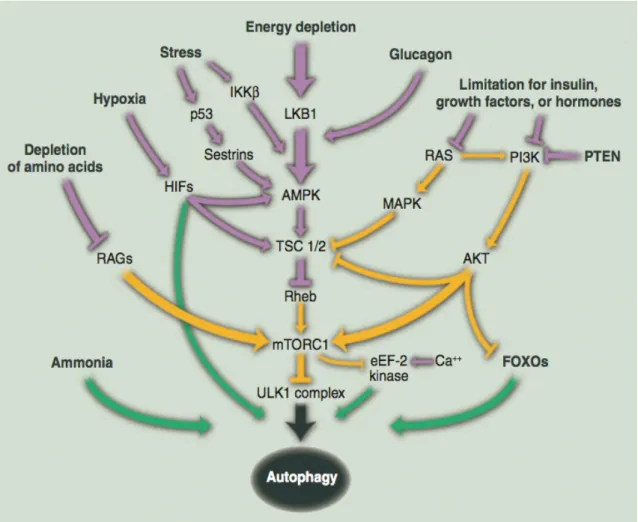
mTORC1 is the better characterized of the two mTOR complexes—mTORC1 and mTORC2. An obvious characteristic of this branch of the pathway is the diversity and number of upstream signals it senses. The mTORC1 pathway integrates inputs from numerous intracellular and extracellular cues, including growth factors, amino acids, stress, energy status and oxygen, to control various major processes such as protein and lipid synthesis and autophagy (Figure 5).