UNIVERSITE LILLE 2 DROIT ET SANTE
FACULTE DE MEDECINE HENRI WAREMBOURG
Année : 2020
T H È S E P O U R L E D I P L O M E D ' É T A T D E D O C T E U R E N M É D E C I N E
Échographie avant un accouchement instrumental : peut-on prédire la
difficulté ?
Présentée et soutenue publiquement le vendredi 19 juin 2020 à 16 heures
au Pôle Recherche
Par Alix PLURIEN
_______________
JURY
Président :
Monsieur le Professeur Damien Subtil
Assesseurs :
Monsieur le Docteur Paul Berveiller
Madame le Docteur Sandy Hanssens
Directeur de Thèse :
Monsieur le Professeur Charles Garabedian
_______________Avertissement
La Faculté n'entend donner aucune approbation aux opinions émises dans les thèses : celles-ci sont propres à leurs auteurs.
L
ISTE DES ABBREVIATIONS
AoP: Angle of progression AUC: area under the curve BMI: body mass index CI: Confidence interval DE: digital examination FHR: fetal heart rate
HPD: head perineum distance
ISUOG: International Society of Ultrasound in Obstetrics and Gynecology LOA: Left occiput anterior
LOP: left occiput posterior LOT: left occiput transverse NA: not applicable
NPV: Negative predictive value OA: occiput anterior
OASIS: obstetric anal sphincter injuries OP: occiput posterior
OR: Odd ratio
OVD : Operative vaginal delivery PPH: post-partum hemorrhage ROA: right occiput anterior ROP: right occiput posterior ROT: right occiput transverse Se: sensibility
Spe: specificity US: ultrasound
Table des matières
Liste des abbreviations ... 4
Résumé ... 6 Introduction ... 8 Article original ... 10 Discussion ... 30 Conclusion... 35 Références bibliographiques ... 36 Annexes ... 40
Annexe 1: Table S1 - Comparison of neonatal characteristics between the two groups. ... 40
Annexe 2: Figure S1: Concordance between digital examination and ultrasound for the presentation ... 41
R
ESUME
Objectif - Déterminer si la variété fœtale, évaluée par échographie, et la distance
tête périnée (HPD), avec ou sans appui sur les tissus mous, est plus prédictive d'un accouchement instrumental (OVD) difficile que le toucher vaginal.
Méthodes - Une étude de cohorte prospective et monocentrique incluant toutes
les OVD de singleton ≥ 34 semaines d’aménorrhée. Les principaux critères d'une OVD difficile étaient basés sur un critère composite : OVD considérée difficile par l’obstétricien, et/ou deux lâchages de ventouse en cas d’utilisation de la ventouse, et/ou un changement d'instrument, et/ou une césarienne pour échec d’OVD.
Résultats - Deux cent quatre-vingt-six OVD ont été incluses, dont 65 (22,7%)
étaient difficiles. L'aire sous la courbe (AUC) pour la prédiction des OVD difficiles par la variété à partir du toucher vaginal ou de l’échographie était de 0,62 (IC à 95% : 0,54-0,70) et 0,66 (IC à 95% : 0,58-0,73), respectivement. Par ailleurs, les AUC de l’HPD sans et avec appui étaient respectivement de 0,59 (IC à 95 % : 0,51-0,66) et 0,60 (IC à 95 % : 0,51-0,68). L’AUC pour l’engagement fœtal par le toucher vaginal était de 0,57 (95 % IC : 0,50-0,63). Les facteurs associés à une OVD difficile étaient les variétés postérieures et transverses (OR : 2,931, IC 95% : 1,640-5,239 ; p = 0,0003). Le seuil de la mesure de l’HPD sans appui était de 37 mm (OR : 2,327, IC 95% : 1,247-4,245 ; p = 0,0080), et avec appui de 17 mm (OR : 2,594, IC 95% : 1,230-5,429 ; p = 0,0114).
Conclusion - Bien que la capacité de prédire les OVD difficiles soit faible avec les
deux méthodes, nos conclusions soutiennent l'idée que l'échographie peut être un complément utile au toucher vaginal. Les résultats indiquent que le choix de la technique de mesure du HPD reste à l’appréciation de l’obstétricien.
Mots clés : échographie ; accouchement instrumental ; distance tête périnée ;
engagement fœtal.
I
NTRODUCTION
L’appréciation de la variété et de l’engagement fœtal pendant le travail par le toucher vaginal reste le gold standard (1–5). De nombreuses études ont montré que le toucher vaginal est subjectif, avec une variabilité intra- et inter-observateurs importante (5–9). Le taux d'erreur pour le diagnostic de la variété fœtale est compris entre 20 et 70 % (5,7,9). Une erreur sur le diagnostic de la variété fœtale peut entraîner un placement inapproprié de la ventouse ou des forceps, ce qui augmente le risque de lésion fœtale et le taux d'échec d’un accouchement instrumental (10–12). La supériorité de l'échographie seule, ou en combinaison avec le toucher vaginal, pour déterminer la variété fœtale a été démontrée par rapport au toucher vaginal seul (2,4,13–16). Dans un article récent, l'ISUOG (International Society of Ultrasound in Obstetrics and Gynecology) recommande d'évaluer systématiquement la variété fœtale par échographie avant un accouchement instrumental (OVD) (7).
Lorsqu'une OVD est nécessaire, l'évaluation de l’engagement de tête fœtale dans le bassin est primordiale. Le toucher vaginal est également subjectif ; Dupuis et al. ont rapporté un taux d'erreur de 30 à 34%, particulièrement en présence d'une bosse séro-sanguine (5). En effet, Vayssiere et al. ont montré que le niveau d’engagement fœtal dans le bassin est un facteur prédictif de succès d’un accouchement vaginal spontané (17). Plusieurs études ont évalué l'utilité de l'échographie pour mesurer le niveau d'engagement fœtal et la probabilité d'un accouchement par voie vaginale (1,7,14,15,18–22). Différentes méthodes ont été proposées : l'angle de progression (AoP), également appelé "angle de descente" (23,24), la distance de progression (25), la distance tête-périnée (HPD) (26), la distance tête-symphyse (27), et la direction de la tête (28).
Cependant, l'évaluation de la difficulté des OVD est également intéressante. En effet, Kasbaoui et al. ont montré qu'une HPD ≥40 mm est associée de manière significative à une OVD difficile selon le critère composite, après ajustement pour la parité, le type de variété et la macrosomie fœtale (Odd ratio, OR: 2,38; intervalle de confiance à 95%, IC: 1,51-3,74 ; p = 0,0002) (20). L’HPD était également un facteur prédictif d’une OVD difficile plus puissant que le toucher vaginal (p = 0,036). Dans leur étude, la sonde abdominale a été appliquée horizontalement sur le périnée, sans appuyer sur les tissus. Néanmoins, dans leurs recommandations, l’ISUOG (ainsi que d'autres études évaluant l’HPD) ont proposé de réaliser cette mesure en comprimant les tissus mous jusqu’au contact du bassin sans entraîner de gêne à la patiente (7,18,29,30).
Par conséquent, l'objectif de cette étude était d'évaluer si la variété fœtale évaluée par échographie et la mesure de l’HPD, avec ou sans appui sur les tissus mous, est plus prédictive d'une OVD difficile que le toucher vaginal.
A
RTICLE ORIGINAL
Ultrasound before operative delivery: Can it predict difficulty?
A. Pluriena, P . Berveillerb,c, E. Drumezd,e, S. Hanssensa, D. Subtila,e , C.
Garabediana,e
Affiliations:
a CHU Lille, Department of obstetrics, F-59000 Lille, France
b Department of Gynecology and Obstetrics, Poissy-Saint Germain Hospital, Poissy,
France
c INRAE, Paris Saclay University, UMR 1198-BREED, RHuMAMontigny-
Le-Bretonneux, France
d CHU Lille, Department of Biostatistics, F-59000 Lille, France
e Univ. Lille, CHU Lille, ULR 2694 - METRICS : Évaluation des technologies de santé
et des pratiques médicales, F-59000 Lille, France
Corresponding author: Alix Plurien
Maternité Jeanne de Flandre Avenue Eugène Avinée 59000 Lille
+33 320446626
ABSTRACT
Objective – To evaluate whether fetal presentation, assessed by ultrasound and
head perineum distance (HPD), with or without soft tissue compression, is more highly predictive of a difficult operative vaginal delivery (OVD) than digital examination.
Methods – A prospective, monocentric cohort study including all singleton OVD
at ≥34 weeks gestation. The principal criteria for a difficult OVD were based on a composite criterion of: the birth attendant considered the OVD difficult, and/or two vacuum device detachments if a vacuum was used, and/or change of instrument, and/or a cesarean delivery for OVD failure.
Results – Two hundred eighty-six OVDs were included, among which 65 (22.7%)
were difficult. The area under the curve (AUC) for predicting difficult OVD according to fetal presentation from digital examination or ultrasound was 0.62 (95% CI: 0.54
–
0.70) and 0.66 (95% CI: 0.58–
0.73), respectively. Moreover, the AUCs of HPD without and with pressure were 0.59 (95% CI: 0.51–
0.66) and 0.60 (95% CI: 0.51–
0.68), respectively. The AUC for digital examination of the fetal station was 0.57 (95% CI: 0.50–
0.63). Factors associated with difficult OVD were posterior and transverse presentations (OR: 2.931, 95% CI: 1.640–
5.239; p = 0.0003), HPD without a pressure threshold of 37 mm (OR: 2.327, 95% CI: 1.247–
4.245; p = 0.0080), and HPD with a pressure threshold of 17 mm (OR: 2.594, 95% CI: 1.230–
5.429; p = 0.0114).Conclusion – Although the ability to predict difficult OVD was low with both
methods, our findings support the notion that ultrasound may be a useful supplement to digital examination. The results indicate that choice of HPD measurement technique remains up to the birth attendant.
Keywords: ultrasound; operative vaginal delivery; head perineum distance; head
INTRODUCTION
Assessing the presentation and station of the fetal head during labor by digital examination remains the gold standard (1–5). Many studies have shown that digital examination is subjective, with both intra- and interobserver variability (5–9). The error rate in evaluating presentation has been reported as ranging from 20
to
70% (5,7,9). An error in evaluating head presentation may result in inappropriate vacuum or forceps placement, increasing the potential for fetal injury and the failure rate of the procedure (10–12). The superiority of ultrasound alone, or in combination with a digital examination, for determining fetal head rotation has been demonstrated compared with digital examination alone (2,4,13–16). ISUOG guidelines recommend systematically evaluating fetal head presentation by ultrasound before operative vaginal delivery (OVD) (7).When OVD is required, assessing the fetal head station in the pelvis is mandatory. Digital examination is also subjective; Dupuis et al. reported an error rate from 30
to
34%, especially in the presence of a caput succedaneum (5). Indeed, Vayssiere et al. showed that the level of fetal station in the pelvis is a predictor of successful vaginal delivery (17). Several studies have assessed the utility of ultrasonography for measuring the degree of fetal head engagement and the probability of vaginal delivery (1,7,14,15,18–22). Different methods have been proposed: angle of progression (AoP), also called the “angle of descent” (23,24), progression distance (25), head perineum distance (HPD) (26), head–symphysis distance (27), and head direction (28).Many studies have evaluated predictions of delivery mode; however, assessing OVD difficulty is also of interest. Indeed, Kasbaoui et al. showed that an HPD ≥ 40 mm is significantly associated with a difficult OVD based on the composite criterion, after
adjustment for parity, presentation type, and fetal macrosomia (odds ratio, OR: 2.38; 95% confidence interval, CI: 1.51
–
3.74; p = 0.0002) (20). HPD was also a more accurate predictor of difficult OVD than digital examination (p = 0.036). In their study, the abdominal probe was applied horizontally to the perineal body without pressing on tissues. Nevertheless, the ISUOG guidelines and other studies evaluating HPD have proposed measuring HPD by compressing soft tissue completely against the pubic bone without causing patient discomfort (7,18,29,30).Therefore, the aim of this study was to evaluate whether fetal presentation assessed by ultrasound and HPD measurement, with or without soft tissue compression, is more highly predictive of a difficult OVD than digital examination.
MATERIAL AND METHODS
Study and eligibility criteria
This prospective, monocentric cohort study was conducted from January 2019 to July 2019 in Lille, France. We included all OVD in singleton pregnancies of ≥34 weeks’ gestation with cephalic presentation. Noncephalic presentations, multiple pregnancies, singleton pregnancies <34 weeks’ gestation, spontaneous deliveries, cesarean deliveries during labor, and patients lacking one measurement were excluded.
Operative vaginal delivery
Before beginning the study, all residents and obstetricians received two months of theoretical and practical training on performing ultrasound-based HPD and presentation assessments. Before each OVD, the birth attendant performed a digital examination to determine fetal head presentation by palpating the sagittal suture and the anterior and posterior fontanels. Fetal head station was assessed based on the relationship between the most distal cranial point and the level of ischial spines. The presence of caput succedaneum was also recorded. Obstetricians and residents were asked to perform a presentation and fetal head station ultrasound systematically before each OVD. The choice of appropriate delivery mode (vacuum, forceps, or cesarean section) was selected by the birth attendant, based on the digital examination with respect to national guidelines (31).
Measurement method
Ultrasonography was carried out via a portable machine (Samsung HM70A). The fetal presentation was determined by an abdominal approach to determine the head and spine positions (7). Then, the abdominal transducer was covered with a sterile glove and positioned horizontally on the perineum, between the labia majora in
Statistical analysis
Categorical variables are expressed as number (percentage). Quantitative variables are expressed as mean (standard deviation) for normally distributed data, or otherwise as median (interquartile range, IQR). Distribution normality was assessed using histograms and the Shapiro–Wilk test. Comparisons between the two study groups, defined by OVD difficulty, were made using the chi-square test or Fisher exact probability test for categorical variables, and using Student’s t test or Mann–Whitney
U test for quantitative variables. Diagnostic performance of digital examination and
HPD was evaluated by calculating the area under the receiver operating characteristic (ROC) curve and compared using the nonparametric approach proposed by DeLong. Optimal threshold values were calculated for HPD, with and without pressure, based on the ROC curves, by maximizing the Youden index. Diagnostic and 95% CI values of the observed optimal and published thresholds were evaluated by calculating sensitivity, specificity, positive and negative predictive values, and positive and negative likelihood ratios. Finally, factors predicting difficult OVD were evaluated using univariate logistic regression models; OR and 95% CI values were calculated as effect sizes. Statistical testing was performed with a two-tailed a level of 0.05. Data were analyzed using the SAS software package, version 9.4 (SAS Institute, Cary, NC).
Ethics
The study was approved by the national committee of research in gynecology and obstetrics (CEROG #2020-OBST-0301, 01/05/2020). Each patient provided informed consent before ultrasound measurements.
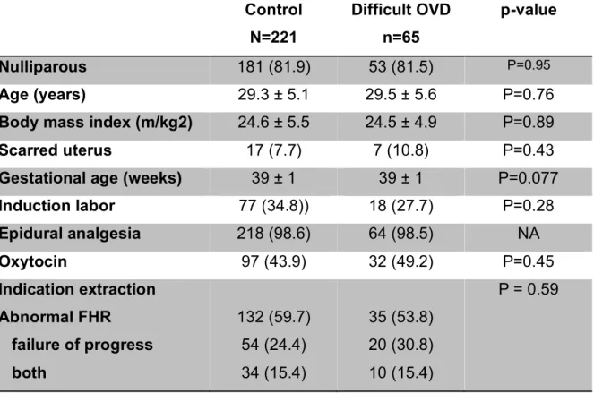
The groups’ demographic and clinical characteristics are presented in Table 1. Maternal characteristics were similar between the two groups. In difficult OVD cases, posterior and transverse presentations were more frequently observed than anterior presentation (determined by ultrasound) (30 [46.2%] vs 50 [22.6%], p < 0.001) and duration was longer in the difficult OVD group (13.8 ± 6.3 vs 7 min ± 3.3, p < 0.001). Postpartum hemorrhage was also observed more frequently in the difficult OVD group (13 [20%] vs 11 [5%], p < 0.001). Vacuum devices were used less often in the difficult OVD group (10 [15,4%] vs 108 [48.9%], p < 0.001). Neonatal characteristics are also presented for the two groups (Annexe 1, Table S1). In the difficult OVD group, neonatal weight was significantly higher (3500 g ± 425 vs 3349 g ± 450, p = 0.014) and the rate of arterial umbilical cord pH < 7.10 was significantly higher (31.8% vs 14.0%, p < 0.001).
Table 1: Comparison of demographic and clinical characteristics between the two
groups Control N=221 Difficult OVD n=65 p-value Nulliparous 181 (81.9) 53 (81.5) P=0.95 Age (years) 29.3 ± 5.1 29.5 ± 5.6 P=0.76
Body mass index (m/kg2) 24.6 ± 5.5 24.5 ± 4.9 P=0.89
Scarred uterus 17 (7.7) 7 (10.8) P=0.43
Gestational age (weeks) 39 ± 1 39 ± 1 P=0.077
Induction labor 77 (34.8)) 18 (27.7) P=0.28 Epidural analgesia 218 (98.6) 64 (98.5) NA Oxytocin 97 (43.9) 32 (49.2) P=0.45 Indication extraction Abnormal FHR failure of progress both 132 (59.7) 54 (24.4) 34 (15.4) 35 (53.8) 20 (30.8) 10 (15.4) P = 0.59
Posterior and transverse presentations (US) 50 (22.6) 30 (46.2) P<0.001 HPD without pressure mean (mm) 40.1 ± 12.2 43.9 ± 13.0 P=0.024
HPD with pressure mean (mm) 21.6 ± 8.8 25.0 ± 10.6 P=0.018 Caput succedaneum 139 (62.9) 33 (50.8) P=0.079 Instruments Forceps Vacuum Both 113 (51.1) 108 (48.9) 0 23 (35,4) 10 (15,4) 32 (49,2) P=0.072 P<0.001 NA
Extraction duration (min) 7 ± 3.3 13.8 ± 6.3 P<0.001
OASIS III or IV 22 (10) 9 (13.8) P=0.38
Bleeding (mL) 201.5 ± 179.5 307.1 ± 299.4 P=0.028
PPH > 500 mL 11 (5) 13 (20) P<0.001
Results presented as number (percentage) or mean+/-standard deviation.
The concordance between digital examination and ultrasound was 66.8%. Figure S1 presents the concordance according to each head presentation type (Annexe 2). The area under the curve (AUC) for predicting difficult OVD according to fetal presentation obtained by digital examination or ultrasound was 0.62 (95% CI: 0.54
–
0.70) and 0.66 (95% CI: 0.58–
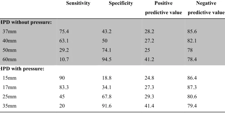
0.73), respectively (Table 2 and Figure 3). There was no significant difference between the two AUC (p = 0.24).From bivariate analyses, the factors associated with a difficult OVD are presented in Table 4. Three criteria were significant: posterior and transverse presentations (OR: 2.931, 95% CI: 1.640
–
5.239; p = 0.0003), HPD without pressure of 37 mm (OR: 2.327, 95% CI: 1.247–
4.245; p = 0.0080), and HPD with pressure of 17 mm (OR: 2.594, 95% CI: 1.230–
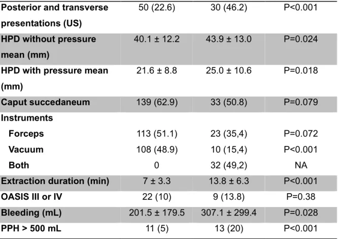
5.429; p = 0.0114).Table 3: Diagnostic values of different thresholds of head perineum distance to
predict difficulty of OVD
Sensitivity Specificity Positive predictive value Negative predictive value HPD without pressure: 37mm 40mm 50mm 60mm 75.4 63.1 29.2 10.7 43.2 50 74.1 94.5 28.2 27.2 25 41.2 85.6 82.1 78 78.4 HPD with pressure: 15mm 17mm 25mm 35mm 90 83.3 45 20 18.8 34.1 67.8 91.6 24.8 27.3 29.3 41.4 86.4 87.3 80.6 79.4
Table 4: Factor predictive of the difficulty of operative vaginal delivery in bivariate
analyses.
Criteria Odd ratio 95% CI P value Posterior and transverse
presentations (US) 2.931 1.640-5.239 0.0003 HPD without pressure 37mm 2.327 1.247-4.345 0.0080 HPD with pressure 17mm 2.594 1.239-5.429 0.0114 Nulliparous 0.976 0.478-1.993 0.9467 Caput succedaneum 1.644 0.941-2.871 0.0806
COMMENTS Main findings
Two HPD assessment methods are described in the literature: with and without soft tissue pressure (7,20). To our knowledge, this is the first study to compare these methods for the prediction of OVD difficulty. Fetal presentation assessed by ultrasound had a higher AUC for predicting OVD difficulty. There was no difference between HPD with or without pressure, for which the optimal thresholds were 37 mm and 17 mm, respectively. As such, the choice of HPD measurement technique remains up to the birth attendant. Overall prediction, whether by digital examination or ultrasound, was low.
Interpretation of results
When OVD is required, it is essential to know the fetal head presentation and station in the pelvis. Clinical diagnosis remains the gold standard but is subjective (1,2,4,10) and remains an important source of error, with presentation diagnosis varying from 20
to
70% (7,9,16). This error risk is higher after one or more hours of active pushing (2,4,26,32). However, these notions of fetal head station and presentation are essential before performing an OVD. Indeed, OVD is associated with a higher risk of maternal and neonatal complications, especially when a second instrument is needed or a cesarean for failed vaginal delivery is necessary (33,34). For the past 15 years, the use of ultrasound has been proposed for predicting the mode of delivery and complications during OVD.One way of improving labor ward practices has been the use of ultrasound to ensure presentation before OVD. Indeed, posterior and transverse presentations are associated with higher rates of perineal lesions and difficult OVD. In our study, we found more posterior and transverse presentations in the difficult OVD group (46.2%
vs 22.6%, p < 0.001). Missed posterior or transverse presentation diagnoses are higher than in anterior presentation (20,34). Akmal et al. found an error rate of 17% for anterior presentation and 46% for posterior and transverse presentations (p = 0.002) (14). In our study, the concordance between digital examination and ultrasound was 66.8%, with a lower concordance in posterior (45.6%) and transverse (left occiput transverse 61.1% and right occiput posterior 37.5%) presentations. In addition, we found that the presentation was more predictive of difficult OVD than was the station, using both ultrasound and digital examinations. Kasbaoui et al. used multivariate analysis to predict the occurrence of difficult OVD with different HPD (without pressure) thresholds (i.e., 40, 50, or 60 mm) to show that posterior or transverse presentation is an independent factor in difficult OVD (20). Finally, the ISUOG recommends the assessment of head presentation by transabdominal ultrasound before OVD (7).
The correct diagnosis of the fetal head station is a prerequisite before instrumental delivery (15,19,20,33,35). Several studies have assessed the utility of different ultrasonography methods for measuring the degree of fetal head station and the probability of vaginal delivery (1,7,14,15,18–22). Kahrs et al. showed that HPD and AoP were significantly associated with spontaneous delivery (AUC = 0.83 [95% CI: 0.77
–
0.89] and 0.75 [95% CI: 0.66–
0.85], respectively). HPD (with firm pressure) £ 20 mm is associated with a high probability of spontaneous vaginal delivery, whereas HPD > 35 mm was associated with cesarean delivery (AUC = 0.83 [95% CI: 0.74–
0.92]) (1). Similarly, Dall’Asta et al. showed that HPD (with pressure, threshold of 15 mm) is also predictive of spontaneous delivery (AUC = 0.74 [95% CI: 0.65–
0.83]) (32). To date, only one study has evaluated the prediction of a difficult OVD, in a prospective design that included 659 patients; using a composite criterion, this group found an association between the occurrence of a difficult OVD and HPD (without pressure), afteradjustment for parity, presentation type, and fetal macrosomia. With a threshold of 40 mm, the AUC was 0.63 (95% CI: 0.58
–
0.69; p < 0.01) with a sensitivity of 73.3% and specificity of 47.6% (20). Our results are consistent with these. Indeed, the optimal HPD threshold without pressure for predicting difficult OVD was 37 mm. In our sample, we used the novel approach of comparing HPD with or without pressure. These AUC for predicting difficult OVD were similar (0.59 with pressure and 0.60 without). Thus, these data do not guide the birth attendant on whether to perform HPD measurements with or without pressure.The HPD measurement is a linear method described as simple, easy, and reproducible (8,20,36). Kasbaoui et al. found a strong correlation between ratings (0.96 [95% CI: 0.95
–
0.97; p < 0.0001]). Interobserver reproducibility remained high in patients with a body mass index (BMI) of 30–
35 (severe obesity) and >35 (morbid obesity), with intraclass correlation coefficients of 0.97 (95% CI: 0.94–
0.98; p < 0.0001) and 0.97 (95% CI: 0.94–
0.99; p < 0.0001), respectively. Herein, we did not evaluate intra- and interrater variability for HPD measurements, which would be interesting to compare in future studies, especially in obese patients. The main limit of HPD is that it cannot be compared directly with the clinical assessment of fetal head station because it does not follow the curve of the birth canal (30). Finally, the ISUOG suggests measuring the fetal head station by transperineal ultrasound before OVD because HPD and AoP are the most reliable sonographic parameters for predicting procedure outcomes (7).In our study, we modified the composite criterion used by Kasbaoui et al., who included an OVD time >10 minutes in the composite criterion for “extraction difficulty” (20). We also found that OVD time was longer in the difficult OVD group than in the control group (13.8 min ± 6.3 vs 7 min ± 3.3, respectively; p < 0.001). The cesarean
section rate in our study for OVD failure was 3.5% (10/286). This is consistent with Dall’Asta et al. and Ramphul et al., who reported rates of 2.75% and 3.9%, respectively, in their nulliparous samples (15,32). In contrast, Kasbaoui et al. reported only 3 cesareans for 659 OVD failure (20). We also found a significant increase in immediate postpartum hemorrhage in the difficult OVD group (13 [20%], versus 11 [5%] in the control group, p < 0.001) and a significant increase in number with neonatal arterial pH < 7.10 (20 [31.8%] versus 31 [14%]; p < 0.001). Similarly, Dall’Asta et al. reported an increased rate of postpartum hemorrhage in their OVD group (p = 0.02) (32). Kahrs et al. had more cases with umbilical arterial pH < 7.10 in their group with HPD > 35 mm (8/40 [20%] versus 2/144 [1.4%] in the HPD < 35 mm group; p = 0.01) (1).
Strengths and weakness
The major strength of this original, prospective study was that we assessed the prediction of difficult OVD by measuring HPD both without and with pressure. These two measurement techniques are described in the literature but to date have never been assessed simultaneously. Moreover, all necessary patient and delivery data were collected prospectively. However, there was a selection bias because presentation and station ultrasound evaluation before OVD was not performed in all patients. Further, the ultrasound measurements were not performed blind, which may have influenced the choice of instruments. In addition, the pressure placed on the ultrasound probe to perform HPD measurement is subjective, even though the method is described in the literature. Moreover, more than 98% of the patients in this study had epidural analgesia. In France, 82.2% of women had an epidural in 2016, compared with 60% in the USA and 30% in the UK (37,38). Thus, the criteria for pressure should not be “until maternal discomfort,” as has been proposed in many studies.
CONCLUSION
Although the prediction of difficult OVD was low using both ultrasound techniques and digital examination, our findings support the notion that ultrasound might be a useful supplement to digital examination for predicting OVD difficulty. Given these findings, the choice of HPD measurement technique remains up to the birth attendant.
D
ISCUSSION
Résultats principaux
Deux techniques de mesure de l’HPD sont décrites dans la littérature : avec et sans appui sur les tissus mous (7,20). À notre connaissance, il s’agit de la première étude qui compare ces méthodes pour la prédiction de la difficulté des OVD. La variété fœtale évaluée par échographie avait une AUC plus élevée pour la prédiction de la difficulté des OVD. Il n'y avait pas de différence entre l’HPD avec ou sans appui, pour lesquelles les seuils optimaux étaient respectivement de 37 mm et 17 mm. Ainsi, le choix de la technique de mesure de l’HPD reste à l’appréciation de l’obstétricien. La prédiction globale, que ce soit par le toucher vaginal ou par échographie, était faible.
Interprétation des résultats
Lorsqu'une OVD est nécessaire, il est essentiel de connaître la variété et le niveau d’engagement dans le bassin du fœtus. Le toucher vaginal reste l’examen de référence mais il est subjectif (1,2,4,10) et est une source d'erreur importante, le diagnostic de variété variant de 20 à 70% (7,9,16). Ce risque d'erreur est plus élevé après une ou plusieurs heures de poussée active (2,4,26,32). Cependant, ces notions d’engagement et de variété fœtale sont essentielles avant d'effectuer une OVD. En effet, celle-ci est associée à un risque plus élevé de complications maternelles et néonatales, notamment lorsqu'un deuxième instrument est nécessaire ou qu'une césarienne pour échec d’accouchement instrumental doit être réalisée (33,34). Depuis une quinzaine d’années, l'utilisation de l'échographie en salle de naissance est proposée pour prédire le mode d'accouchement et les complications lors des OVD.
L’introduction de l’échographie en salle de naissance a permis l’amélioration des pratiques et ainsi de confirmer la variété fœtale avant un accouchement instrumental. En effet, les variétés postérieures et transverses sont associées à des taux plus élevés de lésions périnéales et d’OVD difficiles. Dans notre étude, nous avons trouvé davantage de variétés postérieures et transverses dans le groupe des OVD difficiles (46,2 % contre 22,6 %, p < 0,001). Le taux d’erreur pour les variétés postérieures ou transverses est plus important que pour les variétés antérieures (20,34). Akmal et al. ont trouvé un taux d'erreur de 17 % pour les variétés antérieures et de 46 % pour les variétés postérieures et transverses (p = 0,002) (14). Dans notre étude, la concordance entre le toucher vaginal et l'échographie pour le diagnostic de la variété était de 66,8%, avec une concordance plus faible dans les variétés postérieures (45,6%) et transverses (occipito-iliaque gauche transverse 61,1% et occipito-iliaque droite transverse 37,5%). En outre, nous avons constaté que la variété était plus prédictive d'une OVD difficile que l’engagement, en utilisant à la fois l'échographie et le toucher vaginal. Kasbaoui et al. ont utilisé une analyse multivariée pour prédire l'apparition d’une OVD difficile avec différents seuils d’HPD (sans appui) (c'est-à-dire 40, 50 ou 60 mm). Ils ont montré que les variétés postérieures ou transverses étaient des facteurs indépendants dans les OVD difficiles (20). Enfin, l'ISUOG recommande l'évaluation de la variété fœtale par une échographie trans-abdominale avant une OVD (7).
Le diagnostic correct de la variété est une condition préalable à l'accouchement instrumental (15,19,20,33,35). Plusieurs études ont évalué différentes méthodes en échographie pour déterminer le niveau d’engagement du fœtus et la probabilité d'un accouchement vaginal (1,7,14,15,18–22). Kahrs et al. ont montré que l’HPD et l'AoP étaient significativement associés à un accouchement vaginal spontané (AUC = 0,83 [95% IC : 0,77-0,89] et 0,75 [95% IC : 0,66-0,85], respectivement). L’HPD (avec appui)
à 20 mm est associée à une forte probabilité d'accouchement vaginal spontané, tandis que l’HPD > 35 mm était associée à un accouchement par césarienne (AUC = 0,83 [IC 95% : 0,74-0,92]) (1). De même, Dall'Asta et al. ont montré que l’HPD (avec appui, seuil de 15 mm) est également prédictive d'un accouchement vaginal spontané (AUC = 0,74 [95% IC : 0,65-0,83]) (32). À ce jour, une seule étude a évalué la prédiction d'une OVD difficile, dans une étude prospective qui comprenait 659 patientes. En utilisant un critère composite, cette étude a montré une association entre la survenue d'une OVD difficile et l’HPD (sans appui), après ajustement sur la parité, la variété et la macrosomie fœtale. Avec un seuil de 40 mm, l'AUC était de 0,63 (IC à 95% : 0,58-0,69 ; p < 0,01) avec une sensibilité de 73,3% et une spécificité de 47,6% (20). Nos résultats sont cohérents avec ceux-ci. En effet, le seuil optimal de l’HPD sans appui pour prédire une OVD difficile était de 37 mm. Dans notre étude, nous avons utilisé une nouvelle approche consistant à comparer l’HPD avec et sans appui. Ces AUC pour la prédiction des OVD difficiles étaient similaires (0,59 avec appui et 0,60 sans appui). Ainsi, à la lumière de ces données, l’obstétricien est libre de choisir de réaliser la mesure de l’HPD avec ou sans appui.
La mesure de l’HPD est une méthode linéaire décrite comme simple, facile et reproductible (8,20,36). Kasbaoui et al. ont trouvé une forte corrélation entre deux opérateurs (0,96 95% IC : [0,95-0,97] ; p < 0,0001). La reproductibilité inter-observateur est restée élevée chez les patientes ayant un indice de masse corporelle (IMC) de 30-35 (obésité sévère) et >35 (obésité morbide), avec des coefficients de corrélation intra-classe de 0,97 (IC 95% : [0,94-0,98] ; p < 0,0001) et 0,97 (IC 95% : [0,94-0,99] ; p < 0,0001), respectivement. Dans notre étude, nous n'avons pas évalué la variabilité intra- et inter-observateur pour les mesures de l’HPD. Il serait intéressant de les comparer dans une future étude, en particulier chez les patientes obèses. La principale limite de l’HPD est qu'elle ne peut pas être comparée directement avec
l'évaluation clinique de l’engagement de la tête fœtale car elle ne suit pas la courbe du canal génital (30). Enfin, l'ISUOG suggère de déterminer l’engagement par échographie transpérinéale avant une OVD en mesurant l’HPD ou l'AoP. En effet, ces deux mesures sont les paramètres échographiques les plus fiables pour prédire le mode d’accouchement (7).
Dans notre étude, nous avons modifié le critère composite utilisé par Kasbaoui et al. qui ont inclus une durée d’OVD >10 minutes dans le critère composite "extraction difficile" (20). Nous avons également constaté que la durée de l’OVD était plus longue dans le groupe OVD difficile que dans le groupe contrôle (13,8 min ± 6,3 vs 7 min ± 3,3, respectivement ; p < 0,001). Le taux de césarienne dans notre étude pour l'échec d’OVD était de 3,5% (10/286). Cela correspond aux résultats de Dall'Asta et al. et de Ramphul et al. qui ont rapporté des taux de 2,75% et 3,9%, respectivement, dans leurs échantillons de nullipares (15,32). En revanche, Kasbaoui et al. ont rapporté seulement 3 césariennes pour échec d’OVD sur 659 accouchements instrumentaux (20). Nous avons également constaté une augmentation significative des hémorragies du post-partum immédiat dans le groupe des OVD difficiles (13 [20%], contre 11 [5%] dans le groupe témoin, p < 0,001) et une augmentation significative du nombre de pH artériel < 7,10 (20 [31,8%] contre 31 [14%] ; p < 0,001). De même, Dall'Asta et al. ont constaté une augmentation du taux d'hémorragie du post-partum dans leur groupe d’OVD (p = 0,02)(32). Kahrs et al. ont les mêmes résultats avec un nombre plus important de pH artériel < 7,10 dans leur groupe avec une HPD > 35 mm (8/40 [20%] contre 2/144 [1,4%] dans le groupe HPD < 35 mm ; p = 0,01) (1).
Forces et limites
Le principal atout de cette étude originale prospective était d’avoir évalué la prédiction des OVD difficiles en mesurant l’HPD à la fois avec et sans appui. Ces deux
techniques de mesure sont décrites dans la littérature mais n'ont jamais été évaluées simultanément jusqu'à présent. De plus, toutes les données nécessaires sur les patientes et les accouchements ont été recueillies de manière prospective. Cependant, il y a eu un biais de sélection car l'évaluation échographique de la variété et de l’engagement avant l'OVD n'a pas été effectuée chez toutes les patientes. En outre, les mesures échographiques n'ont pas été effectuées à l'aveugle, ce qui a pu influencer le choix des instruments. De même, la pression exercée sur la sonde d’échographie pour effectuer la mesure du HPD est subjective, même si la méthode est décrite dans la littérature. De plus, plus de 98% des patientes de cette étude avaient une analgésie épidurale. En France, 82,2 % des femmes avaient une péridurale en 2016, contre 60 % aux États-Unis et 30 % au Royaume-Uni (37,38). Ainsi, les critères d’appui ne devraient pas être "jusqu'à l’inconfort maternelle", comme cela a été proposé dans de nombreuses études.
C
ONCLUSION
Bien que la prédiction d'une OVD difficile ait été faible en utilisant à la fois les techniques d'échographie et le toucher vaginal, nos conclusions soutiennent l'idée que l'échographie pourrait être un complément utile au toucher vaginal pour prédire la difficulté des OVD. Compte tenu des résultats, le choix de la technique de mesure de l’HPD reste à l’appréciation de l’obstétricien.
R
EFERENCES BIBLIOGRAPHIQUES
1. Kahrs BH, Usman S, Ghi T, Youssef A, Torkildsen EA, Lindtjørn E, et al. Sonographic prediction of outcome of vacuum deliveries: a multicenter, prospective cohort study. Am J Obstet Gynecol. 2017 Jul;217(1):69.e1-69.e10. 2. Sherer DM, Miodovnik M, Bradley KS, Langer O. Intrapartum fetal head position
II: comparison between transvaginal digital examination and transabdominal ultrasound assessment during the second stage of labor: Fetal head position. Ultrasound Obstet Gynecol. 2002 Mar;19(3):264–8.
3. D. M. S, M. M, K. S. B, O. L. Intrapartum fetal head position I: comparison between transvaginal digital examination and transabdominal ultrasound assessment during the active stage of labor: Fetal head position. Ultrasound Obstet Gynecol. 2002 Mar;19(3):258–63.
4. Chou MR, Kreiser D, Taslimi MM, Druzin ML, El-Sayed YY. Vaginal versus ultrasound examination of fetal occiput position during the second stage of labor. Am J Obstet Gynecol. 2004 Aug;191(2):521–4.
5. Dupuis O, Silveira R, Zentner A, Dittmar A, Gaucherand P, Cucherat M, et al. Birth simulator: Reliability of transvaginal assessment of fetal head station as defined by the American College of Obstetricians and Gynecologists classification. Am J Obstet Gynecol. 2005 Mar;192(3):868–74.
6. Rivaux G, Dedet B, Delarue E, Depret S, Closset E, Deruelle P. Engagement de la tête fœtale : échographie transpérinéale, un nouvel outil diagnostique ? Gynécologie Obstétrique Fertil. 2012 Mar;40(3):148–52.
7. Ghi T, Eggebø T, Lees C, Kalache K, Rozenberg P, Youssef A, et al. ISUOG Practice Guidelines: intrapartum ultrasound. Ultrasound Obstet Gynecol. 2018 Jul;52(1):128–39.
8. Simon E-G, Arthuis C-J, Perrotin F. Engagement de la tête fœtale : ce que l’échographie nous a appris. Gynécologie Obstétrique Fertil. 2014 Jun;42(6):375– 7.
9. Tutschek B, Torkildsen EA, Eggebø TM. Comparison between ultrasound parameters and clinical examination to assess fetal head station in labor: Ultrasound parameters and fetal head station in labor. Ultrasound Obstet Gynecol. 2013 Apr;41(4):425–9.
10. Dupuis O, Silveira R, Dupont C, Mottolese C, Kahn P, Dittmar A, et al. Comparison of “instrument-associated” and “spontaneous” obstetric depressed skull fractures in a cohort of 68 neonates. Am J Obstet Gynecol. 2005 Jan;192(1):165–70.
11. Mola GDL, Amoa AB, Edilyong J. Factors associated with success or failure in trials of vacuum extraction. Aust N Z J Obstet Gynaecol. 2002 Feb;42(1):35–9. 12. Gei A. Brachial Plexus Paresis Associated with Fetal Neck Compression from
Forceps. Am J Perinatol. 2003;20(6):289–92.
13. Sherer DM. Intrapartum ultrasound. Ultrasound Obstet Gynecol. 2007 Aug;30(2):123–39.
14. Akmal S, Kametas N, Tsoi E, Hargreaves C, Nicolaides KH. Comparison of transvaginal digital examination with intrapartum sonography to determine fetal head position before instrumental delivery: Intrapartum sonography. Ultrasound Obstet Gynecol. 2003 May;21(5):437–40.
15. Ramphul M, Ooi P, Burke G, Kennelly M, Said S, Montgomery A, et al. Instrumental delivery and ultrasound : a multicentre randomised controlled trial of ultrasound assessment of the fetal head position versus standard care as an approach to prevent morbidity at instrumental delivery. BJOG Int J Obstet Gynaecol. 2014 Jul;121(8):1029–38.
16. Dupuis O, Ruimark S, Corinne D, Simone T, André D, René-Charles R. Fetal head position during the second stage of labor: Comparison of digital vaginal examination and transabdominal ultrasonographic examination. Eur J Obstet Gynecol Reprod Biol. 2005 Dec;123(2):193–7.
17. Vayssière C, Beucher G, Dupuis O, Feraud O, Simon-Toulza C, Sentilhes L, et al. Instrumental delivery: clinical practice guidelines from the French College of Gynaecologists and Obstetricians. Eur J Obstet Gynecol Reprod Biol. 2011 Nov;159(1):43–8.
18. Eggebø TM, Hassan WA, Salvesen KÅ, Lindtjørn E, Lees CC. Sonographic prediction of vaginal delivery in prolonged labor: a two-center study: Use of ultrasound in prolonged labor. Ultrasound Obstet Gynecol. 2014 Feb;43(2):195– 201.
19. Bultez T, Quibel T, Bouhanna P, Popowski T, Resche-Rigon M, Rozenberg P. Angle of fetal head progression measured using transperineal ultrasound as a predictive factor of vacuum extraction failure: Angle of fetal head progression and vacuum extraction failure. Ultrasound Obstet Gynecol. 2016 Jul;48(1):86–91. 20. Kasbaoui S, Séverac F, Aïssi G, Gaudineau A, Lecointre L, Akladios C, et al.
Predicting the difficulty of operative vaginal delivery by ultrasound measurement of fetal head station. Am J Obstet Gynecol. 2017 May;216(5):507.e1-507.e9. 21. Yonetani N, Yamamoto R, Murata M, Nakajima E, Taguchi T, Ishii K, et al.
Prediction of time to delivery by transperineal ultrasound in second stage of labor: Prediction of time to delivery by TPS. Ultrasound Obstet Gynecol. 2017 Feb;49(2):246–51.
22. Ghi T, Youssef A, Maroni E, Arcangeli T, De Musso F, Bellussi F, et al. Intrapartum transperineal ultrasound assessment of fetal head progression in active second stage of labor and mode of delivery: Second stage fetal head descent and mode
of delivery. Ultrasound Obstet Gynecol. 2013 Apr;41(4):430–5.
23. Barbera AF, Pombar X, Perugino G, Lezotte DC, Hobbins JC. A new method to assess fetal head descent in labor with transperineal ultrasound. Ultrasound Obstet Gynecol. 2009 Mar;33(3):313–9.
24. Kalache KD, Dückelmann AM, Michaelis SAM, Lange J, Cichon G, Dudenhausen JW. Transperineal ultrasound imaging in prolonged second stage of labor with occipitoanterior presenting fetuses: how well does the ‘angle of progression’ predict the mode of delivery? Ultrasound Obstet Gynecol. 2009 Mar;33(3):326– 30.
25. Dietz HP, Lanzarone V. Measuring engagement of the fetal head: validity and reproducibility of a new ultrasound technique. Ultrasound Obstet Gynecol. 2005 Feb;25(2):165–8.
26. Tutschek B, Braun T, Chantraine F, Henrich W. A study of progress of labour using intrapartum translabial ultrasound, assessing head station, direction, and angle of descent: Intrapartum translabial ultrasound. BJOG Int J Obstet Gynaecol. 2011 Jan;118(1):62–9.
27. Youssef A, Maroni E, Ragusa A, De Musso F, Salsi G, Iammarino MT, et al. Fetal head-symphysis distance: a simple and reliable ultrasound index of fetal head station in labor: Head-symphysis distance. Ultrasound Obstet Gynecol. 2013 Apr;41(4):419–24.
28. Henrich W, Dudenhausen J, Fuchs I, Kämena A, Tutschek B. Intrapartum translabial ultrasound (ITU): sonographic landmarks and correlation with successful vacuum extraction. Ultrasound Obstet Gynecol. 2006 Nov;28(6):753– 60.
29. Eggebø TM, Gjessing LK, Heien C, Smedvig E, Økland I, Romundstad P, et al. Prediction of labor and delivery by transperineal ultrasound in pregnancies with prelabor rupture of membranes at term. Ultrasound Obstet Gynecol. 2006 Apr;27(4):387–91.
30. Torkildsen EA, Salvesen KÅ, Eggebø TM. Prediction of delivery mode with transperineal ultrasound in women with prolonged first stage of labor. Ultrasound Obstet Gynecol. 2011 Jun;37(6):702–8.
31. Collège national des gynécologues et obstétriciens français (CNGOF). [Instrumental extractions. Guidelines]. J Gynecol Obstet Biol Reprod (Paris). 2008 Dec;37 Suppl 8:S297-300.
32. Dall’Asta A, Angeli L, Masturzo B, Volpe N, Schera GBL, Di Pasquo E, et al. Prediction of spontaneous vaginal delivery in nulliparous women with a prolonged second stage of labor: the value of intrapartum ultrasound. Am J Obstet Gynecol. 2019;221(6):642.e1-642.e13.
33. Sainz JA, García-Mejido JA, Aquise A, Borrero C, Bonomi MJ, Fernández-Palacín A. A simple model to predict the complicated operative vaginal deliveries using vacuum or forceps. Am J Obstet Gynecol. 2019;220(2):193.e1-193.e12.
34. Ghi T, Dall’Asta A, Masturzo B, Tassis B, Martinelli M, Volpe N, et al. Randomised Italian Sonography for occiput POSition Trial Ante vacuum (R.I.S.POS.T.A.). Ultrasound Obstet Gynecol. 2018 Dec;52(6):699–705.
35. Murphy DJ, Macleod M, Bahl R, Strachan B. A cohort study of maternal and neonatal morbidity in relation to use of sequential instruments at operative vaginal delivery. Eur J Obstet Gynecol Reprod Biol. 2011 May;156(1):41–5.
36. Maticot-Baptista D, Ramanah R, Collin A, Martin A, Maillet R, Riethmuller D. Diagnostic échographique d’engagement de la présentation fœtale. À propos d’une série prospective préliminaire française. J Gynécologie Obstétrique Biol Reprod. 2009 Oct;38(6):474–80.
37. Hu L-Q, Flood P, Li Y, Tao W, Zhao P, Xia Y, et al. No Pain Labor & Delivery: A Global Health Initiative’s Impact on Clinical Outcomes in China. Anesth Analg. 2016;122(6):1931–8.
38. Grant EN, Tao W, Craig M, McIntire D, Leveno K. Neuraxial analgesia effects on labour progression: facts, fallacies, uncertainties and the future. BJOG Int J Obstet Gynaecol. 2015 Feb;122(3):288–93.
A
NNEXES
Annexe 1: Table S1 - Comparison of neonatal characteristics
between the two groups.
Control (n=221) Difficult OVD (n=65) p-value Shoulder dystocia 14 (6.3) 6 (9.2) P=0.41 Neonatal weight (g) 3349 ± 450 3500 ± 425 P=0.014 5 minutes Apgar <7 2 (0.9) 3 (4.6) NA Umbilical arterial pH 7.17 ± 0.07 7.14 ± 0.07 P=0.004 Arterial pH < 7.10 31 (14.0) 20 (31.8) P < 0.001 Lactates (mmol/L) 5.8 ± 1.9 6.3 ± 2.3 P=0.051
Date de Soutenance : Vendredi 19 juin 2020
Titre de la Thèse : Échographie avant un accouchement instrumental : peut-on prédire la difficulté ?
Thèse - Médecine – Lille 2020
Cadre de classement : Gynécologie obstétrique DES + spécialité : Gynécologie obstétrique
Mots-clés : échographie ; accouchement instrumental ; distance tête périnée ;
engagement fœtal.
Objectif - Déterminer si la variété fœtale, évaluée par échographie, et la distance tête
périnée (HPD), avec ou sans appui sur les tissus mous, est plus prédictive d'un accouchement instrumental (OVD) difficile que le toucher vaginal.
Méthodes - Une étude de cohorte prospective et monocentrique incluant toutes les
OVD de singleton ≥ 34 semaines d’aménorrhée. Les principaux critères d'une OVD difficile étaient basés sur un critère composite : OVD considérée difficile par l’obstétricien, et/ou deux lâchages de ventouse en cas d’utilisation de la ventouse, et/ou un changement d'instrument, et/ou une césarienne pour échec d’OVD.
Résultats - Deux cent quatre-vingt-six OVD ont été incluses, dont 65 (22,7%) étaient
difficiles. L'aire sous la courbe (AUC) pour la prédiction des OVD difficiles par la variété à partir du toucher vaginal ou de l’échographie était de 0,62 (IC à 95% : 0,54-0,70) et 0,66 (IC à 95% : 0,58-0,73), respectivement. Par ailleurs, les AUC de l’HPD sans et avec appui étaient respectivement de 0,59 (IC à 95 % : 0,51-0,66) et 0,60 (IC à 95 % : 0,51-0,68). L’AUC pour l’engagement fœtal par le toucher vaginal était de 0,57 (95 % IC : 0,50-0,63). Les facteurs associés à une OVD difficile étaient les variétés postérieures et transverses (OR : 2,931, IC 95% : 1,640-5,239 ; p = 0,0003), Le seuil de la mesure de l’HPD sans appui était de 37 mm (OR : 2,327, IC 95% : 1,247-4,245 ; p = 0,0080), et avec appui de 17 mm (OR : 2,594, IC 95% : 1,230-5,429 ; p = 0,0114).
Conclusion - Bien que la capacité de prédire les OVD difficiles soit faible avec les
deux méthodes, nos conclusions soutiennent l'idée que l'échographie peut être un complément utile au toucher vaginal. Les résultats indiquent que le choix de la technique de mesure du HPD reste à l’appréciation de l’obstétricien.
Composition du Jury :
Président : Professeur Damien Subtil
Assesseurs : Docteur Paul Berveiller, Docteur Sandy Hanssens Directeur de thèse : Professeur Charles Garabedian