MUSCULOSKELETAL
Consensus conference on core radiological parameters to describe
lumbar stenosis - an initiative for structured reporting
Gustav Andreisek&Richard A. Deyo&Jeffrey G. Jarvik&
Francois Porchet&Sebastian F. X. Winklhofer&
Johann Steurer&On behalf of the LSOS working group
Received: 24 February 2014 / Revised: 27 May 2014 / Accepted: 14 July 2014 / Published online: 31 July 2014 # European Society of Radiology 2014
Abstract
Purpose To define radiological criteria and parameters as a minimum standard in a structured radiological report for pa-tients with lumbar spinal stenosis (LSS) and to identify criteria and parameters for research purposes.
Material and methods All available radiological criteria and parameters for LSS were identified using systematic literature reviews and a Delphi survey. We invited to the consensus meeting, and provided data, to 15 internationally renowned experts from different countries. During the meeting, these experts reached consensus in a structured and systematic
discussion about a core list of radiological criteria and param-eters for standard reporting.
Results We identified a total of 27 radiological criteria and parameters for LSS. During the meeting, the experts identified five of these as core items for a structured report. For central stenosis, these were “compromise of the central zone” and “relation between fluid and cauda equina”. For lateral steno-sis, the group agreed that “nerve root compression in the lateral recess” was a core item. For foraminal stenosis, we included“nerve root impingement” and “compromise of the foraminal zone”.
Conclusion As a minimum standard, five radiological criteria should be used in a structured radiological report in LSS. Other parameters are well suited for research.
Key Points
• The five most important radiological criteria for standard clinical reporting were selected
• The five most important quantitative radiological parameters for research purposes were selected
• These core criteria could help standardize the communication between health care providers
Keywords Low back pain . Lumbar spine . Lumbar spinal stenosis . Magnetic resonance imaging . Structured reporting
Introduction
An increasing number of patients suffer from complaints associated with narrowing of the lumbar spinal canal. In patients older than 65 years, the narrowing of the lumbar spinal canal has become the most frequent indication for spine surgery [14].
The North American Spine Society defines degenerative lumbar spinal stenosis as “a condition in which there is di-minished space available for the neural and vascular elements Electronic supplementary material The online version of this article
(doi:10.1007/s00330-014-3346-z) contains supplementary material, which is available to authorized users.
G. Andreisek (*)
:
S. F. X. WinklhoferDepartment of Radiology, University Hospital Zurich, Ramistrasse 100, 8091 Zurich, Switzerland
e-mail: [email protected] S. F. X. Winklhofer
e-mail: [email protected] R. A. Deyo
Oregon Health and Science University, Portland, OR, USA e-mail: [email protected]
J. G. Jarvik
University of Washington, Seattle, WA, USA e-mail: [email protected]
F. Porchet
Schulthess Klinik, Zürich, Switzerland e-mail: [email protected] J. Steurer
Horten Center for patient oriented research and knowledge transfer, University Hospital Zurich, Pestalozzistrasse 24, 8091 Zurich, Switzerland
in the lumbar spine secondary to degenerative changes in the spinal canal” [47]. Since this definition focuses on the abnor-mality of an anatomical space, one might consider anatomical imaging as the method of choice for diagnosis. However, the literature lacks detailed specification of radiological criteria to describe whether or not stenosis is present and parameters to classify the degree of lumbar spinal stenosis [3–5]. Conse-quently, a commonly agreed standard for a structured radio-logical report is not available and radiologists inconsistently use the existing radiological criteria and parameters [3,18].
The lack of well-defined radiological criteria and parame-ters to characterize lumbar stenosis has an important impact on day-to-day clinical communication and also on research. Genevay et al., reported that researchers used varying radio-logical criteria for patients who were included in therapeutic clinical trials for lumbar spinal stenosis [21]. Moreover, a recent review showed that only four of 63 studies described the anatomical abnormality using quantitative parameters [59]; the others used some form of qualitative or semiquanti-tative criteria. However, those were extremely variable [4,21]. Imprecise and varying radiological criteria and parameters limit the interpretability and relevance of clinical studies, such as those assessing the association of radiological criteria and parameters with clinical symptoms and signs, prognosis, or treatment response.
We report the results of a multidisciplinary meeting held October 24-25th, 2012 in Zurich, Switzerland. The aim of the meeting was to define radiological criteria and parameters that should be used as a minimum standard in a structured radio-logical report for patients referred for suspected lumbar spinal stenosis. A further objective was the identification of radio-logical criteria and parameters that might be used for research purposes.
Material and methods
Institutional review board and financial disclosure statement This work did not involve human subjects or animals. Thus, according to national laws and institutional regulations, re-view board (IRB) approval was not necessary. All authors declared that they had no financial interests in the meeting and this communication. All meeting participants received reim-bursement of expenses. The meeting was financed from the annual budget of the Horten Center for Patient-Oriented Re-search and Knowledge Transfer, University Hospital Zurich, Switzerland.
Meeting preparation
As preparatory work for the consensus meeting, we system-atically searched the literature for all published quantitative,
semiquantitative, and qualitative radiological criteria and pa-rameters to characterize lumbar spinal stenosis. Details on the different methods and the corresponding results have been published elsewhere [4, 59]. In addition, a Delphi survey among 21 renowned international experts in spine imaging was performed. A detailed description of the Delphi survey and its results have been published elsewhere [43]. Overall, 27 radiological criteria and parameters were identified during this preparatory work (Table1). They served as the basis for all further discussions during the subsequent consensus meeting. A structured summary of the data including a list of the original literature regarding these 27 criteria and parameters were mailed to the meeting participants two weeks before the meeting. Care was taken that the meeting documents were received and read by all participants.
Recruitment of the meeting participants and moderator For this multidisciplinary consensus meeting, fifteen experts were invited. Experts were from different specialties to assure a balanced discussion with the insights from a variety of medical disciplines. The number of participants (n=15) was similar to other consensus conferences [17,40] in that it was large enough for sufficiently diverse specialty representation but small enough for an efficient meeting. The selection of the experts was based on their clinical and scientific expertise, main field of interest, availability and willingness to make a personal contribution to this meeting. National (n=10) and international experts were invited with international experts coming from the United States of America (n=3), Germany (n=1), and the United Kingdom (n=1). Most, but not all, of the national experts were associated with the multidisciplinary Lumbar Stenosis Outcome Study (LSOS) working group, which in 2010 initiated a large Swiss-based prospective mul-ticenter cohort study focused on diagnosis and outcome of lumbar spinal stenosis [58]. The final group of participants included six fellowship-trained radiologists (four musculo-skeletal and two neuro-radiologists) (G.A.; J.C.; J.H.; J.G.J.; C.W.A.P.; S.F.X.W.), three rheumatologists (B.M.; A.N.; L.W.), two senior spine surgeons (both neurosurgeons by training) (F.R.; F.P.), two senior experts trained in general internal medicine (J.S.; R.A.D.), one physiatrist (J.L.F.) and one senior spine researcher (consultant and physiatrist by training) (A.M.). The median experience of these experts in diagnosing and treating spine patients was 17.5 years (range, 5-30 years). A detailed list of all experts and their affiliations are shown in theAppendix.
To assure an unbiased and balanced allocation of speech time to individual participants, a professional moderator with a medical background (MD by training) chaired the meeting and guided the experts in a structured and systematic discus-sion. The moderator also was responsible for the meeting’s time frame and the successful achievement of the meeting
goals. A two-hour briefing with the moderator was held eight weeks before the meeting.
Structure of the meeting and systematic discussion
The meeting language was English and all participants agreed to have the meeting recorded in writing, and with audiotape and videotape. The moderator operated with the goal of achieving consensus. He explained the aims of the meeting and, as a first step, guided the participants in a discussion about the importance of standard reporting, usefulness of radiological criteria and parameters, the dif-ferent modalities available for lumbar spine imaging, and
possible categorizations for radiological criteria and param-eters. Care was taken to use terminology and definitions of the radiological criteria and parameters according to the corresponding original reports (Table 1). It was not the aim of the meeting to modify or optimize individual criteria and parameters.
As a second step, the moderator asked the participants to identify those criteria and parameters that could be excluded due to overlap with other parameters, being limited to a specific imaging modality, or limited relevance. We excluded other criteria or parameters if they described an underlying cause of lumbar spinal stenosis rather than the stenosis itself. We did not further discuss excluded criteria.
Table 1 List of all 27 radiologi-cal criteria and parameters that were identified during the prepa-ratory work ahead of the consen-sus meeting
Note: §=criteria and parameters excluded due to overlap with other parameters, being limited to a specific imaging modality, lack of relevance or the fact that the criteria or parameter describes an underlying cause of lumbar spinal stenosis rather than the stenosis itself
NO. CRITERION OR PARAMETER CENTRAL STENOSIS, QUANTITATIVE
1§ Antero-posterior diameter of spinal canal [9,20,23,27,31,
33,37,63,65]
2§ Antero-posterior diameter of contrast column (Myelography) [2,9,26,30,57,65]
3 Antero-posterior diameter of dural/thecal sac [30,31] 4 Compression of thecal sac area in % of normal mid-sagittal
diameter [27]
5 Cross-sectional area of dural tube/sac [9,25,36,44,55,56,63] 6§ Ligamentous interfacet distance [27,69]
7§ Transverse diameter of spinal canal [33,63] CENTRAL STENOSIS, QUALITATIVE
12§ Epidural lipomatosis [10,28,34]
13§ Hypertrophy of the ligamentum flavum [22,48] 14§ Disc pathology [11,16,29,32,41,52,64,67] 15 Compromise of the central zone [42] 16 Reduction of posterior epidural fat [51]
17 Redundant nerve roots of the cauda equina [24,46,62] 18 Relation between fluid and cauda equina [38,54] 19 Sedimentation sign [7]
20 Visual assessment of the central spinal stenosis [52] Lateral stenosis, quantitative
8 Lateral recess height [13,61] 9 Depth of lateral recess [15,45] 10§ Lateral recess angle [61] Lateral stenosis, qualitative
21 Compression of the subarticular area [42,53] 22 Nerve root compression in the lateral recess [6] Foraminal stenosis, quantitative
11 Foraminal diameter [8] Foraminal stenosis, qualitative
23 Foraminal nerve root impingement [5,35,50,67] 24 Size and shape of the foramen [52]
25§ Hypertrophic facet join degeneration [1,19,49,60,66,67] [53] 26 Compromise of the foraminal zone [42]
Then, as the third step, the moderator initiated detailed discussion of the remaining radiological criteria and parame-ters. All experts agreed that a point-by-point review would be necessary, and they agreed to create a scoring system for objective evaluation of which criteria or parameters should be used in a standard clinical report. With discussion, the experts recognized that the scoring system for quantitative criteria or parameters should be different from scoring for qualitative criteria or parameters due to the different needs. Thus, we developed two different five-point scoring systems (Online Table1) using the following five measures:
For quantitative radiological criteria or parameters: (a) reproducibility and reliability, (b) feasibility of creating a universal threshold discriminating stenosis from non-stenosis, (c) ease of measurement (e.g., simpler to measure a distance than to calculate an area), (d) ability to account for individual anatomical variation, and (e) correlation with symptoms and outcome.
For qualitative radiological criteria or parameters, the scor-ing included: (a) reproducibility and reliability, (b) presence in the majority of patients (high sensitivity), (c) ease of under-standing (interpretability) by physicians who read the reports, (d) ability to account for individual anatomical variation, and (e) correlation with symptoms and outcome.
Statistical analysis
There was no formal statistical analysis. Results are presented descriptively to allow the reader to follow and understand the core points of the discussion during the meeting.
Results and consensus statement
During the in-depth discussion at the beginning of the meet-ing, all experts agreed that among imaging criteria and param-eters, those based on anatomy and pathoanatomy provide the most important information for diagnosis and management. Anatomy-based criteria and parameters could easily be exported to the different imaging modalities (e.g. CT, MRI, etc). It was noted, however, that the choice of modality might have an impact on the reliability of a radiological criterion or parameter (e.g., reliability of measurement of a bony structure on CT versus MR images). Radiological criteria and parame-ters were distinguished with regard to the relevant anatomical space (central, lateral, and foraminal stenosis) and according to their quantitative or qualitative nature. Subsequent discus-sions and the presentation of results were based on such a categorization.
During the second step, nine parameters were excluded based on the exclusion criteria noted above (Table1) and the remaining eighteen criteria and parameters underwent detailed
point-by-point review by the experts using the aforementioned scoring systems. We summarize the results in Online Table2 and provide additional comments below:
Central stenosis, quantitative and qualitative parameters We did not consider any of the quantitative parameters to be an essential part of standard clinical reports. This was because of a lack of evidence regarding the correlation between the parameter and symptoms or outcome or because acquiring the parameter is difficult during the daily clinical routine. We recognized that even simple measurements are time-consuming and that this often hinders radiologists in applying quantitative parameters on a routine basis. However, the group agreed that the quantitative parameters No. 3, No. 4 and No. 5 might be useful for scientific studies due to their high repro-ducibility [9,25,27,30,31,36,44,55,56,63]. In research, time is a less critical factor. Experts agreed that No. 3 (antero-posterior diameter of dural/thecal sac) is easier to measure but likely less reproducible than No. 4 (compression of thecal sac, in % of normal mid-sagittal diameter) or No. 5 (cross-sectional area of dural sac). The cross-(cross-sectional area of dural sac was considered the most desirable of these three parame-ters if used for a clinical (outcome) study and the experts agreed that it should have the highest priority among research parameters.
The experts selected criterion No. 15, compromise of the central zone, and criterion No.18, relation between fluid and cauda equina [38,42,54] (Fig.1) because both criteria can be applied in the majority of patients with central stenosis; they are easy to understand by physicians who read the reports, and both parameters consider anatomical variation. We did not include criterion No. 19, the sedimentation sign [7], criterion No. 16, reduction of epidural fat [51], and criterion No. 20, visual assessment of central spinal stenosis [52], in the core list, because they were captured in the other parameters or had low clinical relevance.
Lateral stenosis, quantitative and qualitative parameters We did not include quantitative parameters because of their moderate reproducibility/reliability [13,61]. However, param-eters were potentially useful for research purposes. For that, the panel preferred parameter No.8 (lateral recess height) over No. 9 (lateral recess depth), because the first is easier to measure [15,45].
Only two qualitative criteria were present in lateral stenosis [6,42]. The difference between No. 21 (compression of sub-articular area) and No. 22 (nerve root compression in the lateral recess) was unclear to the experts. Because of the fact that No. 22 is easier to understand by physicians reading reports, the panel recommended only the latter (Fig.2). The panel discussed whether criterion No. 21 would be useful for
research but agreed, that due to its similarity to No. 22, it should not be used.
Foraminal stenosis, quantitative and qualitative parameter There was only one quantitative parameter describing foram-inal stenosis, namely foramforam-inal diameter [8]. It originated from an older CT study. Because there is too little data for this parameter using current imaging techniques, and because it did not imply any relation to the size of the nerve root, the panel agreed to exclude this parameter from the core set for standard reporting and also not to recommend this parameter for research.
Criteria No. 23 and No. 26 were selected as they are easy to understand by clinicians, and they consider the individual anatomical variation of patients [5,35,50,67] (Fig.3). As there are four descriptions of No. 23 [5, 35, 50, 67], the participants recommended using the scoring system from
Pfirrmann et al. [50] because it is popular among both radiol-ogists and clinicians.
[Summary Statement] The result of the consensus meeting includes five qualitative criteria for lumbar spinal stenosis that should be used in radiological reports (Table2). The panel recommended using five additional quantitative parameters for clinical research.
Discussion
A panel of experts defined a core set of five radiological criteria that should be included in a radiological report for patients referred with suspected lumbar spinal stenosis. They also identified five additional parameters that might be used for clinical research.
We developed the consensus recommendations to lay a foundation for a standard report in lumbar spinal stenosis to facilitate communication among various health care providers. We based the recommendations on the state of knowledge and available data at the time of the consensus meeting. This Fig. 1 Central stenosis. Axial T2 weighted MR image of the L4/L5
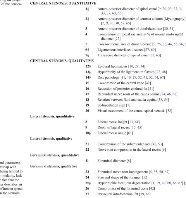
lumbar segment of a 71-year-old patient. According to the final set of core radiological criteria for a structured radiological report in patients with lumbar spinal stenosis, the compromise of the central zone and the relation between fluid and cauda equina should be described using the two classifications of Luire et al. and Schiza et al., respectively. Severe spinal canal stenosis with compromise of the central zone of>2/3 of its normal size due to disc material (moderate stenosis according to Lurie et al,, long arrow) and bilateral hypertrophy of the ligamentum flavum (arrow heads). No rootlets can be recognized and the dural sac demon-strates an almost homogeneous grey signal with no cerebrospinal-fluid signal visible. There is minimal epidural fat present posterior (short arrow). According to the classification of Schizas et al., the relation between fluid and cauda equina can be classified as a type C, severe stenosis
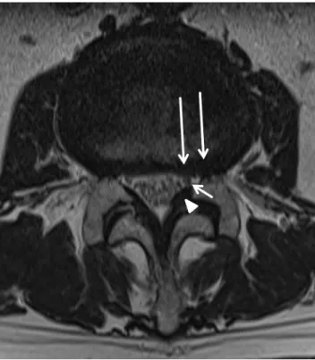
Fig. 2 Lateral stenosis. Axial T2 weighted MR image of the L4/L5 lower lumbar segment of a 67 years-old patient with left-sided accented lateral recess narrowing. According to the final set of core radiological criteria for a structured radiological report, the classification of Bartynski et al., should be used and a grade two compression should be reported. The nerve root on the left side (short arrow) is slightly compressed but with preservation of cerebrospinal fluid around the root in the recess. Reasons for stenosis are a hypertrophy of the ligamentum flavum (arrowhead) and disc material protruding in the lateral recess (long arrows)
included the original terminology and definitions of criteria and parameters, which were used according to their original reports and not changed nor modified. We recognize that as
research continues and more information is obtained, evidence may change and recommendations may have to be revised [40]. In addition, the radiological report for an individual patient, though based on a standard reporting form, can be supplemented with additional information where necessary, depending on the individual circumstances.
The use of a multidisciplinary meeting has advantages and limitations [12,17,40]. An advantage is that the experts have the chance to participate in an open discussion, to learn from each other’s experience, and to contribute to the final consen-sus statement. Participants are exposed to their peers, and experts are required to argue based on facts and published evidence rather than personal experience alone, although the latter is an important part of such a meeting. As much as possible, the panel based their consensus recommendations on published evidence, though in many situations reliable evidence was lacking, resulting in final recommendations that were based on a consensus opinion of the participating experts [40]. Different views are represented due to the expertise of different medical disciplines. This may enhance the accep-tance of results by the scientific community or specialized medical societies or other groups of experts.
An inherent limitation of such a consensus meeting is that any decision is influenced by the composition of the panel [12]. For this Zurich consensus meeting, we aimed for a balance between radiologists and non-radiologists with exper-tise in managing lumbar stenosis. Other limitations include the subjective selection of participants to invite and the imprecise definition of “expert” status. It is inevitable that, when the number of participants must be limited, not all national or international experts can be invited. Other expert groups might have reached different conclusions depending on the composition and size of the group.
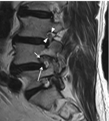
We believe that the results of this Zurich consensus meet-ing are important with regard to clinical care of patients with Fig. 3 Foraminal stenosis. Sagittal T2 weighted MR image of a
73-year-old patient with left-sided foraminal stenosis L4/5. According to the final set of core radiological criteria for a structured radiological report, the compromise of the foramen and the effect on the nerve roots should be described using the two classifications of Lurie et al. and Pfirrmann et al., respectively. Therefore, radiologists should describe the compromise of the foramen of approximately 1/3 of its normal size mainly due to disc protrusion (mild stenosis according to Lurie et al; short arrow). There is also obvious contact of disc material with the nerve root and the normal epidural fat layer around the root (see arrowheads one level above for comparison) is not preserved. This should be reported as a grade 1 impingement according to Pfirrmann et al
Table 2 Final set of core radio-logical criteria that should be used as a minimum standard in a structured radiological report in patients with lumbar spinal ste-nosis (LSS)
Note: * There are two different descriptions of this sign from Schizas et ?al. [54], and from Lee et ?al. [38]. The one from Schizas et ?al., was chosen by the experts to be used because of the higher degree of differentiation
b No. Criteria or Parameter Reference
Central Stenosis
Quantitative -Qualitative
15 Compromise of the central zone [42] 18 Relation between fluid and cauda equina * [54] Lateral Stenosis
Quantitative
-Qualitative 22 Nerve root compression in the lateral recess [6] Foraminal Stenosis
Quantitative -Qualitative
23 Foraminal nerve root impingement [50] 26 Compromise of the foraminal zone [42]
lumbar spinal stenosis. The standardized use of radiological criteria to describe and characterize morphological abnormal-ities in lumbar spinal stenosis could substantially improve day-to-day communication between radiologists and clini-cians. We note that this consensus meeting had no influence on the criteria or parameters themselves. They were not re-vised in any way. It was not the aim to develop new radiolog-ical criteria or parameters. Thus, the advantages and limita-tions of the individual criteria remain and should be consid-ered in any scientific discussion. The Zurich meeting’s main achievement was to discuss and evaluate the individual radio-logical criteria and parameters and to rank their clinical value. Our proposed core set of radiological criteria and parameters may not be the best for all situations and applications, but should be useful for the majority of clinical situations.
The results of this consensus meeting also do not solve the uncertainties in lumbar spinal stenosis that were recently outlined by several authors [3,18]. For example, we did not solve the problem of weak associations between radiological findings and symptoms or the unknown prognostic relevance of the radiological criteria and parameters. However, we be-lieve that consistent use of standard reporting in the future will increase awareness of those problems among physicians and radiologists, and facilitate better research. In addition, stan-dard reports will improve the performance of retrospective clinical observational studies. Prospective clinical studies will also benefit as the comparability of clinical studies increases. In conclusion, as a minimum standard, five core radiological criteria should be used in a radiological report describing lumbar spinal stenosis. Other parameters are well suited for research. Acknowledgments The scientific guarantor of this publication is Gustav Andreisek. The authors of this manuscript declare no relationships with any companies, whose products or services may be related to the subject matter of the article. The authors state that this work has not received any funding. The meeting was financed from the annual budget of the Horten Center, Zurich. No complex statistical methods were necessary for this paper. Institutional Review Board approval was not required because of the nature of the study. Methodology: prospective, multicentre study.
The authors thank Michele Mattle for organizing the meeting, Ursula Meyer for co-organizing the meeting, Dr. med. Sebastian F.-X. Winklhofer for his assistance to the organizers, Dr. med. Alexis Puhan for moderating the meeting, Dr. Christina Gmünder and Dr. Maria Wertli for the written documentation, Reto Kofmehl for the audiovisual recording of the meeting, and all other unnamed coworkers who have supported the meeting.
Alphabetical list of experts who participated in the consensus meeting: Gustav Andreisek, John A. Carrino, Richard A. Deyo, Janna L. Friedly, Jürg Hodler, Jeffrey G. Jarvik, Anne Mannion, Beat Michel, Alexander Nydegger, Christian W.A. Pfirrmann, François Porchet, Florian Ringel, Johann Steurer, Lukas Wildi, and Sebastian F.-X. Winklhofer.
References
1. Abbas J, Hamoud K, Peleg S et al (2011) Facet joints arthrosis in normal and stenotic lumbar spines. Spine 36:E1541–E1546
2. Airaksinen O, Herno A, Turunen V, Saari T, Suomlainen O (1997) Surgical outcome of 438 patients treated surgically for lumbar spinal stenosis. Spine 22:2278–2282
3. Andreisek G, Hodler J, Steurer J (2011) Uncertainties in the diagnosis of lumbar spinal stenosis. Radiology 261:681–684
4. Andreisek G, Imhof M, Wertli M, et al (in press) A systematic review of semi-quantitative and qualitative radiologic criteria for diagnosis of lumbar spinal stenosis. Am J Roentgenol
5. Aota Y, Niwa T, Yoshikawa K, Fujiwara A, Asada T, Saito T (2007) Magnetic resonance imaging and magnetic resonance myelography in the presurgical diagnosis of lumbar foraminal stenosis. Spine 32: 896–903
6. Bartynski WS, Lin L (2003) Lumbar root compression in the lateral recess: MR imaging, conventional myelography, and CT myelography comparison with surgical confirmation. AJNR Am J Neuroradiol 24:348–360
7. Barz T, Melloh M, Staub LP et al (2010) Nerve root sedimentation sign: evaluation of a new radiological sign in lumbar spinal stenosis. Spine 35:892–897
8. Beers GJ, Carter AP, Leiter BE, Tilak SP, Shah RR (1985) Interobserver discrepancies in distance measurements from lumbar spine CT scans. AJR Am J Roentgenol 144:395–398
9. Bolender NF, Schonstrom NS, Spengler DM (1985) Role of com-puted tomography and myelography in the diagnosis of central spinal stenosis. J Bone Jjoint Surgery Am 67:240–246
10. Borre DG, Borre GE, Aude F, Palmieri GN (2003) Lumbosacral epidural lipomatosis: MRI grading. Eur Radiol 13:1709–1721 11. Brant-Zawadzki MN, Jensen MC, Obuchowski N, Ross JS, Modic
MT (1995) Interobserver and intraobserver variability in interpreta-tion of lumbar disc abnormalities. A comparison of two nomencla-tures. Spine 20:1257–1263, discussion 1264
12. Brisse HJ, McCarville MB, Granata C et al (2011) Guidelines for imaging and staging of neuroblastic tumors: consensus report from the International Neuroblastoma Risk Group Project. Radiology 261: 243–257
13. Ciric I, Mikhael MA, Tarkington JA, Vick NA (1980) The lateral recess syndrome. A variant of spinal stenosis. J Neurosurg 53:433–443 14. Deyo RA, Gray DT, Kreuter W, Mirza S, Martin BI (2005) United States trends in lumbar fusion surgery for degenerative conditions. Spine 30:1441–1445, discussion 1446-1447
15. Dincer F, Erzen C, Basgöze O, Özker R, Celiker R (1991) Lateral recess syndrome and computed tomography. Turk Neurosurg 2:30–35 16. Fardon DF (2001) Nomenclature and classification of lumbar disc
pathology. Spine 26:461–462
17. Frates MC, Benson CB, Charboneau JW et al (2005) Management of thyroid nodules detected at US: Society of Radiologists in Ultrasound consensus conference statement. Radiology 237:794–800
18. Friedly JL, Jarvik JG (2012) Agreeing (or not) on how to describe spinal stenosis: expanding a narrow mindset. Radiology 264:3–4 19. Fujiwara A, Tamai K, Yamato M et al (1999) The relationship
between facet joint osteoarthritis and disc degeneration of the lumbar spine: an MRI study. Eur Spine J Off Publ Eur Spine Soc Eur Spinal Deformity Soc Eur Sect Cervical Spine Res Soc 8:396–401 20. Fukusaki M, Kobayashi I, Hara T, Sumikawa K (1998) Symptoms of
spinal stenosis do not improve after epidural steroid injection. Clin J Pain 14:148–151
21. Genevay S, Atlas SJ, Katz JN (2010) Variation in eligibility criteria from studies of radiculopathy due to a herniated disc and of neuro-genic claudication due to lumbar spinal stenosis: a structured litera-ture review. Spine 35:803–811
22. Grenier N, Kressel HY, Schiebler ML, Grossman RI, Dalinka MK (1987) Normal and degenerative posterior spinal structures: MR imaging. Radiology 165:517–525
23. Haig AJ, Geisser ME, Tong HC et al (2007) Electromyographic and magnetic resonance imaging to predict lumbar stenosis, low-back pain, and no back symptoms. J Bone Joint Surg Am 89:358–366
24. Hakan T, Celikoglu E, Aydoseli A, Demir K (2008) The redundant nerve root syndrome of the Cauda equina. Turk Neurosurg 18:204– 206
25. Hamanishi C, Matukura N, Fujita M, Tomihara M, Tanaka S (1994) Cross-sectional area of the stenotic lumbar dural tube measured from the transverse views of magnetic resonance imaging. J Spinal Disord 7:388–393
26. Herno A, Airaksinen O, Saari T (1994) Computed tomography after laminectomy for lumbar spinal stenosis. Patients’ pain patterns, walking capacity, and subjective disability had no correlation with computed tomography findings. Spine 19:1975–1978
27. Herzog RJ, Kaiser JA, Saal JA, Saal JS (1991) The importance of posterior epidural fat pad in lumbar central canal stenosis. Spine 16: S227–S233
28. Ishikawa Y, Shimada Y, Miyakoshi N et al (2006) Decompression of idiopathic lumbar epidural lipomatosis: diagnostic magnetic reso-nance imaging evaluation and review of the literature. J Neurosurg Spine 4:24–30
29. Jarvik JG, Haynor DR, Koepsell TD, Bronstein A, Ashley D, Deyo RA (1996) Interreader reliability for a new classification of lumbar disk disease. Acad Radiol 3:537–544
30. Jonsson B, Annertz M, Sjoberg C, Stromqvist B (1997) A prospec-tive and consecuprospec-tive study of surgically treated lumbar spinal steno-sis. Part I: Clinical features related to radiographic findings. Spine 22: 2932–2937
31. Kalichman L, Cole R, Kim DH et al (2009) Spinal stenosis preva-lence and association with symptoms: the Framingham Study. Spine J Off J N Am Spine Soc 9:545–550
32. Kasdan RB, Howard JL (2008) Neuroimaging of spinal diseases: a pictorial review. Semin Neurol 28:570–589
33. Koc Z, Ozcakir S, Sivrioglu K, Gurbet A, Kucukoglu S (2009) Effectiveness of physical therapy and epidural steroid injections in lumbar spinal stenosis. Spine 34:985–989
34. Kuhn MJ, Youssef HT, Swan TL, Swenson LC (1994) Lumbar epidural lipomatosis: the “Y” sign of thecal sac compression. Comput Med Imaging Graph Off J Comput Med Imaging Soc 18: 367–372
35. Kunogi J, Hasue M (1991) Diagnosis and operative treatment of intraforaminal and extraforaminal nerve root compression. Spine 16:1312–1320
36. Laurencin CT, Lipson SJ, Senatus P et al (1999) The stenosis ratio: a new tool for the diagnosis of degenerative spinal stenosis. Int J Surg Invest 1:127–131
37. Lee BC, Kazam E, Newman AD (1978) Computed tomography of the spine and spinal cord. Radiology 128:95–102
38. Lee GY, Lee JW, Choi HS, Oh KJ, Kang HS (2011) A new grading system of lumbar central canal stenosis on MRI: an easy and reliable method. Skelet Radiol 40:1033–1039
39. Lee S, Lee JW, Yeom JS et al (2010) A practical MRI grading system for lumbar foraminal stenosis. AJR Am J Roentgenol 194:1095–1098 40. Levine D, Brown DL, Andreotti RF et al (2010) Management of asymptomatic ovarian and other adnexal cysts imaged at US: Society of Radiologists in Ultrasound Consensus Conference Statement. Radiology 256:943–954
41. Lurie JD, Doman DM, Spratt KF, Tosteson AN, Weinstein JN (2009) Magnetic resonance imaging interpretation in patients with symp-tomatic lumbar spine disc herniations: comparison of clinician and radiologist readings. Spine 34:701–705
42. Lurie JD, Tosteson AN, Tosteson TD et al (2008) Reliability of readings of magnetic resonance imaging features of lumbar spinal stenosis. Spine 33:1605–1610
43. Mamisch N, Brumann M, Hodler J et al (2012) Radiologic criteria for the diagnosis of spinal stenosis: results of a Delphi survey. Radiology 264:174–179
44. Mariconda M, Fava R, Gatto A, Longo C, Milano C (2002) Unilateral laminectomy for bilateral decompression of lumbar spinal
stenosis: a prospective comparative study with conservatively treated patients. J Spinal Disord Tech 15:39–46
45. Mikhael MA, Ciric I, Tarkington JA, Vick NA (1981) Neuroradiological evaluation of lateral recess syndrome. Radiology 140:97–107 46. Min JH, Jang JS, Lee SH (2008) Clinical significance of redundant
nerve roots of the cauda equina in lumbar spinal stenosis. Clin Neurol Neurosurg 110:14–18
47. North American Spine Society (2007) Evidence-based clinical guide-lines for multidisciplinary spine care., Burr Ridge, IL, USA 48. Park JB, Chang H, Lee JK (2001) Quantitative analysis of
transforming growth factor-beta 1 in ligamentum flavum of lumbar spinal stenosis and disc herniation. Spine 26:E492–E495
49. Pathria M, Sartoris DJ, Resnick D (1987) Osteoarthritis of the facet joints: accuracy of oblique radiographic assessment. Radiology 164: 227–230
50. Pfirrmann CW, Dora C, Schmid MR, Zanetti M, Hodler J, Boos N (2004) MR image-based grading of lumbar nerve root compromise due to disk herniation: reliability study with surgical correlation. Radiology 230:583–588
51. Prasartritha T, Suntisathaporn N, Vathana P, Sriphojanart C (1997) The size of the vertebral canal and the significance of epidural fat in lumbar spinal stenosis. Journal of the Medical Association of Thailand =. Chotmaihet thangphaet 80:247–256
52. Rankine JJ, Hutchinson CE, Hughes DG (1997) MRI of lumbar spondylosis: a comparison of sagittal T2 weighted and three se-quence examinations. Brit J Radiol 70:1112–1121
53. Rothman SLG, Glenn WV (1985) Multiplanar CT of the spine. University Park Press, Baltimore
54. Schizas C, Theumann N, Burn A et al (2010) Qualitative grading of severity of lumbar spinal stenosis based on the morphology of the dural sac on magnetic resonance images. Spine 35:1919–1924 55. Schonstrom N, Willen J (2001) Imaging lumbar spinal stenosis.
Radiol Clin N Am 39:31–53
56. Schonstrom NS, Bolender NF, Spengler DM (1985) The pathomorphology of spinal stenosis as seen on CT scans of the lumbar spine. Spine 10:806–811
57. Sortland O, Magnaes B, Hauge T (1977) Functional myelography with metrizamide in the diagnosis of lumbar spinal stenosis. Acta Radiol Suppl 355:42–54
58. Steurer J, Nydegger A, Held U et al (2010) LumbSten: the lumbar spinal stenosis outcome study. BMC Musculoskelet Disord 11:254
59. Steurer J, Roner S, Gnannt R, Hodler J, LumbSten Research C (2011) Quantitative radiologic criteria for the diagnosis of lumbar spinal stenosis: a systematic literature review. BMC Musculoskelet Disord 12:175
60. Stieber J, Quirno M, Cunningham M, Errico TJ, Bendo JA (2009) The reliability of computed tomography and magnetic resonance imaging grading of lumbar facet arthropathy in total disc replacement patients. Spine 34:E833–E840
61. Strojnik T (2001) Measurement of the lateral recess angle as a possible alternative for evaluation of the lateral recess stenosis on a CT scan. Wien Klin Wochenschr 113(Suppl 3):53–58
62. Suzuki K, Takatsu T, Inoue H, Teramoto T, Ishida Y, Ohmori K (1992) Redundant nerve roots of the cauda equina caused by lumbar spinal canal stenosis. Spine 17:1337–1342
63. Ullrich CG, Binet EF, Sanecki MG, Kieffer SA (1980) Quantitative assessment of the lumbar spinal canal by computed tomography. Radiology 134:137–143
64. van Rijn JC, Klemetso N, Reitsma JB et al (2005) Observer variation in MRI evaluation of patients suspected of lumbar disk herniation. AJR Am J Roentgenol 184:299–303
65. Verbiest H (1979) The significance and principles of computerized axial tomography in idiopathic developmental stenosis of the bony lumbar vertebral canal. Spine 4:369–378
66. Weishaupt D, Zanetti M, Boos N, Hodler J (1999) MR imaging and CT in osteoarthritis of the lumbar facet joints. Skelet Radiol 28:215–219 67. Weishaupt D, Zanetti M, Hodler J, Boos N (1998) MR imaging of the
lumbar spine: prevalence of intervertebral disk extrusion and seques-tration, nerve root compression, end plate abnormalities, and osteo-arthritis of the facet joints in asymptomatic volunteers. Radiology 209:661–666
68. Wildermuth S, Zanetti M, Duewell S et al (1998) Lumbar spine: quantitative and qualitative assessment of positional (upright flexion and extension) MR imaging and myelography. Radiology 207:391– 398
69. Wilmink JT, Korte JH, Penning L (1988) Dimensions of the spinal canal in individuals symptomatic and non-symptomatic for sciatica: a CT study. Neuroradiology 30:547–550