NOTE TO USERS
Université de Sherbrooke
Low-energy electron-induced DNA damage: product
analysis and mechanistic studies of damage in short
oligonucleotides
ByZejun Li
Department of Nuclear Medicine and Radiobiology
Thesis presented to the Faculty of Medicine and Health
Sciences to obtain a diploma of Philosophy Doctorate
(Ph. D.) in Radiobiology
Évaluateurs
Sherbrooke, Québec, Canada
December 2010
Léon Sanche, co-directeur, département de médecine nucléaire et de radiobiologie J. Richard Wagner, co-directeur, département de médecine nucléaire et de radiobiologie Michael Huels, président de jury, département de médecine nucléaire et de radiobiologie
Jean Lessard, évaluateur externe au programme, département de chimie, Université de Sherbrooke
Christopher Wilds, évaluateur externe à l'Université, ' Department of Chemistry and biochemistry, Concordia University
m
NOTICE:
Library and Archives Canada
Published Heritage Branch
395 Wellington Street Ottawa ON K1A ON4 Canada
The author has granted a
non-exclusive license allowing Library and Archives Canada to reproduce, publish, archive, preserve, conserve, communicate to the public by
telecommunication or on the Internet, loan, distrbute and sell theses
worldwide, for commercial or non-commercial purposes, in microform, paper, electronic and/or any other formats.
The author retains copyright ownership and moral rights in this thesis. Neither the thesis nor substantiel extracts from it may be printed or otherwise reproduced without the author's permission. ln compliance with the Canadien Privacy Act some supporting forms may have been removed from this thesis.
While these forms may be included in the document page count, their removal does not represent any loss of content from the thesis.
Canada
Bibliothèque et Archives Canada Direction du Patrimoine de l'édition 395, rue Wellington Ottawa ON K1A ON4 CanadaAVIS:
Y our file Votre référence ISBN: 978-0-494-83336-0 Our file Notre référence ISBN: 978-0-494-83336-0
L'auteur a accordé une licence non exclusive permettant à la Bibliothèque et Archives Canada de reproduire, publier, archiver, sauvegarder, conserver, transmettre au public par télécommunication ou par l'Internet, prêter, distribuer et vendre des thèses partout dans le monde, à des fins commerciales ou autres, sur support microforme, papier, électronique et/ou autres formats.
L'auteur conserve la propriété du droit d'auteur et des droits moraux qui protege cette thèse. Ni la thèse ni des extraits substantiels de celle-ci ne doivent être imprimés ou autrement reproduits sans son autorisation.
Conformément à la loi canadienne sur la protection de la vie privée, quelques formulaires secondaires ont été enlevés de cette thèse.
Bien que ces formulaires aient inclus dans la pagination, il n'y aura aucun contenu manquant.
Dommages à I' ADN induits par les électrons de basse énergie : l'analyse des produits et études des mécanismes de dommage dans des oligonucleotides
courts
Par Zejun Li
Département de médecine nucléaire et de radiobiologie
Thèse présentée à la Faculté de médecine et des sciences de la santé en vue de l'obtention du diplôme de philosophiae doctor (Ph.D.) en radiobiologie, Faculté de médecine et des sciences de la santé, Université de Sherbrooke, Sherbrooke, Québec,
Canada, JlH 5N4
Résumé
L'objectif majeur de notre groupe de recherche est de comprendre le mécanisme de dommage à l' ADN induit par les électrons de basse énergie "secondaire" provenant de radiations ionisantes et sa relation avec la radiosensibilité et la radiothérapie. Le professeur Sanche a développé un nouveau système d'irradiation d'électron de basse énergie avec lequel une relativement grande surface constituée de couches minces d' ADN peut être irradiée avec des électrons mono-énergétiques sous hyper-vide. Ceci permet l'irradiation de l' ADN ciblé et la formation d'une quantité suffisamment grande d' ADN endommagé pour effectuer des analyses chimiques (HPLC, GC/MS et LC/MS-MS) sur les produits générés et ainsi élucider le mécanisme de dommage à l' ADN par les électrons de basse énergie. Mon projet se concentre sur les systèmes simples, dans lesquels des nucléosides ( dThd), nucléotides (pT, Tp et pTp ), oligonucléotides (TT et TTT) et des oligonucléotides modifiés (TSBrUT) sont exposés à des électrons de basse énergie. Les réactions subséquentes sont étudiées par analyses chimiques des produits formés.
Mes études ont révélé trois mécanismes de fragmentation dans l' ADN induits par les électrons de basse énergie : 1) le groupe phosphate terminal a une plus large section efficace dans les dommages induits par les électrons de basse énergie; 2) la capture initiale des électrons de basse énergie et le bris de lien subséquent dans l'anion intermédiaire dépend de la séquence et de l'affinité des électrons de la base; 3)
à 10 eV, un électron peut induire un événement double. La présente étude fourni une base chimique de la formation de bris par la réaction des électrons de basse énergie avec l'ADN.
Low-energy electron-induced DNA damage: product analysis and mechanistic studies of damage in short oligonucleotides
By Zejun Li
Departrnent of Nuclear Medicine and Radiobiology
Thesis presented to the Faculty of Medicine and Health Sciences to obtain a diploma of Philosophy Doctorate (Ph. D.) in Radiobiology, Faculty of Medicine and Health
Sciences, University of Sherbrooke, Sherbrooke, Québec, Canada, JI H 5N4
Abstract
The major objective of our group is to understand the mechanism of DNA damage induced by secondary low-energy electrons (LEE) arising from ionizing radiation and its relationship to radiosensitization and radiotherapy. Prof. Sanche has developed a novel low-energy electron irradiation system in which a relatively large area of thin films of DNA constituents can be irradiated with mono-energetic electrons under ultra high vacuum. This permits the irradiation of target DNA and the formation of sufficient degraded material to allow for chemical analysis (HPLC, GC/MS, and LC/MS/MS) of products remaining on the target surface, so as to elucidate the mechanism of LEE-induced DNA damage. My project focuses on simple systems, in which small DNA components nucleosides (dThd), nucleotides (pT, Tp, pTp), oligonucleotides {TT and TTT) and modified oligonucleotides (T5BrUT) are exposed to low-energy electrons, and the subsequent reactions are studied by chemical analysis of the products.
My studies revealed three mechanisms of LEE-induced fragmentation reactions in DNA: 1) the terminal phosphate group has a larger cross-section in LEE-induced DNA damage; 2) initial LEE capture and subsequent bond breaking within the intermediate anion depend on the sequence and electron affinity of the bases; and 3) at IO eV, one electron might induce double events. This study provides a chemical basis for the formation of DNA strand breaks by the interaction of LEE with DNA.
CONTENTS
List of tables ... .iv
List of figures and schemes ... .iv
List of abbreviations ... vi
Cbapter 1 - Introduction ... 1
1.1 Radiation and people ... 1
1.2 How radiation reacts with the cells ... 1
1.3 Excitation and ionization of radiation ... 2
1.4 Production of secondary LEE ... 3
1.5 Electron interaction with molecules ... .4
1.6 LEE interaction with a molecule XY ... 6
1. 7 "Shape" resonances and "core-excited" resonances ... 8
1.8 DNA damage induced by ionizing radiation ... 9
1.9 DNA damage induced by LEE ... 13
1.10 Experimental design to model LEE-induced damage to DNA ... 14
1.11 Study of LEE-induced DNA damage in the past ... 16
1.12 The new LEE irradiator system in our lab ... 18
1.13 Previous results in our lab ... 20
1.14 Description of this research project ... 21
Chapter II- First article ... 24
Low Energy Electron Induced DNA Damage: Effects of Terminal Phosphate and Base Moieties on the Distribution of Damage. Zejun Li, Yi Zheng, Pierre Cloutier, Léon Sanche and J. Richard Wagner. Journal of the American Chemical Society, 2008, 130( 17), 5612-5613. Chapter III - Second article ... 36
Low-Energy Electron-Induced DNA Damage: Effect of Base Sequence in Oligonucleotide Trimers. Zejun Li, Pierre Cloutier, Léon Sanche, and J. Richard Wagner. Journal of the American Chemical Society, 2010, 132(15), 5422-5427. Chapter IV - Third article ... 60
Low-Energy Electron-Induced DNA Damage: One Electron Induces Double Events in 5-Bromouracil Substituted Oligonucleotide Trimer 5' -TpTpT-3'. Zejun Li, Pierre Cloutier, Léon Sanche and J. Richard Wagner. To be submitted. Chapter V - Further discussion ... 84
Chapter VI - Conclusions and perspectives ... 103
Acknowledgements ... 107
List of tables
Chapter II - First articleTable 1. Yield of Products from LEE Induced DNA Damage ... 32
Chapter III - Second article Table 1. Yield of Products from LEE-Induced DNA Damage ... 48
Table 2. Different Calculations for the Energy Barrier of C-0 Bond Rupture after LEE Attachment. ... 55
Chapter IV - Third article Table 1. Yield of Products from LEE Induced DNA Damage ... 72
Chapter V - Fui1her discussion Table 1. Different calculations on the energy barrier of C-0 and N-C bond rupture after LEE attachment. ... 89
Table 2. Yield ofproducts from LEE-induced DNAdamage in tetramers (10 eV) .... 98
List of figures and schemes
Chapter I - Introduction Figure 1. Schematic diagram of electron-molecule collisions ... 5Figure 2. Pathway "1" when an electron interacts with a molecule XY ... 6
Figure 3. Pathway "II" when an electron interacts with a molecule XY ... 7
Figure 4. Pathway "III" when an electron interacts with a molecule XY ... 8
Figure 5. "Shape" resonance and "core-excited" resonance ... 9
Figure 6. Structures of the four nucleobases: A, T, G, C ... 10
Figure 7. Structure of a nucleotide, thymine-3 '-monophosphate, i.e., Tp ... 11
Figure 8. Schematic diagram of the spin coating system ... 19
Figure 9. General view ofUHV electron irradiator chamber ... 20
Cbapter II - First article Figure 1. Structure of 5'-TpTpT-3' and position(s) of cleavage for N-glycosidic (N-C) and phosphodiester-sugar (C-0) bonds ... 30
Chapter III - Second article Scheme 1. Structure of 5'-TpXpT-3' and Position(s) of Cleavage for the N-glycosidic Bond (1, 2) and Phosphodiester bond (3-6) ... .43
Figure 1. Analysis ofTpTpT radiation products by HPLC/UV detected at 260nm .. .46
Scheme 2. Decay Channels of Transient Negative Ions of DNA Bases Formed at an Initial Electron Energy of Ea ... .49
Scheme 1. Structure of 5' -TpXpT-3' and position(s) of cleavage for the N-glycosidic bond (1), phosphodiester bond (2-5), and C-Br bond cleavage (6) ... 67 Figure 1. Analysis of Tp5BrUpT radiation products by HPLC/UV detected at
260nm ... 70 Figure 2. Comparison of Tp5BrUpT, TpUpT, and TpTpT radiation products by
HPLC/UV detected at 260nm ... 74 Scheme 2. Proposed mechanism of formation of double lesions by the reaction of
one low-energy electron with one TSBrUT molecule ... 78
Chapter V - Further discussion
Figure 1. LEE-induced N-glycosidic bond cleavage in thymidine ... 84 Figure 2. The release of thymine from thymidine as a function of electron energy ... 85 Scheme 1. A proposed mechanism for thymidine N-glycosidic bond break by LEE
bombardment. ... 86 Scheme 2. A proposed mechanism of the LEE-induced single strand bond cleavage
in pyrimidine nucleotide at 3' ... 90 Scheme 3. A proposed mechanism of the LEE-induced single strand bond cleavage
in pyrimidine nucleotide at 5' ... 91 Figure 3. Orbital containing the excess attached electron in thymidine nucleotide for
C-0 bond length value below 1.9
A
(top) and for C-0 bond length value beyond 1.9A
(bottom) ... 91 Figure 4. Phosphodiester bond cleavage sites in oligonucleotide TT ... 93 Scheme 4. Structure of 5'-TpTpT-3' and position(s) of cleavage for the N-glycosidicbond (1), phosphodiester bond (2-5) ... 94 Figure 5. Phosphoester bond (site 1, and 2) and N-glycosidic bond (site 3) cleavage
sites in molecule model pGp ... 97 Figure 6. In gas phase SBrU, different dissociation pathways result in different
negative ion productions after a LEE is attached to it. ... 1 OO Figure 7. Time course of double-lesion products formation by LEE impact.. ... 102
A Ade a.m.u AMI AN Ap aug-cc-p VDZ B3LYP Bru SBrU 5BrUT
c
SClU cm Cp Cyt dA dAdo dAMP dC dCyd dCMP DD DE DEA DFT dG dGuo dGMP DMF DNA dNTPs DSB dT 3', 5'-dTDP dThd 3'-dTMP 5'-dTMP DZP++ E Eo LlEList of abbreviations
Adenine AdenineAtomic mass unit Austin model 1 Acetonitrile
Adenosine-3 '-monophosphate
Augmented, correlation consistent polarized valence double zeta Becke, three-parameter, Lee-Yang-Parr
Bromouracil 5-Bromouracil 5' -5BrUpT-3' Cytosine 5-Chlorouracil Centime ter Cytidine-3' -monophosphate Cytosine 2'-Deoxyadenosine 2'-Deoxyadenosine 2'-Deoxyadenosine monophosphate 2'-Deoxycytidine 2'-Deoxycytidine 2'-Deoxycytidine monophosphate Dipolar dissociation Dissociative excitation
Dissociative electron attachment Density functional theory 2'-Deoxyguanosine 2'-Deoxyguanosine 2'-Deoxyguanosine monophosphate N, N-dimethylformamide Deoxyribonucleic acid Deoxy-ribonucleoside triphosphates Double strand break(s)
Thymidine
Thymidine-3 ',5' -diphosphate Thymidine
Thymidine-3 '-monophosphate Thymidine-5' -monophosphate Double-zeta plus polarization Electron energy
Initial electron energy Transferred electron energy
E E EA e aq Ea e hyd Eox -e pre ESD
e.
5FU FWHM GGC/MS
Gp Gua HOMOHPLC
kcal/mol KE MeV mol MWLC
LC/MS/MS
LEE LUMO mm mL ML mm mM MP2 MS M.W. N2NHE
nm oligos pA pAp pAT p5BrUTEnergy barrier I activation energy Dielectric constant
Electron affmity
Solvated electrons/hydrated electrons Binding energy
Electron re-emitted into the continuum Hydrated electrons
Oxidation potential Prehydrated electrons
Electron stimulated desorption
Electron transfer from the base to the phosphate unit 5-Fluorouracil
Full width at half maximum Guanine
Gas chromatography/ mass spectrometry Guanosine-5' -monophosphate
Guanine
Highest occupied molecular orbital High-perf ormance liquid chromatography Kilo calorie per mole
Kinetic energy Mega electron volts Mole
Molecular weight Liquid chromatography
Liquid chromatography/mass spectrometry/mass spectrometry Low energy electron(s) (0-30 eV)
Lowest unoccupied molecular orbital Minute
Milliliter Monolayer( s) Millimeter
Milli mole per liter
Second order Meller-Plesset perturbation theory Mass spectrometry
Molecular weight Nitrogen gas
Normal hydrogen electrode Nanometer oligonucleotides Adenosine-5' -monophosphate Adenosine-3' ,5 '-diphosphate 5'-pApT-3' 5' -p5BrUpT-3'
pC Cytidine-5' -monophosphate PCM Polarizable continuum model pCT 5'-pCpT-3'
PES Potential energy surface pG Guanosine-5' -monophosphate pGp Guanosine-3 ',5' -diphosphate pGT 5'-pGpT-3' pT Thymidine-5' -monophosphate pTp Thymidine-3' ,5 '-diphosphate pTpTp 5'-pTpTp-3' pTT 5'-pTpT-3' pUT 5'-pUpT-3' pXT 5'-pXpT-3' (X= T, C, A, G) SCF Self-consistent field SE Secondary electron(s) SSB Single strand break(s)
T Thymine TAp 5'-TpAp-3' TAT 5'-TpApT-3' T5BrU 5' -Tp5BrU-3' TSBrUp 5' -TpSBrUp-3' T5BrUT 5'-Tp5BrUpT-3' TCp 5'-TpCp-3' TCT 5'-TpCpT-3' TGp 5'-TpGp-3' TGT 5'-TpGpT-3' Thy Thymine
TMA Transient molecular anion TNI Transient negative ion
Tp Thymidine-3 '-monophosphate TT 5'-TpT-3' TTp 5'-TpTp-3' TTT 5'-TpTpT-3' TTXT 5'-TpTpXpT-3' (X= T, C, A, G) TU 5'-TpU-3' TUp 5'-TpUp-3' TUT 5'-TpUpT-3' TXp 5'-TpXp-3' (X= T, C,A, G) TXTT 5'-TpXpTpT-3' (X= T, C, A, G)
u
UracilUHV
Ultra high vacuumChapter 1 - Introduction
1.1 Radiation and people
Ever since people knew what were radiations and radioisotopes more than 1 OO years ago, people started using them. Nowadays, radiations and radioisotopes are widely used not only in medical examination and treatment, but also in many other fields such as weaporuy, energy, material manufacture, food processing, etc. However, people also suffered greatly from radiation, e.g., the atomic bomb explosion in Hiroshima and Nagasaki in 1945 and the release of radioactive material in Chernobyl nuclear power plant in 1986. So radiation has both good and bad effects. lt is useful in many fields but using it is dangerous and it has harmful effects on people and the environment.
lt is well-known that radiation is bad to the health of people because it damages living cells. But people still use radiations in the medical field and other fields. So understanding the mechanisms of radiation action on cells and tissues is crucial for using radiation and estimating the risk.
1.2 How radiation reacts with the cells
Earlier experiments have shown that the primary sensible sites for cell lethality induced by ionizing radiation are focused in the nucleus. Thus, when ionizing radiation passes through a cell, DNA is the critical target (Steel, 2002; Hall and Giaccia, 2006). DNA is the vehicle for inheritance in cellular life and DNA damage is the critical event in radiation cell killing and mutation. For this reason understanding the processes (chemical reactions) that damage DNA and cause mutations takes on great importance. In this thesis, we deal with damage to DNA induced by secondary LEE arising from ionizing radiation.
DNA is genetic material in the cell that stores hereditary information. When ionizing radiation irradiates the cell, the most significant damage is in DNA. If the damaged DNA is properly repaired, the cell survives and remains a healthy parent cell which gives healthy offspring cells. If the damaged DNA is not properly repaired, it may not properly replicate and may transmit the deficient traits from one generation to another; the cell can die or mutate and induce cancer (von Sonntag, 1987, 2006). However, there are vast differences in the time scale involved in these various events, which lead to cell death. For example, the physical process takes place in less than 10·15 s. The following physical-chemical processes have lifetimes
of about 10·10 to 10·9 s. Finally, the biological expression takes place in a longer time
from a few seconds or minutes to a few years. For example, the oncogenic effect of ionizing radiation may be delayed many years. Radiobiology is the study of the effect of ionizing radiation on biological molecules and living organism, e.g., DNA damage, cell death, mutation, carcinogenesis, etc. The experiments can be performed through chemical methods, physical methods, in vivo or in vitro biological methods, or even through theoretical calculations to predict the processes involved.
1.3 Excitation and ionization of radiation
Any kind of radiation delivers energy to the materials absorbing the radiation. Energy absorption by the material occurs via changes in the molecule or the atom. There are two possible changes when the energy is absorbed by the material. The first is excitation and the second is ionization. If the electron of the outer shell of the atom is raised to a higher energy level, this process is said to be electronic excitation. If sufficient energy is absorbed to overcome the binding energy of the electron and release one or more electrons from the atom, then ionization happens. When the radiation is of sufficient energy to produce ionization, it is called ionizing radiation. In the process of ionization, in solid matter by high energy radiation, many secondary electrons (SE) are produced. If the SE also have enough energy to excite or ionize molecules in the medium, they can further excite or ionize them before they
are thermalized, i.e., before they give their energies to the environment. In the whole process, a large number of LEE (0-30 eV) are produced. lt is reported that about -4x 10⁴ LEE are produced along the radiation track per Me V of deposited energy (La Verne and Pimblott, l 995a, l 995b; Pimblott and La Verne, 2007). The secondary LEE arising from ionizing radiation induced damage to DNA will be the major theme ofthis thesis.
1.4 Production of secondary LEE
We take two kinds of ionizing radiation to ex;plain the production of secondary LEE: an x-ray (or y-ray) photon and a fast charged particle which represents the electromagnetic radiation and particulate radiation.
First, there are two processes which dominate when x-ray photons are absorbed. At high energies (- 0.7-10 MeV), the Compton process dominates. In this process, the photon gives part of its energy to an almost free electron (which means that the binding energy of this electron is very small in comparison with the energy of the photon, i.e., almost negligible) as kinetic energy resulting in a fast electron and a photon with reduced energy. The photon takes part in further interactions. As a result, a large number of relatively fast electrons are produced, and they can further excite or ionize other atoms in the medium and give a large amount of the secondary LEE. At lower energies (E< 0.5 MeV), usually it is the photoelectric absorption processes that become dominant. The electron is ejected from an inner shell of an atom and the photon energy is used to overcome the binding energy of the atomic electron and provide kinetic energy to this ejected electron. Thus, the energy of the incident photon (hv) is shared with the kinetic energy (K.E) of the ejected electron and the binding energy of the electron in its orbit (Es), i.e., K.E=hv - Es. Whether the absorption process is the Compton or the photoelectric process, much of the energy of the absorbed photon is converted into kinetic energy of fast secondary electrons and subsequent secondary LEE.
When the fast Compton or photoelectron or particulate radiation, such as a fast charged particle, passes near molecules, it produces a fast varying electric field and magnetic field on this molecule. This perturbation does not change the energy and momentum of the fast particle very much; thus the energy transfer is like the absorption of electromagnetic radiation by the molecules of the medium as mentioned above. The primary charged particle loses part of its energy, with highest probability of 22 eV (La Verne and Pimblott, 1995a, 1995b; Pimblott and La Verne, 2007; Srdoc et al., 1995). The emission of such low-energy quanta (22 eV) successions represents the energy loss and energy deposition of the charged particle to the molecules. This process can lead to excitation and ionization. The possibility of excitation and ionization should be the same but about 20% of the energy deposited by fast charged particles in organic matter may lead to excitation, and the rest, i.e., 80%, may lead to ionization. The ionization energy is shared as potential energy of the cation and the kinetic energy of secondary LEE. As a result, the largest portion of the energy of primary particle changes to the energy of the secondary electrons (La Verne and Pimblott, 1995a, 1995b; Pimblott and La Verne, 2007). Statistically, about 40% of the energy of the primary radiation goes to the energy of the secondary LEE.
Overall, along the radiation track, many species are created which are cornposed of excited atoms and rnolecules, cations, radicals, and secondary electrons. Secondary electrons are produced abundantly (-4xI04 per MeV of deposited energy),
through energy deposit from the electromagnetic radiations and particulate radiations such as x-rays, y-rays, high-energy electrons, protons, alpha-particles, heavy charged particles, etc. (Cobut et al., 1998). The energy of the secondary electrons has a distribution that lies essentially below 70 eV with a most probable energy below 10 eV (LaVeme and Pimblott, 1995a, 1995b; Pimblott and LaVeme, 2007; Henke et al.,
1977, 1981); these are referred to as secondary LEE.
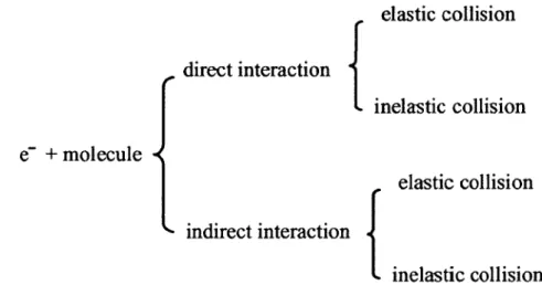
At low energies, electron-molecule collisions can be divided into two main types: direct and indirect interactions (Figure 1). The direct interaction may also be referred to as non-resonant interaction and the indirect interaction may also be referred to as resonant interaction. Direct interaction bas short interaction times. Direct interaction usually occurs at higher electron energies above the ionization threshold of the molecules.
e-
+
molecule
elastic collision
direct interaction {
inelastic collision
elastic collision
indirect interaction {
inelastic collision
Figure 1. Schematic diagram of electron-molecule collisions.The direct and indirect interaction can be divided as elastic and inelastic according to the amount of energy transferred from the electron to the target. In elastic collisions, the loss of electron energy to the target is negligible. In the case of inelastic collision, the loss of electron energy to the target is not negligible. In this case, electronically excited states may be created and followed by subsequent reactions.
If the incoming electron is bound to the target for a relatively longer time than the usual direct interaction time, resonant electron interaction takes place (Palmer and Rous, 1992; Sanche, 1991; Sanche, 1995; Sanche, 2000; Sanche, 2002). In this case, a transient negative ion (TNI) or transient molecular anion (TMA) is formed. This resonant state may be regarded as a previously unfilled orbital of the molecule temporarily occupied by an electron. This process can be described as following: e
corresponds to the energies of transient anions. At this specific energy, i.e., at the resonance energy, usually an increased product yield is observed.
1.6 LEE interaction with a molecule XY
There are three main pathways for a diatomic molecule XY to internet with a LEE (Sanche, 2002; Bass and Sanche, 2004).
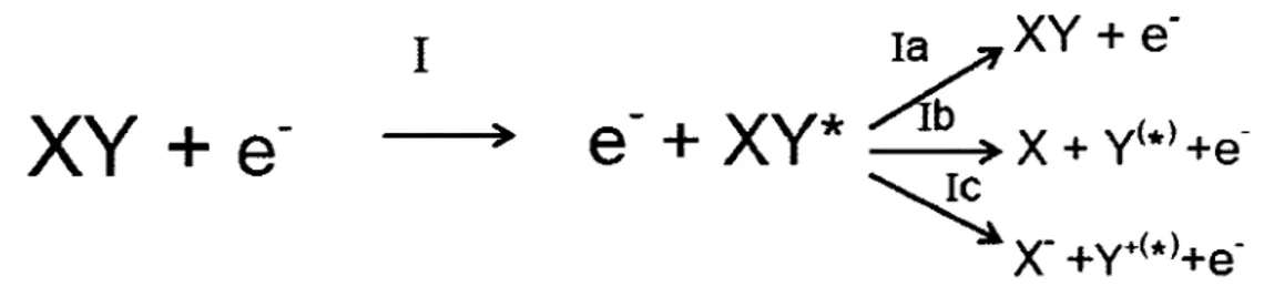
First, an excited neutral state of the molecule XY* may be created by pathway
"1" through the direct electron interaction (inelastic interaction). This process is
illustrated in Figure 2. XY* may disperse its excess energy by emitting a photon and/or transferring its energy to the surrounding environment (pathway la). When the electronically excited neutral state XY* is dissociative, the molecule dissociates in ground state (X
+
Y) or excited fragments (X+
Y*) (pathway lb). Dipolar dissociation (DD) produces a cation(Y+<*»
and an anion (X) (pathway le) if the electron energy is higher than a certain energy threshold ( ~ 14-16 eV).I
)Figure 2 (reprinted Crom Sanche, 2002). Pathway "I" when an electron interacts
with a molecule XY.
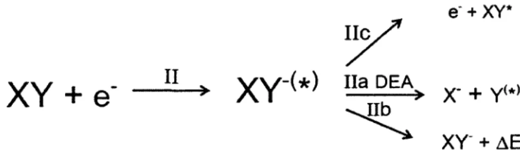
The second major pathway is a resonant interaction. This process is illustrated in Figure 3 (pathway "Il"). Through pathway "Il", an incident electron temporarily attaches to the molecule and a ''TNI" or a "TMA"
(xv-<*»
is produced. Through pathway Ile, the resulting "TNI" or "TMA" may autoionize, which means the electron leaves the molecule and an excited neutral state of the molecule XY* isproduced which may decay through pathway la, lb, or le. The other process, which is also the most important process, is called dissociative electron attacbment (DEA). In this process, the anion may dissociate into a neutral fragment in its excited (Y*) or ground state (Y) and a stable anion (Xl (pathway lla). lt is accepted that the DEA process happens when a LEE interacts with a DNA molecule. During its lifetime, the "TNI" or "TMA" can stabilize by giving its energy (ÂE) to another system (for example, by photon emission to a surrounding medium or by collisional action with another molecule in the medium (process Ilb) and result in a stabilized XY anion, XV-.
y
e·
+
XY*XY +
e-
II
xv-<*>
na
DEA
~
xy-
x-
+
+
y<•>
~EFigure 3 (reprinted from Sanche, 2002). Pathway "Il" when an electron interacts with a molecule XY.
The third pathway is "III". When the incoming electron can directly ionize the molecule resulting in a dissociative cation (XY+<*», it may break up as shown through reaction Illa (X+ y+<*». This process is illustrated in Figure 4 and usually this dissociation channel pathway is non-resonant.
III
Figure 4 (reprinted from Sanche, 2002). Pathway "III" when an electron interacts with a molecule XY.
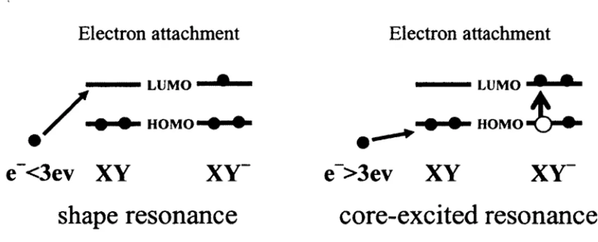
1. 7 "Shape" resonances and "core-excited" resonances
In the resonant process of LEE interaction with a molecule XY, there are two major types of resonances or transient anions involved (Schulz, l 973a, l 973b; Sanche, 2002): one is called "shape" resonance and the other is called "core-excited" resonance. The "shape" resonance is also called a single-particle resonance which means that when the additional electron, usually at lower energies (E < 3eV), goes to a previously unfilled orbital (such as LUMO) of the target (Figure 5). When previously unfilled orbitais (such as LUMO) are occupied by two electrons and the transitory anion is formed, this resonance is termed "core-excited" (Figure S). ln the "core-excited" resonance, the first step is. that one core electron is excited from the ground state (because the incident electron has a higher energy, usually E > 3eV), usually resulting in an electron in the LUMO and a hole in the HOMO. Simultaneously, a core-excited anion is produced by capturing the exciting electron in the LUMO (Sanche, 1991). lt may be also referred to as a one-hole, two-particle state (Figure 5). Also, "core-excited" electron resonance located around 10 eV may play an important role in the fragmentation of DNA during the process of dissociative electron attachment to DNA, because studies have shown that the yield of products are highest with electron energies around 10 eV (Pan et al., 2003; Zheng et al., 2006).
Electron attachment
Electron attachment
/
..
•
--•LUMo....i•-- ---LUMO 9 9 HOMO.t... HOMO • •e-<3ev XY
shape
resonance
core-excited resonance
Figure 5. "Shape" resonance and "core-excited" resonance
Due to limitation of the apparatus in the present study, all the DNA experiments with LEE irradiation are done at the "core-excited" resonance energy region, i.e., around 10 eV. However, even at energies around 10 eV, three decay channels take place, i.e., elastic collision, DEA, and inelastic collision. This will be also discussed in Chapter III. In future experiments, we will focus on the shape resonance energy region, i.e., using 1 eV electrons, which are near the energy of thermalized electron in aqueous solution.
1.8 DNA damage induced by ionizing radiation
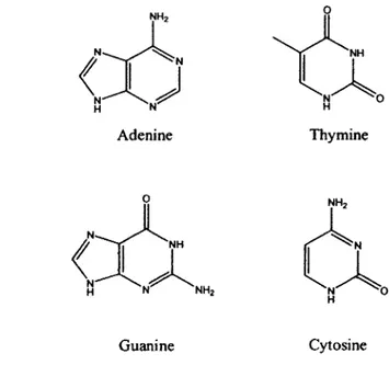
DNA molecule (Deoxyribonucleic acid molecule) is a polymeric macromolecule. The macromolecule consists of nucleotides. A nucleotide is a nucleoside and a phosphate group. A nucleoside consists of a 5-carbon sugar (2-deoxyribose) and a nitrogen containing base, i.e., nucleobase, attached to the sugar. There are four different base moieties referred to as nucleobases in DNA: adenine (A), thymine (T), guanine (G) and cytosine (C). The structures of the four nucleobases A, T, G, C are illustrated in Figure 6.
Adenine Thymine
Guanine Cytosine
Figure 6. Structures of the four nucleobases: A, T, G, C.
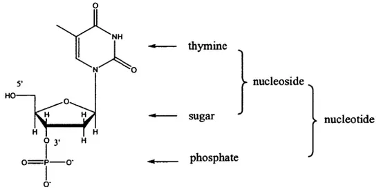
Consequently, there are four fundamental types of nucleosides (dA, dT, dG, dC) and eight fundamental types of nucleotides (pA or Ap, pT or Tp, pG or Gp, pC or Cp). Taking nucleobase thymine as an example, the nucleoside is called thymidine (dT, dThd) and if the phosphate is connected to the 5' of the sugar, the nucleotide is called thymidine-5'-monophosphate (pT) and if the phosphate is connected to the 3' of the sugar, the nucleotide is called thymidine-3' -monophosphate (Tp ). The structure ofthymine-3'-monophosphate, i.e., Tp, is illustrated in Figure 7.
. - thymine 5' nucleoside HO . - sugar nucleotide 0 3• H 1 o=p-o· _ phosphate 1 o·
Figure 7. Structure of a nucleotide, thymine-3 '-monophosphate, i.e., Tp.
A DNA macromolecule consists of two antiparallel single stands of repeated mononucleotides. The repeated mononucleotides are connected one to the next through phosphodiester bonds. The two antiparallel single strands are connected together through hydrogen bonds between A-T and G-C base pairs having the form of a right-handed helix. The base to base 1t stacking between adjacent bases on the same
strand may also be attributed to the stability of the polymer.
DNA molecules are the most important functional and genetic molecules in the cell nucleus. So if DNA is damaged, there will be disastrous results such as cell death and mutation. As mentioned above (Chapter 1.2), DNA is the critical target for ionizing radiation. So the study of radiation induced DNA damage takes on great importance in medicine and radiobiology. Two well-know types of damage induced by ionizing radiation include single-strand break (SSB) and double-strand break (DSB). A break in one strand of the DNA is called a single-strand break. Single-strand breaks are usually repaired rapidly using the opposite strand as a template, and thus, they are relatively benign. The well separated DNA single-strand breaks on bath strands can also be readily repaired and also be thought of as single-strand breaks. But, if the breaks in the two strands are very near, for example,
within one tum of the helix or ten base pairs, or opposite to each other, this case is referred to as a double-strand break (DSB). Because there is no template for the repair, double-strand breaks are believed to be much more lethal than single-strand breaks induced by ionizing radiation. In addition, many other kinds of damages happen such as base damage, base release, DNA-DNA cross-links, DNA-protein cross-links, etc. The combined damage within ten base pairs is the most lethal, which is also called clustered damage or locally multiply damaged sites (LMDS). If the damaged DNA is repaired rapidly and correctly, the cell survives. If the repair is not within sufficient time, for example, within a cell cycle, or is incorrectly repaired (mis-repair), it may result in cell killing, mutation, or carcinogenesis.
Cells also protect themselves through an adaptive response, which means when large radiation exposure is preceded by a small dose, the effect of the large dose is sometimes diminished. This is a protective mechanism because small doses of radiation appear to stimulate protective responses, triggering DNA repair mechanisms and the elimination of severely damaged cells. However, all levels of radiation are considered harmful. For instance, there is no threshold (a level, e.g., a radiation dose, below which there is no observable biological effect) for induction of cancer by radiation. In addition, the phenomenon of bystander effect happens which means that induction of biological effects in cells that are not directly traversed by a charged particle but are in close proximity to cells that are. Thus radiation-induced damage to DNA is a complicated process and it is hard to predict the results. Besides DNA, there are other important targets in cells. For example, radiation damage to cell membrane may also be important because the membrane damage is possibly triggering apoptosis
Both direct ionization and water radiolysis products may lead to the formation of the types of strand breaks, which are called direct action and indirect action of radiation-induced DNA damage. In the direct action, a secondary electron can be produced from absorption of a photon and interaction with the DNA to induce a modification. In the indirect action, the secondary electrons can internet with water
molecules to produce hydroxyl radicals (OH•) or other species such as solvated electrons and H-atoms, which in turn produce DNA damage.
1.9 DNA damage induced by LEE
We know that secondary LEE are the most abundant species induced by ionizing radiation; thus the study of LEE-induced damage to DNA takes on great importance. In 2009, Wang et al. reported their remarkable discovery in the Journal of the American Chemical Society: even free electrons that have lost ail of their kinetic energy also internet with the DNA building blocks (Wang et al., 2009). This work was highlighted in a News & Views article in Nature (Sanche, 2009). By using femtosecond time-resolved laser spectroscopy, the researchers have revealed that adenine and cytosine can effectively trap an prehydrated electron (e-pre) to form stable anions in aqueous solution, and demonstrated that thymidine and especially guanine are highly susceptible to dissociative electron transfer of e -pre. leading to bond dissociation in DNA in aqueous solution. This challenges conventional ideas about radiation-induced DNA damage which suggest that the molecular source of genomic damage induced by the indirect effect in irradiated cells can be attributed to hydroxyl radicals (O'Neill and Fielden, 1993; von Sonntag, 1987; Sanche, 2009). Hydroxyl radicals usually form from the interaction of the water molecules in the cell with high-energy photons or charged particles. This finding indicated another important product of water's radiolysis, i.e., the prehydrated electron, might also be the source of DNA damage and serve as another important culprit.
Thus one can propose that the biological effects of radiation on living organism result not only from the impact of primary quanta on DNA, but also from secondary species that are produced by the primary interactions along thè radiation track. Among the secondary species, LEEs are the most abundant, so it is crucial to determine their action on DNA in cells. One can expect that LEEs will produce novel modifications on DNA molecules because these electrons have a kinetic energy that
can induce resonant interactions, such as the process of dissociative electron attachment (DEA). Different from damage to DNA induced by hydroxyl radicals, which leads to oxidation, LEEs initiate a reductive step at first, followed by reactions that will result in DNA damage.
1.10 Experimental design to model LEE-induced damage to DNA
For the experimental design to study the LEE-induced DNA damage, usually five parts are needed. The first part is an electron gun, which is the most important part and is used for the generation of the LEE beam. By setting diff erent parameters on it, it can emit electrons with a wide range of energies (from a few eV to hundreds of eV and obtain an absolute energy spread of 0.5 eV full width at half maximum). The second part is a chamber, which is used to host the electron gun; an oil-free turbomolecular pump is used to pump the chamber rapidly from atmospheric pressure to the ultra high vacuum (UHV) range (-10-9 Torr). UHV is necessary to protect the
electron gun when it is working and keep away any contaminants. The third part is the LEE bombardment target. Usually it is composed of condensed DNA molecules or their components in the gas or condensed phases. In the case of condensed-phase experiments, the fourth part is the sample holder, which is usually a metal substrate. Up to now, many kinds of metals have been tested but the best and most widely used are either tantalum or gold because experimental results have indicated that DNA was stable on tantalum or gold substrates. For the gas phase experiments, the DNA basic component must be vaporized to interact with the electrons emitted in the chamber. The drawback of the gas phase experiment is the possible decomposition of the sample during the heating process. Also, some big molecules cannot be vaporized so that the system is limited to the study of small molecules. For the solid phase experiments, the sample must be lyophilized on the metal substrate or chemisorbed on a gold substrate, e.g., oligonucleotide self-assembled monolayers (SAM). The fifth part is the product analysis instrument after LEE bombardment. lt can either be located inside or outside the UHV chamber. Many methods of analysis have been
used; for example, mass spectrometry (MS), high performance liquid chromatography (HPLC), gas chromato~aphy-mass spectrometry (GC/MS), liquid
chromatography-mass spectrometry-mass spectrometry (LC/MS/MS), electrophoresis, etc.
As for the LEE bombardment target, oligonucleotide DNA and plasmid DNA are widely studied using the method mentioned above (Zheng et al., 2005; Li et al., 2010; Boudaïffa et al., 2000) because they are similar to natural DNA macromolecules and their damage products are easy to detect and analyze. Oligonucleotide DNA is synthesized artificially. It can be short or long, in single strand form or double strand form. Also, several modified bases, e.g. 5BrU, can be introduced into the DNA strand. Plasmid DNA is usually extracted from microbes ( e.g., E. coli) and it is a natural macromolecule with 2000 - 150,000 base pairs. Short oligonucleotides and their stable decomposition products can be easily analyzed and quantified by HPLC/UV. The structure of decomposition products can be identified by GC/MS and LC/MS/MS. Long oligonucleotide DNA strands and their strand break products can be easily analyzed and quantified by polyacrylamide gel electrophoresis (PAGE) through Maxam-Gilbert sequencing. The smaller the strand break product, the faster it moves in the polyacrylamide gel. Plasmid DNA and its strand break products can be easily analyzed and quantified by agarose gel electrophoresis. There are three forms of plasmid DNA: supercoiled DNA (scDNA), open circular DNA ( ocDNA), and linear DNA. The natural form of plasmid DNA is supercoiled DNA. When a single strand breaks, it becomes open circular DNA ( ocDNA) and when the double strand break happens, it becomes linear DNA. In agarose gel, the order of migration is supercoiled DNA (scDNA) > linear DNA >open circular DNA (ocDNA). In my experiments, I use short oligonucleotide DNA molecules and HPLC/UV analysis for the products.
For now, short DNA single strands (oligonucleotide DNA) can be synthesized by the method of solid-phase synthesis of DNA or the polymer chain reaction (PCR)
using dNTPs (deoxy-ribonucleoside triphosphates: dATP, dGTP, dTTP, dCTP) and enzymes. The short DNA strand is called oligonucleotide. The oligonucleotide can be in its single-stranded or double-stranded configuration (the association of at least six to ten complementary nucleotide base pairs depending on the sequence is required for the stable formation of DNA duplexes in water). The double-stranded DNA achieves contact through hydrogen bonds between A-T and G-C base pairs. By heating or adding some chemical agents, such as urea, the hydrogen bonds between A-T and G-C break, and the double-stranded oligonucleotide denatures leading to a single-stranded oligonucleotide. By cooling down, the denatured single-stranded DNA can be renatured into double-stranded DNA. The oligonucleotide is a good model for studying DNA damage induced by ionizing radiation because it is easy to get and the damage products are easy to analyze. In my experiments, I use short, single-stranded oligonucleotides such as trirners.
1.11 Study of LEE-induced DNA damage in the past
In the past, much experimental effort focused on the interactions of LEE with simple DNA components or DNA macromolecules such as plasmid DNA. These studies include investigation of the basic interactions of LEE with nucleobases (Aflatooni et al., 1998; Scheer et al., 2004; Ptasmska et al., 2005; Gohlke et al., 2003; Abouaf et al., 2003; Hanel et al., 2003; Feil et al., 2004; Huels et al., 1998; Aboul-Carime et al., 2001b, 2004; Denifl et al., 2004; Klyachko et al., 1999), ribose derivatives (Antic et al., 1999; Antic et al., 2000; Breton et al., 2004; Huels et al., 2004; Lepage et al., 1998; Ptasillska et al. 2004, Park et al., 2006), oligonucleotides and plasmid DNA (Dugal et al., 2000; Aboul-Carime et al., 2000a; Huels et al., 2003; Pan et al., 2003; Martin et al., 2004; Ray et al., 2005; Folkard et al., 1993; Nogues et al., 2004). Meanwhile, theoretical simulations using ab initio or density functional theory (DFT) calculations have also been performed to predict the process involved (Anusiewicz et al., 2004; Bao et al., 2006; Barrios et al., 2002; Berdys et al., 2004; Gu et al., 2005, 2006, 2007, 2009, 2010; Kobytecka et al., 2008, 2009; Kumar and Sevilla,
2007; Li et al., 2002, 2003, 2006, 2007; Richardson et al., 2004, Schyman and Laaksonen, 2008; Simons, 2006, 2007; Voityuk et al., 2001; Voityuk, 2009; Wesolowski et al., 2001; Wetmore et al., 2001). These studies support the ability of LEE bombardrnent to induce DNA damage and suggest that a resonance process, i.e., dissociative electron attachment (DEA), happens, and that a stable anion forrns followed by fragmentation of the anion and formation of radical fragments. The technique of electron stimulated desorption (ESD) has been used to study the reaction of LEE with condensed-phase DNA components together with mass spectrometry (MS) detection of small primary radicals and ions (<100 a.m.u.) desorbing from the surface of the solid target upon LEE bombardment under ultra high vacuum (UHV) (Huels et al., 1998, 2003; Aboul-Carime et al., 2001b, 2004; Denifl et al., 2004; Klyachko et al., 1999; Antic et al., 2000; Ptasinska et al., 2004; Dugal et al., 2000; Pan et al., 2003). The existing LEE techniques are only capable of analyzing degradation products trapped in or desorbed from a condensed film, i.e., positive ion, negative ion, and neutral species can be measured. However, most of the non-volatile radiation products, which remain on the surface, have not been identified, e.g., strand break products, base reduction products, etc. There is a missing link to elucidate the chemical steps from the initial electron-initiated reactions to the final damage. Therefore, it is necessary by coupling chemical analysis with LEE irradiation to obtain information of the structure of the final products. The objective of this research project is to study chernical modifications of DNA induced by LEE, starting with basic DNA components.
For these reasons, a novel sarnple spin coating and LEE irradiator system has been set up in our group that can prepare a uniform film of biomolecules and used to bombard the inside surface of a cylinder target. With this configuration, we are able to irradiate a surface of a hundred times ( 10-50 µg) larger than with a conventional electron gun, and therefore, provide a sufficient amount of degraded material for preliminary chemical analysis of nonvolatile products outside the vacuum. After recovery from the substrate, the sample can be analyzed by high-pressure liquid
chromatography (HPLC}/UV, GC/MS, or LC/MS/MS for detection ofboth the strand break products (Zheng et al., 2005; Li et al., 2010) and the base reduction products (Park et al., 2010), so as to elucidate the mechanism of LEE-induced DNA damage.
1.12 The new LEE irradiator system in our lab
Recently, a novel spin coating and LEE gun irradiator system was developed in our lab (Zheng et al., 2004b). The difference of this system is that a large area of thin film of the sample surface (26 cm2) and a relatively large amount ofbiomolecules can be bombarded (10-50 µg) with LEE, providing sufficient degraded material for chemical analysis of the nonvolatile products. This novel system includes a spin coating system and an electron gun irradiator and is described briefly below.
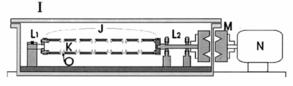
The biomolecules to be irradiated are spin coated onto the inner surface of the tantalum cylinders (Figure 8). Seven tantalum cylinders are packed together with Teflon spacers. The sample solution is injected independently onto the inner surface of each cylinder through the apertures between the spacers. The sleeve of cylinders was put into a chamber and rotated magnetically outside the chamber to an angular velocity up to around 1500 rpm. At this speed the liquid is distributed onto the inner surface of each cylinder uniformly. With pumping, the pressure inside the chamber rapidly drops from atmospheric to 2-5 Torr. During this procedure, the liquid freezes onto the tantalum substrate. When the pressure drops below the 2-5 Torr range, sublimation can be assumed to be completed and then the pressure rapidly reaches 100 mTorr. Usually, this step takes between 30 and 60 minutes depending on the solvent used. During this procedure, the sample can be assumed to be distributed uniformly onto the inner surface of the cylinders.
1
,,--··----
- J - ---·-..
N
Figure 8. Schematic diagram of the spin coating system. /- vacuum chamber,
J- tube holder, K- sample substrate, L1 and L.r-ball-bearing shafts, M-magnetic
coupling, N--electric motor, 0--Teflon spacer.
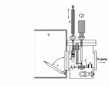
The LEE gun irradiator was put in an ultra high vacuum (UHV) chamber driven by an oil-free turbomolecular pump allowing the chamber to be pumped rapidly from atmospheric pressure to the 10-9 Torr range without significant
hydrocarbon contamination (Figure 9). The LEE gun irradiator can irradiate the inner surface (26 cm2) of a tantalum cylinder with 3-130 eV electrons having an absolute
energy spread of 0.5 eV full width at half maximum. The cylinders are mounted on a rotatable circular platform. Rotation of the cylinders secured to the platform allows each sample to be bombarded by the electron gun at a specific energy and current for a given time. The UHV chamber can be opened by a quick access port to a dry Nr purged glove box for the recovery of the sample in it. After irradiation, the tantalum cylinders are mounted with Teflon caps for recovery with water. Finally, the resulting solution is analyzed by HPLC/UV, GC/MS, or LC/MS/MS.
I
BTopump
---+
Figure 9. General v1ew of UHV electron irradiator chamber. A-electron gun,
B-linear drive, C-rotatable disk used as cylinder support, D-electron current detector, E--cylindrical sample substrate, F--quick access port, G-glove box sealed under a N2 atmosphere, H-ion gauge.
1.13 Previous results in our lab
Using this system and HPLC/UV, GC/MS analysis, Zheng et al. in our lab first irradiated a relatively simple DNA nucleoside, thymidine (Zheng et al., 2004a). The results show that LEE irradiation efficiently induces N-glycosidic bond cleavage in thymidine and gives thymine as a major product.
Furthermore, two oligonucleotide tetramers (CGTA and GCAT) were irradiated with monoenergetic LEE. The products included non-modified nucleobase, nucleoside and nucleotide fragments resulting from the cleavage of phosphodiester and N-glycosidic bonds of each tetramer (Zheng et al., 2005). This is consistent from results with thymidine. In addition, the distribution of non-modified products suggests a mechanism of damage involving initial electron attachment to nucleobase moieties,
followed by electron transfer to the sugar-phosphate backbone, and subsequent dissociation of the phosphodiester bond. These results demonstrate that the phosphodiester bond breaks by a distinct pathway in which the negative charge localizes on the phosphodiester bond giving rise to non-modified fragments with an intact terminal phosphate group. These studies support electron transfer of LEE from the base moiety to the sugar-phosphate backbone in DNA as suggested by theoretical studies (Simons, 2006). Such a mechanism of damage suggests that the capture of non-thermalized electrons by DNA bases followed by the formation of strand breaks may be an important factor in radiation induced DNA damage in living cells.
1.14 Description of this research project
The main objective of the present research project is to elucidate the chemical aspects of the mechanism of DNA single strand breaks induced by LEE using the newly-developed LEE gun irradiator system and small DNA components as model substrates and direct chemical analysis (HPLC/UV analysis) of the products remaining on the substrate as the basis for DNA damage.
Our initial study examines the effect of terminal phosphate and base moieties on LEE-induced DNA damage, which constitutes the frrst article ( chapter II). The DNA model compounds are monomers (pT, Tp, pTp) and dirners (pTpT, TpTp, pTpTp) and a trimer TpTpT. First, we find that the presence of terminal phosphate groups in monomers (pT, Tp, pTp) and dirners (pTpT, TpTp, pTpTp) increases overall damage by 2-3-fold while it decreases N-C and C-0 bond cleavage by 2-10-fold. This suggests that the capture of LEE directly by the terminal phosphate does not contribute to N-C and C-0 bond cleavage. Second, we find that terminal bases appear to shield the internai base from damage, resulting in a bias of damage toward the termini. In summary, the presence of terminal phosphate base moieties greatly affects the distribution of LEE-induced damage in DNA model compounds.
investigated; it constitutes the second article ( chapter III). The model compounds include TXT, where X represents one of the four normal bases of DNA (thymine (T), cytosine (C), adenine (A), and guanine (G)). The results indicated that when trimers were irradiated with LEE (10 eV), the total damage decreased 2-fold in the following order: TTT > TCT > TAT > TGT. The release of nonmodified nucleobases mainly occurred from the terminal sites of trimers (i.e., T) whereas the release of central nucleobases was minor (C) or not at all detected (A and G). This study indicates that the initial LEE capture and subsequent bond breaking within the intermediate anion depend on the sequence and electron affinity of the bases, with the most damage attributed to the most electronegative base, T.
In chapter IV, the model is still the oligonucleotide trimer TXT, but X represents 5-bromouracil, a potential radiosensitizer, i.e., a 5-bromouracil substituted trimer TTT in the second place. The results show that, when TSBrUT is irradiated with LEE (10 eV), TUT is the major product. Substitution of 5-bromouracil with the central thymine of TTT increases the total damage to trimer by at least 50%. This change leads to an increase in both C-N bond and C-0 bond cleavage compared to TTT. These findings support the hypothesis that 5-bromouracil is a strong electron captor and LEE is initially captured by the base and then transferred from the base either to the C-N bond leading to base release or to the C-0 bond leading to phosphodiester bond cleavage. Most importantly, a relatively large percentage of fragments arising from N-C and C-0 bond cleavage contained uracil rather than 5-bromouracil (e.g., uracil, pUT and TUp) are detected, indicating that a single 10 eV electron induces double events, i.e., Br-C and C-N cleavage or Br-C and C-0 cleavage. These findings suggest that LEE from ionizing radiation induce multiply damaged sites in DNA especially when it contains modified bases such as 5-bromouracil that have a high cross-section for electron capture.
Overall, my studies have helped elucidate the mechanism of LEE-induced fragmentation reactions in DNA: the terminal phosphate group has a larger
cross-section in LEE-induced DNA damage; initial LEE capture and subsequent bond breaking within the intermediate anion depend on the sequence and electron affmity of the bases; at 10 eV, one electron can induce double events. The present study provides a chemical basis for the formation of strand breaks by the reaction of LEE withDNA.
Chapter II - First article
Low Energy Electron Induced DNA Damage: Effects of Terminal Phosphate and Base Moieties on the Distribution of Damage.
Zejun Li, Yi Zheng, Pierre Cloutier, Léon Sanche and J. Richard Wagner. Journal of the American Chemical Society, 2008, 130(17), 5612-5613.
1 did all the experiments in this paper and wrote the first version of the manuscript. Dr. Yi Zheng taught me how to use the machine and Mr. Pierre Cloutier helped me to repair the machine when it had problems. Prof. Léon Sanche and Prof. J. Richard Wagner are my joint supervisors and helped me to revise the manuscript and to submit it.
Résumé
Les électrons de basse énergie (EBE) induits des lésions à l'ADN par attachement dissociatif, ce qui implique la libération d'une base (rupture de la liaison N-glycosidique (N-C), et la formation de cassures (rupture de la liaison phosphodiester du sucre (C-0). La détermination de l'effet des EBE au phosphate terminal et aux bases a été évaluée par une exposition à des composés modèle d'ADN dans la phase condensée, suivie d'une analyse par HPLC-UV des produits restant à la surface. Tout d'abord, la présence de groupes terminaux de phosphate dans les monomères (PT, Tp, PTP) et des dimères (pTpT, TPTP, pTpTp) augmente en général les dommages de l'ordre de 2-3 fois, tandis qu'elle diminue 2-10 fois la rupture de la liaison N-C et la liaison C-0. Ceci suggère que la capture directement de I'EBE par le phosphate terminal ne contribue pas à la rupture de la liaison N-C et la liaison C-0. Deuxièmement, les bases terminales semblent protéger la base interne de dommages, entraînant un biais de dommages vers le terminus. En résumé, la présence de la fraction terminale de base de phosphate affecte sérieusement la distribution des dommages dans les composés modèle d'ADN induite par les EBE.
Mots clés: dommage à l' ADN, électrons de basse énergie, HPLC-UV
SYNOPSIS TOC
'-0Y Enerav Electrons 1+15 eV> - - - DNAOamage
~Thy·
~
ThyO...f.
9-(
Hd-ovy
s;Thy
n-Y-o
,0 HO OH Nucleobase_____..
---PhosphateBase Release (-Thy) 5' and 3' C-0 bond cleavage (Tp, pTpT, TpT, TpTp, pT plus modified fragments)
ABSTRACT
Low energy electrons (LEE) induce DNA damage by dissociative electron attachment, which involves base release (N-glycosidic bond (N-C) cleavage) and the formation of strand breaks (phosphodiester-sugar bond (C-0) cleavage). The effect of terminal phosphate and base moieties was assessed by exposing DNA model compounds to LEE in the condensed phase followed by HPLC-UV analysis of products remaining on the surface. First, we report that the presence of terminal phosphate groups in monomers (pT, Tp, pTp) and dimers (pTpT, TpTp, pTpTp) increases overall damage by 2-3-fold while it decreases N-C and C-0 bond cleavage by 2-J 0-fold. This suggests that the capture of LEE directly by the terminal phosphate does not contribute to N-C and C-0 bond cleavage. Second, we report that terminal bases appear to shield the internai base from damage, resulting in a bias of damage toward the termini. In summary, the presence of terminal phosphate base moieties greatly affects the distribution of LEE induced damage in DNA model compounds.
The transfer of energy from ionizing radiation to molecules generates large quantities (-105/MeV) of low energy electrons (LEE, <30 eV).1 These species react
with DNA and its components, leading to various ionic and radical fragments and strand breaks as one type of final damage. 2 Previously, we showed that LEE induces
N-glycosidic (N-C) cleavage, leading to base release ( e.g., release of thymine from thymidine), and phosphodiester-sugar (C-0) cleavage, leading to fragments containing a terminal phosphate group and as yet unidentified sugar fragments. 3'4
The most likely step leading to C-0 cleavage involves the decay of transient radical anions of the phosphate P=O x* orbital to the strongly dissociative C-0 cr* orbital.5
lt is unclear, however, whether initial phosphate P=O n* radical anions are created from (1) initial electron capture by the base moiety followed by transfer to the phosphate or (2) direct electron capture by the phosphate.6
•7 Here, we show that the
presence of terminal phosphate groups enhances total damage but decreases C-N and C-0 cleavage, suggesting that the latter steps in the formation of damage occur by initial electron capture by the base rather than the phosphate group.
In this study, we focus on LEE induced damage within a series of small DNA model compounds ( dThd, pT, Tp, pTp, TpT, pTpT, TpTp, pTpTp, and TpTpT; counterion =Na+; Figure 1). Details of the irradiation system have been described.8
Briefly, the above compounds were spin-coated on the inside surface of tantalum cylinders (3.2 cm x 2.5 cm diameter) to obtain a thin solid film (2.5 nm) of 4 to 5
monolayers. The compounds were then bombarded with -1016 monoenergetic
electrons at ambient temperature under a vacuum of 10-9 Torr. The electron energy
was selected to coïncide with the peak of resonance ( 11 eV) using a dose within the linear regime of damage
as
shown for dThd (Figures SI and S2). After irradiation, the samples were transferred to a N2-purged glovebox, dissolved in aqueous solution,and lyophilized to dryness under vacuum. The parent and modified compounds were quantified by HPLC-UV as shown for TpT (Figure S3). The identity and amount of products were based on comparison of their retention and absorption properties with authentic standards. In addition, the analysis of some products was confirmed by
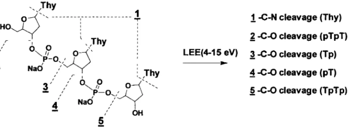
Figure 1. Structure of 5'-TpTpT-3' and position(s) of cleavage for N-glycosidic
(N-C) (1) and phosphodiester-sugar (C-0) bonds (2-5). The structure of other DNA model components consists of monomers (pT, Tp, pTp) and dimers (pTpT, TpTp, pTpTp). Na was the counterion.
treatment with alkaline phosphatase.
1
-C-N cleavage (Thy)~ -C-0 cleavage (pTpT)
LEE(4-15 eV) A-C-0 cleavage (Tp)
~ -C-0 cleavage (pT)
.§ -C-0 cleavage (TpTp)
The yield of LEE induced products is given in Table 1. The results show that the addition of terminal phosphates causes considerable increases in total damage as estimated by HPLC-UV (note that total damage includes all losses of initial molecules). The total damage for monomers increased from 47 to 129 to 162 for thymine containing zero, one, and two terminal phosphate groups, respectively. A similar trend was observed for the series of dinucleotides. The total damage for dinucleotides increases from 141 to 267 to 321 for TpT, pTpT, and pTpTp, respectively.
These results indicate that terminal phosphate groups efficiently capture 11 eV electrons and that these events cause considerable damage. This effect may be related to the primary structure of phosphate or to other factors, such as changes in the conformation of molecules, changes in the intermolecular ordering of molecules, or to the presence of H20 bonded to phosphate groups. Recent experiments with thin
very large cross section (10-15 cm2
)9 for LEE (7-12 eV) induced fragmentation,
which is much larger than that of the furyl ring. 10
•11 Thus, the increase in total
damage upon the addition of terminal phosphates may be attributed to increases in the initial electron capture by phosphate.
In sharp contrast to total damage, the presence of terminal phosphate leads to a marked decrease in base release (N-C) and phosphodiester-sugar (C-0) cleavage. For example, N-C cleavage decreased from 9.5 to 4.9 to 0.9 in monomers and from 23.4 to 6.4 to 1.7 in dimers for zero, one, and two phosphate groups (Table 1). Similarly, the presence of terminal phosphate decreased C-0 cleavage in dimers (12.9 to 9.4 to 3.8). The same trend was observed taking values of damage normalized to the size of the molecule (Table l; values in parentheses). lnterestingly, a similar decrease in N-C bond cleavage occurs for other nucleosides and mononucleotides of DNA (dAdo and dAMP, dCyd and dCMP, and dGuo and dGMP; unpublished results).
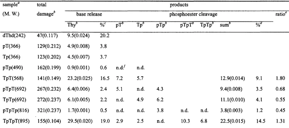
Table 1. Yield of Products from LEE Induced DNA Damage sample0 (M. W.) dThd(242) pT(366) Tp(366) pîp(490) Tpî(568) pTpî(692) Tpîp(692) total
damageb base release Thy6 47(0.117) 9.5(0.024) 129(0.212) 4.9(0.008) 123(0.202) 4.5(0.007) 162(0.199) 0.9(0.001) 20.2 3.8 3.7 0.6
n.d.1
141(0.149) 23.2(0.025) 16.5 7.2 267(0.232) 6.4(0.006) 272(0.237) 6.1(0.005) 2.4 5.1 2.2 n.d. pîpîp(816) 321(0.237) 1.7(0.001) 0.5 n.d. TpTpT(895) 155(0.104) 29.5(0.020) 19.0 2.9 n.d. 5.7n.d.
4.9 n.d. 2.5 pîpb 4.3 6.2 3.8n.d.
products phosphoester cleavage pTpf' Tpîp6 sum6 n.d. 10.3n.d.
6.8 12.9(0.014) 9.4(0.008) 11.1(0.010) 3.8(0.003) 22.5(0.015) 9.1 3.5 4.1 1.2 14.5 1.80 0.68 0.55 0.45 1.31a DNA samples and fragments written from 5' to 3' with p indicating the position of terminal phosphate groups (MW in parentheses). Total
damage includes all losses of initial targeted molecules based on HPLC-UV analysis (see Supporting lnfonnation for details). The values are expressed as a ratio of damaged molecules to 1000 initial target molecules and are the average of three independent experiments; SD