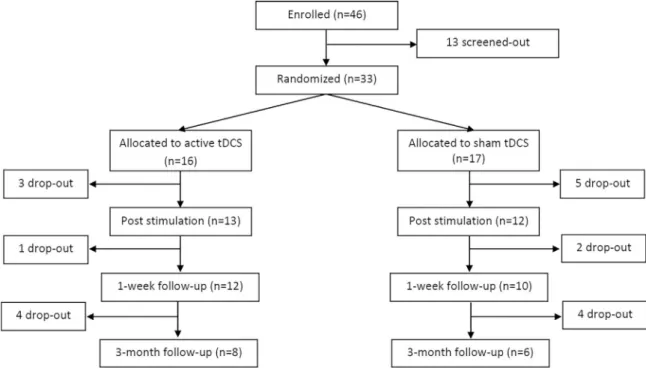
Delayed pain decrease following M1 tDCS in spinal cord injury: A randomized controlled clinical trial.
Texte intégral
Figure
Documents relatifs
They are respectively the Standard and Poor's 500 Composite Index (S&P) and the equally weighted (USEW) and value weighted (USVW) market portfolio of all stocks
We conduct an automatic and manual evaluation of the difficulties of processing these domains for part-of-speech prediction, and introduce a pilot study to determine whether
A primitive field is in the form of a couple (Name, Type) where Type corresponds to a data type like Integer, Boolean and String; it can be multivalued. A complex field
We describe how we have handled the 3 main problems in automatic testing reactive and realtime software like SBIMS: (i) the generation of relevant input data for testing,
Les auteurs doivent également s'assurer qu'ils ont dûment reconnu toute contribution au financement de la recherche, ainsi que de toute contribution significative au travail
La Revue recherche et encourage aussi la publication des travaux de chercheurs internationaux, stimulant ainsi, à travers le monde, le dialogue entre spécialistes sur des
Nous pouvons donc conclure que les données de la littérature tendent à confirmer l’intérêt d’une prise en charge orthophonique dès le stade précoce de la maladie
