in Great All reserved
BIOLOGICAL ACTIVITIES OF PHTHALOCYANINES-XI.
PHOTOTOXICITY
OF SULFONATED ALUMINUM
HAMSTER CELLS
NAPHTHALOCYANINES TOWARDS V-79 CHINESE
BENOIT
PAQUETTE,
HASRAT
ALI, REJEAN
LANGLOIS
and JOHAN
E.
V A NLIER*
M R C Group in the Radiation Sciences, Faculty of Medicine, University of Sherbrooke,Sherbrooke, Quebec, Canada J I H 5N4 (Received 17 April 1989; uccepied 14 September 1989)
Abstract-The phototoxicity of sulfonated aluminum naphthalocyanines towards V-79 Chinese hamster cells is investigated. T h e disulfonated naphthalocyanine exhibits similar photostability, but better cell penetrating properties than the tetrasulfonated dyes. T h e capacity of the naphthalocyanines to generate singlet oxygen is comparable to that of the corresponding phthalocyanines. However, in contrast to the phthalocyanine dyes, the sulfonated aluminum naphthalocyanines show very little phototoxicity towards the V-79 cells, suggesting close association with non-vital cell constituents or extensive formation of photoinactive adducts and aggregates.
INTRODUCTION
Photodynamic therapy (PDT)t has been proposed
as an alternative treatment modality to complement
conventional protocols in the management of malig-
nant tumors. Specific accumulation of non-toxic
dyes and localized application of therapeutic red
light allows for a higher selectivity of PDT protocols
as compared to conventional therapies of tumors.
The photosensitizer preparation currently used in
clinical trials of PDT consists of a mixture of hem-
atoporphyrin derivatives (HPD); a preparation
enriched in the active components is marketed as
Photofrin
IITM.
The effectiveness of PDT could be
enhanced by the use of sensitizers which are more
selectively retained by tumors and which absorb
more strongly towards the red end of the light spec-
tum.
To date, using the latter approach, numerous
porphin analogs with red-shifted absorption maxima
have been evaluated as possible second generation
sensitizers for PDT (for a review see van Lier,
1988). Among them, naphthalocyanines (Nc) (Fig.
1) are of particular interest, distinguishing them-
selves by a strong absorption band at about 760 nm
which corresponds to a further 100 nm red-shift as
compared to the structural analogous phthalocyan-
ines (Pc) (Fig.
2 ) .
The latter dyes have been studied
extensively for their potential use in PDT (for
reviews see Spikes, 1986; Ben-Hur, 1987; van Lier
*To whom correspondence should be addressed. PAbbreviations: D M F , N,N-dimethylformamide; HPPI,
3a-hydroperoxy- 1,2,3,3a, 8,8a -hexahydropyrroIo[2,3b] indole-2-carboxylic acid; Nc, naphthalocyanine; NcS,, mono- to tetrasulfonaphthalocyanine; PBS, phosphate buffered saline; Pc, phthalocyanine; PcS,,, mono- to tetrasulfophthalocyanine; P D T , photody- namic therapy; @, molecules formed per absorbed pho- ton.
et a [ . ,
1988; van Lier
andSpikes, 1989). At
750-800 nm light penetration of tissue is two fold
better than at the highest absorption band of hem-
atoporphyrin derivatives, e.g.
630 nm (Anderson
and Parrish, 1982). Although the synthesis of Nc
was reported by Bradbrook and Linstead as long
ago as 1936, this dye was only recently suggested
for use in PDT (Spikes, 1986). Some photophysical
and photochemical studies
o nNc's and their water-
soluble sulfonated derivatives (NcS), and particu-
larly their capacity to generate
lo2,
suggest their
potential value for PDT (McCubben and Phillips,
1986; Firey and Rodgers, 1987; Firey et al., 1988).
To further investigate the potential of Nc as a photo-
sensitizers for PDT, we evaluated the photodynamic
properties of aluminum naphthalocyanines, sulfon-
ated to different degrees, using the following para-
meters: (a) capacity to photooxidize tryptophan in
homogeneous solution, (b) uptake by V-79 Chinese
hamster cells in vitro, (c) photostability in V-79 cells
and (d) toxicity and photocytotoxicity towards V-
79 cells.
MATERIALS AND METHODS
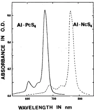
Sulfonated aluminum phthalocyanines (AI-PcS,,) were prepared as previously described (Ali el a / . , 1988). The corresponding naphthalocyanines were prepared in a simi- lar manner. T h e spectral properties of the monomeric dyes, i.e. in 100% MeOH, were: AI-PcS, A,.,, 674 nm. E 19 x 10' M-I c m - ' . , AI-NcS, A,,,, 771 nrn, c 16 x 10' M - ' crn-I.
Mono- io ietrasulfonared uluminum naphihalocyanine (A/-NcS). Chloroaluminum naphthalocyanine (AICI-Nc) was prepared by condensation of 2.3-dicyanonaphthalene and AICI, (anhydrous) at 27@-280"C for 1 h . The sulfon- ation procedure was adapted from Linstead and Weiss
(1950). Briefly, AlCI-Nc (200 mg) was dissolved in 5 me oleum (fuming H2S0, containing 15% free SO,) at 50°C with stirring. The mixture was kept at this temperature
for 45 min whereafter it was poured onto crushed ice. The resulting dark green precipitate was filtered and washed with IN HCI (10 mP), redissolved in IN NaOH (15 m t )
314 BENOIT PAQUETTE et al.
Al
- N c S ~
Al
- P c S ~
Figure 1. Chemical structure of sulfonated aluminium naphthalocyanine (AI-NcS) and sulfonated aluminum
phthalocyanine (AI-PcS). R = SO; or H.
I
IWAVELENGTH
IN
nm
Figure 2. Absorption spectra of AI-PcS, and AI-NcS, (3 ) L M ) in MeOH/H,O (95:5) in a I cm path quartz
cuvette.
and filtered again in order to remove insoluble impurities, including non-sulfonated naphthalocyanine. The filtrate was neutralized with I N HCI and analyzed by HPLC, revealing the presence of tetra-(major) and trisulfonated (minor) products. In order to obtain mono- and disulfon- ated products, AICI-Nc (100 mg) was dissolved in 1.5 mY
of oleum and 1.5 mY H,SO, (sp. gr. 1.84). The reaction mixture was kept at 75-80°C for 1-4 h (depending on the degree of sulfonation required), poured onto crushed ice and worked up in the usual manner.
Mixed AI-NcS (100 mg) in 100 mY of water were frac- tionated by chromatography on a 30 cm long x 2 cm i.d. glass column packed with C-18 reverse phase, particle size 25-40 pin (Macherey-Nagel, Diiren, Germany). Elution was carried out with a stepwise gradient of 0-100% MeOH in 10 mM sodium phosphate buffer, pH 5.0. AI-NcS sul-
fonated to different degrees were collected, concentrated under vacuum, adsorbed on a small amount of reverse phase (C-18) packing, washed with water to remove salts and eluted with MeOH (25-70%) in water. The compo- sition of the various purified AI-NcS,, fractions was determined by HPLC in an identical manner as described for the analogous AI-PcS,-, (Ah et al., 1988), except that
NcS were detected by their absorption at 700 nm. Briefly, this system consists of a 25 cm (0.94 cm i.d.) reverse phase column packed with ODS-2 spherisorb, 5 p m (CSC, Montreal) operated at 2 mY min-' with a linear gradient (55 min) from 0 to 95% MeOH in 10 mM sodium phos- phate buffer, pH 5. Retention times of the AI-NcS,, were similar to those observed for the corresponding AI-PcS,, and ranged from 20 to 25 min for the tetra-, 25 to 35 min for the tri-, 35-55 min for the di-, and 55 to 65 min for the monosulfonated derivatives.
Determination of the degree of sulfonation. In addition to HPLC analysis, the average degree of sulfonation of the various AI-NcS fractions was determined via quantifi- cation of the ratios between sulfonated and nonsulfonated naphthalimide fragments derived from the oxidative degradation of the Nc macrocycle (Ali ef al., 1988). For
this assay about I mg of AI-NcS was dissolved in 1.8 mY of DMF followed by the addition of 0.2 mY HNO, (conc.). The solution was heated (50-90°C) until the characteristic blue-green color disappeared and then neutralized with 1N NaOH. An alternative degradation procedure which results in the formation of fewer secondary products and which is applicable to both PcS and NcS dyes, involves the use of ammonium cerium(1V) nitrate as an oxidizing agent. In the latter case, approx. 1 mg of the dye was dissolved in a few mY of MeOHIH,O (1:l) followed by the addition of a few mg of the cerium salt. The mixture was heated (50-90°C) until the color disappeared. Product analysis after both degradation procedures was performed on a reverse phase HPLC column (25 cm long x 0.94 cm i.d.) packed with ODs-2 on 5 p m spherisorb (CSC, Mon- treal), operated at 2 melmin with 0.1% trifluoracetic acid in water (5 min) followed by a 30 min linear gradient from this solvent to 100% MeOH. Phthalimide and naphthalim- ide products are well separated in this system and quant- ified by manual integration of their absorption peaks at 215 nm. Retention times include 3-sulfophthalimide 15 min, 4-sulfophthalimide 16 min, phthalimide 28 min, sul- fonaphthalimide 22 min and naphthalimide 32 min.
Phorocytotoxicity assay. Cell survival of Chinese ham- ster lung fibroblasts (line V-79) was determined using a colony forming assay as described by Brasseur et al. (1985). Cells were maintained in growth medium (MEM) supplemented with 10% fetal bovine serum, 1% L-gluta- mine and 1% vitamins (Flow lab). Cells in log phase were plated in 60 mm Petri dishes with growth medium and incubated for 3 h at 37°C in 5% CO, to allow cell attach- ment. The number of cells plated was adjusted such as to yield 200 colonies after treatment. The cells were rinsed with PBS and incubated for 1 h in the dark at 37°C with 1
m t
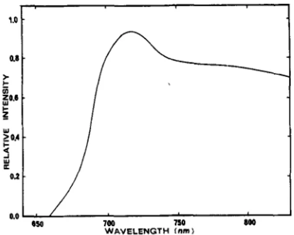
of medium containing 1 % serum and 10-100 p M dye. After removal of the dye and washing with PBS, the cells were exposed at room temperature for 4 rnin to red light from a 500 W tungstedhalogen lamp equipped with a 10 cm water filter and a red filter (LL-7OO-S-J579, Corion). The emission spectrum is presented in Fig. 3. The fluence over the absorption peak of the photoactive monomeric dye (751-791 nm) was 27 kJ/m2. After photo- treatment, the cells were re-fed with growth medium and incubated at 37°C in 5% CO, for 6 7 days whereafter colonies were counted. Experiments were repeated twice using four dishes per concentration point.Cell uptake and photostability. About 4 X 10' Chinese hamster lung fibroblast cells (line V-79) were plated in 60 mm Petri dishes. After an incubation period of 3 h under
5Y0
C 0 2 , to allow cell attachment, 70 )LM AI-NcS in 1 ml of medium containing 1% serum was added and cells were incubated for 1 additional hour in the absenceof light. After removal of the medium, cells were washed three times with PBS and irradiated 8 min with red light
to receive a fluence of 72 kJim' (calculated over the 751-791 nm absorbance peak of the photoactive mono- meric dye). Cells were detached with 600 p t trypsin- EDTA 0.25% (4-5 rnin incubation) and dishes were
Figure 3. Emission spectrum of the light source used in the photocytotoxicity and in virro photostability assays.
washed with 600 pt' PBS. Cells were collected by centifug- ation in 1.S mt' plastic tubes ( 5 rnin, 600 g) and resus- pended in 200 p t of buffer (Tris 0.2 M , MgClz 10 mM,
CaCl,, 1 mM, p H 7.8). After three freeze-thaw cycles in liquid nitrogen, cell debris was incubated in the dark overnight with 0.5 unit of proteinase K (Sigma) and diluted in 100 pf of Tris buffer. D N A was digested for 2 h at 37°C with 1 unit of micrococcal nuclease (Wor- thington). To eliminate hydrophobic and ionic interactions with cellular fragments, SO pt' D M F was added whereafter the mixture was incubated for 30 min, followed by the addition of SO pt of 1.0 N N a O H and a 10 rnin incubation period at 37°C. The fluorescence of the diluted cell extracts (0.5 to 1.0%) in a mixture of MeOHIH,O (95:s) was measured after a 1 h stabilization period. AI-NcS and AI-PcS fluorescence was measured with a SLM-Aminco SPF-S00C spectrofluorometer ( AI-NcS: A,, := 760 nm, A,,,, = 790 nm; Al-Pcs: A,, = 660 nm, A,, = 690 nm) and concentrations were calculated by means of standard curves. T h e extraction efficiency was evaluated by adding a known amount of dye after the freeze-thaw treatment of the cells followed by the usual workup.
Quantification of L-tryptophan peroxides. The interac- tion of L-tryptophan with '0, in homogeneous aqueous solution yields a pair of characteristic hydroperoxides which have been characterized as the isomeric cis and
trans 3a-hydroperoxy-l,2,3,3a,8.8a-hexahydropyrrolo[2,
3b]indole-2-carboxylic acids (HPPI) (Langlois et a[., 1986). HPPI is readily quantified by a colorimetric procedure using Fe'+ and xylenol orange. Fez+ is oxidized to yield a Fe,'-xylenol orange complex with a A,,,fi., at 540 nm (Gupta, 1973). We have previously used this reaction to compare the ' 0 , generating capacity of differently sulfon- ated Ga-PcS and shown that the @(HPPI) is directly proportional to the @(lo,) (Wagner et al., 1987). T h e reagent was prepared fresh before use by mixing the fol- lowing stock solutions: 10 parts of 4 x M ferrous ammonium sulfate hexahydrate in 0.1 N H,SO,, one part of lo-' M xylenol orange tetrasodium salt and nine parts of purified water (conductivity < 0.25 z (I-' cm-'1.L-Tryptophan (2.0 m M ) and the AI-PcS o r AI-NcS dye (3.5 ( L M ) in MeOHIH,O (95:5), saturated with 0, by purging for 5 min. were irradiated for 1 h with red light, Prior to use, dye solutions were purified twice on a 1 ml' kelex column (Sigma, dry mesh SC-100) in order to remove traces of metal ions. The irradiation apparatus consisted of a 1000 W xenon lamp collimated by a double condensing lens, a 10 cm cooled water filter, a Spectral Energy monochromator (model GM 252) mounted with a G M A 152 quartz focusing sleeve at the entrance, a red filter (Ealing 26-4390) and a lens to focus light on a quartz cuvette (1 cm path length). T h e amount of light absorbed was determined with an Optikon radi-
ometer (model 88 XLC) fitted with a model 300 probe, calibrated via Reinekers salt actinometry. O n e milliliter of the irradiated sample was mixed with 2 m t of reagent and absorbance at 540 nm was measured after 1 h. Hydro- peroxide (HPPI) formation was quantified taking E = 2.68 x lo4 M - ' cm--l at 540 nm for the Fe3+-xylenol orange complex. Values were corrected for photodegrad- ation of the NcS and PcS dyes, which was determined from dye absorption before and after irradiation.
RESULTS AND DISCUSSION
Effect of the degree of sulfonation of Al-NcS
and Al-PcS
onphototoxicity
The effect of two AI-NcS fractions, sulfonated
todifferent degrees,on V-79 cell survival is presented
in Fig. 4. The AI-NcS3., fraction, containing mainly
tri- and tetrasulfonated dye, showed little photo-
cytotoxicity with 73.7% cell mortality at
70
p M .A
substantial dark toxicity, accounting for
20% of the
cell killing,
was observed. The
low phototoxicity of
this highly sulfonated dye is in agreement with earl-
ier observations on analogous AI-PcS preparations.
Under similar experimental conditions a high
LDuo
of 38
p Mwas reported for AI-PcS3,, (Paquette
et
al., 1988) while Ga-PcS4 showed only slight photo-
toxicity, even at elevated concentrations
of100
p M(Brasseur
etal., 1987).
Surprisingly, the hydrophobic AI-NcS,
,qfraction
showed only 19% cell death at
100
p M ,
either with
or without exposure to red light. This contrasts
Concentration
in
pMFigure 4. Survival of V-79 cells incubated for 1 h at 37°C with AI-NcS,,,
( A )
and AI-NcS,,, ( 0 ) followed by exposure to red light. The colony forming assay was316
BENOIT
PAQUETTEet
a1strikingly with our earlier observations on the anal-
ogous AI-PcS2, which induced 90% cell death at
1.7
p M(LD,,)
after exposure to red light, without
detectable dark toxicity (Paquette et al., 1988). Our
failure to observe photocytotoxicity with the sulfon-
ate naphthalocyanines could result from a number
of factors, including lack of cell uptake
of
the dyes,
high intracellular photodecomposition or inef-
ficiency to generate phototoxic products. These
possible explanations are addressed in the following
studies.
Cell
uptake and photostability
In view of the instability
of
the Nc dyes under
extreme alkaline conditions, we monitored the
fluorescence intensity for possible changes after the
addition of NaOH during the analytical procedure.
No variations were observed during the 10 min
incubation period. However, cell extracts were not
neutralized with HCI since this was found to lower
the reproducibility of the assay. At higher dye con-
centrations a green precipitate occurred, but at the
dye concentration used in our protocol, combined
with the final 1 h incubation period in 95% MeOH,
this problem was eliminated. Extraction efficiencies
varied between 85 and 100%.
Cell
uptake
Cell uptake of the AI-NcS and AI-PcS are pre-
sented in Table 1. Incubation conditions were
adjusted to those used in the photocytotoxicity
assay. It is evident from these data that the less
hydrophobic AI-NcS~.~
and AI-PCS~.~
fractions,
which contain mainly tri- and tetrasulfonated dyes,
accumulate poorly in V-79 cells. Accordingly, the
low level of photocytotoxicity observed with highly
sulfonated Nc or Pc dyes appears to reflect their
inability to cross the cell membrane. Absence of
AI-PCS~,~
in V-79 cells, even at elevated dye concen-
trations and prolonged incubation periods, was pre-
viously confirmed by fluorescence microscopy
(Paquette et
al.,
1988). In vitro, such polar dyes
accumulate only on the cell surface where they may
exert their phototoxic effect.
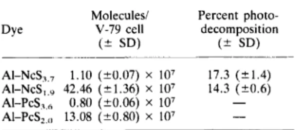
Table 1. Cell uptake and photodecomposition of
AI-NcS and AI-PcS*
Molecules/ Percent photo-
Dye V-79 cell decomposition
( 2 SD) ( 2 SD)
AI-NcS,,, 1.10 (20.07) X lo7 17.3 (k1.4)
AI-NcS,,, 42.46 (21.36) X lo7 14.3 (?0.6)
AI-P~s?,, 0.80 (k0.06) x 107 -
AI-PcS,,,, 13.08 (20.80) X
lo7
-
*Chinese hamster V-79 cells were incubated with70 K M of the Nc or Pc dye at 37°C for 1 h followed by a 8 min exposure to red light. Dye uptake by the cells was determined before and after irradiation.
In contrast, the more hydrophobic AI-NcS,
.9frac-
tion accumulated into V-79 cells 38 times better
than the highly sulfonated AI-NcS3,, fraction and
3.2 times more efficiently than the equivalently sul-
fonated AI-PcS2 fraction (Table 1). This high cellu-
lar accumulation likely reflects the distinct amphi-
philic property of the assymetric mono- and di-
sulfonated dyes. Accordingly, the lack of phototox-
icity of the mainly disulfonated Al-NcSl,y fraction
does not result from lack of cell penetrating proper-
ties of the dye.
Intracellular photostability
The same procedure was followed as described
for the cell uptake studies except that the irradiation
period was prolonged from 4 to 8 min (72 kJ/m2).
After extraction, the amount of dye was measured
by its fluorescence, and recovered dye levels were
compared to those from non irradiated cells (Table
1). The AI-NcS3,, and AI-NCS,,~
fractions showed
similar intracellular photostability, 17.3 and 14.3%
dye decomposition upon cell irradiation, respect-
ively. Since 2.7 times less red light fluence (27 kJI
m2) was used during the photocytoxocity assay, this
limited photodecomposition cannot account for the
lack of photocytotoxicity exhibited by the AI-NcS
preparations. Thus, the apparent photostability of
the sulfonated naphthalocyanines most likely
reflects extensive cellular aggregation of the dye,
which in turn would explain the lack of a photocy-
toxic response.
L-
Tryptophan oxidation
Chemical quenching
of
lo2by L-tryptophan fol-
lowed by quantitation of the resulting hydroperox-
ide products (HPPI) is an indirect way to measure
the capacity of the various dye preparations to gen-
erate
loz.AI-PcS2 gave a (P(HPP1) of 1.38
x(Table 2), which is within the range of earlier
Q,values reported for a series of Ga-PcS (Wagner et
al., 1987). The AI-NcS~,~
and AI-NCS~,~
fractions
exhibited only slightly lower efficiencies to oxidize
L-tryptophan with Q,(HPPI) of 0.89
xlou3 and
0.85 x
respectively. Furthermore, the
1.6
times lower efficiency for
lo2
production of the
AI-NCS~.~
vsthe AI-PcSZ, should, at the cellular
level, largely be compensated for by the 3.2 times
Table 2. Quantum yield for the for- mation of L-tryptophan oxidation products (HPPI) after exposure of L- tryptophan to AI-NcS or AI-PcS and
red light
AI-NcSA.7 0.89
AI-NcS, ,9 0.85
higher cell uptake
of
the former. Thus, lack
of
phototoxicity
of
disulfonated A1-NcS towards V-79
cells cannot be explained by limited cell uptake, by
intracellular stability, nor by lack of '02
generating
properties of this dye. These data suggest that the
lower sulfonated Al-NcS are highly aggregated in
the intracellular environment to yield photoinactive
complexes or, alternatively, that these dyes are
strongly associated with non-vital cellular com-
ponents which readily quench any
lo2
generated.
CONCLUSION
In these studies with
V-79 Chinese hamster cells,
we have evaluated phototoxicity, toxicity, cell
uptake, intracellular stability, and the capability to
generate
loz,of AI-NcS and Al-PcS sulfonated to
different degrees. Whereas all underlying par-
ameters leading to photosensitized cell killing are
similar for both the naphthalo- and phthalocyanine
dyes, the actual photocytoxicity of the two related
classes of dyes was strikingly different. While both
A1-PcS2
or A1-NcS2 were good generators of
lo2,showed good cell uptake and sufficient intracellular
photostability, only the Pc dye exhibited strong pho-
tocytotoxicity. Thus, lack of photoactivity of the
A1-NcS2 must involve another variable, such as
close association with non-vital cell constituents or
formation of photoinactive adducts and aggregates.
The lack of in vitro activity does not exclude possible
in
vivo
activities of the naphthalocyanine dyes since
the latter does not necessarily require neoplasmic
cell uptake. Furthermore, the lower photostability
of the Nc dyes could limit unwanted secondary
effects such as skin toxicity. Finally, the availability
of inexpensive diode lasers, which deliver light a t
wavelengths compatible with the Q band absorption
of naphthalocyanines, combined with the above
considerations, warrants further
in
vivo studies on
the potential of Nc dyes as photosensitizers for the
P D T of cancer.
REFERENCES
Ali, H., R. Langluis, J . R. Wagner, N. Brasseur, B. Paquette and J . E . van Lier (1988) Biological activities of phthalocyanines-X. Synthesis and analyses of sul-
fonated phthalocyanines. Phorochem. Phorobiol. 47, 7 13-71 7.
Anderson, R. R. and J. A. Parrish (1982) Optical proper- ties of human skin. In The Science of Photomedicine (Edited by J. D. Regan and J . A. Parrish), pp. 147-194. Plenum Press, New York.
Ben-Hur, E . (1987) Photochemistry and photobiology of phthalocyanines: new sensitizers for photodynamic ther- apy of cancer. In From Photophysics to Photobiology (Edited by A. Favre, R. Tyrrell and J . Cadet), pp. 407-420. Elsevier, New York.
Bradbrook, E. F. and R . P. Linstead (1936) Phthalocyan- ines. Part VIII. 1,2-naphthalocyanines. J . Chem. SOC. 1744-1748.
Brasseur, N., H. Ah, D. Autenrieth, R. Langlois and J . E. van Lier (1985) Biological activities of phthalocyanines-111. Photoinactivation of V-79 Chinese hamster cells by tetrasulfophthalocyanines. Photochem. Photobiol. 42, 515-521.
Brasseur, N., H. Ali, R. Langlois and J. E. van Lier (1987) Biological activities of phthalocyanines-VII. Photoinactivation of V-79 Chinese hamster cell by selec- tively sulfonated gallium phthalocyanines. Phorochern. Photobiol. 46, 739-714.
Firey, P. A , , W. E. Ford, J. R . Sounik, M. E. Kenney and M. A . J . Rodgers (1988) Silicon naphthalocyanine triplet state and oxygen: a reversible energy-transfer reactions. J . A m. Chem.
SOC.
110, 7626-7630. Firey, P. A. and M. A. Rodgers (1987) Photo-propertiesof a silicon naphthalocyanine: a potential photosensi- tizer for photodynamic therapy. Phorochem. Photobiol. Gupta, B. L. (1973) Microdetermination techniques for
H,O, in irradiated solutions. Microchem. J . 18,363-374. Langlois, R., H. Ah, N. Brasseur, R. Wagner and J . E.
van Lier (1986) Biological activities of phthalocyanines- IV. Type I1 sensitized photooxidation of L-tryptophan and cholesterol by sulfonated metallo phthalocyanines. Photochem. Photobiol. 44, 117-125.
van Lier, J . E. (1988) New sensitizers for photodynamic therapy of cancer. In Light in Biofogy and Medicine (Edited by R. H. Douglas, J. Moan and F. Dall'Acqua), Vol. 1, pp. 133-140. Plenum Press, New York. van Lier, J. E., N. Brasseur, B. Paquette, J . R. Wagner,
H. Ah, R. Langlois and J . Rousseau (1988) Phthalocy- anines as sensitizers for photodynamic therapy of can- cer, In Photosemitisarion: Molecular and Medical Aspects (Edited by G . Moreno, R. H. Pottier and T. G.
Truscott). NATO AS1 Series H, Vol. 15, pp. 435-444. Plenum Press, New York.
van Lier, J . E. and J. D. Spikes (1989) The chemistry, photophysics and photosensitizing properties of phthal- ocyanines. In Photosensitizing Compounds: their Chem- istry, Biology and Clinical Use (Edited by T. J . Dough- erty, G. H. Bock and S. Harnett), Ciba Foundation Symposium 146, pp. 17-32. Wiley, Chichester. Linstead, R. P. and F. T. Weiss (1950) Phthalocyanines
and related compounds. Part XX. Further investigations on tetrabenzoporphin and allied substances. J . Chem. McCubbin, I. and D. Phillips (1986) The photophysics and photostability of zinc (11) and aluminum (111) sulfonated naphthalocyanines. J . Photochem. 34, 187-195. Paquette, B. H., Ali, R. Langlois and J . E. van Lier
(1988) Biological activities of phthalocyanines-VIII. Cellular distribution in V-79 Chinese hamster cells and phototoxicity of selectively sulfonated aluminum phthal- ocyanines. Photochem. Phorobiol. 47, 215-220. Spikes, J . D . (1986) Phthalocyanines as photosensitizers
in biological systems and for photodynamic therapy of tumors. Photochem. Photobiol. 43, 691-700.
Wagner, J . R., H . Ali, R. Langlois, N. Brasseur and
J. E. van Lier (1987) Biological activities of phthalocyanines-VI. Photooxidation of L-tryptophan by selectively sulfonated gallium phthalocyanines: sin- glet oxygen yields and effects of aggregation. Phoro- chem. Photobiol. 45. 587-595.
45, 535-538.