HAL Id: tel-02874581
https://tel.archives-ouvertes.fr/tel-02874581
Submitted on 19 Jun 2020HAL is a multi-disciplinary open access
archive for the deposit and dissemination of sci-entific research documents, whether they are pub-lished or not. The documents may come from teaching and research institutions in France or abroad, or from public or private research centers.
L’archive ouverte pluridisciplinaire HAL, est destinée au dépôt et à la diffusion de documents scientifiques de niveau recherche, publiés ou non, émanant des établissements d’enseignement et de recherche français ou étrangers, des laboratoires publics ou privés.
New computational approaches for investigating the
impact of mutations on the transglucosylation activity of
sucrose phosphorylase enzyme
Mahesh Velusamy
To cite this version:
Mahesh Velusamy. New computational approaches for investigating the impact of mutations on the transglucosylation activity of sucrose phosphorylase enzyme. Bioinformatics [q-bio.QM]. Université de la Réunion, 2018. English. �NNT : 2018LARE0045�. �tel-02874581�
Thesis submitted to the Université de la Réunion
for award of Doctor of Philosophy in Sciences
speciality in bioinformatics
New computational approaches for investigating the
impact of mutations on the transglucosylation activity of
sucrose phosphorylase enzyme
Mahesh VELUSAMY
Thesis to be presented on 18th December 2018 in front of the jury composed of
Mme Isabelle ANDRE, Directeur de Recherche CNRS, INSA de TOULOUSE, Rapporteur M. Manuel DAUCHEZ, Professeur, Université de Reims Champagne Ardennes, Rapporteur M. Richard DANIELLOU, Professeur, Université d'Orléans, Examinateur
M. Yves-Henri SANEJOUAND, Directeur de Recherche CNRS, Université de Nantes, Examinateur Mme Irène MAFFUCCI, Maître de Conférences, Université de Technologie de Compiègne, Examinateur Mme Corinne MIRAL, Maître de Conférences HDR, Université de Nantes, Examinateur
M. Frédéric CADET, Professeur, Université de La Réunion, Co-directeur de thèse M. Bernard OFFMANN, Professeur, Université de Nantes, Directeur de thèse
ெ ்்ந்ி ிை ு்ூுத்
ெ ்யாம் ெ ்த உதி்ு ை யகு் ானகு் ஆ்ற் அிு. -ிு ்ு ் ுதி் அ்ப் ுுக், எனு த்ைத ே ு ாி, தா் க்ூி, அ்ண் ு்ு்ுமா், த்ைக பா்பா, ஆ்தா ுு ்மா், ீனா, அ்ண் ்ுக், ெபிய்பா, ெபிய்மா ம்ு் உுுைணயா் இு்த அைண்ு ந்ப்கு்ு ்எனு மனமா்்த ந்ி. இ்த ஆ் ி்ைக ுுைம அை3 த்ு ுுுத்்காரண், எனு ஆ் ி்ைக இய்ுன் ேபராிிய் ெப்னா்் ஆஃ்ேம். ப்ே ு துண்கி் நா் மனதாு், ெபாுளாதார அளிு் க்3்ி் இு்தேபாு, என்ு இ்ெனாு த்ைதயாகே இு்ு எ்ைன பா்்ு்ெகா்3ா். ுி்பாக, எனு ூ்றா் ஆ்ு இுிி், அ ்்ு எ் ளே ா தி்ப்3 க3ைமக் ம்ு் ிர்ிைனக் இு்தாு், அைத்ெபாு்பு்தாு, அ ் என்ு ெ ்த ெபாுளாதார உதி, ப்கைB்கழக பிு ம்ு் இதர ி் ாக ்ப்த்ப்3 உதிகு்ு எ்ன ைகமா்ு ெகாு்தாு் ஈ3ாகாு. ேபராிிய் ஃிரெ3ி் ேக3் ம்ு் ேபராிிய் ிி் ா்3் ஆிேயாரு இைண ேம்பா்ை ்ு் எ் உளமா்்த ந்ி. ேபராிிய். ய்்-ெH்ி ேனு ்ு அ ்கேள உ்குை3ய ிிி ு்3ள ா்ுத், ேமாெ3்ேB் ்ப்த்ப்3 ு்ியமான பி்ுைரக் ம்ு் எ்ுுய ஆ்ு க்ுைர எுு த்கான உ்குை3ய அைன்ு ஊ்கமான ா்்ைதகு்ு் ி்க ந்ி. 3ா்3். ிி் அ்னா்் ம்ு் ிேயான் Hாஃ்ேம் ஆிய இு ு்ு் ுேராேம்் ்ப்த்ப்3 உ்குை3ய உதி்ு ி்க ந்ி. 3ா்3். ்ெ3பாேன ே3ேB்ே யா அ ்கேள உ்குை3ய ிு ிு ெதாி்ு்ப ்ப்த்ப்3 உதிகு்ு ந்ி. எ் ுதBா் ஆ்ி், ீூியி் த்ு் ி ம்ு் இதர ு்ியமான ிகைள ெ ்ு த்த ு்ு் ாி ுு்ப்தா்க், ேநாெய்ி மிி் ம்ு் ேபராிிய் பாிி் கா்ெ3ிய் ஆிேயாு்ு எ் ிர் தா்்த ந்ி. ேமு், ிூிய் நா்கி் என்ு மற்க ுியாத ிைனுகைள ெகாு்த ந்ப்க் ேமகBா்கா, அினா், ே ாு, ேம்், மு்கா, ெக்், அேராரா, ூி, ேUா, மி், ிைர் அ்ணா, எி, எ்ினா, ிி, அிBா, ராUா், அண்யா ம்ு் அி் ீூிய் தி் ெ ா்தகு்ு் எ்ேBாு்ு் எ் மனமா்்த ந்ி. ுி்பா ெ ்ி் அ்ணா, ு்தி அ்ி, Uனி ு்ி ம்ு் Hிதா, உ்குை3ய அளி்Bா அ்ு ம்ு் அ்கைர்ு ெரா்ப ந்ி.பாி்B த்ுறு்ு ீு ுு்ு, அ்த நா்கைள இிதாகு், மற்குியாத துண்களாகு் மா்ுன த்ே ் த்ி, பாBா அ்ணா, ி்ப்், ி, ெபி்் அைன ்கு்ு் எ் மனமா்்த ந்ி. ிிஎ்ி பாி் ஆ் க க ஊிய்கு்ு், ுி்பாக ேபராிிய். ேக்தி் எ்்ே ெப்் அ ்கி் மாுB் ு்ப்கைள் ப்ிய பயு்ள அிுைரகு்ு் ம்ு் ிு. ிூெப்் அ ்கி் ெதாி்ு்ப ெதா3்பான உதி்ு் எ் மனமா்்த ந்ி. எ்ேனா3, நா்ே3் நா்கைள ெபா்னான துண்களாக மா்ியி் ெபு் ப்ு ி்ப ்க் ெபனா்் ே3ி், ேUாக், 3ா்3். ஐயனா் ெ ்ிே ் ம்ு் மாுி. ுி்பாக, ெபனா்் ே3ி், ேUாH் ெH்ி்் ஆிய இு ி் ெதாி்ுைற ீிிBான ப்கி்ு ம்ு் உு க்பு்ுத் ெதா3்ுை3ய அிுைரக் எனு ுைன ் பி்ி் ுிுகி் ெபிய ப்ு ி்ிறு. இு ு்ுேம எனு மனமா்்த ந்ி. எ்ுை3ய ஆ்ிB்ைத ி பா்்த எ் ேதாிக் ிிகா, ிேனகா ம்ு் ுரி அைன ு்ு் ந்ி. இ ்கேளாு, எ் ேமB அ்கைற ம்ு் அளி்Bா அ்ு கா்ிய தாரி, ி்ரா்கா, எி், ிஃப், ் ா் ம்ு் இமாு்ு் எ்ுை3ய மணமாண்்த ந்ி. இைத தி்்ு, எ்ுை3ய பி்ி மாண ்கி் எ்ே\ா ைB பய்பா்ி்்கான ப்கி்ி்ு மனமா்்த ந்ி. 3ா்3். ெHௌ3ா ெப்ெH்ி-ேமா்ராி, ேபராிிய். பாிி் ிேளயி ம்ு் 3ா்3். யெ ா்ி் ே ரு் இு ு்ு், உ்குை3ய அளி்Bா அ்ி்ு் ம்ு் எ்ீு ெகா்3 தி்ப்3 அ்கைர்ு் ி்க ந்ி. ஆர்பகாB்ி், எ் ிறைம ீு ந்ி்ைக ை ்ு, எ்ைன இ்த ுைன ் ப்3்பி்ு்ு பி்ுைர்த ேபராிிய் ிெரெ3ி் ேக3், ேபராிிய். எ். யி்றா, ிு. ேகா்்்ி் ுணீB் ம்ு் 3ா்3். ி்ிய் ஆிேயாு்ு எ் மனமா்்த ந்ி. எ்ுை3ய ுதBா் ஆ்ி், ுத் ஒு மாத காB் எ்ிு ஆ்ு ூ3்ி் இு்ு ே ைB ெ ்ய அுமி்த ேபராிிய். எ். ீி ா ் அ ்கு்ு எ் மனமா்்த ந்ி. எ்ுை3ய மி்ுி்க ூ்த மாண ் ு ரா் ம்ு் இதர எ்ிு க ந்ப்கு்கி் உதி்ு் எ் ந்ி. எ்ுை3ய ிெ3் ேதாிக் ெ ்ி, ிேBாினா, ீி ம்ு் ுமருு அ ்கி் ஆ்ு க்ுைர ுு்கம ிபா்்த் ம்ு் ெ ்்ந்ி ூுத் ஆ்ிB் மா்ற்ி்கான ப்கி்ி்ு, எ் மனமா்்த ந்ி. எனு நB் ிு்ிக் அ்ு்Bா, ேமாக், கேண், க்ண், ஆ்்ி, கUா, ிி, ே ாு, ுு, ப்ி, க்ூி ந்ப்க் ம்ு் அைன்ு ஆ் க ந்ப்கி் ஊ்க்ி்ு், அ்ி்ு் எ் மனமா்்த ந்ிிைன இ்த மி்்ியான துண்ி் ெதிி்ு ெகா்ிேற். -மேக் ே ு ாி
Acknowledgements
Assistance given by those who ne’er received our aid, Is debt by gift of heaven and earth but poorly paid
-Thiruvalluvar First and foremost, I would like to extend my sincere gratitude to lord murugan, my family for holding my hands through things that make me nervous and for all the shoves when I needed them. They include my father Velusamy, my mother Kasthuri, my brother Muthukumar, my sister Pappa, my grandmother Kuruvammal, Meena, my brother Shanmugaraj, Periyappa and Periyamma.
I am also deeply indebted to my advisor, Professor Bernard Offmann who was the pillar of support through the toughest emotional and financial times of my Doctoral studies. His amazing guidance and care as an academic father were instrumental in motivating me towards success and without him this thesis would not have been possible. I would also like to extend my gratitude to Professor Frederic Cadet and Asst. Prof. Philippe Charton for their co-supervision. I have learnt so much from the three of them, and not just in terms of academics.
I would like to thank my thesis committee members Professor Yves-henri Sanejouand and Professor. Catherine Etchebest for giving moral support, valuable suggestions and immense ideas helped me to improve my research day by day. Also, my special thanks to Professor Richard Daniellou, Mme Irène Maffucci, Mme Corinne Miral for accepting to be part of my jury members.
I offer my profound gratitude to thank Professors Manuel Dauchez and Isabelle Andre for taking their precious time to review my thesis. Also, my special thanks to our collaborator Professor Tom Desmet and his student Dr. Tom Verhaeghe, whose thesis work served as basics for mine. Dr. Philippe Arnaud and Dr. Lionel Hoffmann cannot thank you enough for all the insightful contributions and wonderful help with GROMACS. My special thanks to Dr. Stephane Téletchea for his many technical advices.
My thesis project took me to several places, one and the initial being Region La Reunion. I would like to thank the entire Mr. Muthuswami and family, Noelly Madeleine and Professor Fabrice Gardebien, with my accommodation and housing in La Reunion. I also cherish the wonderful support and happy memories, that I had with my friends Avinash, Somasekar, Mekala, Max, Marushka, Gaile, Aurora, Julie, Joana, Marine, Brice, Eddy, Elvina, Vigna, Akila, Anamya. A
special mention to my siblings and friends from the AFIR Reunion Tamil community, brother Senthil, sister Sundari, kids Janani and Haritha for the abounding love and care they shared on me. My sincere gratitude goes to brothers Tharvesh, Bala, Gilbert, Sasidhar and Felix, who helped with my accommodation in Paris and gave me many happy memories. My heartfelt thanks to all the colleagues at DSIMB, Paris, especially Mr. Hubert, who provided many technical helps.
I wholeheartedly appreciate all the contribution of Benoit in helping me in with dissertation and his guidance in parameterization work. I am also eternally grateful to Johann, Iyanar for sharing their wisdom with me and providing priceless technical advices during the final year of my Ph.D. in Nantes. Especially, my heartfelt thanks to Johann for his help in parameterization and plotting puckering states. I am also greatly indebted to the help rendered by my friends Sneha, Surbhi and Lipika who shared an important part of my joys and sorrows in Nantes and helped me with my communication and proofreading of my thesis. Many thanks to my friends Maruthi, Dharani, Sitraka, Enys, Typhaine, Chao, Alka, Harsh, Deepak and Iman who showered their love and affection on me. I am also obliged to appreciate the help of the internship students for helping me with the ENZO. I would like to say a very warm thank you to Dr. Houda Benhelli-Mokrani, Professor Fabrice Fleury, Dr. Yvonnick Cheraud and all the members of UFIP for all your graciousness and kindness towards me.
My personal thanks to Professor Yathindra Narayanarao, Mr. Goldsmith Kunaseelan and Dr. Lilian Olivieri who pushed me towards this opportunity and had played a pivotal role in shaping my educational career. I would also like to express my sincere gratitude to Professor Srinivasan Narayanan who provided me the opportunity to work in the MBU research unit for a month and my senior Yuvaraj and MBU colleagues who greatly supported me there. Many thanks to my friends Selvi, Philomina and Kumaraguru who helped communicate this dissertation better. Finally, I would like to sincerely thank all my well-wishers, Abdhulla, Mohan, Ganesh, Kannan, Aarthy, Kaja, Nivetha, Sonu, Guru, Jeevitha, IST, my school, college and research group friends for their inspiration, motivation and encouragement throughout my doctoral studies.
This work was supported by the Conseil Reginal de La Reunion in the form of grant to Mahesh VELUSAMY Tier N° D2014029434, Allocations Régionales de Recherche de Doctorate-session 2014. Further, I would like to thank for the computational resources provided by Université de Nantes, CNRS UMR 6286, UFIP, 2 rue de la Houssinière, 44322 Nantes Cedex 3, France., Université de La Réunion, INSERM UMR-S 1134, DSIMB, 15 Avenue René Cassin, La Reunion, France., Université Paris Diderot, INSERM UMR-S 1134, DSIMB, 6 rue Alexandre Cabanel, 75739 Paris, France.
அ்்பி்ிேற் ...
எ் ெப்ேறா்
, ேகாதர், ேகாதி,
ுு ்மா் ஆ்தா
,
ீனா
ேபராிிய்
. எ். ய்ி்ிரா, ேபராிிய்.
ெப்னா்் Hாஃ்மா்
எ் ந்ப்க் ம்ு் உறின்க்
ெத் ்தா் ஆகா ெதிு் ுய்ித்
ெம் ு்த் ூி து்
.
-ிு ்ு ்To…
my Parents, Broher, Siser, kuruvammal grandma, Meena
Prof. N. Yahindra, Prof. Bernard Ofmann
my riends and relaives
hough fae-divine should make your labour vain;
Efort its labour's sure reward wil gain.
Table of Contents
Synopsis...18
Synopsis in English...18
Synopsis in French...21
Chapter 1 Literature review...23
1. GLYCOSYLATION: From Conventional Methods To Enzymatic Synthesis...23
1.1 Carbohydrates, Glycosides, and Glycoconjugates...23
1.2 Chemical synthesis of glycosides...24
1.3 CAZymes: Carbohydrate-Active enZymes...26
2. SUCROSE PHOSPHORYLASE...27
2.1 Classification Scheme...27
2.2 General properties...28
2.3 Crystal structure of SP and their associated reaction mechanism...28
2.4 Conformational changes during sucrose conversion...32
2.5 Interest of sucrose phosphorylases...35
3. GENERAL OBJECTIVES OF THESIS...37
4. TOWARDS ENZyme Optimization (ENZO)...39
4.1 Selection of essential residues and design of a smart library...40
4.2 Generating libraries of mutated sequences...43
4.3 Modelling of variants and validation of models...44
4.4 Molecular dynamic simulations of mutant models...46
4.4.1 Calculation of electronic properties...47
4.4.2 Carbohydrate force fields and employed software for simulations...48
4.5 Molecular docking of mutant models...48
Chapter 2 Parameterization Of The Glucosylated Aspartate Residue And Their Successive Applications...50
ABSTRACT...51
1. INTRODUCTION...52
2. MATERIALS AND METHODS...56
2.1. Construction of the glucosylated aspartate (DGC) residue...56
2.2. ESP partial atomic charge calculation...57
2.3. Derivation of DGC parameters using CGenFF following implementation in both MODELLER and GROMACS...57
2.4. Additional refinements on the problematic dihedral phases...58
2.5. Obtention of DGC parameters using GLYCAM06 forcefield and implementation in GROMACS...59
2.6. Obtention of DGC forcefield parameters using GAFF for the AMBER ff99sb-ILDN and implementation in GROMACS...60
2.7. Benchmarking of the modified CHARMM22 forcefield extended with DGC parameters within MODELLER...60
2.8. Benchmarking of the AMBER ff99sb-ILDN forcefield extended with the DGC force field parameters within GROMACS...61
2.9. Cremer-Pople puckering and binding site analysis...62
3. RESULTS AND DISCUSSION...63
3.1. Parameters of DGC residue...63
3.2. Cremer-Pople puckering reference plot...64
3.3. Refined dihedral phase...65
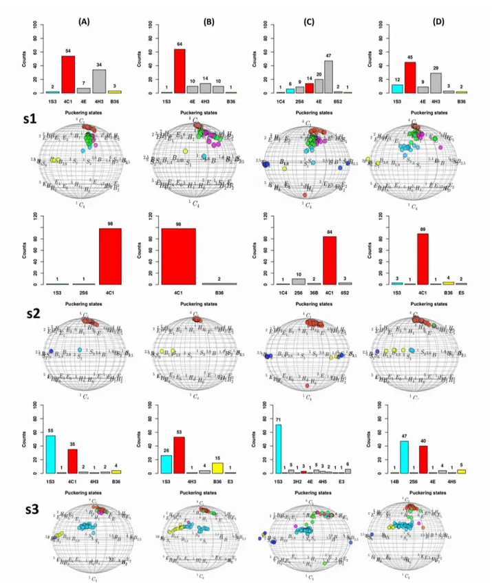
3.6. Molecular dynamics of DGC embedded structures...70
3.6.1. Results of CHARMM36 parameters...71
3.6.2. Results of a general AMBER forcefield (GAFF) parameters alone...72
3.6.3. Results of mixed (GAFF and GLYCAM-06) parameters...73
3.6.4. Results of GLYCAM-06 parameters along with glucosyl-threonine analogs...74
4. CONCLUSIONS...77
5. SUPPLEMENTARY INFORMATION...78
Chapter 3 Rational explanation for observed transglucosylation regio-selectivity patterns of variants sucrose phosphorylase: perspectives for engineering rare sugars...89
Abstract...90
1. Introduction...91
2. MATERIALS AND METHODS...94
2.1 Dataset of sucrose phosphoryalse variants...94
2.2 Modelling of sucrose phosphorylase variants...94
2.3 Benchmarking the role of environment...95
2.4 Modelling of sucrose phosphorylase variants...96
2.5 Supplementary experiments of mutate modeling...97
2.6 Modeling of sucrose phosphorylase variants using automated comparative modeling using automodel...98
2.7 Molecular docking of sucrose phosphorylase variants against glucose...99
2.8 Construction of sucrose phosphorylase database and identification of reference product binding poses...99
2.9 Statistical analysis for productive orientations...100
3 Results...101
3.1 The glucosylated form of aspartate residue...101
3.2 The preferred optimization region for sampling variants...101
3.3 Validation of mutate models and sampling assessment...102
3.4 Molecular docking results and sucrose phosphoryalse database...103
3.5 Regioselectivity analysis of mutate models and the respective docking poses...106
3.6 Validation of automodels and sampling assessment...109
3.7. Regioselectivity analysis of automodels and the respective docking poses...110
4. Discussion...112
6. Supplementary informations...114
Chapter 4 ENZO: An Automated Computational Tool For Engineering Sucrose Phosphorylase Enzyme And Their Homologues...125
ABSTRACT...126
1. INTRODUCTION...127
1.1 ENZO: An overview of the pipeline...129
1.2 ENZOWP1: Mutagenesis schemes for generating mutant sequence...130
1.3 ENZOWP2: Modeling of the 3D protein structures of the variants and its wild type sequence...133
1.4 ENZOWP3: Energy minimizations of the 3D models...134
1.5 ENZOWP4A/B: Molecular docking of ligands in a protein structure model...135
1.6 ENZO: The web interface...135
2. MATERIALS...137
2.1 Listing of the work packages inputs...137
2.2 Required softwares and hardware...138
2.3 Equipment: setup...139
3. PROCEDURE...140
3.1 Accessing to the ENZO web interface...140
3.2 Generate a mutants library...141
3.4 Energy minimization / molecular dynamics simulations of the mutant models...143
3.5 Molecular docking of acceptors in the minimized/non-minimized models...144
4. TROUBLESHOOTING...144
5. TIMING...145
6. ANTICIPATED RESULTS...146
7. SUPPLEMENTARY INFORMATIONS...147
Chapter 5 Conclusion and future perspectives...151
1. DISCUSSION AND CONCLUSIONS...151
2. EXPLORATION OF THE SUCROSE PHOSPHORYLASE MUTATIONAL LANDSCAPE FOR ENGINEERING APPLICATIONS...153
2.1 Construction of smart library of variants...155
2.1.1 Selection of starting structure...155
2.1.2 Annotation of sucrose phosphorylase structure...156
2.1.3 Identification of hot spot residues following filtration...159
2.1.4 Towards smart library based on stability and experimental data...162
2.2 Generation of 3D models of smart library following characterization of individual mutants using molecular docking...165
3. FUTURE PERSPECTIVES...168
List of Tables
Chapter 1Table 1: Classification schemes for sucrose phosphorylases...27 Table 2: List of interactions between highly conserved residues and the glucosyl moiety of sucrose phosphorylase of Bifidobacterium adolescentis...31 Table 3: The list of mutants that alter the selectivity of BaSP towards kojibiose and nigerose
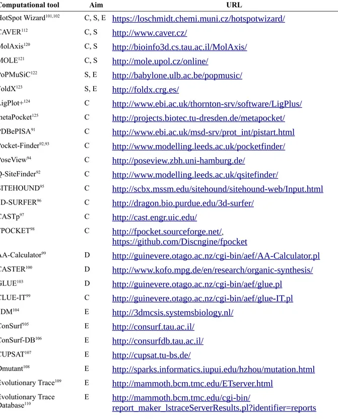
production...37 Table 4: Comparison of the three classical different enzyme engineering approaches...40 Table 5: List of available computational tools for smart library design...41 Table 6: The four different protein engineering strategies of Hotspots Wizard for the identification of hotspot residues...41 Table 7: Comparison of methods employed for quantum mechanic calculations...46 Chapter 2
Table 1: Benchmarking results of DGC forcefield parameters using MODELLER...68 Table 2: Distribution of individual puckering states of glucosyl moiety covalently linked to Asp nucleophile residue during MD simulations...70 Table S1: List of the three tested MODELLER parameter sets...78 Table S2: List of CGenFF parameters along with penalty score and the type of parameters...78 Table S3: The list of DGC residue atom numbers, atom names, atom types and their respective ESP partial atomic charges...80 Table S4: The list of four known covalent intermediate structures from glycoside hydrolase family 13 with ß-D-Glucose substrate covalently linked to a nucleophile Asp residue...81 Chapter 3
Table 1: The list of mutants that alter the selectivity of BaSP towards kojibiose and nigerose
production...93 Table 2. The statistics for number of models generated through the Mutate model method along with details of mutants and wild type...97 Table 3: The statistics for number of models generated through the Automodel method along with details of used soft sphere restraint weights, mutants and wild type...98 Table 4: Global analysis of binding modes of α/β-D-Glucose acceptors based on mutate model...107 Table 5: Global analysis of binding modes of α/β-D-methylglucosides based on mutate model....107 Table 6: The detailed analysis of binding modes of α/β-D-Glucose acceptors based on
mutate model ...108 Table 7: Global analysis of binding modes of α/β-D-Glucose acceptors based on automodel...111 Table 8: Global analysis of binding modes of α/β-D-methylglucosides based on automodel...112 Table 9: The detailed analysis of binding modes of α/β-D-Glucose acceptor
based on automodel...112 Table 10: The detailed analysis of binding modes of α/β-D-Glucose acceptors based on automodel and mutate model...113 Table S1: The list of tested mutants and their associated total number models and docking poses with respect to α/β-D-Glucose acceptors...116 Table S2: The list of tested mutants and their associated total number models and docking poses with respect to α/β-D-methylglucosydase acceptors...117 Table S3: Classical DOPE SCORE analysis for productive orientation with respect to β-D-Glucose acceptor...117 Table S4: The frequency of productive binding poses based on RMSD with respect to β-D-Glucose acceptor...118 Table S5: The frequency, Average (Avg) and Standard deviation (STD) of productive poses based on highest binding energy with respect to β-D-Glucose acceptor...120 Table S6: The frequency of productive binding poses based on top rank pose with respect to β-D-Glucose acceptor...121
Table S7: Comparison of puckering states (1S3,4C1, B36) counts across the models of 11 different experiments based soft sphere restraint weight...122 Table S8: The distribution of RMSD at soft sphere weight 10 with respect to the wild types 2gdv_A and 5c8b...125 Table S9: The list of tested mutants and their associated total number models and docking poses with respect to α/β-D-Glucose acceptors...125 Table S10: The list of tested mutants and their associated total number models and docking poses with respect to α/β-D-methylglucosidase acceptors...126 Chapter 4
Table 1: ENZO pipeline example files, modeller libraries and modified forcefield that are
incorporated with DGC residue...140 Table 2: The list of trouble shooting from the case studies...146 Chapter 5
Table 1: The list of available 3D structures of sucrose phosphorylase from Bifidobacterium adolescentis where 1R7A and 2GDV is wild-type active form and 2GDU is
inactive mutant form...157 Table 2: Comparison of four different engineering strategies of HotspotWizard to identify potential the mutable positions and design library...158 Table 3: The list of initial functional hot spots...161 Table 4: The list of 20 positions from the initial 39 functional hot spots (FUH) following three filtrations...162 Table 5: The details of stability criteria’s along with the range of ΔΔG values and the corresponding number of mutants associated...165
List of Figures
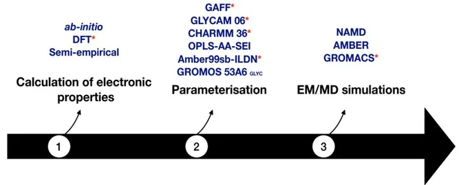
Chapter 1Figure 1: Examples of monosaccharides (A), disaccharides (B) and polysaccharides (C)...24 Figure 2: (A) Conversion of glucose to methyl glucoside in Fischer glycosidation using (TMS-CL) trimethylsilyl chloride as the catalyst. (B) Synthesis of alkyl or aryl O-glycosides in Koenigs-Knorr glycosidation reaction upon substituting the glycosyl halides by alcohol or phenols in the presence of heavy metals or Lewis acids...25 Figure 3: Illustration of four different types of CAZymes owing to their respective reactions with sucrose...26 Figure 4: Structure of bacterial sucrose phosphorylases...29 Figure 5: Schematic representation of the main structural rearrangements observed in sucrose phosphorylase from Bifidobacterium adolescentis (BaSP) during sucrose conversion...33 Figure 6: The homologues and mutant version of sucrose phosphorylase structures...35 Figure 7: Possible transglucosylation products of sucrose phosphorylases when glucose is used as acceptor...36 Figure 8: Overview of the different approaches used to reach the objectives of the thesis...39 Figure 9: Typical scheme followed for the study of protein-carbohydrate complexes using molecular dynamics simulation...46
Chapter 2
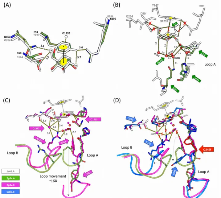
Figure 1: The role of glucosylated aspartate residue (termed as DGC) in the reaction mechanism catalyzed by sucrose phosphorylase. ...52 Figure 2: (i) The structural representation of the sucrose phosphorylase reaction (left) and (ii) their consecutive structural rearrangements (top right) along with the list of comparative structures (iii) (bottom right)...54 Figure 3: Illustration of the protocol used in parameterization and implementation of DGC residue using CHARMM22 (a), GAFF (b), GLYCAM (c) and CHARMM36 (d)...56 Figure 4: Comparison of native (blue) and constrained (green) conformations of DGC residues.. .58 Figure 5: The representation of DGC residue...63 Figure 6: Reference Cremer-Pople puckering plots...64 Figure 7: Comparison of four selective structure and their key interactions. (A) Corresponds to the superimposition of 1s46_A (white) and 2gdv_A (green)...66 Figure 8: The distribution of individual puckering states and their respective energy spots on Cremer-Pople puckering chart...69 Figure 9: Analysis of puckering states of glucosyl moiety covalently linked to Asp during MD simulations.(CHARMM36)...71 Figure 10: Analysis of puckering states of glucosyl moiety covalently linked to Asp during MD simulations.(GAFF)...72 Figure 11: Analysis of puckering states of glucosyl moiety covalently linked to Asp during MD simulations.( GAFF and GLYCAM-06)...73 Figure 12: Analysis of puckering states of glucosyl moiety covalently linked to Asp during MD simulations.(GLYCAM-06 + glucosyl-threonine analogs)...75 Figure 13: Interactions observed in MD snapshots key for determining the puckering states of the covalently-linked glucosyl moiety...76 Figure 14: The graphical representation of distance variations of two important interactions with respect to three selective puckering states...77 Figure S15: The distributions of dihedral values using 100 models at two different phases 0.00 (A) and 109.4 (B)...82 Figure S16: Multiple structure alignment of four known covalent intermediate binding sites of glycoside hydrolase 13 family...82 Figure S17: The presence of HBONDS and van der waals interactions associated between all the individual binding site residues and ß-D-Glucose of four covalent intermediate structures...83
Figure S18: The presence of HBONDS associated with water molecules, binding site residues and
ß-D-Glucose of four covalent intermediate structures...84
Figure S19: The equilibrations (NVT, NPT), radius of gyration and Root Mean Square Deviation of DGC incorporated covalent intermediate structures over 500-ps, 1ns and 100ns molecular dynamics simulation using CHARMM36 parameters...85
Figure S20: The graphical representation of the stability of system at equilibration (NVT and NPT) and the equilibrations (NVT, NPT), radius of gyration and Root Mean Square Deviation of DGC incorporated covalent intermediate structures over 500-ps, 1ns and 100ns molecular dynamics simulation using GAFF parameters...86
Figure S21: The equilibrations (NVT, NPT), radius of gyration and Root Mean Square Deviation of DGC incorporated covalent intermediate structures over 500-ps, 1ns and 100ns molecular dynamics simulation using combination of both GLYCAM and GAFF parameters...87
Figure S22: The equilibrations (NVT, NPT), radius of gyration and Root Mean Square Deviation of DGC incorporated covalent intermediate structures over 500-ps, 1ns and 100ns molecular dynamics simulation using GLYCAM06 parameters...88
Chapter 3 Figure 1: Overview of the three different phases of our methodological approach used in this study...94
Figure 2: Representation of single DGC residue...101
Figure 3: Comparison of different different optimization environment in the context of replicating Tyr344 repositioning (on top) and sampling size based on RMSD distribution (bottom)...102
Figure 4: Comparison of three different protocol used in the sampling of selective SP variants....103
Figure 5: The reference productive poses for α-D-glucose and β-D-glucose acceptors...105
Figure 6: Towards the implementation of global optimization in mutate models...108
Figure S7: The distribution of RMSD and DOPE SCORE of mutate models and their respective wild types. The distribution shown for all the kojibiose and nigerose selectivity mutants with the number of 100 models...114
Figure S8: The reference productive poses for α-D-Methylglucosidase and β-D-Methylglucosidase acceptors...117
Figure S9: Docking of β-D-glucose in to the double optimized mutant (Q345F) model shows the consequence of losing minimal distance between His234-DGC192(O2). It leads to form narrow binding site and doesn’t allows the glucose acceptor inside the pocket...120
Figure S10: The distribution of puckering states (1S3,4C1, B36) based on the Cremer-Pople puckering energy plot (shown only for software restraint weight 10). (Cyan: 1S3; Red: 4C1; Yellow: B36 which is identical puckering state of 1S3)...121
Figure S11: The distribution of RMSD (with respect to Model no1) and DOPE SCORE at soft sphere weight 10...121
Figure S12: Distance scheme (in ranges) for regioselectivity analysis on mutate models...123
Figure S13: Distance scheme (in ranges) for regioselectivity analysis for automodel method...124
Chapter 4 Figure 1: The list of available modules of the ENZO web application. (DGC: Glucosylated covalent aspartate residue; STD: standard aspartate residue; WP: Work Packages)...128
Figure 2: The home page of the ENZO web interface...136
Figure 3: Graphical representation of the ENZO...136
Figure 4: Example of job submission page of a given work package...137
Figure 5: Overview of the ENZO architecture and the external directories containing the MODELLER (A) and GROMACS (B) libraries which have been modified to accommodate the parameters of the glucosyl-aspartate (DGC) residue...140
Figure 6: Example access to the outputs from the job submission page (A) or from the general user workspace page (B)...142
Figure S8: Example of input submission forms of ENZOWP2A (A), ENZOWP2B (B), ENZOWP2C (C) (the inputs were uploaded from local system)...148 Figure S9: Example of input submission forms of ENZOWP2A (A), ENZOWP2B (B), ENZOWP2C (C) (the inputs were redirected output from previous packages)...149 Figure S10: Example input submission (redirected output from previous packages) forms of
ENZOWP3 (A), ENZOWP4A (B) and ENZOWP4B (C)...150
Chapter 5
Figure 1: Comparison of sucrose phosphorylase structures from Bifidobacterium adolescentis based on superimposition (A) and RMSD deviation based on multiple structure alignment (B)...156 Figure 2: Graphical representation of protein engineering strategies namely functional hot spots (A), stability hot spots corresponding to sequence conservation (B) and correlated hotspots (C). (SC: sequence conservation)...158 Figure 3: The identified catalytic pocket (left) and tunnel (right) using Hotspot Wizard3.0...158 Figure 4: Cross validation of identified 24 hot spots with experimental data...163 Figure 5: The over view of mutant library after the three filtrations steps and stability prediction.165 Figure 6: Specific activities of selective positions which are experimentally shown to have less activity for sucrose and Glucose-1-phosphate71...166
Figure 7: The comparison of docking results of sucrose compounds (rigid and flexible) against wild type (1R7A) and mutant version (2GDU) of SP using AutoDockVina and Vina-Carb...167 Figure 8: The future pipeline for regioselectivity prediction...168
List of Schemes
Chapter 1
Scheme 1: The type of carbohydrates along with few examples...23 Scheme 2: Schematic representation of sucrose phosphorylase double displacement reaction
mechanism...30
Chapter 2
Scheme 1: Large panel of rare disaccharide sugars associated with sucrose phosphorylase
transglucosylation function using glucose acceptors...92 Scheme 2: Illustration of statistical schemes for identify the productive docking poses...100
Chapter 3
Scheme 1: A workflow of ENZO pipeline for engineering sucrose phosphorylase enzyme as well as general protein mutation, structure and functional associated studies...129 Scheme 2: Example usage of the scanning (A) and the site saturation mutagenesis (B)...130 Scheme 3: Examples of usage of the random mutagenesis functionality with respect to the use of a user-defined mutation rate (Cii) or a default random seed (Ci)...131 Scheme 4: Examples of usage of the site directed (D) and combinatorial mutagenesis (E)
functionalities...132
Chapter 5
Scheme 1: Overview of the modelling pipeline used to engineer the regioselectivity of sucrose phosphorylases...154
Synopsis
Synopsis in English
To date, the applications of carbohydrates for the industrial exploitation is enormous because of their abundance in nature with versatile group of organic compounds. They are widely used in various industries such as food (low-calorie sweeteners, prebiotics), cosmetics (sulfated polysaccharides from seaweeds), pharmaceuticals (building blocks for anti-cancer and anti-viral drugs), bio-degradable packaging materials (PLA-polylactide from the renewable resources such as starch and sugar), textiles and paper etc. Conjugating carbohydrates to another molecule also commonly used for improve its stability, solubility and bioavailability. However, the advances in commercial exploitation of interesting carbohydrates and glycosylated compounds are hindered by their insufficient amount in nature. Therefore, an efficient alternative synthesis technologies that use readily available starting materials with less expensive are highly desirable.
In that respect, compared to other commonly used traditional chemical technologies (Koenigs-knorr reactions, Fischer glycosidation etc.). Enzyme-mediated synthesis of glycosylated compounds is a better alternative and eco-efficient because of their regio- and stereospecificity. In accordance, sucrose phosphorylase (EC 2.4.1.7) enzyme is one of the most interesting biocatalyst for exploiting industrial applications due to its unique transglucosylation function with broad acceptor specificity and its sucrose utilization as a donor substrate. However, the use of sucrose phosphorylase is hampered by its poor specificity or selectivity towards any alternative acceptors. Afore concerns have been improved by the combination of insilico and in vitro techniques. There are no efficient computational strategies available to enhance the existing protocols by directly incorporating covalent glucosylated aspartate residue in the computational theoretical structures.
The derivation of non-standard amino acid residue forcefield parameters is often a cumbersome task to perform prior to computational modeling and simulations. Thus, the successful addition of a new residue type in forcefield libraries is undoubtedly necessary for further computational studies of proteins carrying such modified residues. In Chapter 2, we present our strategy to derive the force field parameters of the glucosylated aspartate residue (DGC) as observed in the crystal structure of the sucrose phosphorylase enzyme from Bifidobacterium adolescentis. Accordingly, we parametrized the DGC residue for both CHARMM and AMBER ff99sb-ILDN force fields and implemented these parameters within MODELLER and GROMACS respectively. Subsequently, we have also shown that our parameters were efficient in terms of reproducing the models as in crystal structures using modeller and MD simulations.
In Chapter 3, we have applied our modified forcefield and parameters to provide a rational explanation on the observed switch of transglucosylation regioselectivity of some recently reported sucrose phosphorylase variants. Towards the end, we built models of selective variants that were shown to change the regioselectivity of the enzyme towards rare sugars such as kojibiose and nigerose production. All the models were built in covalent intermediate form using both mutate model and automodel methods in combination with different optimization protocols. Subsequently, molecular docking studies were conducted on the wild type enzyme and the respective variants against both α/β-D-glucose to predict the preferred orientation of the acceptors in the +1 site. The preferred orientations of α/β-D-glucose in this +1 site were compared and we have shown that our studied variants indeed displayed a preferred binding mode for the acceptor that could explain the selectivity for maltose, kojibiose and nigerose production.
Subsequently in Chapter 4, an automated web application called ENZO is presented for the Glyco-enzymology community with the list of packages incorporated with DGC residue. By which one can create a plethora mutant library for sucrose phosphorylase enzyme from a given FASTA file (WP1) and perform a set of experiments on them. These involve modeling of variants (WP2), energy minimization (WP3) and molecular docking (WP4A/B) of mutant models. It is noteworthy that ENZO modules have been designed to work with both standard aspartate (D) residue as well as modified glucosylated aspartate residue (DGC). As a viable outcome, a step-by-step guidance according to the standardized protocol (Chapter 3) is presented for the community to screen the putative mutants of sucrose phosphorylase using ENZO. Further, we envisage that this can be also be useful to a broader community working in the field of protein engineering.
Lastly, in Chapter 5, we employed our ENZO web tool to conduct large-scale mutagenesis experiments to gain in-depth insights on how mutations impaired sucrose binding, alter binding modes of the α and β anomers of glucose or methyl-glucoside, hence alter regioselectivity. In this regard, we have also tried to incorporate external tools such as HotspotWizard, FoldX respectively for selecting hotspot residues and predicting (de)stabilizing mutations to design pertinent libraries of variants for use in ENZO. Further, the results of respective works are considered in larger framework and future prospects in the existing field of glycobiology are discussed.in in-depth insights on how mutations impaired sucrose binding, alter binding modes of the α and β anomers of glucose or methyl-glucoside, hence alter regioselectivity. In this regard, we have also tried to incorporate external tools such as HotspotWizard, FoldX respectively for selecting hotspot residues and predicting (de)stabilizing mutations to design pertinent libraries of variants for use in ENZO. Further, the results of respective works are considered in larger framework and future prospects in the existing field of glycobiology are discussed.in in-depth insights on how mutations impaired
sucrose binding, alter binding modes of the α and β anomers of glucose or methyl-glucoside, hence alter regioselectivity. In this regard, we have also tried to incorporate external tools such as HotspotWizard, FoldX respectively for selecting hotspot residues and predicting (de)stabilizing mutations to design pertinent libraries of variants for use in ENZO. Further, the results of respective works are considered in larger framework and future prospects in the existing field of glycobiology are discussed.
Synopsis in French
De nos jours, l’application des glucides pour l’exploitation industrielle est considérable, car ceux-ci sont abondants dans la nature et possèdent une variété de groupes de composés organiques. Ils sont largement utilisés dans des industries variées, telles que l’agroalimentaire (sucrants à faible calorie, prébiotiques), la cosmétique (polysaccharides sulfatés extraits d’algue), la pharmaceutique (élaboration de médicaments anti-cancer et anti-virale), des matériaux d’emballage bio-dégradable (PLA-polylactide extrait de ressources renouvelables telles que l’amidon et le sucre), le textile, le papier, etc. Très souvent, les glucides sont combinés à d’autres molécules pour améliorer leur stabilité, leur solubilité et leur bio disponibilité. Cependant, les avancées dans l’exploitation commerciale des glucides et de composés glycosylés d’intérêts sont limitées par leur quantité insuffisante dans la nature. Par conséquent, une technologie de synthèse alternative efficace, utilisant un matériau de départ facile à obtenir et à prix rentable, est hautement désirable.
A cet effet, comparées aux autres technologies chimiques traditionnelles (Réaction de Koenigs-knorr, Glycosylation de Fischer, etc.), la synthèse à médiation enzymatique de composés glycosylés est une meilleure alternative et possède aussi une éco efficacité à cause de leur régio- et stéréospécificité. Ainsi, l’enzyme de la sucrose phosphorylase (EC 2.4.1.7) est, grâce à sa fonction de transglucosylation unique avec de multiples accepteurs spécifiques et aussi son utilisation du sucrose comme donneur de substrat, l’un des biocatalyseurs le plus intéressant pour être appliqué en industrie. Cependant, l’utilisation de la sucrose phosphorylase est limitée par sa faible activité et sa faible stabilité envers d’autres accepteurs alternatifs. Récemment, ce problème a pu être amélioré, et ce en combinant les techniques in silico et in vitro. Il n’y a pas de stratégie informatique efficace et disponible pour améliorer les protocoles déjà existant dans le but d’incorporer directement les résidus aspartate glycosylés, de manière covalente, dans les structures théoriques informatiques. La dérivation de paramètre du champ de forces d’acide aminé non standard est souvent une lourde tâche afin d’effectuer les modélisations et simulations informatiques. Ainsi, l’addition réussie d’un nouveau type de résidu dans les bibliothèques de champ de forces est sans aucun doute nécessaire pour faire des études informatiques plus approfondis de protéines contenant ces résidus modifiés. Dans le chapitre 2, nous présentons notre stratégie pour dériver les paramètres du champ de forces du résidu aspartate glycosylé (DGC), comme observé dans une structure cristallographique de l’enzyme sucrose phosphorylase chez Bifodobacterium adolescentis. Par conséquent, nous avons paramétré le résidu DGC pour les deux champs de forces CHARMM et AMBER ff99sb_ILDN et avons incorporé ces paramètres respectivement dans MODELLER et GROMACS. Par la suite, nous
avons également montré que nos paramètres étaient efficaces en termes de répétabilité, comme dans la structure cristallographique, en utilisant les modélisateurs et les simulations MD.
Dans le chapitre 3, nous avons appliqué notre champ de forces modifié et ses paramètres pour fournir une explication rationnelle sur le switch observé de la régiosélectivité de la transglucosylation de sucrose phosphorylase variantes récemment signalés. Vers la fin, nous avons construit des modèles de variants sélectionnés qui ont montré un changement dans la régiosélectivité d’enzymes envers des sucres rares comme la kojibiose et la nigerose. Tous les modèles ont été construit sur des formes covalentes intermédiaires en utilisant à la fois un modèle mutant et des méthodes de modèles automatisés en combinaison avec différents protocoles d’optimisation. Ainsi, les études de docking moléculaire ont été mené sur une enzyme sauvage et sur ses variants respectifs, dirigés contre α/β-D-glucose afin de stimuler l’entrée dans son accepteur en son site +1. Les orientations préférentielles des α/β-D-glucose dans le site +1 ont été comparé et en effet, nous avons montré que tous les variants étudiés ont montré un mode de liaison préférentiel avec l’accepteur, ce qui pourrait expliquer la sélectivité pour la production de maltose, de kojibiose et de negeriose.
Par la suite, au chapitre 4, un site internet automatisé, appelé ENZO, est présenté avec la liste des packages incorporée avec le résidu DGC, pour la communauté des glycoenzymologistes. Ainsi, peut se créer une bibliothèque abondante de mutants pour le sucrose phosphorylase obtenu à partir d’une séquence FASTA (WP1) et s’effectuer une série d’expérience sur eux. Cela implique des modélisations des variants (WP2), la conservation d’énergie (WP3) et le docking moléculaire (WP4A/B) des modèles mutants. Il est intéressant de savoir que les modules ENZO ont été élaboré pour fonctionner avec à la fois un résidu aspartate standard et un résidu asparatyl glycosylé (DGC). Comme résultat viable, un protocole standardisé décrivant pas à pas les étapes (Chapter 3) est présenté à la communauté pour cribler les mutants putatifs de sucrose phosphorylase en utilisant ENZO. De plus, nous envisageons que cela puisse être également utile pour une communauté plus large travaillant dans l’ingénierie des protéines. Enfin, dans le chapitre 5, nous avons utilisé notre outil web ENZO pour entreprendre des expériences de mutagénèse à grande échelle dans le but de mieux comprendre comment les mutations altèrent la liaison au sucrose, modifient les modes de liaison des anomères α et β du glucose ou de glucosides méthylés, changeant ainsi la régiosélectivité. A cet effet, nous avons aussi essayé d’incorporer des outils externes tels que HotspotWizard et FlodX afin de choisir respectivement des résidus sensibles et prédire des mutations (de)stabilisantes pour élaborer des bibliothèques pertinentes de variants dans l’utilisation d’ENZO. Pour finir, nous discutons des résultats des travaux pour un cadre plus large et de futures perspectives dans le domaine existant de la glycobiologie.
Chapter 1
Literature review
1. GLYCOSYLATION: From Conventional Methods To Enzymatic Synthesis
1.1 Carbohydrates, Glycosides, and Glycoconjugates
Carbohydrates are also known as sugars and consist of aldehydes and ketones with multiple hydroxyl groups. They are further classified as monosaccharides, disaccharides, oligosaccharides, and polysaccharides (Scheme 1). Monosaccharides are the simplest form of sugars that usually contains three to seven carbon atoms. A monosaccharide classically adopts either a linear or ring-shaped structure (Figure 1A). Di- or oligosaccharides consist of 2-10 monosaccharide units linked by glycosidic bonds upon condensation. Sucrose, lactose and maltose are the most common disaccharides which are composed of two monomeric units connected by a glycosidic bond (Figure 1B). Similarly, inulin and cyclodextrins are some of the best-known examples of oligosaccharides. Polysaccharides are made out of more than 10 monosaccharide units and are also known as glycans. They exist in homo/hetero forms of monosaccharides and can form either branched (e.g., starch, glycogen) or unbranched (e.g., cellulose in Figure 1C) structures1. A glycosidic bond is a type of
covalent bond that can also link monosaccharides or oligosaccharides to non-sugar groups (aglycones) to form glycosides1. Similarly, carbohydrate molecules that are chemically linked to
other compounds such as proteins or lipids called glycoconjugates2.
Scheme 1: The type of carbohydrates along with few examples.
Glycosides and glycoconjugates are abundantly available on earth. They are involved in a range of functions. For instance, oligosaccharides can perform as signaling molecules in plants (e.g., oligosaccharins) while they can also trigger the immune system in animals (e.g., human milk oligosaccharides)3,4. Polysaccharides are well known to act as an energy source (e.g., amylose) or as
various recognition motifs such as the blood group determinants or to provide surfactant properties (e.g., rhamnolipids or sophorolipids)6. Besides, linking sugar moieties can alter activity of their
respective aglycones: glycosylation is known to alter the spectrum of the activity of antibiotic7 and
in the case of naringin, it affects the bitterness of grapefruits8.
Figure 1: Examples of monosaccharides (A), disaccharides (B) and polysaccharides (C).
1.2 Chemical synthesis of glycosides
The above examples demonstrate that the synthesis of glycosidic compounds is of significance for the pharmaceutical, cosmetics and food industries. Glycosides are classically synthesized using conventional chemical techniques. The widely used Fischer glycosidation involves a reaction
between an aldose/ketose and an alcohol in the presence of an acid catalyst (Figure 2A). In the extensively used Koenigs-Knorr method (on of the oldest protocol, used since from 1901), alkyl/aryl O-glycosides are synthesized in the presence of heavy metals or Lewis acids following the substitution of glycosyl halides by alcohol/phenols (Figure 2B). In general, glycosyl chlorides and bromides are used in the Koenigs-Knorr reaction. Only recently were iodides used as glycosyl donors. Apart from these methods, there are also many other glycosidation reactions that make use of different donor substrates such as glycosyl acetates, glycosyl trichloroacetimidates, and thioglycosides9–13.
Figure 2: (A) Conversion of glucose to methyl glucoside in Fischer glycosidation using (TMS-CL)
trimethylsilyl chloride as the catalyst. (B) Synthesis of alkyl or aryl O-glycosides in Koenigs-Knorr glycosidation reaction upon substituting the glycosyl halides by alcohol or phenols in the presence of heavy metals or Lewis acids.
However, achieving selective formation of a glycosidic bond using the above methods is still an uphill walk with many drawbacks. For instance, preparation of glycosyl halides in Koenigs-Knorr reaction require severe conditions (e.g., thermally not stable) and also experience with hydrolysis. Besides, the co-activators used in Koenigs-Knorr method is often toxic and explosive. On the other hand, the reliability of protection strategies involved in other synthetic pathways are also very poor and consist of multistep protocol resulting in overall low yields with a lot of waste13–22.
1.3 CAZymes: Carbohydrate-Active enZymes
Enzymes that display activity towards formation or breakdown of glycosidic bonds are collectively called "Carbohydrate-Active enZymes" (CAZymes). Different enzymes are at play behind the chemical reactions that breakdown glycosidic bonds: hydrolysis is performed by glycoside hydrolases, phosphorolysis by glycoside phosphorolases, redox reactions by lytic polysaccharide mono-oxygenases and beta-elimination by polysaccharide lyases23. On the other hand,
glycosyltransferases and transglycosidases are the enzymes usually found behind the formation of glycosidic bonds24.
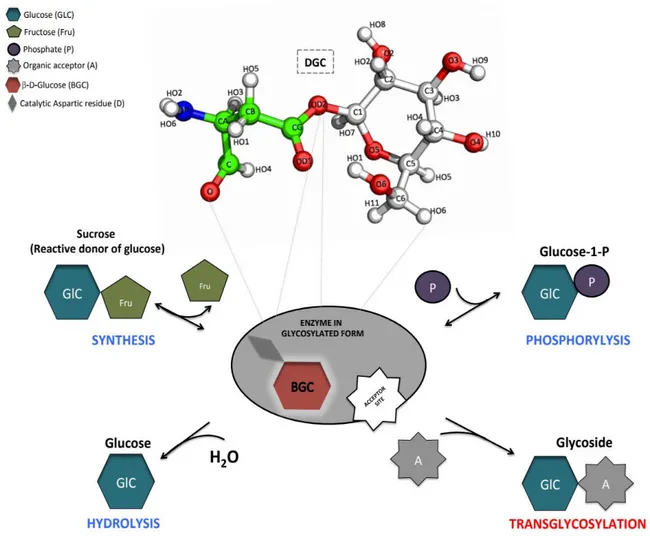
Figure 3: Illustration of four different types of CAZymes owing to their respective reactions with
sucrose.
Synthesis of sucrose by sucrose synthase (GT) which can be further converted into amylose by amylosucrase, a transglycosidase (TG) or converted to either glucose-1-phosphate by sucrose phosphorylase (GP) or glucose by sucrose hydrolase (GH).
In general, these CAZymes are known to catalyze glycosyl transfers and are classified into four different categories25 (see Figure 3). First, a large number of glycosylation reactions in nature is
exploited by glycosyltransferases (GTs) that commonly use nucleotide-activated sugars as glycosyl donor. Similarly, synthesis of saccharides by transglycosidases (TGs) consist of the transfer of glycone moieties between carbohydrate chains. On the other hand, Glycoside Hydrolases (GHs) and Glycosyl Phosphorylases (GPs) catalyze the degradation of glycosidic bonds using the acceptor substrates water and inorganic phosphate respectively. With respect to synthetic applications,
CAZymes are reputed to be as an efficient as the chemical synthetic reactions mentioned above and even sometimes a better alternative. Indeed, it has been shown that by the use of appropriate enzymes, glycosylation reactions generated up to five times less waste and up to fifteen times higher space-time yield, accordingly improving the eco-efficiency tremendously26,27. In that respect,
sucrose phosphorylase (SP) is probably one of the most interesting class of biocatalysts for industrial applications28. In this thesis, we have considered SP’s as a study model. This class of
enzyme is further described in the following sections.
2. SUCROSE PHOSPHORYLASE
2.1 Classification Scheme
Sucrose phosphorylase (SP) is a class of bacterial enzymes that plays a crucial role in sucrose metabolism and is notoriously known for reversibly catalysing the phosphorolysis of sucrose: sucrose + phosphate = D-fructose + α-D-glucose-1-phosphate29,30. This reaction leads to the
production of essential glucose moieties from sucrose. According to the classification of IUBMB and enzyme commission, SP belongs to the class of transferases and particularly to the glycosyltransferases/hexosyltransferases subgroup that catalyze the transfer of hexosyl groups. It comes under the enzyme number EC 2.4.1.731.
Table 1: Classification schemes for sucrose phosphorylases
IUBMB and Enzyme commission* CAZy database#
EC 2.4.1.7 Enzyme entry GH13 Family (Glycoside hydrolase) EC 2 Class (Transferases) GH13-18 Subfamily
EC 2.4 Subclass (glycosyltransferases) EC 2.4.1 Sub-subclass (Transfer hexosyl group)
*IUBMB International Union of Biochemistry and Molecular biology; #
CAZy: Carbohydrate-Active enZymes
It is placed in the CAZy database in the retaining glycoside hydrolase family 13 (GH13) also known as α-amylases family23,32. This family mainly contains α-amylases but is also associated with a
specific group of enzymes that use sucrose as substrate or catalyze transglucosylation reactions from amylose. Because of its large size, GH13 family is divided into subfamilies on the basis of their sequence and functional similarities. Sucrose phosphorylases narrow down to a distinctive subfamily named GH13_1832. The classification of SP’s is summarized in Table 1.
2.2 General properties
In early 1940s, an intracellular enzyme SP was independently discovered in Leuconostoc mesenteroides33–37 and Pleomonas/Pseudomonas saccharophila by Kagan (1942) and Doudorff
(1943) teams30 respectively. Subsequently, it was found in many other microorganisms such as
Streptococcus mutants38–40, Bifidobacterium adolescentis41, Bifidobacterium longum42, Lactobacillus
acidophilus43,44, and in the meta-genomes analyses of sucrose-rich environments45. In the
above-mentioned species, most of them contained a ~400-500AA functional monomer or dimer that catalyze an equilibrium reaction with an equilibrium constant Keq of about 5.6 at 30°C, pH 7.046.
SP’s can thus be utilized for both assembly and breakdown of sucrose. In vivo, the enzyme has a catabolic role in the cell because of the higher concentration of phosphate compared to glucose-1-phosphate28. Besides, since the phosphorolysis reaction catalyzed by SP’s does not require ATP and
yields products that can be directly be used in glycolysis, it can be viewed as an energy-saving cellular process29. The optimal temperatures (30-37°C) and pH (6.0-7.5) of bacterial SP’s reflect the
growth conditions of their hosts exception made of SP from Bifidobacterium adolescentis which has an optimal temperature of 58°C47.
2.3 Crystal structure of SP and their associated reaction mechanism
Sucrose phosphorylases are 54-60 kDa enzymes comprised of 480-504 residues48,49. As shown
biochemically, they are generally monomeric while sucrose phosphorylase from Bifidobacterium adolescentis (BaSP) is homodimeric41. The only SP three-dimensional structures solved by X-ray
crystallography available to date are that of BaSP50. The wild-type (PDB 1R7A and 2GDV)
enzyme as well as two mutated forms (PDB 2GDU and 5C8B) all crystallize in its homodimeric form. Each chain is characterized by four domains named A, B, B' and C (Figure 4A). Domain A forms the major part of the monomeric structure and is characterized by a (β/α)8-barrel fold that
constitutes the catalytic domain. Subsequently, the architecture of the catalytic site is maintained by loops from domains B and B’ which additionally contains two short antiparallel β-sheets/α-helices and coil regions respectively. The corresponding loops also play a role in substrate specificity. Dimer interactions occur mainly at the level of the B domain. Domain B’ controls the size of the substrate access tunnel and oligosaccharide binding50. The three domains A, B and B’ are common
to all GH13 enzymes whereas the domain C is unique to sucrose active members. This domain contains typically 5 stranded antiparallel β-sheets and play a crucial role in the stabilization of the catalytic domain and facilitate substrate binding51–53. However, the function of this domain remains
obscure in sucrose phosphorylase. Interestingly, in one of the four crystal structures of the native enzyme (PDB 2dgv), one of the two chains features the covalent glycosyl-enzyme intermediate (chain A in Figure 4B) while the other features non-covalent (chain B in Figure 4C) forms. It reflects the general mechanism of the enzyme. Indeed, enzymes from the glycosyl hydrolase family 13 undergo a double-displacement reaction mechanism via the formation of covalent glucosyl-enzyme intermediate54.
Figure 4: Structure of bacterial sucrose phosphorylases.
Shown in (A) is the overall three-dimensional structure of sucrose phosphorylase from Bifidobacterium adolescentis (PDB 1R7A) featuring its constitutive domain organization. In (B) is shown the catalytic site displaying the covalent glucosyl-enzyme intermediate as observed in chain A of PDB 2GDV and in (C) the catalytic site of the non-covalent form of the enzyme bound to glucose, the product resulting of the hydrolysis of the sucrose substrate. Catalytic residues (D192 and E232) are shown in green sticks, conserved residues among sucrose-active enzymes in yellow sticks, and glucosyl moiety as white ball/stick.
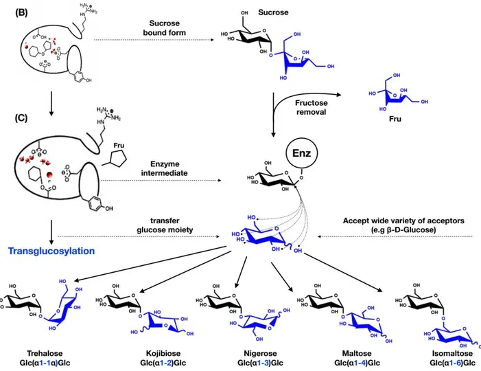
SP also follows the same reaction mechanism (see Scheme 2)55. The reaction is initiated by the
concomitant nucleophilic attack of the anomeric carbon of the glucosyl moiety and protonation of the oxygen of the glycosidic linkage. The nucleophilic attack is performed by a conserved aspartic acid (identified as Asp192 in BaSP) while the protonation is carried out by a conserved general acid/base catalyst or proton donor (Glu232 in BaSP)23,55. This leads to the formation of the
covalently linked glucosyl-enzyme intermediate and release of fructose. Subsequently, in presence of excess inorganic phosphate as substrate acceptor and following some conformational changes in the acceptor site, glucose is transferred on the phosphate moiety and release of glucose-1-phosphate occurs. Owing to the mechanism and presence of competing water in the acceptor site, hydrolysis occurs as a side reaction but at low level (1-5%)43. This indicates that the catalytic site of SP’s has
Scheme 2: Schematic representation of sucrose phosphorylase double displacement reaction mechanism.
In red is shown the covalent glucosyl-enzyme intermediate with an aspartate residue covalently linked to glucose via a glycosidic bond.
The above-mentioned mechanism and the identification of residues central in the mechanism were confirmed by the obtention of a structure of BaSP featuring the covalent glucosyl-enzyme intermediate55 and following site-directed mutagenesis studies. Mutation of Asp192 to alanine
resulted in the loss of wild-type function56,57. Apart from the catalytic residues, there are other
conserved residues (Figure 4B and C) that were determined to be crucial for the reaction mechanism as well as in the maintenance of binding site architecture. For instance, Asp290 is one such residue and is known for its "transition state stabilization" role and its contribution in the catalytic triad50,51,55,58. Besides, two highly conserved phenylalanine residues (Phe53 and Phe156) in
the donor site of BaSP are crucial as they respectively serve as a hydrophobic platform and allow the formation of a hydrophobic sandwich59–61. They contribute for the strong electrostatic cation-Π
interaction that stabilizes by 15 kJ/mol the oxocarbenium-ion-like transition state. Mutation of these aromatic residues reduces the catalytic activity three fold and increases KM for sucrose. Apart from
those, residues that favor substrate binding are His88, Arg190, Asp50, Arg399, Ala193, and Leu341 for glucosyl moiety62 (see Table 2), Tyr196, Asp342, Gln345, Ala193, and Leu341 for the fructosyl
part63 and Arg135 and Tyr344 for phosphate acceptor63.
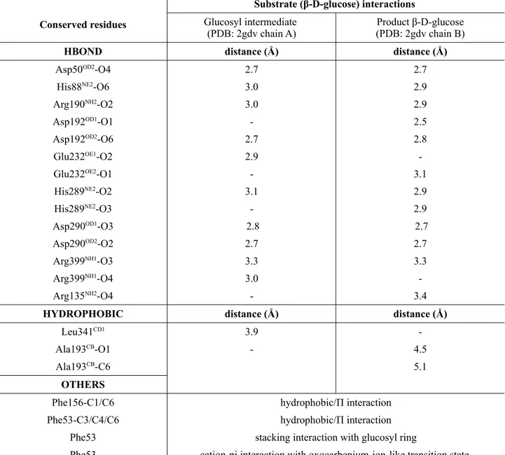
Table 2: List of sucrose phosphorylase interactions. Shown between highly conserved residues and
the glucosyl moiety in both the covalent glucosyl-enzyme intermediate (Figure 4B) and non-covalent (Figure 4C) form of sucrose phosphorylase of Bifidobacterium adolescentis. All the given interactions are according to PDB structure 2gdv.
Conserved residues
Substrate (β-D-glucose) interactions
Glucosyl intermediate (PDB: 2gdv chain A)
Product β-D-glucose (PDB: 2gdv chain B)
HBOND distance (Å) distance (Å)
Asp50OD2
-O4 2.7 2.7
His88NE2-O6 3.0 2.9
Arg190NH2-O2 3.0 2.9
Asp192OD1-O1 - 2.5
Asp192OD2-O6 2.7 2.8
Glu232OE1-O2 2.9
-Glu232OE2-O1 - 3.1
His289NE2-O2 3.1 2.9
His289NE2-O3 - 2.9
Asp290OD1 -O3 2.8 2.7 Asp290OD2 -O2 2.7 2.7 Arg399NH1-O3 3.3 3.3 Arg399NH1-O4 3.0 -Arg135NH2-O4 - 3.4
HYDROPHOBIC distance (Å) distance (Å)
Leu341CD1
3.9
-Ala193CB-O1 - 4.5
Ala193CB-C6 5.1
OTHERS
Phe156-C1/C6 hydrophobic/Π interaction Phe53-C3/C4/C6 hydrophobic/Π interaction
Phe53 stacking interaction with glucosyl ring
Phe53 cation-pi interaction with oxocarbenium-ion-like transition state
2.4 Conformational changes during sucrose conversion
It has been shown that SP undergoes significant structural changes during the conversion of sucrose55. These structural rearrangements are delimitated by five distinct conformations (A to