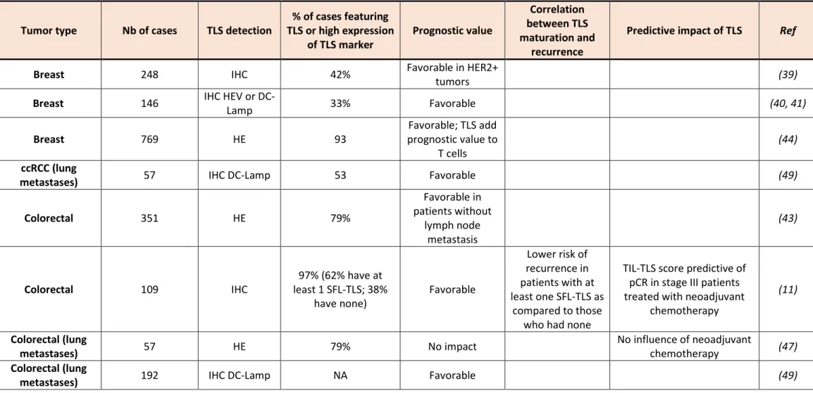
Table 1. Characteristics of TLS in human cancers
Tumor type Nb of cases TLS detection TLS or high expression % of cases featuring
of TLS marker Prognostic value
Correlation between TLS maturation and
recurrence
Predictive impact of TLS Ref
Breast 248 IHC 42% Favorable in HER2+ tumors (39)
Breast 146 IHC HEV or DC-Lamp 33% Favorable (40, 41)
Breast 769 HE 93 prognostic value to Favorable; TLS add
T cells (44)
ccRCC (lung
metastases) 57 IHC DC-Lamp 53 Favorable (49)
Colorectal 351 HE 79% Favorable in patients without lymph node metastasis (43)
Colorectal 109 IHC least 1 SFL-TLS; 38% 97% (62% have at
have none) Favorable
Lower risk of recurrence in patients with at least one SFL-TLS as
compared to those who had none
TIL-TLS score predictive of pCR in stage III patients treated with neoadjuvant
chemotherapy
(11) Colorectal (lung
metastases) 57 HE 79% No impact No influence of neoadjuvant chemotherapy (47)
Colorectal (lung
Hepatoblastoma 12 APC-HB, 15 non APC-HB HE and IHC CD3
12/12 APC-HB have PFL-TLS; 3/15 non APC-HB have PFL-TLS and 12/15 have E-TLS
NA 11/11 APC-HB have PFL-TLS after neoadjuvant chemotherapy; 0/5 APC-HB prechemotherapy have TLS (96)
Hepatocellular 273 HE 47% (56% E-TLS, 33% PFL-TLS, 11%SFL-TLS) Favorable
Lower risk of recurrence and survival in patients
with PFL and SFL-TLS as compared to
those who had E-TLS (35) Hepatocellular 462 HE 34% Favorable lower risk of recurrence and shorter survival in patients with increased maturation stages (34) GIST 118 and 69 HE 44,9%; (33%, 5.9%, 5.9%), 47,8; (43%, 4,3%, 4,3%) Favorable
TLS presence associated with
lower imatinib resistance (38) Liver metastases of
colorectal cancer 65 IHC C20 69% Favorable neoadjuvant chemotherapy TLS prognostic after (48)
Lung 74 IHC DC-Lamp NA Favorable (33)
Lung 151 IHC DC-Lamp and CD8 Favorable After neoadjuvant chemotherapy (53)
Lung 362 IHC DC-Lamp 54% Favorable (14)
Lung squamous 138 HE >95% Favorable
Prognostic impact lost in patients treated by neoadjuvant chemotherapy;
TLS decrease upon corticosteroid treatment
(12)
Melanoma 82 IHC DC-Lamp and CD3+T
cells NA Favorable (42) Melanoma 22 IHC CD20 and H&E, CD20/CD21/C D23; multiplex IF NA NA Vast majority of SFL-TLS in R patients (primary tumors and extranodal metastases) Increased TLS densities associated with response to
nivolumab and ipilimumab, particularly on early on
treatment samples
(16)
Melanoma 177 (15 primary tumors) 44% (11% with SFL-TLS) Favorable (15)
Oral (squamous) 80 IHC 21% Favorable (37)
Ovary 147 IHC DC-Lamp and CD20 50% Favorable (36)
Ovary (metastases) 172 IHC 20% Favorable (30)
Pancreas 308 HE 16% Favorable (46)
Pancreas 104 IHC CD20 NA Favorable (45)
RCC 18 IHC CD20 and H&E, CD20/CD21/C D23 and multiplex IF
NA NA associated with response to Increased TLS densities
nivolumab (16) Soft tissue sarcoma IHC CD3/CD20 12%; E-TLS 60.5%, PFL-TLS 21.1%; SFL-PFL-TLS 18.3% of all TLS analysed differences between histologies UPS having only 16.7%
of E-TLS.
Tertiary Lymphoid Structures and B cells: clinical impact and therapeutic modulation in cancer
Catherine Sautès-Fridman*1, Johanna Verneau1, Cheng-Ming Sun1, Marco Moreira1, Wei-Wu
Chen3, Maxime Meylan1,2, Florent Petitprez2, Wolf Herman Fridman1
1Centre de Recherche des Cordeliers, Sorbonne Université, Inserm, Université de
Paris, F-75006, Paris, France.
1 Centre de Recherche des Cordeliers, 15 rue de l’Ecole de Médecine, 75006, Paris, France
2Programme Cartes d’Identité des Tumeurs, Ligue Nationale contre le Cancer, F-75013, Paris,
France.
2Ligue Nationale contre le Cancer, 69 rue Corvisart, 75013, Paris, France
3National Taiwan University Hospital and Graduate Institute of Oncology National Taiwan
University College of Medicine, Taipei, Taiwan
*corresponding author, catherine.fridman@crc.jussieu.fr Highlights (3 to 5, 85 char incl space each)
TLS are active sites for the generation and activation of adaptive immune responses in tumors. B cells and TLS have a favorable impact on patient’s prognosis and response to immunotherapy.
The impact of the T cells infiltrate on the prognostic value of B cells is tumor dependent. Chemotherapy and immunotherapy induce TLS formation.
B cells may function via antigen-presentation, antibody production and generation of memory cells.
Key words 6 : tertiary lymphoid structures, B cells, adaptive anti-tumor response, prognosis, clinical outcome, immunotherapy
Abbreviations foot note page 1
TLS: tertiary lymphoid structures ; BC : breast carcinoma, CRC : colorectal carcinoma, GBM : glioblastoma, ccRCC : clear cell renal cell cancer, NSCLC : non squamous cell lung cancer, STS : soft tissue sarcoma
Abstract
Tumors progression is under the control of an heterogeneous microenvironment composed of immune cells, fibroblasts, blood and lymphatic vessels, in which T cells have been demonstrated to be major actors, through their cytotoxic and cytokine producing effector functions and their long term memory that protects against metastasis. In this scenario, lessons from mouse models taught that B cells exert a protumoral role, via macrophage-dependent activation of inflammation. However, it became progressively evident from studies in patients with human cancers that the anti-tumor responses can be generated and controlled in tertiary lymphoid structures (TLS) that concentrate most of the intratumoral B cells and where B cells can differentiate into plasma cells and memory cells. Furthermore, recent studies demonstrated that the presence in tumors of B cells and TLS are associated with favorable outcome in patients treated by immunotherapy, unraveling TLS as a new predictive marker of anti-tumor response human cancers. This review encompasses the characteristics and functions of TLS and of B cells in human tumors, their prognostic and theranostic impact and summarizes the mouse models used to induce TLS neogenesis in tumors.
Tumors form an ecosystem in which malignant cells are entangled in a tumor microenvironment (TME) containing cells which interact directly and/or indirectly through chemokine and cytokine production with the tumor cells. Both tumor promoting and tumor containing forces are exerted by the TME. Fibroblasts, as well as the majority of myeloid cells (MDSC (myeloïd-derived suppressor cells), M2 macrophages, neutrophils, mast cells), and some lymphocytic (Treg, Th17) populations favor tumor growth whereas most T cell populations (Th1, Tfh, T cytotoxic) yield anti-tumor effects. The impact of NK and B cells is context dependent. It is striking that the efficient anti-tumoral immune responses are exerted by CD8+ cytotoxic T cells particularly dense in tumors with high mutational burden, such as
MSI (microsatellite instable) cancers, UV induced melanomas or with gene modifying adducts such as in lung cancers. It is also striking that these cancers are the most responsive to immunotherapy with immune checkpoint inhibitors (ICI) such as antibodies against CTLA-4, PD-1 or PD-L1. Thus, adaptive immune responses towards tumor-associated antigens are not only a major mechanism of tumor control but are also targets for efficient immunotherapies. The question of the generation of anti-tumor immunity and its impact in cancers with low mutational burden is therefore of prominent interest. Draining lymph nodes (LN) are obvious sites for the generation of anti-tumor immunity but increasing evidence demonstrate that it could also take place in the TME where the tumor-associated chronic inflammation promotes the formation of Tertiary Lymphoid Structures (TLS). The latter are very similar to Secondary Lymphoid Organs (SLO) found in reactive LN [1]. Both are composed of a T cell zone and a B cell zone forming a germinal center (GC) in mature TLS and SLO. Inside TLS, antigen-presenting cells (APC) interact with T and B cells where, in conjunction with Th and Tfh cells, they instruct T and B cells towards tumor-associated antigens, inducing their activation, proliferation and differentiation into memory and effector T cells and into memory B cells and antibody producing plasma cells. Importantly, as for SLO, TLS are surrounded by High Endothelial Venules (HEV) allowing a direct entry of naïve T and B cells, thus escaping the immunosuppressive and inflammatory milieu of the rest of the TME. Podoplanin+ lymphatic
vessels producing CCL21, a chemokine that could favor emigration of lymphocytes from TLS to draining lymph nodes, are also located close to TLS [2]. Altogether these observations support the concept that the TME, as the reactive LNs, can be considered as an anti-cancer « lymphoid » organ.
1. Characteristics of Tertiary Lymphoid Structures in human cancers: 1.1 TLS formation and composition
TLS presence is associated with antigen persistence and chronic inflammation in diseased tissues. Most of what is known on the mechanisms of TLS formation has been learned from sites of chronic infections, inflammatory diseases, chronic graft rejection and mice invalidated for genes involved in LN formation. Thus, TLS are formed in inflamed sites, starting upon contact between IL-7-secreting stromal cells acting as lymphoid tissue organizer cell (LTo) and a lymphoid tissue inducer cell (LTi) in an environment with CXCL13, a B cell chemoattractant expressed in LN [3–6]. Tissue resident monocytic cells, Th17 or B cells can function as LTi [7– 9] CCL21 and CXCL12 participate in lymphocytes recruitment, while CXCL13 and CCL19, together with adhesion molecules, govern the structural organization of the forming TLS [10]. The infiltrating immune cells form aggregates with a T cell rich zone where mature DC present MHC-Cl II peptides to CD4+ T cells, promoting their activation, proliferation and differentiation
into T effector cells secreting cytokines, including Tfh expressing the CXCL13 receptor, CXCR5. The Tfh migrate to the CXCL13-rich B cell zone where they induce the activation, proliferation and differentiation of antigen-specific B cells into plasmablasts or their migration into follicles to form GCs where B cells undergo class switch recombination (CSR). TLS include a series of heterogenous structures from lymphoid aggregates of T and B cells to structures including follicles of B cells. Three TLS “maturation” steps have been identified recently in CRC [11] and SCLC [12] and soft tissue sarcoma [13], according to the expression level of two markers expressed in B cell follicles, CD21 on follicular dendritic cells, capable to bind C3d complexes fixed on immune complexes and CD23 on follicular dendritic cells as well as on unswitched B cells. This classification allows the ranking of TLS from the early lymphoid aggregates, CD21
-CD23- (E-TLS), to primary follicle-like TLS (PFL-TLS) containing a network of CD21+ follicular
dendritic cells (FDC) and secondary follicle-like TLS (SFL-TLS) containing a GC including a network of CD21+CD23+ follicular dendritic cells (FDC).
1.2 TLS govern T cell education within tumors
The TLS-rich tumors are more infiltrated by CD4+ T cells and CD8+ T cells of both naïve and
effector memory phenotypes as shown in several tumor types (NSCLC, BC, melanoma), reflecting T cell entry and differentiation into tumors.
The activation and differentiation of T cells within TLS allows to profoundly modulate their functionality in the TME outside TLS. Thus, in Non-Small Lung Cancer (NSCLC), the number of T cells, particularly CD8+ T cells is higher in TLS rich than in TLS poor tumors. Moreover, in TLS
rich tumors, TLS have a high expression of genes linked to activation and Th1 orientation, T cell chemotaxis, as well as in cytotoxicity, characteristic of « good » prognostic signatures. Analysis of micro-dissected TLS in NSCLC revealed strong correlations between the mature dendritic cell marker DC-LAMP and Th1 polarization, and cytotoxicity set of genes, demonstrating that T cell differentiation occurs within TLS [14]. The spatial influence of TLS on T cell activation and polarization in the TME was further evidenced by transcriptomic analyses [14–16]. In melanoma, TLS were found to contain CD4+ T cells whereas CD8+ T cells
are located outside, but the presence of TLS was in all cases associated with tumor associated-CD8+T cells. Whether CD8+ T cells are activated directly by cross-presenting mature DC such
as in anti-viral responses or need CD4+ T cell help remains to be determined. Little is known
about the precise influence of TLS the expression of inhibitory molecules in the TME. Negative correlations have been found between densities of TLS and PD1+ or PD-L1+ cells in ccRCC [17]
and HCC [9] and TLS presence is associated with lower expression of PD-1, Tim3, GZB genes in melanoma [15], whereas an association between TLS presence and expression of PD-1 and Lag3 and Tim3 has been observed in BC [18].
1.3 B cells and TLS
In contrast to T cells which are often scattered within tumors sometimes in close contact with tumor cells, most B lymphocytes, localize together forming clusters with varying size, cellular composition and maturation degree with T cells, from a loose aggregate juxtaposing a T cell cluster in E-PFL, to denser follicular aggregates in PFL- and SFL- TLS. In SFL-TLS, CD20+ B cells
are located within and at the periphery of GCs. Similarly to GCs in SLOs, TLS-GCs are characterized by a dark and a light zone. The dark zone is dense, contains proliferating Ki67+
B cells and expresses BCl6, the master regulator of GC reaction, and activation-induced deaminase (AID), the enzyme regulating class switch and somatic hypermutation. In the light zone, which has a lesser cell density, B cells establish contacts with the network of CD21+/CD23+ FDC that sequester antigen via immune complexes, allowing clonal selection of
micro-dissected follicles, compared with peripheral B cells, reveals clonal amplification, suggesting the development of an antigen-driven B-cell response occurring within TLS-GC [19–25]. Those B cells that successfully bind antigen receive CD40-mediated help from Tfh, survive and exit the GC as long-lived plasma cells or memory B cells. Indeed, CD4+, PD-1Hi, ICOS+, CXCR5+ Tfh
have been detected in GC from BC and STS [13,26]. In BC, Tfh cells were found to produce not only CXCL13 but also IL-21, an essential cytokine for B cell activation, expansion and terminal differentiation as well as for CSR [26,27]. Their presence correlates with that GC-B cells. They are located close to a population of CD4+PD-1+ICOS+(iCTLA4+)FOXP3+ Treg which may
influence their expansion [26]. An additional subset of PD-1Hi CD8+ T cells that secrete CXCL13
was also detected in the light B cell zone in NSCLC tumors [28]. Plasma cells (CD138+) located
at the periphery of GC have been detected in NSCLC, BC and ovarian cancers [21,26,29]. In NSCLC, a significant correlation between the percentages of PCs and GC-B cells has been observed, suggesting that most PCs were differentiated within TLS [29,30]. Notably B cells populations differ in HNSCC tumors according to HPV infection. In HNSCC HPV+ tumors, B cells
display centroblast phenotypes characteristic of GC B cells from tonsils and putative interactions between B cells and CD4+ Tfh cells, unlike B cells found in HPV- tumors which
present plasmablasts or switched memory B cells only and lack these interactions [31]. These findings may highlight the key role of exposure to infectious agents in TLS formation.
In situ and single cell RNAseq analyses have shown that within TLS, a fraction of the affinity matured B cells also undergo AID-dependent CSR that regulates isotype switching [15,29]. Recent works suggest that in SLO this process could occur prior to differentiation of B cells into GC B cells or plasmablasts, rather than within GC as previously suggested [32]. At which step of TLSs maturation does CSR occur remains an open question. Scattered mature DC characterized by expression of the DC-Lamp marker can be detected at each TLS maturation step, in close contact with the T cells and in some B cell follicles, allowing local antigen presentation [22,33].
Tumors contain variable proportions of TLS and of their different maturation steps [11– 13,15](Table 1). The proportion of TLS varies according to tumor types from around 10%-20% in HCC [34,35], ovarian cancer [36] and oral squamous cancer [37], 50% in NSCLC [14], GIST [38], breast [39–41], melanoma [15,16,42], up to be present in more than 80% of tumors such as in CRC [11,12,43] and lung squamous cell carcinoma [12] or TNBC [44] primary tumors. Discordant results have been found in pancreatic tumors [45,46]. In any case, these
percentages should be interpreted with caution, because of the use of different technologies for detection of TLS. TLS have been detected in metastases in CRC [47,48], ovarian cancer [30] and ccRCC [49]. Notably, variable degrees of expression levels of the 12 chemokines gene signature characteristic of TLS [50] was also observed in samples of the 9980 tumors from 30 cancer types of the TCGA cohorts, from low levels in tumors located in immune-privileged sites (glioblastoma, ocular melanoma) to high levels in tumors present in mucosal tissues such as CRC or lung cancers [2]. The proportion of mature TLS also seems to vary in the different cancer types. Thus SFL-TLS can represent either a small proportion of TLS such as in GIST (10%), HCC and melanoma (11%) or STS (18%) or a higher one such as in CRC [11] (Table 1). These results need to be confirmed using a uniformed TLS detection method. Further investigations are needed to better understand the different roles of chemokines involved in the process of TLS formation and maturation in human tumors as well as the context of the tissue microenvironment which may influence such patterns.
2. Clinical impact of TLS in cancer 2.1 Prognostic impact of TLS
Since TLS are likely sites of generation of anti-tumor immunity [51], their impact on cancer outcome increasingly becomes a focus of many analyses. Early after their discovery in the TME of human cancers, their prognostic impact has been questioned. In NSCLC, BC, ovarian cancer, it has been reported that higher TLS densities correlate with longer PFS and OS. In NSCLC, TLS rich tumors have a Th1/cytotoxic functional orientation and high density of CD8+ T cells
correlates with longer PFS and OS. In TLS poor tumors, the favorable prognostic impact of CD8+ T cells was decreased [14]. It was later found that the density of B cells follicles present
in TLS were also associated with PFS and OS [29]. These findings have been extended to a large array of cancers and a favorable prognostic impact has been reported in SCLC [12], BC [39,40,44], CRC [11,43], GIST [38], HCC [34,35], melanoma [15,16,42], pancreatic cancers [45,46,52], oral squamous [37] and ovarian cancer [30,36] (Table 1). After neoadjuvant chemotherapy, a similar TLS pattern was found that the one found in chemotherapy-naive patients, with comparable densities of tumor-infiltrating CD8+and DC-LAMP+cells, a similar
A few studies have compared the prognostic impact of TLS at different maturation stages (Table 1). In CRC, were a high proportion of TLS+ tumors have been detected (97%) as
compared to the other cancers, a significant lower risk of recurrence (9,5%) was found in patients (62%) who had at least one SFL-TLS as compared to 28,5% in the 38% who did not have any GC-harboring TLS. Low SFL-TLS was associated with a 4-fold higher risk of recurrence after multivariable adjustment for age, ECOG performance status, stage and adjuvant chemotherapy [11]. In HCC, where TLS have been detected in 47% tumors, the few patients presenting primary and secondary follicles seem to have a lower risk of recurrence than those with loose aggregates [35]. In GIST, significant differences in survival and recurrence were also observed between patients with E-TLS, PFL-TLS and SFL-TLS, SFL-TLS been associated with more favorable disease outcome [38]
Hepatocellular carcinoma (HCC) represents a special case of tumors in terms of etiology and microenvironment. HCC generates and develops in cirrhotic liver in which chronic inflammation is induced and maintained by alcohol, HBV, HCV infection or NASH. TLS are thus present in the stroma, but not in healthy nodules, in the inflammatory liver long before the malignant transformation and persists there during the different steps of liver carcinogenesis. In this environment, some hepatic nodules are transformed, and different stages have been characterized, progressing from low grade dysplasic nodules (LGDN) to high grade dysplasic nodules (HGDN) to early HCC (eHCC) to small and progressive HCC (spHCC) [54]. A recent publication analyzed the TLS content and immune activation occurring inside nodules at these different steps. Absent in healthy nodules and in LGDN, TLS number peaks at HGDN and still persist at later stages of carcinogenesis, together with intra-nodular immune activation. TLS found at these early stages are immature, in the form of lymphoid aggregates with no full B GC-containing B cell zone. Indeed, the presence of these immature TLS correlates with immune activation but strikingly with markers of inflammation (IL1β, IL6, IL32, LTΒ), immune suppression (TGFβ, IL10R, LILRΒ2) and immune exhaustion (CTLA-4, PD-1, TIM3, PD-L2) as well as the membrane expression of PD-L1 [55]. Therefore, in the early stages of liver carcinogenesis, full TLS formation aborts and cannot sustain an efficient immune reaction thus allowing tumor progression. In progressive HCC, the situation appears to be different. TLS present in the cirrhotic stroma may serve as niche for metastatic tumor cells, favoring metastasis and their density correlates with poor prognosis [9]. In contrast, the density of tumor-associated TLS, particularly when they are mature with a fully formed GC, correlates
with recurrence-free and Overall Survival [35]. The mechanisms underlying these different behaviors such as the presence of T or B regulatory cells are being explored, and liver carcinogenesis represent an interesting model for other cancers developing in a chronically inflamed tissue.
2.2 Prognostic impact of B cells
The prognostic impact of B cells has been analyzed independently of that of TLS. Although mouse studies militated to support a deleterious role for B cells, in human high densities of CD20+ B cells in the TME correlate with good prognosis in the majority of cancers including
Biliary tract cancer [56], BC [57], CRC [58–60], HCC [61,62], Head and Neck cancer [63], cutaneous [64] and metastatic melanoma [15,16], ovarian cancer [65,66], STS [13], and urothelial urinary bladder cancer [67](Table 2). Expression of B cell signatures were also found positively associated with survival in BC, lung cancer, melanoma and STS [13,15,16,68,69] whereas no significant impact on survival was found in ccRCC TCGA data set [16]. In CRC, densities of CD138+ PCs and of IGKC+ (immunoglobulin kappa chain) cells were also positively
associated with prognosis [70][59]. IGKC was consistently associated with metastasis-free survival across different molecular subtypes in BC, NSCLC and colorectal cancer [68,71]. The presence of infiltrating PCs was found to be relevant for the association with prognosis in ovarian cancer [30].The prognostic impact of macrophages attenuates that of B cells in STS [72] and B cell density was associated with poor prognosis in ccRCC [73].
Notably, the impact of T cells infiltrate on the prognostic value of B cells seems to be tumor dependent. In STS, a tumor type where CD8 and cytotoxic signatures did not correlate with survival, B cell lineage signature was found associated with longer OS independently of CD8+
Tcell and cytotoxic cell signatures [13]. In ovarian cancer, tumors positive for both CD8 and CD20 TIL showed markedly greater disease-specific survival compared with those positive for CD8 TIL alone suggesting that CD20 TIL can potently enhance the antitumor effect of CD8 TIL in this tumor type [21]. In CRC, a high correlation was found between CD20+ B cells and CD8+
T cells, CD20+ B cells only slightly increased the prognostic value of CD8+ T cells. In HCC, a
correlation between CD20+ B cells and CD8+ or CD3+ T cells was also observed and patients
with low density of both subsets [61,62]. In melanoma as well, co-occurrence of tumor associated CD8+ T cells and CD20+ B cells are the best prognosticator of survival [15]. In this
tumor type, TLS contain predominantly CD4+ T cells whereas CD8+ T cells are located outside
TLS and single cell analyses showed that B cell rich tumors contain more CD4+ and CD8+ T cells
with naïve and memory-like characteristics (expressing TCF7 and IL7R) as compared to B cell poor samples [15]. In HNSCC, an RNA signature identifying Tfh interacting with GC-B cells was associated with better survival in HPV+ patients [31].
The positive influence of CD20+ cells including PCs on CD8+ T cells infiltrate on patient’s survival
occurring in many cancer has been interpreted as a reflection of a direct cooperation between CD20+ B cells and CD8+ T cells [21,74]. Tumor specific antibodies could greatly facilitate antigen
uptake and cross-presentation to CD8+ T cells has been described for NY-ESO-1 [75]. the
expression of CD40 by B cells in the TME may further enhance the help of CD4+ T cell for
activation of CD40L+CD8+ T cells as shown in anti-viral responses [76,77] or enhance CD4+ T
cell help. The mechanistic processes sustaining presentation of MHC Class I-associated peptides to CD8+ T cells in anti-tumor responses need comprehensive investigations. Another
possibility would be that DC-LAMP+ mature DC shown to be capable of enhancing the
favorable prognostic value of CD20+ B cells such as in NSCLC play a role via cross presentation
of antigen activated by immune complexes to CD8+T cells. A correlation between B cells,
DC-LAMP+ DC and the expression of granzyme A and perforin has been observed in omental
metastases of ovarian cancer [22].
A few studies addressed the question of the role of B regulatory cells in human cancers. The frequencies of CD19+IL-10+ Breg correlates with shorter OS and is prognostic factor
independent of clinicopathologic characteristics in GC [78] and the coexistence of Breg with Treg correlates with shorter metastasis free survival in BC [79].
Early studies based on the differential expression of IgD and CD38 showed that memory B cells represented the predominant B-cell subset in fresh tumors, the second major population being PCs [29]. Both non class switched CD27+IgM+ and class switched (CD27+IgM- and CD27
-IgM-) memory phenotypes have been detected in ovarian cancer omental metastases [22].
Recent in depth phenotypic and molecular analyses in melanoma tumors [16,80] showed that TIL B cells contain a mixture of B cells including immature, activated, GC and memory phenotypes and of plasmablasts and plasma cells, refining their previous characterization in
NSCLC and high serous ovarian cancer. A subset of BC, medullary carcinoma of the breast, is characterized by high numbers of oligoclonal plasma cells [24].
In HCC, the density of TIL-B cells correlates with the density of apoptotic tumor cells identified by activated caspase-3+ tumor cells whereas the density of TIL-B cells inversely correlates with
the density of proliferating tumor cells identified by their expression of Ki-67, suggesting an anti-proliferative and proapoptotic effect directed towards tumor cells which may occur in situ [62]. One study showing that human B cells from healthy donors can secrete granzyme A and B upon stimulation by IL-21 and that this process is enhanced by BCR or TLR9 stimulation suggest that a direct action of B cells on tumor cells cannot be excluded [81]. A series of indirect mechanisms could sustain the anti-tumor effect of TIL-B cells. The TIL-B cells express MHC Class II (HLA-DR) and costimulatory molecules (CD80, CD86, CD40) for cooperation with CD4+ T cells. Functional analyses show that they are capable of responding to antigen
stimulation ex vivo, to allow antigen presentation to CD4+ T cells [15,82,83]. Two populations
of CD69+DR+ TIL-B cells, differing on the expression of CD27 and CD21 markers have been
described in NSCLC, the coculture with CD4+ T cells of the positive ones inducing activation of
T cells into an T effector phenotype secreting IL-2/IFN-g whereas the negative ones inducing a Treg phenotype [82].
The TIL-B cells secrete antibodies when cultured ex vivo, producing IgM, IgG and IgA antibodies. Antibodies directed against tumor antigens such as LAGE-1, MAGE antigens and NY-ESO-1 were detected in supernatants of TIL-B cells from half of the patients in NSCLC [29]. Antibodies against the tumor-associated antigen MUC1 overexpressed in tumors under an unglycosylated form and against ganglioside GD3, CEA, MUC1 and FN1 have been detected in BC and ovarian cancer [22,24,83,84]. These antibodies may have opposite effects, depending on their isotype and on the tumoral context. Indeed, IgG antibodies may favor ADCC exerted by macrophages or NK cells against tumor cells. The IgG antibodies may also result in the formation of immune complexes which could be taken up by DCs. It is well established that immune complexes are far more efficient to present antigen from DC to T cells (1000 times less antigen needed) than antigen alone [85]. This mechanism may be operating in tumors with low mutational burden, such as STS, diminishing the threshold of antigen needed to induce the anti-tumoral response. The IgG antibodies may also have a deleterious effect by binding the first complement component C1q produced by M2 macrophages. In the presence
of more distal complement components (C1r, C1s, C4, C2, C3…) produced by tumor or other cells in the TME, C3 is cleaved, promoting inflammation and angiogenesis resulting in deleterious clinical outcome. It is the case in ccRCC in which complement is activated intratumorally by tumor bound IgG binding macrophage-produced C1q, which activates the classical complement cascade utilizing tumor cell produced complement components resulting in sustained inflammation but not tumor cell killing by the C5b-C9 lytic complex since tumor cells have high levels of complement inhibitors [86,87].
In general, the IgA isotype is often characteristic of the Breg/Treg circuit that regulate mucosal inflammation, Treg cells producing transforming growth factor-β (TGFβ), which mediates isotype class switching to IgA [88]. In HCC, IgA+ B cells inhibit cytotoxic T cells responses that
prevent hepatocarcinogenesis in the inflamed liver [89]. Bregs producing IL-10 and CD4+Foxp3+ Treg have been detected within TLS in BC and both cell populations correlate
with each other and with shorter metastasis free survival in BC patients [79]. Recent findings suggest that exosomes expressing PD-L1 can mediate immunosuppression locally upon interaction with PD1+ immune cells [90]. In GBM, the capacity of B cells to suppress CD8+ T cell
activation and acquisition of an effector phenotype may depend upon transfer of membrane-bound PD-L1 originating from MDSC [91]. In GC, where a suppressive population of CD19+CD24hiCD27+ B cells inhibiting the production of IFN-g by CD4+ T cells has been detected
ex vivo, the frequency of CD19+IL-10+ Bregs correlates with shorter OS and is prognostic factor
independent of clinicopathologic characteristics [78]. Further investigations are needed to better understand these circuits, including the role of PD-L1 expressed by Bregs and which may closely associate with their immunoregulatory function [89,92].
Thus, although favorable in most cases, the impact of B cells and antibodies is modulated by the other components of the TME.
2.3 Theranostic impact of TLS and B cells
The presence and density of TLS and B cells correlate with responses to various anti-cancer therapies.
2.3.1 Chemotherapy :
Early studies analysis of the expression of metagene signatures of proliferating B cells (60 genes) showed an association with metastasis free survival in node-negative BC (n = 965) [68].
Further studies identified IGKC as the representative marker of good prognosis in this metagene and of response to neoadjuvant chemotherapy in data from BC (1800 tumors), NSCLC (1000 tumors) and CRC (500 tumors) [71]. An eight gene Tfh signature, as well as the CXCL13 gene and a Th1 cell signature predicted complete pathological response in patients with BC treated by neoadjuvant chemotherapy [27]. Similar findings were reported in patients with TNBC or with HER2+ BC treated with adjuvant trastuzumab [93,94]. Neo-adjuvant
chemotherapy results in the induction of an ICOSL+/CCR2+ B cell subset as well as TLS in the
responding tumors in BC, especially TNBC. The proposed mechanism of action is that chemotherapy, by increasing antigen release from dying tumor cells and promoting inflammation, provokes the activation of the complement component C3 which activates the Complement Receptor 2 (CR2) bearing B cells to express ICOSL, resulting in activation of Th1 and cytotoxic T cells [95]. The proportions of TLS were also found to be higher and predictive of response in group of patients with pancreatic cancer treated by neoadjuvant therapy than in those treated by surgery first [52]. In Hepatoblastoma (HB) pathological reviewing identified massive intratumor TLS of the primary follicle like type, containing both lymphocytes and antigen-presenting cells in all APC (Adenomatous Polyposis Coli germline mutated) - HB (11) cases who received cisplatin-based neoadjuvant chemotherapy but not in five pre-chemotherapy samples (four paired biopsies and one patient resected without chemotherapy), indicating that these TLS are induced by chemotherapy up to an incomplete maturation stage. The number of lymphocytes outside TLS was also increased after cisplatin treatment. Notably all APC-HB had complete remission after chemotherapy. All non-mutated tumors also exhibited TLS but with a majority of E-TLS, the frequency of PFL-TLS being lower as compared to the mutated tumors (3/15 versus 11/11), suggesting that APC alterations have a major role in the induction of an active tumor immune response after cisplatin chemotherapy [96].
2.3.2 Immunotherapy:
TLS and B cells are induced by immunotherapy and their increase may be associated with therapeutic responses. Notably, desmoplastic melanoma, a tumor type characterized by high frequencies of TLS, exhibit a high response rate to PD-1 blockade [97]. Treatment with Ipilimumab, an anti-CTLA-4 antibody, combined with a demethylating agent which favors
tumor cell immunogenicity, highly increased the B cell infiltrate in melanoma tumors [98]. In melanoma and RCC, ICI increases TLS density and infiltration of B cells in tumors from patients responding to PD-1 and/or CTLA-4 blockade, while the increase of T cells did not correlate with response [16]. The effect is more pronounced on early treatment samples suggesting that immune infiltrate seems far more predictive of the response to ICI than assessment of pretreatment samples [16]. Using MCP-counter, a tool which quantifies infiltrating immune and non-immune cells in the TME from bulk transcriptomic data, STS have been classified in 5 immune classes (SIC), SIC E being highly infiltrated by B and T cells. Fifty percent of patients with SIC E tumors, characterized, as opposed to the other SIC, by the presence of TLS, respond to immunotherapy with Pembrolizumab, an anti-PD-1 antibody [13]. In melanoma, the presence and densities of intratumoral TLS and B cells correlate with responses to ICI with anti-PD-1 and anti-CTLA-4 antibodies, independently [16] or in conjunction with [15] CD8+ T
cells. In RCC as well increased TLS densities are associated with response to nivolumab [16]. The vast majority of TLS in responding patients with melanoma were SFL-TLS and contained both CD4+ and CD8+ T cells [16]. It is thus tempting to postulate that the presence of fully
mature TLS in a tumor highly favors the responses to ICI [99]. A memory B cell signature and a TLS gene signatures also correlated with response in patients treated with anti-PD-L1 antibodies in urothelial cancer [100]. Regarding B cell secreted antibodies, anti-melanoma Ig were found in the serum of patients responding to ICI [101] and anti-HPV antibodies were detected in the serum of patients responding to PD-1 blockade [102].
3. Therapeutic modulation of TLS and B cells
3.1 Preclinical cancer mouse models for TLS formation
TLS and B cells are not only markers of therapeutic responses; they also exert anti-tumor activities that could be therapeutically induced and modulated [103]. For this purpose, pre-clinical models are critical. Unfortunately, the fast-growing transplanted tumor models rarely harbor TLS and thus, can hardly be used for this purpose. TLS have been found in more slowly developing models, more representative of the human situation such as the P53- induced lung cancer model developed by Tyler Jacks.
Different induction models of TLS are established, mainly in lung models due to the natural high immune infiltrate of lungs. First, in a mouse model of small cell lung cancer (SCLC), the gene p107 was identified as a tumor suppressor gene and loss of this gene using the CRISPR-Cas9 technology significantly accelerates tumor progression. Numerous TLS were observed in the TME in lungs of the mice infected with sgRNAs targeting p107 indicating an increase in immune cell infiltration in sgp107 tumors compared to controls [104]. Another study showed that Treg ablation in a spontaneous mouse model of tumor (KrasLox-STOP-LOX-G12D/WT; p53Flox/Flox (KP)) was associated with TLS formation in lung cancer. In this mouse model, increased rate of costimulatory ligand expression by DCs and effector T cell proliferation and responses were observed leading to tumor destruction [105], suggesting that Treg depletion might be a promising method of in disrupting TLS development and preventing tumor progression.In the liver, TLS are associated with chronic hepatitis.The HCC mouse model developed by Finkin et al [9], displayed TLS which were defined as niches for malignant hepatocyte progenitor cells, which may lead to tumor recurrence in a complex cellular and cytokine environment. The IKKβ(EE)Hep model provided functional information for TLS,
showing that they provide a unique microenvironment supporting growth of tumor progenitor cells.
3.2 Induction of TLS neogenesis
3.2.1 Use of immune cells for TLS neogenesis
Other studies highlighted the importance of immune cells in TLS neogenesis. For instance, activated CD3+CD4+ T cells can interact with DCs in a Hashimoto thyroiditis mouse model,
resulting in TLS formation [106]. Th17 are associated with TLS formation in several human diseases [107]. In a mouse model of experimental autoimmune encephalomyelitis (EAE), Th17 cells can induce TLS formation, which is in turn dependent on IL-17 and podoplanin [8,108]. Delivery of DCs engineered to express the T cell specific T box transcription factor T-bet into sarcoma tumour lesions (MCA205 xenografts) of mice promotes lymphocyte infiltration, Th1 cell skewing and the development of TLS and slows tumour growth [109,110].
3.2.1 Use of stromal cells for TLS neogenesis
Different mouse models of TLS were created by implanting lymph node-derived stromal cells that express markers of fibroblast reticular cells. The subcutaneous injection of stromal cell
line induces TLS that attract infiltration of host immune cell subsets and can improve the anti-tumour immune response against MC38 colon cancer xenografts in mice with a significant suppression of tumor growth in TLS-bearing compared with control mice. In addition, TILs from TLS-bearing mice demonstrated significantly higher IFN-γ release than that in control, suggesting the presence of TLS could improve the antitumor activity of TILs in adjacent MC38 tumors [111].
Another model of lymph nodes-derived stromal cells use transfection of LTα pulsed with dendritic cells to induce the formation of TLS. In order to increase the induction rate of TLS in this model, the injection is performed in collagen sponges containing additional cytokines allowing the establishment of proper lymphoid structure after 3 weeks [112].
3.2.2 Use of cytokines and chemokines for TLS neogenesis
Over-expression of lymphokines, chemokines or factors are also used in conditional transgenic mice [113,114] by adenovirus delivery [115], or via tissue engineering and biomaterials delivery [112]. The development of TLS in tumours including the use of LTβ receptor (LTβR) ligands (LIGHT-VTP) that induces the formation of intra-tumoral TLS and prolongs survival in combination with immune checkpoint inhibitors in mice [116]. In addition, the treatment of an autoantibody-mediated cardiac allograft mouse model, with an inhibitory LTβR-Ig fusion protein abolished TLS formation and markedly inhibited effector antibody responses [117]. The induction of CXCL13 can be locally increased by the transgenic expression of 6 and IL-6R forming lymphoid structures in lungs [118]. IL-7 overexpression can also increase the amount of LTi cells in the spleen, pancreas and salivary gland [119]. Furthermore, TLR 1 and 2 ligands (Pam3CSK4) [120] as well as TLR4 ligand (LPS) [121] are also used for TLS genesis in vivo in several mice models of cancers [122]. Further development of mouse models is essential for a better understanding of TLS neogenesis in cancer and for preclinical studies aiming to unravel new therapeutic targets originating from TLS.
Conclusions
Recent studies unveiled the importance of B cells and TLS in control of tumor progression and in patient’s response to immunotherapy with immune check point inhibitors. These advances
open new perspectives for treatment of patients with immune desertic and/or low mutation burden tumors and unravel new predictive markers for immunotherapies.
Acknowledgements
Funding This work was supported by INSERM, Sorbonne Université, Université de Paris, CARPEM (Cancer Research for Personnalized Medecine) programme of the Sites Integrés de Recherche sur le Cancer (SIRIC), LabeX Immunooncology
References
[1] M.-C. Dieu-Nosjean, J. Goc, N.A. Giraldo, C. Sautès-Fridman, W.H. Fridman, Tertiary lymphoid structures in cancer and beyond, Trends Immunol. 35 (2014) 571–580. https://doi.org/10.1016/j.it.2014.09.006.
[2] C. Sautès-Fridman, F. Petitprez, J. Calderaro, W.H. Fridman, Tertiary lymphoid structures in the era of cancer immunotherapy, Nature Reviews Cancer. 19 (2019) 307–325. https://doi.org/10.1038/s41568-019-0144-6.
[3] C.D. Buckley, F. Barone, S. Nayar, C. Bénézech, J. Caamaño, Stromal cells in chronic inflammation and tertiary lymphoid organ formation, Annu. Rev. Immunol. 33 (2015) 715– 745. https://doi.org/10.1146/annurev-immunol-032713-120252.
[4] S. Nayar, J. Campos, M.M. Chung, L. Navarro-Núñez, M. Chachlani, N. Steinthal, D.H. Gardner, P. Rankin, T. Cloake, J.H. Caamaño, H.M. McGettrick, S.P. Watson, S. Luther, C.D. Buckley, F. Barone, Bimodal Expansion of the Lymphatic Vessels Is Regulated by the Sequential Expression of IL-7 and Lymphotoxin α1β2 in Newly Formed Tertiary Lymphoid Structures, J. Immunol. 197 (2016) 1957–1967.
https://doi.org/10.4049/jimmunol.1500686.
[5] F. Barone, D.H. Gardner, S. Nayar, N. Steinthal, C.D. Buckley, S.A. Luther, Stromal Fibroblasts in Tertiary Lymphoid Structures: A Novel Target in Chronic Inflammation, Front Immunol. 7 (2016) 477. https://doi.org/10.3389/fimmu.2016.00477.
[6] G.W. Jones, D.G. Hill, S.A. Jones, Understanding Immune Cells in Tertiary
Lymphoid Organ Development: It Is All Starting to Come Together, Front Immunol. 7 (2016) 401. https://doi.org/10.3389/fimmu.2016.00401.
[7] J. Rangel-Moreno, D.M. Carragher, M. de la Luz Garcia-Hernandez, J.Y. Hwang, K. Kusser, L. Hartson, J.K. Kolls, S.A. Khader, T.D. Randall, The development of inducible bronchus-associated lymphoid tissue depends on IL-17, Nat. Immunol. 12 (2011) 639–646. https://doi.org/10.1038/ni.2053.
[8] A. Peters, L.A. Pitcher, J.M. Sullivan, M. Mitsdoerffer, S.E. Acton, B. Franz, K. Wucherpfennig, S. Turley, M.C. Carroll, R.A. Sobel, E. Bettelli, V.K. Kuchroo, Th17 cells induce ectopic lymphoid follicles in central nervous system tissue inflammation, Immunity. 35 (2011) 986–996. https://doi.org/10.1016/j.immuni.2011.10.015.
Goossens, S. Nakagawa, G. Gunasekaran, M.E. Schwartz, M. Kobayashi, H. Kumada, M. Berger, O. Pappo, K. Rajewsky, Y. Hoshida, M. Karin, M. Heikenwalder, Y. Ben-Neriah, E. Pikarsky, Ectopic lymphoid structures function as microniches for tumor progenitor cells in hepatocellular carcinoma, Nature Immunology. 16 (2015) 1235–1244.
https://doi.org/10.1038/ni.3290.
[10] C. Pitzalis, G.W. Jones, M. Bombardieri, S.A. Jones, Ectopic lymphoid-like structures in infection, cancer and autoimmunity, Nat. Rev. Immunol. 14 (2014) 447–462.
https://doi.org/10.1038/nri3700.
[11] F. Posch, K. Silina, S. Leibl, A. Mündlein, H. Moch, A. Siebenhüner, P. Samaras, J. Riedl, M. Stotz, J. Szkandera, H. Stöger, M. Pichler, R. Stupp, M. van den Broek, P. Schraml, A. Gerger, U. Petrausch, T. Winder, Maturation of tertiary lymphoid structures and recurrence of stage II and III colorectal cancer, Oncoimmunology. 7 (2017).
https://doi.org/10.1080/2162402X.2017.1378844.
[12] K. Siliņa, A. Soltermann, F.M. Attar, R. Casanova, Z.M. Uckeley, H. Thut, M. Wandres, S. Isajevs, P. Cheng, A. Curioni-Fontecedro, P. Foukas, M.P. Levesque, H. Moch, A. Linē, M. van den Broek, Germinal Centers Determine the Prognostic Relevance of Tertiary Lymphoid Structures and Are Impaired by Corticosteroids in Lung Squamous Cell Carcinoma, Cancer Res. 78 (2018) 1308–1320. https://doi.org/10.1158/0008-5472.CAN-17-1987.
[13] F. Petitprez, A. de Reyniès, E.Z. Keung, T.W.-W. Chen, C.-M. Sun, J. Calderaro, Y.-M. Jeng, L.-P. Hsiao, L. Lacroix, A. Bougoüin, Y.-M. Moreira, G. Lacroix, I. Natario, J. Adam, C. Lucchesi, Y.H. Laizet, M. Toulmonde, M.A. Burgess, V. Bolejack, D. Reinke, K.M. Wani, W.-L. Wang, A.J. Lazar, C.L. Roland, J.A. Wargo, A. Italiano, C. Sautès-Fridman, H.A. Tawbi, W.H. Fridman, B cells are associated with survival and immunotherapy response in sarcoma, Nature. 577 (2020) 556–560. https://doi.org/10.1038/s41586-019-1906-8.
[14] J. Goc, C. Germain, T.K.D. Vo-Bourgais, A. Lupo, C. Klein, S. Knockaert, L. de Chaisemartin, H. Ouakrim, E. Becht, M. Alifano, P. Validire, R. Remark, S.A. Hammond, I. Cremer, D. Damotte, W.-H. Fridman, C. Sautès-Fridman, M.-C. Dieu-Nosjean, Dendritic cells in tumor-associated tertiary lymphoid structures signal a Th1 cytotoxic immune
contexture and license the positive prognostic value of infiltrating CD8+ T cells, Cancer Res. 74 (2014) 705–715. https://doi.org/10.1158/0008-5472.CAN-13-1342.
[15] R. Cabrita, M. Lauss, A. Sanna, M. Donia, M.S. Larsen, S. Mitra, I. Johansson, B. Phung, K. Harbst, J. Vallon-Christersson, A. van Schoiack, K. Lövgren, S. Warren, K. Jirström, H. Olsson, K. Pietras, C. Ingvar, K. Isaksson, D. Schadendorf, H. Schmidt, L. Bastholt, A. Carneiro, J.A. Wargo, I.M. Svane, G. Jönsson, Tertiary lymphoid structures improve immunotherapy and survival in melanoma, Nature. 577 (2020) 561–565. https://doi.org/10.1038/s41586-019-1914-8.
[16] B.A. Helmink, S.M. Reddy, J. Gao, S. Zhang, R. Basar, R. Thakur, K. Yizhak, M. Sade-Feldman, J. Blando, G. Han, V. Gopalakrishnan, Y. Xi, H. Zhao, R.N. Amaria, H.A. Tawbi, A.P. Cogdill, W. Liu, V.S. LeBleu, F.G. Kugeratski, S. Patel, M.A. Davies, P. Hwu, J.E. Lee, J.E. Gershenwald, A. Lucci, R. Arora, S. Woodman, E.Z. Keung, P.-O. Gaudreau, A. Reuben, C.N. Spencer, E.M. Burton, L.E. Haydu, A.J. Lazar, R. Zapassodi, C.W.
Hudgens, D.A. Ledesma, S. Ong, M. Bailey, S. Warren, D. Rao, O. Krijgsman, E.A. Rozeman, D. Peeper, C.U. Blank, T.N. Schumacher, L.H. Butterfield, M.A. Zelazowska, K.M. McBride, R. Kalluri, J. Allison, F. Petitprez, W.H. Fridman, C. Sautès-Fridman, N. Hacohen, K. Rezvani, P. Sharma, M.T. Tetzlaff, L. Wang, J.A. Wargo, B cells and tertiary lymphoid structures promote immunotherapy response, Nature. 577 (2020) 549–555. https://doi.org/10.1038/s41586-019-1922-8.
[17] N.A. Giraldo, E. Becht, F. Pagès, G. Skliris, V. Verkarre, Y. Vano, A. Mejean, N. Saint-Aubert, L. Lacroix, I. Natario, A. Lupo, M. Alifano, D. Damotte, A. Cazes, F. Triebel,
G.J. Freeman, M.-C. Dieu-Nosjean, S. Oudard, W.H. Fridman, C. Sautès-Fridman,
Orchestration and Prognostic Significance of Immune Checkpoints in the Microenvironment of Primary and Metastatic Renal Cell Cancer, Clin. Cancer Res. 21 (2015) 3031–3040. https://doi.org/10.1158/1078-0432.CCR-14-2926.
[18] L. Buisseret, S. Garaud, A. de Wind, G. Van den Eynden, A. Boisson, C. Solinas, C. Gu-Trantien, C. Naveaux, J.-N. Lodewyckx, H. Duvillier, L. Craciun, I. Veys, D. Larsimont, M. Piccart-Gebhart, J. Stagg, C. Sotiriou, K. Willard-Gallo, Tumor-infiltrating lymphocyte composition, organization and PD-1/ PD-L1 expression are linked in breast cancer,
Oncoimmunology. 6 (2017) e1257452. https://doi.org/10.1080/2162402X.2016.1257452. [19] A. Cipponi, M. Mercier, T. Seremet, J.-F. Baurain, I. Théate, J. van den Oord, M. Stas, T. Boon, P.G. Coulie, N. van Baren, Neogenesis of Lymphoid Structures and Antibody Responses Occur in Human Melanoma Metastases, Cancer Res. 72 (2012) 3997–4007. https://doi.org/10.1158/0008-5472.CAN-12-1377.
[20] S.R. Selitsky, L.E. Mose, C.C. Smith, S. Chai, K.A. Hoadley, D.P. Dittmer, S.J. Moschos, J.S. Parker, B.G. Vincent, Prognostic value of B cells in cutaneous melanoma, Genome Med. 11 (2019) 36. https://doi.org/10.1186/s13073-019-0647-5.
[21] J.S. Nielsen, R.A. Sahota, K. Milne, S.E. Kost, N.J. Nesslinger, P.H. Watson, B.H. Nelson, CD20+ Tumor-Infiltrating Lymphocytes Have an Atypical CD27− Memory
Phenotype and Together with CD8+ T Cells Promote Favorable Prognosis in Ovarian Cancer, Clin Cancer Res. 18 (2012) 3281–3292. https://doi.org/10.1158/1078-0432.CCR-12-0234. [22] A. Montfort, O. Pearce, E. Maniati, B.G. Vincent, L. Bixby, S. Böhm, T. Dowe, E.H. Wilkes, P. Chakravarty, R. Thompson, J. Topping, P.R. Cutillas, M. Lockley, J.S. Serody, M. Capasso, F.R. Balkwill, A Strong B-cell Response Is Part of the Immune Landscape in Human High-Grade Serous Ovarian Metastases, Clinical Cancer Research. 23 (2017) 250– 262. https://doi.org/10.1158/1078-0432.CCR-16-0081.
[23] H.A. Schlößer, M. Thelen, A. Lechner, K. Wennhold, M.A. Garcia-Marquez, S.I. Rothschild, E. Staib, T. Zander, D. Beutner, B. Gathof, R. Gilles, E. Cukuroglu, J. Göke, A. Shimabukuro-Vornhagen, U. Drebber, A. Quaas, C.J. Bruns, A.H. Hölscher, M.S.V.
Bergwelt-Baildon, B cells in esophago-gastric adenocarcinoma are highly differentiated, organize in tertiary lymphoid structures and produce tumor-specific antibodies,
OncoImmunology. (2018).
https://www.tandfonline.com/doi/abs/10.1080/2162402X.2018.1512458 (accessed December 30, 2018).
[24] J.A. Coronella, C. Spier, M. Welch, K.T. Trevor, A.T. Stopeck, H. Villar, E.M. Hersh, Antigen-Driven Oligoclonal Expansion of Tumor-Infiltrating B Cells in Infiltrating Ductal Carcinoma of the Breast, The Journal of Immunology. 169 (2002) 1829–1836.
https://doi.org/10.4049/jimmunol.169.4.1829.
[25] S. Nzula, J.J. Going, D.I. Stott, Antigen-driven clonal proliferation, somatic
hypermutation, and selection of B lymphocytes infiltrating human ductal breast carcinomas, Cancer Res. 63 (2003) 3275–3280.
[26] C. Gu-Trantien, E. Migliori, L. Buisseret, A. de Wind, S. Brohée, S. Garaud, G. Noël, L.D. C.V., J.-N. Lodewyckx, C. Naveaux, H. Duvillier, S. Goriely, D. Larsimont, K. Willard-Gallo, CXCL13-producing TFH cells link immune suppression and adaptive memory in human breast cancer, JCI Insight. 2 (2017). https://doi.org/10.1172/jci.insight.91487. [27] C. Gu-Trantien, S. Loi, S. Garaud, C. Equeter, M. Libin, A. de Wind, M. Ravoet, H. Le Buanec, C. Sibille, G. Manfouo-Foutsop, I. Veys, B. Haibe-Kains, S.K. Singhal, S. Michiels, F. Rothé, R. Salgado, H. Duvillier, M. Ignatiadis, C. Desmedt, D. Bron, D.
Larsimont, M. Piccart, C. Sotiriou, K. Willard-Gallo, CD4+ follicular helper T cell infiltration predicts breast cancer survival, J. Clin. Invest. 123 (2013) 2873–2892.
[28] D.S. Thommen, V.H. Koelzer, P. Herzig, A. Roller, M. Trefny, S. Dimeloe, A. Kiialainen, J. Hanhart, C. Schill, C. Hess, S.S. Prince, M. Wiese, D. Lardinois, P.-C. Ho, C. Klein, V. Karanikas, K.D. Mertz, T.N. Schumacher, A. Zippelius, A transcriptionally and functionally distinct PD-1 + CD8 + T cell pool with predictive potential in non-small-cell lung cancer treated with PD-1 blockade, Nature Medicine. 24 (2018) 994–1004.
https://doi.org/10.1038/s41591-018-0057-z.
[29] C. Germain, S. Gnjatic, F. Tamzalit, S. Knockaert, R. Remark, J. Goc, A. Lepelley, E. Becht, S. Katsahian, G. Bizouard, P. Validire, D. Damotte, M. Alifano, P. Magdeleinat, I. Cremer, J.-L. Teillaud, W.-H. Fridman, C. Sautès-Fridman, M.-C. Dieu-Nosjean, Presence of B cells in tertiary lymphoid structures is associated with a protective immunity in patients with lung cancer, Am. J. Respir. Crit. Care Med. 189 (2014) 832–844.
https://doi.org/10.1164/rccm.201309-1611OC.
[30] D.R. Kroeger, K. Milne, B.H. Nelson, Tumor-Infiltrating Plasma Cells Are Associated with Tertiary Lymphoid Structures, Cytolytic T-Cell Responses, and Superior Prognosis in Ovarian Cancer, Clin. Cancer Res. 22 (2016) 3005–3015. https://doi.org/10.1158/1078-0432.CCR-15-2762.
[31] A.R. Cillo, C.H.L. Kürten, T. Tabib, Z. Qi, S. Onkar, T. Wang, A. Liu, U. Duvvuri, S. Kim, R.J. Soose, S. Oesterreich, W. Chen, R. Lafyatis, T.C. Bruno, R.L. Ferris, D.A.A. Vignali, Immune Landscape of Viral- and Carcinogen-Driven Head and Neck Cancer, Immunity. 52 (2020) 183-199.e9. https://doi.org/10.1016/j.immuni.2019.11.014.
[32] J.A. Roco, L. Mesin, S.C. Binder, C. Nefzger, P. Gonzalez-Figueroa, P.F. Canete, J. Ellyard, Q. Shen, P.A. Robert, J. Cappello, H. Vohra, Y. Zhang, C.R. Nowosad, A. Schiepers, L.M. Corcoran, K.-M. Toellner, J.M. Polo, M. Meyer-Hermann, G.D. Victora, C.G. Vinuesa, Class-Switch Recombination Occurs Infrequently in Germinal Centers, Immunity. 51 (2019) 337-350.e7. https://doi.org/10.1016/j.immuni.2019.07.001.
[33] M.-C. Dieu-Nosjean, M. Antoine, C. Danel, D. Heudes, M. Wislez, V. Poulot, N. Rabbe, L. Laurans, E. Tartour, L. de Chaisemartin, S. Lebecque, W.-H. Fridman, J. Cadranel, Long-term survival for patients with non-small-cell lung cancer with intratumoral lymphoid structures, J. Clin. Oncol. 26 (2008) 4410–4417. https://doi.org/10.1200/JCO.2007.15.0284. [34] H. Li, J. Wang, H. Liu, T. Lan, L. Xu, G. Wang, K. Yuan, H. Wu, Existence of intratumoral tertiary lymphoid structures is associated with immune cells infiltration and predicts better prognosis in early-stage hepatocellular carcinoma, Aging (Albany NY). 12 (2020). https://doi.org/10.18632/aging.102821.
[35] J. Calderaro, F. Petitprez, E. Becht, A. Laurent, T.Z. Hirsch, B. Rousseau, A. Luciani, G. Amaddeo, J. Derman, C. Charpy, J. Zucman-Rossi, W.H. Fridman, C. Sautès-Fridman, Intra-tumoral tertiary lymphoid structures are associated with a low risk of early recurrence of hepatocellular carcinoma, Journal of Hepatology. 70 (2019) 58–65.
https://doi.org/10.1016/j.jhep.2018.09.003.
[36] I. Truxova, L. Kasikova, M. Hensler, P. Skapa, J. Laco, L. Pecen, L. Belicova, I. Praznovec, M.J. Halaska, T. Brtnicky, E. Salkova, L. Rob, R. Kodet, J. Goc, C.
Sautes-Fridman, W.H. Sautes-Fridman, A. Ryska, L. Galluzzi, R. Spisek, J. Fucikova, Mature dendritic cells correlate with favorable immune infiltrate and improved prognosis in ovarian carcinoma patients, J Immunother Cancer. 6 (2018) 139. https://doi.org/10.1186/s40425-018-0446-3. [37] A.M. Wirsing, I.K. Ervik, M. Seppola, L. Uhlin-Hansen, S.E. Steigen, E. Hadler-Olsen, Presence of high-endothelial venules correlates with a favorable immune
microenvironment in oral squamous cell carcinoma, Mod. Pathol. 31 (2018) 910–922. https://doi.org/10.1038/s41379-018-0019-5.
[38] Q. Lin, P. Tao, J. Wang, L. Ma, Q. Jiang, J. Li, G. Zhang, J. Liu, Y. Zhang, Y. Hou, W. Lu, R. Xue, H. Tong, Tumor-associated tertiary lymphoid structure predicts postoperative outcomes in patients with primary gastrointestinal stromal tumors, Oncoimmunology. 9
(2020) 1747339. https://doi.org/10.1080/2162402X.2020.1747339.
[39] X. Liu, J.Y.S. Tsang, T. Hlaing, J. Hu, Y.-B. Ni, S.K. Chan, S.Y. Cheung, G.M. Tse, Distinct Tertiary Lymphoid Structure Associations and Their Prognostic Relevance in HER2 Positive and Negative Breast Cancers, The Oncologist. 22 (2017) 1316–1324.
https://doi.org/10.1634/theoncologist.2017-0029.
[40] L. Martinet, I. Garrido, J.-P. Girard, Tumor high endothelial venules (HEVs) predict lymphocyte infiltration and favorable prognosis in breast cancer, Oncoimmunology. 1 (2012) 789–790. https://doi.org/10.4161/onci.19787.
[41] L. Martinet, I. Garrido, T. Filleron, S. Le Guellec, E. Bellard, J.-J. Fournie, P.
Rochaix, J.-P. Girard, Human solid tumors contain high endothelial venules: association with T- and B-lymphocyte infiltration and favorable prognosis in breast cancer, Cancer Res. 71 (2011) 5678–5687. https://doi.org/10.1158/0008-5472.CAN-11-0431.
[42] A. Ladányi, J. Kiss, B. Somlai, K. Gilde, Z. Fejős, A. Mohos, I. Gaudi, J. Tímár, Density of DC-LAMP+ mature dendritic cells in combination with activated T lymphocytes infiltrating primary cutaneous melanoma is a strong independent prognostic factor, Cancer Immunol Immunother. 56 (2007) 1459–1469. https://doi.org/10.1007/s00262-007-0286-3. [43] G. Di Caro, F. Bergomas, F. Grizzi, A. Doni, P. Bianchi, A. Malesci, L. Laghi, P. Allavena, A. Mantovani, F. Marchesi, Occurrence of tertiary lymphoid tissue is associated with T-cell infiltration and predicts better prognosis in early-stage colorectal cancers, Clin. Cancer Res. 20 (2014) 2147–2158. https://doi.org/10.1158/1078-0432.CCR-13-2590.
[44] M. Lee, S.-H. Heo, I.H. Song, H. Rajayi, H.S. Park, I.A. Park, Y.-A. Kim, H. Lee, G. Gong, H.J. Lee, Presence of tertiary lymphoid structures determines the level of tumor-infiltrating lymphocytes in primary breast cancer and metastasis, Modern Pathology. (2018). https://doi.org/10.1038/s41379-018-0113-8.
[45] G.F. Castino, N. Cortese, G. Capretti, S. Serio, G. Di Caro, R. Mineri, E. Magrini, F. Grizzi, P. Cappello, F. Novelli, P. Spaggiari, M. Roncalli, C. Ridolfi, F. Gavazzi, A. Zerbi, P. Allavena, F. Marchesi, Spatial distribution of B cells predicts prognosis in human pancreatic adenocarcinoma, Oncoimmunology. 5 (2016) e1085147.
https://doi.org/10.1080/2162402X.2015.1085147.
[46] N. Hiraoka, Y. Ino, R. Yamazaki-Itoh, Y. Kanai, T. Kosuge, K. Shimada, Intratumoral tertiary lymphoid organ is a favourable prognosticator in patients with pancreatic cancer, Br. J. Cancer. 112 (2015) 1782–1790. https://doi.org/10.1038/bjc.2015.145.
[47] T. Schweiger, A.S. Berghoff, C. Glogner, O. Glueck, O. Rajky, D. Traxler, P. Birner, M. Preusser, W. Klepetko, K. Hoetzenecker, Tumor-infiltrating lymphocyte subsets and tertiary lymphoid structures in pulmonary metastases from colorectal cancer, Clinical & Experimental Metastasis. 33 (2016) 727–739. https://doi.org/10.1007/s10585-016-9813-y. [48] A. Meshcheryakova, D. Tamandl, E. Bajna, J. Stift, M. Mittlboeck, M. Svoboda, D. Heiden, S. Stremitzer, E. Jensen-Jarolim, T. Grünberger, M. Bergmann, D. Mechtcheriakova, B cells and ectopic follicular structures: novel players in anti-tumor programming with prognostic power for patients with metastatic colorectal cancer, PLoS ONE. 9 (2014) e99008. https://doi.org/10.1371/journal.pone.0099008.
[49] R. Remark, M. Alifano, I. Cremer, A. Lupo, M.-C. Dieu-Nosjean, M. Riquet, L. Crozet, H. Ouakrim, J. Goc, A. Cazes, J.-F. Fléjou, L. Gibault, V. Verkarre, J.-F. Régnard, O.-N. Pagès, S. Oudard, B. Mlecnik, C. Sautès-Fridman, W.-H. Fridman, D. Damotte, Characteristics and clinical impacts of the immune environments in colorectal and renal cell carcinoma lung metastases: influence of tumor origin, Clin. Cancer Res. 19 (2013) 4079– 4091. https://doi.org/10.1158/1078-0432.CCR-12-3847.
[50] D. Coppola, M. Nebozhyn, F. Khalil, H. Dai, T. Yeatman, A. Loboda, J.J. Mulé, Unique Ectopic Lymph Node-Like Structures Present in Human Primary Colorectal
https://doi.org/10.1016/j.ajpath.2011.03.007.
[51] M.-C. Dieu-Nosjean, N.A. Giraldo, H. Kaplon, C. Germain, W.H. Fridman, C. Sautès-Fridman, Tertiary lymphoid structures, drivers of the anti-tumor responses in human cancers, Immunol. Rev. 271 (2016) 260–275. https://doi.org/10.1111/imr.12405.
[52] S. Kuwabara, T. Tsuchikawa, T. Nakamura, Y. Hatanaka, K.C. Hatanaka, K. Sasaki, M. Ono, K. Umemoto, T. Suzuki, O. Sato, Y. Hane, Y. Nakanishi, T. Asano, Y. Ebihara, Y. Kurashima, T. Noji, S. Murakami, K. Okamura, T. Shichinohe, S. Hirano, Prognostic
relevance of tertiary lymphoid organs following neoadjuvant chemoradiotherapy in pancreatic ductal adenocarcinoma, Cancer Sci. 110 (2019) 1853–1862.
https://doi.org/10.1111/cas.14023.
[53] R. Remark, A. Lupo, M. Alifano, J. Biton, H. Ouakrim, A. Stefani, I. Cremer, J. Goc, J.-F. Régnard, M.-C. Dieu-Nosjean, D. Damotte, Immune contexture and histological
response after neoadjuvant chemotherapy predict clinical outcome of lung cancer patients, Oncoimmunology. 5 (2016) e1255394. https://doi.org/10.1080/2162402X.2016.1255394. [54] International Consensus Group for Hepatocellular NeoplasiaThe International
Consensus Group for Hepatocellular Neoplasia, Pathologic diagnosis of early hepatocellular carcinoma: a report of the international consensus group for hepatocellular neoplasia, Hepatology. 49 (2009) 658–664. https://doi.org/10.1002/hep.22709.
[55] M. Meylan, F. Petitprez, L. Lacroix, L. Di Tommaso, M. Roncalli, A. Bougoüin, A. Laurent, G. Amaddeo, D. Sommacale, H. Regnault, J. Derman, C. Charpy, F. Lafdil, J.-M. Pawlotsky, C. Sautes-Fridman, W.H. Fridman, J. Calderaro, Early hepatic lesions display immature tertiary lymphoid structures and show elevated expression of immune inhibitory and immunosuppressive molecules, Clin. Cancer Res. (2020). https://doi.org/10.1158/1078-0432.CCR-19-2929.
[56] B. Goeppert, L. Frauenschuh, M. Zucknick, A. Stenzinger, M. Andrulis, F. Klauschen, K. Joehrens, A. Warth, M. Renner, A. Mehrabi, M. Hafezi, A. Thelen, P. Schirmacher, W. Weichert, Prognostic impact of tumour-infiltrating immune cells on biliary tract cancer, Br. J. Cancer. 109 (2013) 2665–2674. https://doi.org/10.1038/bjc.2013.610.
[57] S.M.A. Mahmoud, A.H.S. Lee, E.C. Paish, R.D. Macmillan, I.O. Ellis, A.R. Green, The prognostic significance of B lymphocytes in invasive carcinoma of the breast, Breast Cancer Res. Treat. 132 (2012) 545–553. https://doi.org/10.1007/s10549-011-1620-1.
[58] G. Bindea, B. Mlecnik, W.-H. Fridman, J. Galon, The prognostic impact of anti-cancer immune response: a novel classification of cancer patients, Semin Immunopathol. 33 (2011) 335–340. https://doi.org/10.1007/s00281-011-0264-x.
[59] J. Berntsson, J. Eberhard, B. Nodin, K. Leandersson, A.H. Larsson, K. Jirström, Expression of programmed cell death protein 1 (PD-1) and its ligand PD-L1 in colorectal cancer: Relationship with sidedness and prognosis, Oncoimmunology. 7 (2018) e1465165. https://doi.org/10.1080/2162402X.2018.1465165.
[60] S. Edin, T. Kaprio, J. Hagström, P. Larsson, H. Mustonen, C. Böckelman, K. Strigård, U. Gunnarsson, C. Haglund, R. Palmqvist, The Prognostic Importance of CD20+ B
lymphocytes in Colorectal Cancer and the Relation to Other Immune Cell subsets, Sci Rep. 9 (2019) 19997. https://doi.org/10.1038/s41598-019-56441-8.
[61] J.-Y. Shi, Q. Gao, Z.-C. Wang, J. Zhou, X.-Y. Wang, Z.-H. Min, Y.-H. Shi, G.-M. Shi, Z.-B. Ding, A.-W. Ke, Z. Dai, S.-J. Qiu, K. Song, J. Fan, Margin-infiltrating CD20(+) B cells display an atypical memory phenotype and correlate with favorable prognosis in
hepatocellular carcinoma, Clin. Cancer Res. 19 (2013) 5994–6005. https://doi.org/10.1158/1078-0432.CCR-12-3497.
[62] M. Garnelo, A. Tan, Z. Her, J. Yeong, C.J. Lim, J. Chen, K.H. Lim, A. Weber, P. Chow, A. Chung, L.L.P. Ooi, H.C. Toh, M. Heikenwalder, I.O.L. Ng, A. Nardin, Q. Chen, J.-P. Abastado, V. Chew, Interaction between tumour-infiltrating B cells and T cells controls the
progression of hepatocellular carcinoma, Gut. 66 (2017) 342–351. https://doi.org/10.1136/gutjnl-2015-310814.
[63] C.M.L. van Herpen, R. van der Voort, J.A.W.M. van der Laak, I.S. Klasen, A.O. de Graaf, L.C.L. van Kempen, I.J.M. de Vries, T.D. Boer, H. Dolstra, R. Torensma, J.H. van Krieken, G.J. Adema, P.H.M. De Mulder, Intratumoral rhIL-12 administration in head and neck squamous cell carcinoma patients induces B cell activation, Int. J. Cancer. 123 (2008) 2354–2361. https://doi.org/10.1002/ijc.23756.
[64] K. Garg, M. Maurer, J. Griss, M.-C. Brüggen, I.H. Wolf, C. Wagner, N. Willi, K.D. Mertz, S.N. Wagner, Tumor-associated B cells in cutaneous primary melanoma and improved clinical outcome, Hum. Pathol. 54 (2016) 157–164.
https://doi.org/10.1016/j.humpath.2016.03.022.
[65] K. Milne, M. Köbel, S.E. Kalloger, R.O. Barnes, D. Gao, C.B. Gilks, P.H. Watson, B.H. Nelson, Systematic analysis of immune infiltrates in high-grade serous ovarian cancer reveals CD20, FoxP3 and TIA-1 as positive prognostic factors, PLoS ONE. 4 (2009) e6412. https://doi.org/10.1371/journal.pone.0006412.
[66] P.P. Santoiemma, C. Reyes, L.-P. Wang, M.W. McLane, M.D. Feldman, J.L. Tanyi, D.J. Powell, Systematic evaluation of multiple immune markers reveals prognostic factors in ovarian cancer, Gynecol. Oncol. 143 (2016) 120–127.
https://doi.org/10.1016/j.ygyno.2016.07.105.
[67] A.A. Zirakzadeh, P. Marits, A. Sherif, O. Winqvist, Multiplex B Cell Characterization in Blood, Lymph Nodes, and Tumors from Patients with Malignancies, The Journal of
Immunology. 190 (2013) 5847–5855. https://doi.org/10.4049/jimmunol.1203279.
[68] M. Schmidt, D. Böhm, C. von Törne, E. Steiner, A. Puhl, H. Pilch, H.-A. Lehr, J.G. Hengstler, H. Kölbl, M. Gehrmann, The humoral immune system has a key prognostic impact in node-negative breast cancer, Cancer Res. 68 (2008) 5405–5413.
https://doi.org/10.1158/0008-5472.CAN-07-5206.
[69] M.D. Iglesia, J.S. Parker, K.A. Hoadley, J.S. Serody, C.M. Perou, B.G. Vincent, Genomic Analysis of Immune Cell Infiltrates Across 11 Tumor Types, J. Natl. Cancer Inst. 108 (2016). https://doi.org/10.1093/jnci/djw144.
[70] J. Berntsson, M.C. Svensson, K. Leandersson, B. Nodin, P. Micke, A.H. Larsson, J. Eberhard, K. Jirström, The clinical impact of tumour-infiltrating lymphocytes in colorectal cancer differs by anatomical subsite: A cohort study, Int. J. Cancer. 141 (2017) 1654–1666. https://doi.org/10.1002/ijc.30869.
[71] M. Schmidt, P. Micke, M. Gehrmann, J.G. Hengstler, Immunoglobulin kappa chain as an immunologic biomarker of prognosis and chemotherapy response in solid tumors,
Oncoimmunology. 1 (2012) 1156–1158. https://doi.org/10.4161/onci.21653.
[72] P. Tsagozis, M. Augsten, Y. Zhang, T. Li, A. Hesla, J. Bergh, F. Haglund, N.P. Tobin, M. Ehnman, An immunosuppressive macrophage profile attenuates the prognostic impact of CD20-positive B cells in human soft tissue sarcoma, Cancer Immunol. Immunother. 68 (2019) 927–936. https://doi.org/10.1007/s00262-019-02322-y.
[73] E. Sjöberg, M. Frödin, J. Lövrot, A. Mezheyeuski, M. Johansson, U. Harmenberg, L. Egevad, P. Sandström, A. Östman, A minority-group of renal cell cancer patients with high infiltration of CD20+B-cells is associated with poor prognosis, Br. J. Cancer. 119 (2018) 840–846. https://doi.org/10.1038/s41416-018-0266-8.
[74] M.C.A. Wouters, B.H. Nelson, Prognostic Significance of Tumor-Infiltrating B Cells and Plasma Cells in Human Cancer, Clin Cancer Res. 24 (2018) 6125–6135.
https://doi.org/10.1158/1078-0432.CCR-18-1481.
[75] S. Gnjatic, D. Atanackovic, E. Jäger, M. Matsuo, A. Selvakumar, N.K. Altorki, R.G. Maki, B. Dupont, G. Ritter, Y.-T. Chen, A. Knuth, L.J. Old, Survey of naturally occurring CD4+ T cell responses against NY-ESO-1 in cancer patients: correlation with antibody