HAL Id: tel-00333530
https://tel.archives-ouvertes.fr/tel-00333530
Submitted on 23 Oct 2008HAL is a multi-disciplinary open access archive for the deposit and dissemination of sci-entific research documents, whether they are pub-lished or not. The documents may come from teaching and research institutions in France or abroad, or from public or private research centers.
L’archive ouverte pluridisciplinaire HAL, est destinée au dépôt et à la diffusion de documents scientifiques de niveau recherche, publiés ou non, émanant des établissements d’enseignement et de recherche français ou étrangers, des laboratoires publics ou privés.
Muscle Receptors For External Control
Milan Djilas
To cite this version:
Milan Djilas. Interpretation of Sensory Information From Skeletal Muscle Receptors For External Control. Automatic. Université Montpellier II - Sciences et Techniques du Languedoc, 2008. English. �tel-00333530�
T H E S E
pour obtenir le grade de
DOCTEUR DE L'UNIVERSITE MONTPELLIER II
Formation doctorale: SYSTEMES AUTOMATIQUES ET MICROELECTRONIQUES Ecole Doctorale: INFORMATION, STRUCTURES ET SYSTEMES
présentée et soutenue publiquement par
Milan DJILAS le 13 octobre 2008
Titre:
INTERPRETATION DES INFORMATIONS SENSORIELLES DES RECEPTEURS
DU MUSCLE SQUELETTIQUE POUR LE CONTROLE EXTERNE
INTERPRETATION OF SENSORY INFORMATION FROM SKELETAL MUSCLE RECEPTORS FOR EXTERNAL CONTROL
JURY
Jacques LEVY VEHEL Directeur de Recherches, INRIA Rapporteur Xavier NAVARRO Professeur, Université Autonome de Barcelone Rapporteur André CROSNIER Professeur, Université Montpellier II Examinateur
Régis GUILLEMAUD Ingénieur, CEA Léti / Minatec Examinateur
Serge PICAUD Directeur de Recherches, INSERM Examinateur
Guy CATHEBRAS Professeur, Université Montpellier II Directeur de Thèse
Christine AZEVEDO-COSTE Chargé de Recherches, INRIA Co-encadrante
This manuscript has been written to document my research work as a doctoral candidate in the DEMAR team at the Laboratory of Computer Science, Microelectronics and Control in Montpellier (LIRMM), during the period from October 2005 to October 2008.
DEMAR (D´embulation et Mouvement Artificiel) is a joint project between the French National Institute in Computer Science and Control (INRIA), the French National Cen-ter for Scientific Research (CNRS), and Universities of Montpellier I and II. The team’s research activities are modeling and controlling the human sensory motor system, and im-planted neuroprosthetic devices.
International collaboration between DEMAR and the Center for Sensory-Motor Inter-action (SMI) at the University of Aalborg in Denmark exists since 2003, and the research proposal for this thesis is the result of this collaboration. The proposal concerns the reha-bilitation of movement of paralyzed limbs through functional electrical stimulation (FES), an interdisciplinary field that requires joint efforts of scientists with backgrounds in neuro-physiology, robotics, and microelectronics. The objective of the project is to explore the possibility of using information from sensory nerve fibers of muscle receptors as feedback in closed-loop FES systems. Challenges involve theoretical, experimental, and technical aspects.
Financial support for the duration of the thesis was insured by INRIA on the basis of funding provided by the European Aeronautic Defense and Space (EADS) corporate foundation.
I would like to express my gratitude to everyone at the Laboratory of Computer Science, Robotics and Microelectronics in Montpellier (LIRMM) and its director, Michel Robert, for welcoming me and having me here for the last three years. I am also grateful to the European Aeronautic Defense and Space company (EADS) and their foundation for the funding provided for this thesis. Without their financial support, this thesis would have not been possible.
I wish to express my deepest and sincere gratitude to my supervisors Christine Azevedo-Coste, permanent research scientist in the DEMAR project team, part of the French National Institute for Research in Computer Science and Control (INRIA), Guy Cath´ebras, professor at the University of Montpellier II, and Ken Yoshida, associate profes-sor at the Department of Biomedical Engineering at Indiana University - Purdue University Indianapolis (IUPUI), and also Associate Professor at the Center for Sensory-Motor Inter-action at the Department of Health Science and Technology at Aalborg University, for their never failing interest in discussing and commenting on research directions and manuscript drafts, and also for helping out with the animal experiments. Special thanks goes to David Guiraud, project leader of the DEMAR team, for his invaluable advise and support through-out the thesis. I would like to thank and J´erˆome Bourien, at the Institute of Neurosciences in Montpellier, for his valuable advise.
I sincerely thank Xavier Navarro, professor at the Autonomous University of Barcelona, Jacques L´evy V´ehel, permanent research scientist in the APIS project team at INRIA, R´egis Guillemaud, head of the Laboratory of Microelectronics for Health (LE2S) at the French Atomic Energy Commission in Grenoble (CEA-LETI), Andr´e Crosnier, professor at the University of Montpellier II, and Serge Picaud, director of research at the French National Institute for Health and Medical Research (INSERM), for kindly accepting to be jury mem-bers at my thesis defense.
I wish to thank the staff at the two locations where animal experiments took place, for their technical assistance: Ole Sørensen, Torben Madsen, Jens Sørensen and Henrik
Montpellier. Additional thanks goes to Mathijs Kurstjens for helping out with experiments and giving me a crash course in how to make nerve cuff electrodes, at the Bioelectronics Laboratory at the Center for Sensory-Motor Interaction at Aalborg University in Denmark. I also thank Thierry Duquesne and Olivier Tempier, for providing me with access to the robotics and microelectronics workshops, and for their technical assistance. Special thanks also to Annie Aliaga and Elisabeth Greverie, for their help with solving the maze of French administration.
I thank my colleagues and friends at the lab, for providing a positive and stimulat-ing work environment: Carla Aguiar, Andreea-Elena Ancuta, Yousra Ben Zaida, Mourad Benoussaad, Antonio Bo, Vincent Bonnet, Floor Campfens, Lotfi Chikh, David Corbel, Michel Dominici, Lionel Gouyet, Mitsuhiro Hayashibe, J´er´emy Laforet, Chao Liu, Jos´e-Marconi Rodrigues, Sebastien Lengagne, Kevin Loquin, Baptiste Magnier, Ionut Olaru, Rog´erio Richa, Ashvin Sobhee, Guillaume Souquet, Jean-Mathias Spiewak, Walid Zarrad, and Dingguo Zhang. Thank you all again. It has been a pleasure.
A big thanks to a few very special people outside the lab: Maria Papaiordanidou, Szy-mon Stoma, and Karine Dennis. Thank you for your friendship.
Lastly, and most importantly, I would like to thank my family, brother Marko and par-ents Slavko and Dana, for their support and encouragement throughout my graduate stud-ies. To them I dedicate this thesis.
The topic of this thesis was the rehabilitation of movement of paralyzed limbs through functional electrical stimulation (FES). The objective of the project was to explore the possibility of using information from sensory nerve fibers of muscle receptors as feedback of the closed-loop control of FES systems using intrafascicular peripheral nerve electrodes. Acute animal experiments were performed to record afferent muscle spindle responses to passive stretch. The recordings were performed using the new thin-film Longitudinal Intra-Fascicular Electrode (tfLIFE), developed by Dr. Ken Yoshida at Aalborg University in Denmark. A first-order model of muscle spindle response to passive muscle stretch was proposed that manages to capture the non-linear properties of the afferent neural activity. Moreover, estimation of muscle state from the recorded multi-channel ENG provided more robust results compared to using single-channel recordings.
For the abovementioned model to be usable in a estimator of muscle state, the rate of change of muscle length during movement must have negligible effect on model param-eters. A neural spike detection and classification scheme was developed for the purpose of isolating sensory neural activity of muscle receptors having minimal sensitivity to the velocity of muscle motion. The algorithm was based on the multi-scale continuous wavelet transform using complex wavelets. The detection scheme outperformes the commonly used simple threshold detection, especially with recordings having low SNR. Results of classifi-cation of units indicate that the developed classifier is able to isolate activity having linear relationship with muscle length, which is a step towards on-line model-based estimation of muscle length that can be used in a closed-loop FES system with natural sensory feedback. One of the main issues limiting the interpretation of ENG data is the low level of the neural signal compared to the level of noise in the recordings. Our hypothesis was that shielding the implant site would help improve signal-to-noise level. Experimental results from a preliminary study we had performed indicate that placing a standard cuff electrode around the tfLIFE active sites increases the level of ENG signal in the recordings.
Key words: Neural Engineering, Neural Prostheses, Biomedical Signal Processing,
Functional Electrical Stimulation, Natural Sensory Feedback, Control Strategies.
Preface i
Aknowledgements iii
Abstract v
1 Introduction 1
1.1 Restoring Movement with Functional Electrical Stimulation . . . 2
1.2 Closed-Loop FES using Natural Sensory Feedback . . . 2
1.3 Problem Statement . . . 3
1.4 Contribution . . . 4
1.5 Thesis Outline . . . 4
2 Control of Movement 7 2.1 Neuroanatomical Basis for Control of Movement . . . 7
2.1.1 The Central Nervous System . . . 7
2.1.2 The Neuron . . . 8
2.1.3 Membrane Excitation and the Action Potential . . . 10
2.1.4 Some Characteristics of Nerve Conduction . . . 12
2.2 Skeletal Muscles as Actuators of Movement . . . 14
2.2.1 Muscle Length and Force . . . 14
2.2.2 Muscle Function . . . 15
2.3 Sensory Systems for Control of Movement . . . 15
2.3.1 Muscle Spindles . . . 16
2.3.2 Golgi Tendon Organs . . . 17
2.3.3 Muscle Receptors Information Encoding . . . 17
2.4 Injury to Sensory-Motor Systems . . . 19
2.4.1 Cerebro-Vascular Infarction (Stroke) . . . 19 vii
2.5 Neuroprostheses for Restoration of Movement . . . 21
2.5.1 Instrumentation for FES Systems . . . 22
2.5.2 Control Systems for Movement . . . 23
2.5.3 Recording Nerve Activity . . . 24
2.5.4 Interfacing the Peripheral Nerve . . . 25
3 Modeling the Afferent Nerve Activity to Estimate Muscle State 31 3.1 Introduction . . . 31
3.2 Acute Animal Experiments . . . 33
3.2.1 Animal Preparation . . . 33
3.2.2 Electrode Preparation . . . 36
3.2.3 Data Acquisition System . . . 38
3.2.4 Muscle Stretch Protocol . . . 38
3.3 Modeling the Nerve Response . . . 39
3.3.1 Noise Processing . . . 39
3.3.2 Quantifying Neural Activity . . . 40
3.3.3 Linear Model Approximation . . . 41
3.3.4 First-Order Model Approximation . . . 43
3.4 Results . . . 43
3.5 Discussion . . . 47
3.5.1 Model Parameters . . . 47
3.5.2 Application in Closed-Loop FES . . . 48
3.6 Summary . . . 49
4 Detection and Classification of Neural Action Potentials 51 4.1 Introduction . . . 51
4.2 State of the Art in Neural Spike Sorting . . . 52
4.2.1 Neural Spike Detection . . . 52
4.2.2 Classification of Units . . . 53
4.2.3 Multiple Channels . . . 56
4.3 Neural Spike Detection . . . 56
4.3.1 Choice of Complex Wavelet Family and Scale Factors . . . 57
4.3.2 Algorithm Implementation . . . 60
4.4 Classification of Action Potentials . . . 61
4.6 Results . . . 67
4.7 Discussion . . . 70
4.7.1 Cluster Centroid Comparison for Different Stretch Rates . . . 70
4.7.2 Aggregate Compared to Single-Cluster Activity . . . 71
4.7.3 Application in Closed-Loop FES . . . 72
4.8 Summary . . . 73
5 Improving SNR in Longitudinal Intraneural Recordings 75 5.1 Introduction . . . 75
5.2 Shielding the Implant Site with a Double Layer . . . 76
5.2.1 Artifact Field Gradient Inside the Shield . . . 77
5.2.2 Experimental Evaluation . . . 78
5.2.3 Nerve and Artifact Signal Components . . . 80
5.3 Results . . . 82
5.4 Discussion . . . 84
5.4.1 Increased Neural Signal Level with an Insulating Shield . . . 84
5.4.2 Electrode-Tissue Impedance and Level of Muscle Artifact . . . 87
5.5 Summary . . . 89
6 Conclusion and Final Remarks 91 6.1 Conclusion . . . 91
6.2 Remaining Challenges . . . 92
6.2.1 Reflex Regulation of Muscle Spindle Sensitivity . . . 92
6.2.2 Muscle Fatigue . . . 93
6.3 Perspectives . . . 94
7 Resum´e en Franc¸ais 95 7.1 Introduction . . . 95
7.2 Mod`ele d’Activit´e Nerveuse Aff´erente Pour Estimer l’Etat d’un Muscle . . 96
7.3 D´etection et Classification des Potentiels d’Action Neurale . . . 100
7.4 Am´elioration du SNR Dans des Enregistrements Intra-Neuraux . . . 105
7.5 Conclusions . . . 109
A.2 Fabrication and Assembly . . . 128
Introduction
Injuries to both central and peripheral nervous system can seriously disrupt normal move-ments. Stroke can destroy neurons in cortical and subcortical areas. Multiple sclerosis or spinal cord injury can block transmission of signals to motor neurons. Clearly, a “cure” in the form of complete repair of the damaged neural structures with restoration of normal bodily functions would be the ideal, but it is not achievable at this time.
Much interest and optimism was generated in the 1980s when it was shown that portions of peripheral nerves could be used as tissue grafts with the potential to “bridge the gap” of a complete spinal transection [Richardson et al., 1980]. Stem cells transplant therapy is another approach [Zigova et al., 2002]. In the last decade there have been several reports of successful regeneration of certain spinal pathways in rats, apparently resulting in improve-ments of motor function after partial or even complete spinal transections [Bregman et al., 1995], [Cheng et al., 1996], [Olson, 1997], [Kim et al., 1999], [Brosamle et al., 2000], [Ramon-Cueto et al., 2000]. However, though neural regeneration using tissue bridges can certainly be achieved, it is far from clear whether functional connections are made between descending axons and neurones below the level of lesion. Restored function, when it oc-curs, may result from a facilitated recovery of local neuronal circuits rather than a restored flow of commands in descending pathways [Jones et al., 2001], [Pearson, 2001].
Taking the optimistic view that some combination of tissue bridging, stem-cell implants [Ribotta et al., 2000], [Slawinska et al., 2000] and growth factors and molecules that block factors inhibitory to regeneration [Marcoux and Rossignol, 2000] will become a clinical reality in the future, it remains almost certain that the functions of daily life will only be partially restored. In the light of this, there is clearly a continuing need for assisting technologies [Prochazka et al., 2001], which should be seen as complementary rather than concurrent research areas.
Closed-loop controller Musculoskeletal system Reference trajectory Stimulation Actual trajectory S Error Sensors -+
Figure 1.1: Organization of a classical closed-loop control functional electrical stimulation system. From Popovi´c and Sinkjær [2000].
1.1
Restoring Movement with Functional Electrical
Stim-ulation
The 1960s saw the birth of functional electrical stimulation (FES) for the restoration of movement in partially paralyzed people [Vodovnik et al., 1965], [Vodovnik et al., 1967]. FES is a technique for restoring function of paralyzed muscles by applying electrical stimulation pulses to sensory-motor systems via electrodes which are placed on the skin, or implanted. Forces generated in muscles activated by FES can be graded by vary-ing parameters of stimulation pulses in order to restore function. Typical FES applica-tions include correction of drop-foot [Haugland and Sinkjær, 1995], [Lyons et al., 2002], [Hansen et al., 2003], [Hansen et al., 2004], hand grasp in stroke and paraplegic pa-tients [Haugland et al., 1999], [Popovi´c and Sinkjær, 2000], [Inmann and Haugland, 2004], standing and walking in paraplegic patients [Guiraud et al., 2006], and bladder and bowel voiding [Jezernik and Sinkjær, 1998], [Johnston et al., 2005].
1.2
Closed-Loop FES using Natural Sensory Feedback
When FES is used for restoration of movement, closed-loop control is desirable, in order to correct for disturbances and unavoidable musculoskeletal modeling errors. Joint angle and force transducers provide signals from which the current state of the system can be estimated and fed back to the controller (Fig. 1.1). Closed-loop controllers are classically error driven, i.e. they respond to the trajectory error which is determined by comparing the actual and the reference trajectory.
In order to provide complex motor actions such as walking or grasping, closed-loop FES systems require feedback from many sensors. Artificial force and joint angle trans-ducers are difficult to embbed and are insufficiently biocompatible and reliable for imple-mentation [Zou et al., 2003], [Roetenberg et al., 2007]. They can be used as a source of
information during the stimulation of the neuromuscular system but they present problems such as the need for frequent calibration, difficulties to position, and cosmetic unaccept-ability due to sensor dimensions and appearance. The latter can be solved by making the sensors fully implanted [Loeb et al., 2000], [Loeb et al., 2003], [Guiraud et al., 2006]. This is particularly suitable when the stimulator and electrodes are implanted.
With the advent of advanced implanted prosthetic interfaces, natural sensors are being explored as an alternative source for feedback information. By directly interfacing the peripheral nerves, it is possible to record signals from natural sensors distributed throughout the body [Haugland and Hoffer, 1994], [Hoffer et al., 1996], [Strange and Hoffer, 1999], [Sinkjær, 2000]. Muscle spindles are one type of natural sensor. Their main function is to signal changes in the length of the muscle within which they reside [Kandel et al., 2000]. Changes in the muscle length are associated with changes in the angles of the joints that the muscles cross. Therefore, their activity could be used to provide feedback information about muscle state in a closed-loop FES system [Yoshida and Horch, 1996].
1.3
Problem Statement
One of the main challenges in systems using natural sensory feedback is the lack of a fully reliable and sufficiently selective neural interface for recording of natural sensors activity. The neuroelectric activity recorded with state-of-the-art neural interfaces is a mixture of signals from several adjacent neurons and noise. To improve selectivity, electrode active sites size must be reduced, which unfortunately implies the increase of background noise in the recordings. Consequently, the nerve signal level is in the order of a magnitude of the background noise, leading to low signal-to-noise ratio (SNR). This, in turn, impedes the performance of closed-loop FES systems where muscle spindle nerve activity is used as feedback. Due to the lack of recording selectivity and low SNR, state of the art systems are still unable to access information from individual sensory fibers, but only their aggregate activity [Yoshida and Horch, 1996]. This leads to limited applicability of such systems (limited motion speed and range).
Moreover, state of the art closed-loop systems using muscle receptor neural activity for feedback, such as the system of Yoshida and Horch [1996], are based on PID controllers. PID gains are especially difficult to tune when dealing with muscle and do not allow for coping with complex muscle behaviour and properties, including fatigue. Model-based closed-loop control would allow for having more advanced control laws designed that could eventually provide more robust control to face perturbations and fatigue.
1.4
Contribution
In this thesis we use a promising new multi-electrode structure for FES: the microfabricated multi-site thin-film Longitudinal Intra-Fascicular Electrode (tfLIFE) array that is realized as a patterned thin film on polyimide structure [Yoshida et al., 2000]. The electrode had not yet been fully tested in experimental conditions and it was the first objective of our work to test the new electrode in vivo, in recordings of multi-channel ENG in acute animal prepara-tions. Throughout the thesis more than 20 acute animal experiments have been performed, where in almost all the tfLIFE was successfully implanted and ENG data acquired.
The second objective was to improve the state of the art in closed-loop control of FES using muscle spindle activity as feedback. A model of muscle spindle neural response to passive muscle stretch was developed which, in combination with a developed novel spike sorting algorithm, provides a basis for an on-line estimator of muscle state in a closed-loop FES system, where the estimator would feed back to the controller the information about the length of the muscle in which the spindles reside in. The novel method for classification based on spike signatures in wavelet space performs equally well as the best state of the art methods, but, unlike other methods, requires no additional computation time because it uses data obtained directly from the detection step of the algorithm. Classification of muscle receptor action potentials manages to produce classes having firing rates that are less dependant on muscle motion velocity compared to the case where aggregate firing of all fibers is used. This should provide a larger range of muscle motion in which the developed model could be used as a basis for a future on-line estimator of muscle state in a closed-loop FES system. We have also shown that using multiple channels of ENG, now available with the tfLIFE, provides a more robust estimation of muscle length compared to estimation based on a single channel of ENG.
A novel shielding technique was also proposed for improving SNR in signal acquisition. Several recording configurations were experimentally evaluated and the results indicate that placing a cuff electrode around the tfLIFE implant site improves the SNR in recordings made using intraneural longitudinally placed electrodes.
During the course of the work, three papers have been accepted and presented at inter-national conferences, and one journal paper has been submitted. Work on two more journal papers is underway and will be submitted as soon as possible.
1.5
Thesis Outline
Chapter 2 starts with a brief overview of the neuroanatomical basis and sensory-motor systems relevant for natural control of movement. Pathology of these systems is given for
the most common injuries of the central nervous system. Eventually, the chapter concludes with an introduction to neuroprostheses for the restoration of movement and an overview of principles and instrumentation for recording of activity from the peripheral nervous system. Chapter 3 introduces the FES control scheme adopted in this thesis and reports the work on modeling the muscle spindle nerve response to passive muscle stretch, required for eventually building the a model-based estimator of muscle state. Acute animal experiments are described first, followed by the modeling part. Model evaluation and discussion of results are presented last.
Chapter 4 reports the work on an algorithm for decomposing the mixture of signals from multiple nerve fibers into separate sources. The algorithm is divided into two parts: 1) neural spike detection and 2) classification of detected action potentials. The developed method was evaluated on synthesized data and its performance compared to existing meth-ods. Eventually, the algorithm was evaluated on experimentally recorded data. The chapter concludes with the discussion of results.
Chapter 5 reports the work on a shielding technique for improving the signal-to-noise ratio in recording neural activity using intra-neural longitudinal electrodes. The method is described and evaluated in experimental conditions. Results are discussed and the chapter concludes with suggestions of how the shielding structure could be improved.
Chapter 6 gives the global final remarks and suggests possible directions for future work. The final chapter gives the summary of the thesis in French.
Control of Movement
2.1
Neuroanatomical Basis for Control of Movement
In this section we recall the principles and mechanisms of human motor control that are necessary for understanding the following chapters. This thesis is on the border between engineering and biology worlds and some readers may not be familiar with physiology. Most of the background presented in this chapter are extracts from Popovi´c and Sinkjær [2000] and Kandel et al. [2000].
2.1.1
The Central Nervous System
The central nervous system consists of the brain and the spinal cord. The spinal cord receives information from receptors in the skin, joints and muscles from the trunk and limbs, and it is the final station of issuing commands for movement. In the spinal cord there is an orderly arrangement of motor and sensory nuclei, controlling the limbs and trunk. In addition to nuclei, the spinal cord is connected to the peripheral nervous system through afferent pathways for sensory information to flow from the body to the brain and efferent pathways for commands necessary for motor control to descend from the brain to the body. Thus, afferent nerve fibers carry information to the central nervous system and efferent nerve fibers carry command out of the central nervous system.
A transverse section of the spinal cord shows that it is organized in a butterfly-shaped centralized gray area, where the cell bodies of the centralized neurons are located, and a surrounding region of the white matter that contains afferent and efferent axons (Fig. 2.1). The gray matter is divided into the dorsal horn and the ventral horn. This is where the cell bodies of motor neurons that innervate the skeletal muscles are located. Sensory fibers carrying information about perception enter the spinal cord at the dorsal side. The cell
Figure 2.1: Major anatomical features of the spinal cord. The left side depicts a cell stain of the gray matter and the right side a fiber stained section. The ventral horn contains motor neurons whereas the dorsal horn contains sensory neurons. From Kandel et al. [2000].
bodies of these fibers are in the dorsal root ganglia, located just outside the spinal cord. The information from these fibers is projected into the cerebral cortex, where it is used for the perception of limb position.
Large regions of the cerebral cortex are committed to movement and sensation (Fig. 2.2). The primary motor cortex has neurons that project directly to the spinal cord. The pri-mary sensory cortex receive information from peripheral receptors with only a few synapses interposed. Surrounding the primary areas are the higher order sensory and motor areas. These areas process more complex aspects of a sensory modality or motor function. The purpose of the higher order sensory areas is to achieve greater analysis and integration of information coming from the primary sensory areas. In contrast, the flow of informa-tion from the motor areas is in the opposite direcinforma-tion. Higher order motor areas distill complex information about a potential motor action and relay it to the primary motor cor-tex, which is the site from which voluntary movement is initiated [Kandel et al., 2000], [Popovi´c and Sinkjær, 2000]. Damage to any of these areas can leed to sensory-motor deficiencies and disruption of the normal control of movement.
2.1.2
The Neuron
The nervous system is composed of two major types of cells: neurons and glia. Neurons integrate and convey information and glia provide support for the neurons. A generic neu-ral cell or neuron consists of four morphologically and physiologically distinct parts: a
Figure 2.2: The lateral surface of the human brain shows the regions of the primary sensory and motor cortices, and the higher-order motor and sensory cortices. From Kandel et al. [2000].
receiving pole, a terminal transmitting pole, an intervening conducting segment, and a cell body or soma (Fig. 2.3). The neuron possess two types of protoplasmic processes extend-ing outward from the soma: dentrites and axons. The processes vary in length and in the amount and extent of their branching. The dendrites are usually multiple, short and highly branched. They constitute the receiving pole of the cell. The axons are usually single, long, and although one or more collateral branches may occur, relatively unbranched except at their ends. The axon is responsible for both conduction of excitation and its transmission to other cells.
An axon generates action potentials (nerve impulses) and conducts them from the re-ceiving part of the cell to the transmitting region. It is a delicate cylinder of neural cy-toplasm with a limitting membrane, the axolemma. It varies in length and in diameter in different types of neurons. Axons are enclosed in a cellular sheath of lipid material, the myelin sheath, which serves to electrically insulate individual axons from one another. Small axons, which are invested by only a single layer of sheath cell process, are called “unmyelinated” fibers. Large axons are enclosed with more numerous sheathing layers
Figure 2.3: Structure of a neuron. From Martini [2005].
formed by one or more windings of the sheath cell process. The myelin sheath of larger ax-ons is segmented, rather than continuous, and a single sheath cell encloses each segment. The length of the segments and the thickness of the myelin are quite consistent for neu-rons of a given diameter. Large axons have longer segments (1 to 2 mm long) and thicker sheaths. The segments are separated by short unmyelinated gaps, called Ranvier nodes. Neurons can have axon diameters ranging from 0.5 µm in small unmyelinated fibers to about 20µm in the largest myelinated fibers.
The cell body of the neuron is the metabolic center of the cell. If severed from the cell body, a neuronal axon soon degenerates because these metabolic processes are interrupted. Neurons can be classified as either receptor neurons or synaptic neurons, based on the type of input they receive. Receptor neurons are specialized to react to specific types of stimuli, and their dendritic parts are appropriately adapted in structure. Synaptic neurons receive information from other neurons by means of synaptic transmission.
A number of different kinds of stimuli may excite neurons. The normal stimulus for synaptic neurons is the action upon their membranes of chemical transmitters released by other neurons. Stimulation of receptor neurons is normally provided by chemical, thermal, mechanical, and electromagnetic stimuli.
2.1.3
Membrane Excitation and the Action Potential
Inside the cell membrane of a neuron, there is a slight negative charge with respect to the outside. The cause is a slight excess of positive ions outside the cell membrane and a
Figure 2.4: Phases of the action potential: depolarization, repolarization and hyperpolar-ization. From Martini [2005].
slight excess of negative ions inside the cell membrane. This unequal charge distribution is created by differences in the permeability of the membrane to various ions as well as by active mechanisms that transport ions accross the membrane. The resulting potential across the cell membrane is referred as the transmembrane potential or resting potential when the cell is undisturbed.
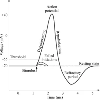
All neural activity begins with a change in the resting potential. A stimulus produces a localized temporary change in the transmembrane potential, which decreases with distance. It is called a graded potential. If the graded potential is sufficiently large it produces an action potential in the membrane of the axon. An action potential is an electrical impulse that is propagated across the surface of the membrane and that does not diminish as it moves away from its source.
When a biological cell or patch of membrane undergoes an action potential, or elec-trical excitation, the polarity of the transmembrane voltage swings rapidly from negative to positive and back. An action potential involves a depolarization, a re-polarization and finally a hyperpolarization (or ”undershoot”). The period of membrane hyperpolarization is called the refractory period of the nerve fiber (to be explained in the next section). The different phases of the action potential can be seen on Fig. 2.4.
The action potential spreads actively across the neuron, with an ”all-or-none” response. In this response, the action potential is activated to depolarization, or not at all.
The transmembrane voltage changes that take place during an action potential result from changes in the permeability of the membrane to specific ions, the internal and exter-nal concentrations of which cells maintain in an imbalance. In the axon fibers of nerves, depolarization results from the inward rush of sodium ions, while repolarization and hy-perpolarization arise from an outward rush of potassium ions. Calcium ions make up most or all of the depolarizing currents at an axon’s presynaptic terminus, in muscle cells and in some dendrites.
Changes in membrane permeability and the onset and cessation of ionic currents reflect the opening and closing of voltage-gated ion channels, which provide portals through the membrane for ions. Residing in and spanning the membrane, these proteins sense and respond to changes in transmembrane potential.
The depolarization phase of an action potential is due to the opening of voltage-gated ion channels, either sodium channels or calcium channels or a combination of both, de-pending on the particular membrane. Sodium ions and calcium ions are positively charged. Cells use chemical energy to pump sodium and calcium ions out across the cell surface membrane. When a voltage-gated sodium channel or calcium channel opens, positively charged ions move into the cell. Voltage-gated sodium channels automatically gate shut after about a millisecond. Calcium-mediated action potentials can be much longer in dura-tion. The re-polarization phase of an action potential is due to the opening of voltage-gated potassium channels. Cells normally keep the concentration of potassium ions high inside cells. When voltage-gated potassium channels open, positively charged potassium ions move out of the cell, causing the membrane potential to return to a negative inside poten-tial.
In myelinated fibers the action potential travels from one location in the cell to another, but ion flow occurs only at the nodes of Ranvier. As a result, the action potential signal jumps along the axon, from node to node, rather than propagating smoothly, as they do in unmyelinated axons. The clustering of voltage-gated sodium and potassium ion channels at the nodes permit this behavior, called saltatory conduction.
2.1.4
Some Characteristics of Nerve Conduction
Refractory Periods
As the action potential travels along the fiber surface, it consists of a wave of negativity followed by an area of gradually recovering positivity. While an area is in its reverse (active) state, it is absolutely refractory and cannot be restimulated. During recovery, the membrane is relatively refractory. During this period, intense or sustained stimuli can re-stimulate the original site.
Frequency of Impulses
Neurons normally carry trains of impulses. In general, natural stimuli are of sufficient duration to reactivate the membrane after the absolute refractory period. A single electric shock may produce a single action potential, but only because its duration does not outlast the absolute refractory period of the fiber. The stronger the stimulus, the the earlier will it re-excite, the shorter will be the time between impulses, hence, the greater the frequency.
Velocity of Conduction
Velocity of conduction depends not only on the myelination but also on the diameter of the fiber. It can be fairly accurately said that the conduction velocity is proportional to the diameter of the axon. The largest motor and sensory nerve fibers, with diameters near 20 µm, have conduction velocities up to 120 m/s. In small unmyelinated fibers, the ve-locities range from 0.7 to 2 m/s. Large fibers not only conduct more rapidly than small fibers, but characteristically have lower stimulus thresholds and larger spikes with shorter duration.
Classification of Nerve Fibers
Efferent nerve fibers are classified into three major groups, A, B, and C, on the basis of their conduction velocity. Group C contains the unmyelinated postganglionic fibers and group B the small myelinated fibers. Group A includes the large, rapidly conducting myelinated fibers. Group A has been further divided into four subgroups: alpha (α), beta (β), gamma (γ), and delta (δ), based on the velocity and diameter. Sensory nerve fibers have been separately classified according to diameter into groups I, II, III, and IV, with corresponding velocities. In order to avoid confusion, use of alphabetical designations is restricted to efferent fibers and the numerical designations to afferent fibers.
Neural Adaptation
Neural adaptation, or sensory adaptation, is a change over time in the responsiveness of the sensory system to a constant stimulus. It results in a decrease of neural firing rate over time.
2.2
Skeletal Muscles as Actuators of Movement
The human motor system comprises three interrelated anatomical systems: the muscle system, which supplies the power to move the skeleton; the nervous system, which directs and regulates the activity of the muscles. Muscle pairs are situated across joints, being attached at two or more points to bones via tendons. Movement is produced by shortening of the muscle, which brings the muscle ends closer to each other, ultimately changing the joint across which it acts.
The most distinguishing characteristic of muscle is contractility. By contractility, refer-ence is made to the capacity of the muscle to produce force between its ends. Relaxation is the opposite of contraction. It is entirely passive. Both relaxation and contraction progress from zero to maximal values over a finite time. Neither is instantaneous.
2.2.1
Muscle Length and Force
The most obvious property of the muscle is its capacity to develop force against resistance. The length of the muscle at the time of activation markedly affects its ability to develop force and to perform external work. When a muscle contracts, the contractile material itself shortens, but whether the muscle shortens or not depends on the relation of the internal force developed by the muscle to the external force exerted by the resistance or load.
Two general types of muscle contractions are distinguished according to the length change, induced by the relationship between internal and external forces: isometric and isotonic. If the internal force generated by the contractile components does not exceed the external force of the resistance and if no change of muscle length occurs during the contraction, the contraction is isometric. If the constant internal force produced by the muscle is maintained during the movement, the contraction is isotonic.
The initial length of the muscle, i.e. its length at the time of stimulation, influences the magnitude of its contractile response to a given stimulus. A stretched muscle contracts more forcefully than when it is unstretched at the time of activation. The force produced by skeletal muscle can be differentiated into 2 types, active and passive, their sum comprising the muscle total force. The relationship of force to length can be presented graphically in a form of a force-length curve, in which a force in an isolated muscle is plotted against a series of muscle lengths from less that to greater than the resting length. (Fig. 2.5).
Within physiological limits, the greater the initial length, the greater the passive elastic force of the muscle will be. Maximal active force is produced at the so called optimal length of the muscle. In general, optimal length is close to the maximal body length of the muscle, i.e. the greatest length the muscle can attain in the normal living body. It is about
Length
Force
1 2
Figure 2.5: Illustration of what the force vs. length curve for isolated muscle looks like: 1) passive elastic tension; 2) total force. The dashed line marks the muscle length at which contraction force is maximal.
1.2 to 1.3 times the rest length of the muscle. Force capability is less at shorter and longer lengths.
2.2.2
Muscle Function
Motor skill and all forms of movement result from the interaction of muscular force, grav-ity and any other external forces that act upon skeletal levers. The muscles rarely act alone. Rather, groups of muscles interact in many ways so that the desired movement is accom-plished. This interaction may take many different forms so that a muscle may serve in a number of different capacities, depending on the movement. Whenever a muscle causes movement by shortening, it is functioning as a mover or agonist. The muscles whose action oppose that of the agonists, are called antagonists.
2.3
Sensory Systems for Control of Movement
Sensory receptors are highly specialized neural structures that receive information about the external world. Various external stimuli that impinge upon our bodies are transformed into neural signals at these neural structures. Sensory information is coming from the en-vironment and from within our bodies (e.g. blood vessels, the actions of skeletal muscles). To distinguish the systems that convey signals from these different sources, the sensory systems are divided into three categories: exteroceptive, proprioceptive, and interoceptive. Exteroceptive sensors are sensitive to external stimuli and include vision, audition, skin sensation, and some chemical senses. Proprioceptive sensors provide information about
the relative position of the body in space. Interoceptive sensors are concerned with internal bodily events such as blood pressure and the concentration of glucose in the blood. In the following we will limit ourselves to proprioceptive sensors. Muscle receptors in particular. Muscles and joints contain a variety of receptors. Some inform the central nervous system about the length of the muscle, others detect its tension, and still other respond to pressure and noxious stimuli. Among these receptors, two have been most thoroughly stud-ied: muscle spindles and Golgi tendon organs. Although both of these receptors discharge when the muscle is stretched, differences in their anatomical arrangement within the mus-cle are reflected in the differences in information they convey to the central nervous system. Muscle spindles, arranged in parallel to the muscle fibers, provide information about the length of the muscle. Golgi tendon organs are arranged in series with the muscle fibers and inform the nervous system of the tension exerted by the muscle on its tendon insertion to the bone.
2.3.1
Muscle Spindles
Mammalian muscle spindles are receptors that are distributed throughout the fleshy parts of skeletal muscles. Each spindle, which consists of an encapsulated group of specialized muscle fibers, is tapered at each end and expanded at its center in a fluid-filled capsule. Within this capsule the terminal branches of afferent nerve fibers entwine the muscular elements (Fig. 2.6).
The small muscle fibers within the spindle are called intrafusal fibers. They do not contribute to the overall tension of the muscle, but regulate the excitability of the spin-dle afferents by mechanically deforming the receptors. Intrafusal fibers are innervated by small motor cells of the ventral horn called gamma motorneurons. The large skeletal mus-cle fibers that do develop substantial musmus-cle tension are called extrafusal fibers and are innervated by large alpha motorneurons of the ventral horn of the spinal cord.
Muscle spindles contain two types of intrafusal muscle fibers called nuclear bag fibers and nuclear chain fibers after the arrangement of nuclei in their equatorial region. The bag fibers have nuclei clustered in groups of two and three. The nuclear chain fibers have nuclei in a single row. The bag and chain fibers also differ in the kind of contraction they exhibit. Bag fibers produce slow contractions, whereas chain fibers produce fast (twitch) contractions.
There are two types of afferent terminals in muscles spindles: primary and secondary. Primary endings innervate every single intrafusal fiber within a spindle, while secondary endings lie almost exclusively on nuclear chain fibers.
Figure 2.6: The main components of the muscle spindle are intrafusal muscle fibers, af-ferent sensory fiber endings, and efaf-ferent motor fiber endings. The sensory fiber endings spiral around the the central regions of the intrafusal fibers and are responsive to stretch of these fibers. From Kandel et al. [2000].
2.3.2
Golgi Tendon Organs
The Golgi tendon organ is a slender capsule approximately 1 mm long and 0.1 mm in diameter. Each organ is in series with about 15-20 extrafusal skeletal muscle fibers that enter the capsule through a tight fitting collar. The muscle fibers terminate in musculo-tendonious junctions after entering the capsule and give rise to collagen fiber bundles that become braided and run the length of the capsule (Fig. 2.7). An afferent nerve fiber enters the capsule in the middle and branches many times so that the axons of the afferent fiber become twisted within the braids of the collagen fiber bundles. When the skeletal muscle fibers contract, they cause the collagen fibers to contract, resulting in compression of the afferent nerve fibers and, thereby, their firing. Thus, the organization of the collagen fiber bundles makes them sensitive to small changes of muscle tension.
2.3.3
Muscle Receptors Information Encoding
Muscle stretch receptors encode information about muscle length, tension and velocity of stretch. Loading the muscle activates both the tendon organ and the muscle spindle recep-tors. Contraction further stretches the tendon organs. On the other hand, contraction of the extrafusal muscle fibers makes the intrafusal fibers go slack, unloading the spindle so it is
Figure 2.7: When a Golgi tendon organ is stretched, the afferent axon is compressed by collagen fibers and its rate of firing increases. From Kandel et al. [2000].
no longer stretched. As a result, when the muscle is passively stretched, the afferent fibers from either the tendon organ or the spindle would increase their rate of discharge. In con-trast, when the muscle actively contracts, the tendon organ further increases its discharge, but the spindle decreases or ceases altogether.
The primary and secondary afferents in the muscle spindles respond quite differently to passive muscle stretch [Matthews and Stein, 1969a]. Both fiber types respond to static (steady-state) stretch, but they respond differently to the dynamic phase of stretch. Primary endings are very sensitive to the dynamic phase of stretch, whereas the secondary endings are not. The secondary endings mainly encode information about the length of the muscle, whereas the primary endings encode mainly information about the rate of change in length. The dynamic sensitivity of the primary endings is largely due to the mechanical properties of the nuclear bag fibers.
There are two type of gamma motorneurons. One type innervates nuclear bag fibers (gamma dynamic orγd) and the other type innervates nuclear chain fibers (gamma static
or γs). The reason for the names dynamic and static is that these gamma motorneurons
regulate the sensitivity of spindle afferents either to dynamic or static phases of stretch. An important role of the gamma system is to allow the spindle to maintain a high sensitivity over a wide range of muscle lengths during reflex and voluntary contractions [Barker et al., 1970].
Golgi tendon organs have a relatively high activation thresholds which is why it is believed that they, together with their inhibitory spinal connections, function as a safety device to prevent muscle damage during excessive strain [Houk and Simon, 1967]. For this reason muscle spindles are thought to have a more important role in proprioception, as
they are continuously operating transducers.
Muscle receptors show sensory adaptation. Afferent fiber firing rates drop to the value which is 40-70% of the initial firing rate. Moreover, the rate of adaptation seems to be indipendent from stimulus intensity [Adrian and Zotterman, 1926].
2.4
Injury to Sensory-Motor Systems
Injury to any part of the organs and tissues responsible for movement may have many, very different consequences. In the following we give a very short overview.
2.4.1
Cerebro-Vascular Infarction (Stroke)
The term stroke, or cerebro-vascular accident, refers to the neurological symptoms and signs, usually focal and acute, which result from diseases involving blood vessels. Strokes are either occlusive (due to closure of a blood vessel) or hemorrhagic (due to bleeding from a vessel). Insufficiency of blood supply is called ischemia. If it is temporary, symptoms and signs may clear with little or no pathological evidence of tissue damage. Ischemia reduces blood supply, thereby deprives tissue from oxygen, glucose, and prevents the removal of potentially toxic metabolites such as lactic acid. When ischemia is sufficiently severe and prolonged, neurons and other cellular elements die. Depending on which region of the cen-tral nervous system is affected, the consequences can be different: concen-tralateral weakness, sensory loss, impaired spacial perception, and other not directly related to movement.
2.4.2
Spinal Cord Injuries
Spinal cord injuries or diseases are a frequent reason of disability and result in total and partial obstruction of flow of both sensory and motor information being instrumental for normal life. Spinal cord injuries are most often caused by trauma, especially following motor vehicle or sport accidents. The resulting syndrome depends on the extent of direct injury of the cord or compression of the cord by displaced vertebrae or blood clots. In extreme cases trauma may lead to complete or partial transsection of the spinal cord. Le-sions of the spinal cord give rise to motor and sensory symptoms that are often related to a particular sensory or motor segmental level of the spinal cord (Fig. 2.8).
Tetraplegia refers to impairment or loss or motor and/or sensory function in the cervical segments of the spinal cord due to damage of neural elements within the spinal canal. It results in the impairment of function in the arms as well as in the trunk, legs and pelvic or-gans. Paraplegia is the impairment or loss of motor and/or sensory function in the thoracic,
Figure 2.8: The spinal cord at different levels. It is divided in 4 major regions: cervical, thoracic, lumbar, and sacral. Spinal nerves at the cervical levels innervate the head, neck, and arms. Nerves at the thoracic level innervate the upper trunk, while lumbar and sacral spinal nerves innervate the lower trunk, back and legs. From Kandel et al. [2000].
lumbar or sacral (but not cervical) segments of the spinal cord. With paraplegia arm func-tioning is spared, but, depending on the level of injury, the trunk, legs, and pelvic organs may be involved. Terms quadriparesis and paraparesis describe incomplete lessions, where many functions are preserved.
2.4.3
Multiple Sclerosis
Multiple sclerosis (MS) is an autoimmune condition in which the immune system attacks the central nervous system, leading to demyelination. It affects the areas of the brain and spinal cord known as the white matter. More specifically, MS destroys oligodendrocytes which are the cells responsible for creating and maintaining a fatty layer, known as the myelin sheath, which helps the neurons carry electrical signals. MS results in a thinning or complete loss of myelin and, less frequently, the cutting (transection) of the neuron’s extensions or axons. When the myelin is lost, the neurons can no longer effectively conduct their electrical signals. The name multiple sclerosis refers to the scars (scleroses - better known as plaques or lesions) in the white matter. Loss of myelin in these lesions causes some of the symptoms, which vary widely depending upon which signals are interrupted. Almost any neurological symptom can accompany the disease. Those related to movement include changes in sensation (hypoesthesia), muscle weakness, abnormal muscle spasms, and difficulties with coordination and balance.
2.5
Neuroprostheses for Restoration of Movement
A neuroprosthesis is a system for replacing or augmenting a function that is lost or dimin-ished because of injury or disease of the nervous system. The basic principle for operation of a neuroprosthesis is the stimulation of neuro-muscular tissue, i.e. activation of sensory and/or motor system. Advanced neuroprosthesis are bidirectional, also capable of sensing neuroelectric activity either in the central either in the peripheral nervous system. Func-tional electrical stimulation (FES) is at this time the essence of neuroprosthesis. FES elicits controlled neural activation by delivering low level electrical currents.
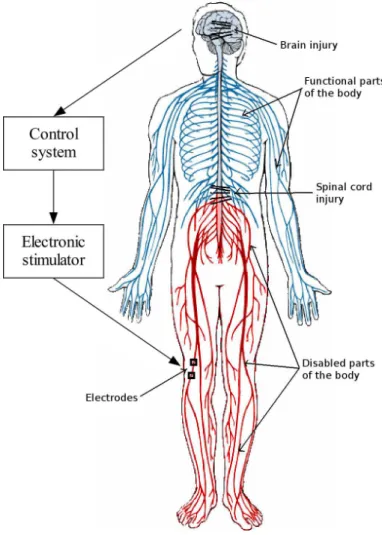
Fig. 2.9 shows the principle of operation of a neuroprosthesis. After an injury or disease of the central nervous system, parts of the body remain functioning normally, but other parts of the body will be paralyzed. Many muscles will remain connected to the CNS below the level of injury. They are innervated, but not under volitional control. These muscles can be used for movement restoration. At the same time many sensory pathways remain intact below the level of injury. Neuroprostheses can be considered as a bypass of the damaged
Figure 2.9: Principle of operation of neuroprostheses. Components of the system are the control system, stimulator, sensory feedback (not shown) and electrodes.
sensory-motor structure. Basically, the FES system consists of an interface to the sensory and motor systems of the body and a controlable stimulator.
2.5.1
Instrumentation for FES Systems
A functional diagram of the FES system (Fig. 2.10) shows the main components required for restoring motor function after injury of the central nervous system. The stimulator receives control signals, generates trains of pulses of electrical charge, and delivers those to the the excitable tissue via electrodes.
Each of the components has to be selected or built based on the specific application, e.g. lower extremities upper extremities, single-channel, multi-channel, transcutaneous,
Command interface Control System Electronic Stimulator Electrodes Paralyzed muscles Movement Sensors Decision to move
Figure 2.10: Instrumentation for FES: command interface, control system, electronic stim-ulator, electrodes and sensors. Decision to move is at the voluntary level of the user.
Open-loop controller Musculoskeletal system Reference trajectory Stimulation Actual trajectory
Figure 2.11: Organization of an open-loop control system. From Popovi´c and Sinkjær [2000].
percutaneous, and fully implanted systems.
2.5.2
Control Systems for Movement
Open-loop controllers can be designed to function without knowledge of the actual plant trajectory as shown in Fig. 2.11. The open-loop controller delivers command signals based on precomputed sequences. If there is any perturbation in the trajectory, an error will occur, to which the system will not react. The precomputed sequences are patterns of stimulation for the appropriate muscles. Regardless of design method and implementation, the per-formance of any open-loop control system will probably be inadequate, since disturbances will cause performance to deviate significantly and lead to undesired behavior.
To correct for disturbances, a closed-loop controller with ongoing knowledge of the effects of the disturbance, or modification of muscle performance due to fatigue, must be designed. Joint angle and force transducers provide signals from which the current state of the system can be estimated and fed back to the controller (Fig. 2.12). Closed-loop controllers are classically error driven – they respond to the trajectory error which is determined by comparing the actual and the desired trajectories. PID controllers are not an entirely appropriate solution and it is difficult to tune the gain factors.
Model-based closed-loop controllers would be more robust. They require sensors and a dynamic model of the system to continuously recalculate the desired trajectory in order
Closed-loop controller Musculoskeletal system Reference trajectory Stimulation Actual trajectory S Error Sensors -+
Figure 2.12: Organization of a closed-loop control system. From Popovi´c and Sinkjær [2000]. Model-based controller Musculoskeletal system Desired trajectory Stimulation Actual trajectory Sensors
Figure 2.13: Organization of a model-based control system. From Popovi´c and Sinkjær [2000].
to accomplish the task (Fig. 2.13). For example, during walking, if the body is following a desired path, the model-based controller uses the same muscle stimulation pattern as an open-loop controller. If the body deviates from the desires path, the model-based controller generates a new stimulation pattern which, according to the model, should maintain or restore stable walking. Accounting for change of muscle properties and fatigue would also be desirable.
2.5.3
Recording Nerve Activity
Advanced implanted neuroprostheses aim to take advantage of both recording and activat-ing properties of the neural interface to restore sensory-motor functions. Measurement of nerve signals implies a recording chain that can be broken into three subsystems: biology, interface, and signal conditioning system. The biology system comprises the peripheral nervous system, responsible for driving information from/to the central nervous system to/from the periphery. The interface, i.e. the electrodes, senses and translates chemoelec-tric potentials, which result from elecchemoelec-trical activity of cells. The signal conditioning system is the electronic apparatus that allows recording nerve signals as potentials. A classical sig-nal conditioning scheme can be seen on Fig. 2.14.
Figure 2.14: Basic configuration of a biopotential recording system. From Bronzino [2000].
The measurements involve voltages at very low levels, with high source impedances and superimposed high level interference signals and noise. The signals need to be amplified to be compatible with devices such as displays, recorders, or A/D converters for computer-ized equipment. Amplifiers adequate to measure these signals have to satisfy very specific requirements. They have to provide amplification selective to the physiological signal, re-ject superimposed noise and interference signals, and guarantee protection from damages through voltage and current surges for both patient and electronic equipment. Visual rep-resentation of recorded neuroelectric activity is called the electroneurogram (ENG).
2.5.4
Interfacing the Peripheral Nerve
A transverse section of a nerve is illustrated on Fig. 2.15. Nerve axons travel in bundles through the body. These bundles are encapsulated in fibroconnective tissue. Entire nerve bundles are surrounded by the epineurium, which bears most of the mechanical tension applied to the nerve. Branching from the epineurium and dividing the nerve bundle into fascicles is the perineurium, which bears the elongation load. Finally each individual axon is surrounded by the endoneurium. Each axon, or nerve fiber, can be seen as an individual information channel.
There are two basic designs of peripheral nerve recording electrodes: extraneural and intraneural electrodes. Extraneural electrodes are generally wrapped around the circum-ference of the nerve and commonly named cuff electrodes. Less invasive than intraneural electrodes, they permit recording from a whole nerve and do not provide access to infor-mation from a small group of neurons. Cuff electrodes come in a variety of configurations, but they all have a longitudinal opening to allow installation on the nerve without dam-aging it [Andreasen and Struijk, 2002], [Yoo and Durand, 2005], [Navarro et al., 2005].
Figure 2.15: The structure of a peripheral nerve. From Horch and Dhillon [2004].
Figure 2.16: Cuff electrode. It is wrapped around the circumference of the nerve. The closed cuff is shown on the left, and the open on the right. From Hoffer and Kallesøe [1999].
They can also be in a form of a spiral [Naples et al., 1988] and have multiple contacts [Grill and Mortimer, 1996], [Stieglitz et al., 2003]. The standard cuff is made out a poly-mer and the electrodes within the cuff are made out of metal, usually being circumferential around the inner surface of the cuff (Fig. 2.16).
Cuff electrodes are a proved method for chronic recording from the peripheral nervous system. The first chronic studies were done in the 1970s by Hoffer [1975] and Stein et al. [1978]. For more than a decade now it has been used in many studies where information from whole-nerve recordings were extracted to provide event detection. A recent review has been published by Struijk et al. [1999]. One example is recording from cutaneous sensors for the detection of object slippage while being held [Hoffer et al., 1991] and ob-taining skin contact force information [Hoffer et al., 1989]. Human trials have also also
Figure 2.17: Longitudinal intrafascicular electrode. The electrode invades the epineurium and perineurium, and placed longitudinally inside a nerve fascicle. Courtesy of Dr. Ken Yoshida.
been conducted [Sinkjær et al., 1991]. Muscle spindle afferent activity was recorded to provide natural sensory feedback in studies by [Haugland and Hoffer, 1994], [Jensen et al., 2002b], [Micera et al., 2001], [Riso et al., 2000], [Strange and Hoffer, 1999]. Due to its poor selectivity and low signal-to-noise ratio, the digital signal processing algorithms used to decode afferent nerve activity fail to provide reliable information to be used for feedback [Upshaw and Sinkjær, 1998].
An alternative to the cuff is the longitudinal intrafascicular electrode (LIFE). It is de-signed to be implanted within the body of the peripheral nerve to ultimately serve as a neu-ral interface for use in applications such as FES. They are placed in intimate contact with the nerve fibres to form a highly selective implanted neural interface (Fig. 2.17). When used for stimulation, they demonstrate topological selectivity in axonal recruitment, and when used for recording, they pick up neural activity from a relatively small population of fibres to enable the resolution of single fibres [Goodall and Horch, 1992], [Goodall et al., 1993], [Malmstorm et al., 1998], [Yoshida and Stein, 1999], [Chemineau et al., 2004].
The first LIFE was produced by Ken Horch and Andy Schoenberg of the University of Utah [Schoenberg et al., 1987]. Malagodi et al. [1989] did the first stimulation and record-ings. Nannini and Horch [1991] did the first major work on electrical stimulation using LIFEs. The first work with recording in chronic animals was done by Lefurge et al. [1991] and Goodall et al. [1991]. Results from their six-month study showed that intrafascicular electrodes can be used on a chronic basis to acquire neural activity.
Figure 2.18: (A) Schematic of the tf-LIFE. Each half of the structure has a ground electrode (GND), an indifferent electrode (L0, R0) and four recording sites (L1-4, R1-4). (B) The tfLIFE is folded by the central line, so both branches can be closely apposed. (C) View of the proximal part of the electrode. (D) Higher magnication view showing the four active sites made by Pt sputtering. (E) Photograph of the whole system. The tf-LIFE is attached at the ending part to a ceramic connector for nerve recording/stimulation. The loop between the two branches of the tfLIFE is connected to a Kevlar lament that in turn is glued to a tungsten needle for insertion in the nerve. From Lago et al. [2007].
McNaughton and Horch [1992] developed the first dual channel recording with LIFEs. Yoshida and Horch [1993] did the first work with multichannel stimulation. It has been shown that stimulation through them can be used to selectively stimulate nerve fascicles as well as subfascicular regions within the nerve in the cat animal model. Yoshida and Horch [1996] followed up this work by closing the control loop.
In an effort to create LIFEs with smaller, more precise, and more reproducible active sites, a flexible micromachined polymer structure was designed to replace the key com-ponent of the electrode, the fine wire in contact with the endoneural tissue that forms the neural interface of the electrode. Yoshida et al. [2000] developed the first thin-film multi-channel LIFE structure. The thin-film LIFE was later developed through work with the Fraunhofer IMBT in Ingbert, Germany. The latest version of the electrode is the micro-fabricated multi-site thin-film Longitudinal Intra-Fascicular Electrode (tfLIFE) array that is realized as a patterned thin film on polyimide structure [Hoffmann and Koch, 2005]. It allows recording up to 8 channels of ENG (Fig. 2.18).
In the meantime, the polyLIFE was being developed by Lawrence et al. [2003] and Lawrence et al. [2004], who did a series of experiments to show their acute and chronic feasibility in rabbits. Based on this work, Dhillon et al. [2004] and Dhillon and Horch [2005] implanted an advanced version of the polyLIFE in amputee human subjects for a short term trial. They showed the proof of concept of using LIFEs to elicit sensations and record volition from electrodes implanted semi-chronically in nerve stumps of human upper extremity amputee volunteers.
Modeling the Afferent Nerve Activity to
Estimate Muscle State
3.1
Introduction
Changes in muscle length are associated with changes in the angle of the joint that the muscle crosses. Thus, activity from muscle spindles can be used to sense relative positions of body segments. These sensors remain intact and active below the level of lesion in spinal cord injured patients. Therefore, their activity could be used to provide feedback information about muscle state in a closed-loop control FES system, if on-line extraction of reliable information from neural activity in a form usable by the controller is possible.
Recently, Stein et al. [2004] have shown how populations of sensory receptors could encode limb position and how the firing of a small number of neurons can be used to decode the position of the limb. In their study, microelectrode arrays have been used to record impulses simultaneously from individual nerve cells in the dorsal root ganglia (DRG) of the cat. They showed that a small number of informative neurons (68) could account for over 80% of the variance. The drawback of this technique is the risk associated to the surgery needed to place the electrodes at the DRG. Recording activity from peripheral nerves is less invasive; it is classically done using cuff electrodes which sense the aggregate activity of the whole nerve fibers. We propose to use intrafascicular electrodes which allow solving the lack of selectivity of the cuff electrodes.
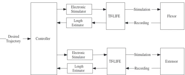
Closed-loop control of joint movement using neuromuscular stimulation adopted in this thesis is based on the approach developed by Yoshida and Horch [1996], where a pair of single-channel LIFE’s was implanted in the nerves innervating the joint agonist/antagonist muscle pair. The idea is to have the two electrodes working in opposite modes, e.g. while
Flexor Extensor TF-LIFE TF-LIFE Stimulation Stimulation Recording Recording Electronic Stimulator Electronic Stimulator Length Estimator Length Estimator Controller Desired Trajectory
Figure 3.1: Scheme of FES closed-loop control of joint movement where a pair of tfLIFE electrodes is implanted in nerves innervating the the joint agonist/antagonist muscle pair.
one electrode is stimulating the agonist, the other is recording muscle spindle activity from the antagonist, and vice versa (Fig. 3.1). This way the recording electrode is always sensing neural activity from a passively stretched muscle.
For estimating muscle length from this activity the authors used a look-up table. Esti-mation of muscle length was fed back to the controller and used to modulate the stimulation pulse train parameters to minimize ankle position error. With such a control scheme, the need for simoultaneous recording and electrical stimulation of the same muscle is avoided. Electrical stimulation in close proximity of the recording electrode produce large stimu-lation artifacts which corrupt the recorded signals. The control scheme also allows using passive mechanical muscle models for estimating muscle state, instead of complex model-ing of active muscle.
This approach proved to be applicable as feedback in on-line closed-loop control in restrictive conditions (limited motion speed and range), most probably due to the unac-counted variation of dynamic sensitivity of the sensory endings. The purpose of the study was to assess the feasibility of using muscle spindles as a sensor in a closed-loop FES sys-tem, rather than to build an optimized controller, per se, a simple PI (proportional-integral) controller was implemented to modulate the pulse width of a constant amplitude, fixed frequency stimulus pulse train.
If a valid analytical model, linking muscle spindle afferent nerve activity and muscle length, could be found for the locomotor range of ankle motion (in terms of both range of motion and motion speed), it would allow for having an on-line model-based estimator of muscle length and advanced control laws can be designed. Our aim is to develop a model,

![Figure 2.15: The structure of a peripheral nerve. From Horch and Dhillon [2004].](https://thumb-eu.123doks.com/thumbv2/123doknet/7702554.245866/39.892.199.670.193.495/figure-structure-peripheral-nerve-horch-dhillon.webp)