Université de Sherbrooke
BIOCHEMICAL AND GENETIC ANAL YSIS OF RNA PROCESSING AND DECA Y
Par Ghada Ghazal
Département de Microbiologie et d'infectiologie
Thése présentée à la Faculté de médecine et des sciences de la santé en vue de l'obtention du garde de
philosophiae doctor (Ph.D.) en Microbiologie
Membres du Jury:
Dr. Nicholas Proudfoot, raporteur externe à l'université Dr. François Bachand, raporteur externe au programme
Dr. Raymund Wellinger, président et directeur du département Dr. Sherif Abou Elela, directeur de thèse
l+I
NOTICE:
Library and Archives Canada
Published Heritage Branch
395 Wellington Street Ottawa ON K1A ON4 Canada
The author has granted a
non-exclusive license allowing Library and Archives Canada to reproduce, publish, archive, preserve, conserve, communicate to the public by
telecommunication or on the Internet, loan, distribute and sell theses
worldwide, for commercial or non-commercial purposes, in microform, paper, electronic and/or any other formats.
The author retains copyright ownership and moral rights in this thesis. Neither the thesis nor substantial extracts from it may be printed or otherwise reproduced without the author's permission. ln compliance with the Canadian Privacy Act some supporting forms may have been removed from this thesis.
While these forms may be included in the document page count, their removal does net represent any loss of content from the thesis.
•
••
Canada
Bibliothèque et Archives Canada Direction du Patrimoine de l'édition 395, rue Wellington Ottawa ON K1A ON4 CanadaAVIS:
Your file Votre référence ISBN: 978-0-494-62832-4
Our file Notre référence ISBN: 978-0-494-62832-4
L'auteur a accordé une licence non exclusive
permettant à la Bibliothèque et Archives
Canada de reproduire, publier, archiver, sauvegarder, conserver, transmettre au public par télécommunication ou par l'Internet, prêter, distribuer et vendre des thèses partout dans le
monde, à des fins commerciales ou autres, sur
support microforme, papier, électronique et/ou autres formats.
L'auteur conserve la propriété du droit d'auteur et des droits moraux qui protège cette thèse. Ni la thèse ni des extraits substantiels de celle-ci ne doivent être imprimés ou autrement
reproduits sans son autorisation.
Conformément à la loi canadienne sur la
protection de la vie privée, quelques
formulaires secondaires ont été enlevés de cette thèse.
Bien que ces formulaires aient inclus dans
la pagination, il n'y aura aucun contenu
TABLE OF CONTENTS
TABLE OF CONTENTS ... 1
LIST OF FIGURES AND TABLES ... Ill ABREVIATIONS ... IV Résumé Summary INTRODUCTION ... 1
1. Regulation of gene expression ... 1
2. Transcription ... 3
3. RNA Maturation ... : ... 5
3.1 Capping ... 7
3.2 Splicing ... 8
3.3 Termination and 3'end formation ... 10
4. RNA turnover and degradation ... 17
5 .. Ribonucleases ... 19
5.1 Exoribonucleases ... 20
Ali eukaryotes ... 22
5.2 Endoribonucleases ... 23
6. RNase Ill family ... 25
6.1 Protein structure of budding yeast RNase Ill ... 26
6.2 Cellular functions of yeast RNase 111 ... 30
7. Aim of the project ... 32
ARTICLE 1 ... 34
Préambule ... 34
Summary ... 35
Préambule ... 104 Summary ... 105 ARTICLE 3 ... 155 Préambule ... 155 Summary ... 156 DISCUSSION ... 209
1. A flexible mechanism of substrate ·selectivity increases the spectrum of Rnt1 p cleavage targets ... 209
1.1 The AGNN tetraloop sequence is not essential for RNA cleavage 211 1.2 Contribution of the stem sequence to cleavage efficiency ... 213
1.3 The helical rulers cleavage mechanism re-defined ... 214
1 .4 Flexible protein conformation adopts to changes in the substrate structure ... 216
2. The architecture of the pre-snoRNA transcript defines the processing mechanism ... 219
2.1 Rnt1 p cleavage is essential for the processing of polycistronic snoRNA ... 220
2.2 ls the cleavage of the monocistornic RNA an evolutionary relie of gene clusters or a mechanism for quality contrai? ... 221
2.3 Processing of intronic snoRNA and the regulation of pre-mRNA splicing ... 222
3. New model for transcription termination ···'··· 224
3.1 Transcription termination of non-polyadenylated RNA ... 224
3.2 Transcription termination de pendent RNA decay ... 229
4. Towards an integrated mode of gene expression ... 231
ACKNOWLEDGMENTS ... 234
REFERENCES ... 235
LIST OF FIGURES AND TABLES
Figure 1. Schematic representation of co-transcriptional processing ... 6
Figure 2. Models for polyadenylation dependent RNA stability ... 12
Figure 3. Transcription termination by RNA polymerase 11. ... 16
Table 1. Summary of exoribonuclease superfamilies ... 22
Figure 4: Schematic representation of the RNase Ill family ... 27
Figure 5. Rnt1 p mechanism of action ... 29
Figure 6. Rnt1 p Interaction with stem-loop capped with either NGNN or AAGU tetraloop ... 218
bp CPSF CTD CUT dsRBD dsRBP ETS mRNA nt N-term NUCD PAZ Polll Pre-mRNA Pre-rRNA rDNA ABREVIATIONS Base pair
Cleavage polyadenylation specific factors
Carboxy-terminal demain
Cryptic unstable transcript
Double stranded RNA binding demain
Double stranded RNA binding protein
External transcript Messenger RNA nucleotide N-terminal demain Nuclease demain PIWl/Argonaute/Zwille Polymerase Il Premature mRNA
premature ribosomal RNA
RN Ai RNA interference
RNP Ribonucleoprotein
snRNP Small nuclear ribonucleoprotein
Université de Sherbrooke
L'ANALYSE BIOCHIMIQUE ET GÉNÉTIGUE DE LA MATURATION ET LA DÉGRADATION DE L' ARN
Par Ghada Ghazal
Département de Microbiologie et d'infectiologie
Thése présentée à la Faculté de médecine et des sciences de la santé en vue de l'obtention du garde de
philosophiae doctor (Ph.D.) en Microbiologie
L'expression des gènes est le conduit par lequel l'information génétique est traduite dans les phénotypes cellulaires. Récemment, il a été démontré que le programme de l'expression des gènes dans les cellules de mammifères est régi, au moins en partie par l'expression d'ARN double brin court (ARNdb). Ce mode de régulation des gènes est influencé par un grand groupe de protéines de liaison à l'ARN double brin qui peuvent soit stabiliser ou déclencher la dégradation de l'ARN double brin. En effet, les ribonucléases (RNases) spécifiques à l'ARN double brin jouent un rôle important dans l'expression des gènes. Dans la plupart des eucaryotes, les membres de la famille des RNase Ill spécifiques ~ l'ARNdb déclenchent la dégradation de
l'ARN et initient la réponse immune de la cellule. Un défaut dans l'activité de la RNase Ill (DICER) inhibe l'expression des gènes et favorise le développement du cancer. D'autre part, la surexpression de la RNase Ill bloque l'infection virale. Cependant, très peu est connu sur la fonction de
gestion domestique des RNases Ill chez les eucaryotes et le mécanisme par lequel ils font la distinction entre les espèces d'ARN cellulaire et l'infection virale. Cette thèse pave la voie sur la manière dont les ARNdbs sont choisis pour être clivés et démontre leur contribution dans le mécanisme de l'ARN en utilisant la levure comme modèle d'étude. Initialement, les déterminants de réactivité de la RNase Ill chez la levure (Rnt1 p) ont été identifiés in vitro et utilisés pour étudier l'impact global de Rnt1 p sur la maturation des ARNs non-codants. Les résultats indiquent que Rnt1 p est nécessaire pour la maturation de tous les petits ARN nucléolaires (snoRNAs) impliqués dans la méthylation de l'ARNr et ils identifient un nouveau rôle de Rnt1 p dans la maturation des snoRNAs introniques. Il a été démontré que le clivage de Rnt1 p contribue à
coordonner l'expression de certaines protéines ribosomales et des snoRNA contenus dans leurs introns. La maturation du snoRNA à partir' de l'ARN pré-messager bloque l'expression du gène hôte, alors qu'en retardant la maturation du snoRNA, celle-ci se séroule sur l'intron excisé ce qui permet l'expression des deux gènes. De cette façon, la cellule peut coordonner soigneusement la quantité de protéines ribosomales et de snoRNAs requises pour la biogénèse des ribosomes. En outre, l'analyse globale de la maturation des snoRNAs a identifié de nouveaux signaux de clivage de Rnt1 p qui ne présentent pas un motif de séquence conservé.
Cette constatation a conduit à la conclusion que Rnt1 p utilise une vaste combinaison de motifs structuraux pour identifier ses substrats et augmenter ainsi le nombre de cibles potentielles de dégradation in vivo. Pour évaluer
cette possibilité, une nouvelle recherche de motifs pouvant être clivés par Rnt1 p a été effectuée. Fait intéressant, de nombreux signaux de clivage de Rnt1 p ont été identifiés dans des régions intergéniques qui n'encodent aucun transcrit d'ARN connus. ln vivo, les résultats démontrent que Rnt1 p est capable de terminer la transcription des ARNms non-polyadenylés et participe
à
un mécanisme de surveillance contre la continuation de transcription (read-through). Cette découverte démontre un lien direct entre la Rnt1 p et la machinerie de la transcription des ARN messagers, et prévoit un nouveau mécanisme de la terminaison de la transcription indépendante de la polyadénylation. Ensemble, les travaux décrits dans cette thèse présentent un exemple de la façon dont les RNase Ill chez les eucaryotes identifient leurs substrats et présentent un modèle dans lequel la transcription de l'ARN, sa maturation et sa stabilité sont liés.Mots clés : dsRNA, RNases, Rnt1 p, snoRNA, transcription termination, 3'end formation
BIOCHEMICAL AND GENETIC ANAL YSIS OF RNA PROCESSING AND DECA Y
Par Ghada Ghazal
Département de Microbiologie et d'infectiologie
Thèse présentée à la Faculté de médecine et des sciences de la santé en vue de l'obtention du garde de
philosophiae doctor (Ph.D.) en Microbiologie
Gene expression is the conduit by which genetic information is connected into cellular phenotypes. Recently, it was shown that gene expression in mammalian cells is governed, at least in part, by the expression of short double stranded RNA (dsRNA). This mode of gene regulation is influenced by a large group of dsRNA binding proteins that could either stabilize or trigger the degradation of dsRNA. lndeed, double stranded RNA (dsRNA) specific ribonucleases (RNases) play an important raie in regulating gene expression. ln most eukaryotes, members of the dsRNA specific RNase Ill family trigger RNA degradation and initiate cellular immune response. Disruption of human RNase Ill (Dicer) deregulates fetal gene expression and promotes the development of cancer. However, very little is known about the housekeeping function of eukaryotic RNase Ill and the mechanism by which they distinguish between exogenous and endogenous cellular RNA species. This thesis elucidates how dsRNAs are selected for cleavage and demonstrates their contribution to RNA metabolism in yeast as model eukaryote. lnitially, the reactivity determinants of yeast RNase Ill (Rnt1 p) were identified in vitro and
used to study the global impact of Rnt1 p on the processing of non-coding RNA. The results indicate that Rnt1 p is required for the processing of all small nucleolar RNAs (snoRNAs) involved in rRNA methylation and identify a new role of Rnt1 p in the processing of intronic snoRNAs. lt was shown that Rnt1 p cleavage helps to coordinate the expression of some ribosomal protein genes hosting intronic snoRNAs. Direct snoRNA processing from the pre-mRNA blacks the expression of the host gene, while delayed snoRNA processing from the excised intron allows the expression of both genes. ln this way, the cell can carefully calibrate the amount of snoRNA and ribosomal proteins required for ribosome biogenesis. ln addition, a global analysis of snoRNA processing identified new form~ of Rnt1 p cleavage signais that do not exhibit a conserved sequence motif but instead use a new RNA fold to recruit the enzyme to the cleavage site. This finding led to the conclusion that Rnt1 p may use a wide combination of structural motifs to identify its substrates and thus increases the theoretical number of potential degradation targets in vivo. To evaluate this possibility, a new search for snoRNA independent Rnt1 p cleavage targets was performed. lnterestingly, many Rnt1 p cleayage signais were identified in intergenic regions devoid of known RNA transcripts. ln vivo, it was shown that Rnt1 p induce the termination of non-polyadenylated transcripts and functions as a surveillance mechanism for transcription read-through. This finding directly links Rnt1 p to the transcription machinery and provides a new mechanism for polyadenylation independent transcription termination. Together the work described in this thesis presents an example
of how eukaryotic RNase Ill may identify its substrates and present a case study where transcription, RNA processing and stability are linked.
Key Words: dsRNA, RNases, Rnt1 p, snoRNA, transcription termination, 3'end formation
INTRODUCTION
1. Regulation of gene expression
Modern cells àre defined by their gene sequence but shaped by their protein make up (Herbert and Rich, 1999a). The transformation of genetic information from deoxyribonucleic acid sequence (DNA) into protein is an essential and tightly controlled process termed "gene expression" (Granneman and Baserga, 2005; Hinnebusch, 1990). ln its simplest form, gene expression is a passive conduit of stored genetic information with little influence on the phenotypic outcome (Herbert and Rich, 1999b ). However, even in the simplest of live forms like viruses, the process leading to protein production can greatly influence the organism's function and may even determine its chance to survive (Katze and Agy, 1990; Naryshkin et al., 1998; Stoltzfus and Madsen, 2006). Therefore, cells have developed a highly refined mechanism to contrai the expression time and amount of each gene and used it to fine-tune the accumulation of any particular protein at a specific time (Haile and Papadopoulou, 2007; Harrison, 1990; lzawa and lnoue, 2009). Regulation of gene expression provides cells with the flexibility they need to face changes in their environment and increase the versatility of their protein functions (Hengge-Aronis, 2002; Marles-Wright and Lewis, 2007; Wassarman, 2002). For example, yeast genes involved in glucose metabolism are expressed in the presence of glucose, while those required for gluconeogenesis are repressed (Gelade et al., 2003). ln bacteria, regulation of gene expression is
mostly a response mechanism to rapidly changing environment (Klaenhammer et al., 2007). ln contrast, changes in mammalian gene expression respond to the need for cell specialization and differentiatiàn (Harrison, 1990).
The mechanism regulating gene expression varies depending on the organism and gene function. ln bacteria, gene regulation is ingrained in the genome structure (Rocha, 2008). For example, genes with related function are clustered into "operons" to allow coordinated expression of proteins with interdependent functions (Rocha, 2008). ln eukaryotes, the genome structure and mechanism of gene expression is drastically different than that of bacteria (Mateos-Langerak et al., 2007). Genes are normally not organized by function and transcription is physically separated from translation by the nuclear membrane. Eukaryotic genes need not only to include information about their transcriptional program but also need to embed in the RNA information that dictates its stability, export, translatability and the nature of the protein it produces (Zhai et al., 2008). lmpairing any of these steps may signal RNA degradation and abort the expression process. Therefore, in eukaryotes co-regulation of proteins cannot be achieved by a simple switch, a single factor
qr
even a single step of gene expression. For simplicity, eukaryotic gene regulation is often separated into four classes; 1) transcriptional, 2) posttranscriptional, 3) translational and 4) posttranslational gene regulation (Nolan and Cogoni, 2004 ). Transcriptional gene regulation influences the overall amount of the primary gene products and is often used as a master onand off switch of gene expression. ln reality, however, it is important to note that eukaryotic gene regulation is an integrated process where one level of gene expression affects the other.
2. Transcription
The most direct way to contrai the expression of a gene is to regulate its rate of transcription; that is, the rate at which RNA polymerases transcribe genes into messenger RNA (mRNA) (Westholm et al., 2008). The basic mechanism of transcription is the same in ail organisms where DNA dependent RNA polymerases recognize a specific DNA sequence and use it to polymerize free nucleotides into ribonucleic acid chains (Lee and Young, 2000). The main difference between bacterial and eukaryotic transcription machinery is in the number of RNA polymerases and the associated transcription factors. ln Bacteria, ail genes are transcribed by the same RNA polymerase (Balleza et al., 2009), whereas eukaryotes use three different nuclear polymerases (RNA Pol 1-111) (Chambon, 1975; Roeder and Rutter, 1970). These polymerases differ in the number and type of subunits they contain, as well as the class of RNAs they transcribe; that is, RNA Pol 1 transcribes ribosomal RNAs (rRNAs) (Kuhn et al., 2007), RNA Pol Il (Meyer et al., 2009) transcribes RNAs that will become messenger RNAs (mRNAs) and also small regulatory RNAs, whereas RNA Pol Ill transcribes small RNAs such as transfer RNAs (tRNAs). Because RNA Pol li transcribes protein-encoding genes, it has been the main
target of transcriptional regulation. Transcription begins with the binding of the polymerase to the promoter region which is essential for correct positioning and assembly of Pol 11 and the general transcription factors in a state termed preinitiation complex (PIC). Next, a marked conformational change allowing the active Pol Il to open the template strand of the promoter and starts the initiation of transcription. After synthesis of -30 bases of RNA, the 5' end of the RNA is modified by adding a cap that consists of a modified guanine nucleotide. Pol Il then releases its contacts with the core promoter and the rest of the transcription machinery and enters the transcription elongation phase (Figure 1 ). Factors that promote productive RNA chain synthesis, RNA processing, RNA export and chromatin modification can all be recruited to elongating Pol Il (Bentley, 2002). A key step of the transition of Pol Il to the elongation mode of RNA synthesis is an extensive phosphorylation of the RNA polymerase Il tail, carboxy-terminal do main "CTD" (Figure 1 ). This C-terminal domain consists of a long tandem array of repeated seven-amino-acid sequences, containing two serines (serine 2, serine 5) per repeat that can be phosphorylated'. ln addition phosphorylation of a third serine in position 7 was recently identified and its impact on transcription is currentlybeing investigated (Kim et al., 2009). As transcription precedes the two major sites of phosphorylation (serine 2, serine 5), predominate, and the CTD of the Pol Il undergoes conformational changes to recruit the termination factors (Bentley, 2005; Zorio and Bentley, 2004; Bentley, 1999).Transcription termination is an important process as it enhances gene expression by
facilitating polymerase recycling and thus maintains a pool of available polymerase (Oye and Proudfoot, 1999). Once transcribed, RNA is .normally processed to produce the mature form and either exported to the cytoplasm for translation or assembled into functional RNP in the nucleus (RNP).
3. RNA Maturation
RNA maturation is the process by which a nascent RNA is transformed into a stable functional form. For a mRNA this means capping, polyadenylation, and removal of intronic sequence through splicing (Meyer et al., 2009). On the other hand, for non-coding RNA like small nuclear RNA (snRNA) or ribosomal RNA (rRNA) maturation means the removal of transcribed spacers, modification of the 5' and 3' ends, and assembly into an active RNA protein complex (Lafontaine and Tollervey, 1995; Nazar, 2004; Reddy and Busch, 1983). ln both cases, the aim of this process is to remove non-functional sequence, ensure the quality of the transcribed RNA and increase the versatility of RNA functions. Each step of RNA maturation involves a complex machinery of RN~ and protein factors capable of specifically recognize its
target and modify it according to a pre-determined and precise program (Fischer et al., 1991; Maxwell and Fournier, 1995; Wahl et al., 2009)
Serine S pbospborylation Serine 2 phosphorylation Initiation ....,cc,,..,,=,.,,.,.... ... ...,... Elongation ... __ ... _._.,.. Ten1ùnation
Capping Cleavage/polyadenylation
/\
\
·'--- ---AAAAAAAAAAA,,
C8Pfltll!I FectOISFigure 1. Schematic representation of co-transcriptional processing. Processing factors interact with the Pol Il machinery via the carboxy-terininal domain (CTD) of the largest subunit of RNA Pol Il. Capping enzymes are recruited to the 5'ends of genes. As Pol Il traverses the gene, splicing factors associate with the transcription complex. Phosphorylation of Ser2 and Ser5 residues in the CTD heptaq repeats is indicated in yellow circles. Exon numbers are marked in colors. lntrons are shown in black boxes. The red star represents the cap structure (adapted from Zorio and Bentley, 2004).
3.1 Capping
Capping is a specific form of 5' end modification that occurs during transcription by RNA polymerase Il (Pol 11). RNA produced by Pol 1 or Ill are not capped and these polymerases do not associate with capping enzymes (Gu and Lima, 2005). ln general caps confer stability to mRNAs by protecting them from digestion by exonucleases. However the crucial raie of the 5' cap of the mRNAs is to position the ribosome to initiate translation through the binding of the initiation factor CBPI. ln fact, some viruses such as poliovirus prevent capped cellular mRNAs from being translated into proteins. This enables poliovirus to take over the protein synthesizing machinery in the infected cell to make new viruses (Thompson and Samow, 2000). The 5' cap is generated by the addition of a guanosine to the extreme 5' end of the nascent mRNA by the guanylyl transferase enzyme, this guanosine later converted into 7-methylguanosine by the guanine methyltransferase (Furuichi and Shatkin, 2000; Gu and Lima, 2005; Shuman, 2001 ). These dimeric capping enzymes are recruited to the phosporylated carboxy-terminal domain (CTD) of the Polymerase Il at the early stages of RNA synthesis (Figure 1)
(Viladevall et al., 2009). After the RNA is capped, elongation factors required for splicing and termination are recruited to the CTD of the Pol li.
3.2 Splicing
Splicing is a process by which intervening sequence (introns) are removed and the protein encoding fragments (exons) are joined together to generate mature mRNA ready for translation (Rio, 1993; Umen and Guthrie, 1995). This process if performed by a large RNA protein complex (Spliceosomal complex) that ensures the fidelity and efficiency of intron removal (Wahl et al., 2009). Splicing allows cells to swap exons during development and thus modify protein sequence and function as the cellular functions change (lrimia et al., 2009; Mattaj and Hamm, 1989; Rio, 1993). ln addition, splicing is also believed to contribute to genome complexity and increase the diversity of protein functions through the process of alternative splicing (Boue et al., 2003; Kim et al., 2008; Kriventseva et al., 2003; Park and Graveley, 2007). lndeed most human genes are now believed to be alternatively spliced (Wang et al., 2008).
Although introns are often considered disposable junk DNA, it is becoming increasingly clear that information in these non-coding sequences can directly or indirectly affect gene expression (Le Hir et al., 2003). lntrons can influence every level of RNA metabolism from transcription (Finkbeiner, 2001) to RNA stability and thus may have a major impact on cell function and fitness. For example, mutations in conserved intron sequences may lead to several
human diseases like the neurodegenerative disorders Friedreich ataxia, (Baralle et al., 2008; Lewandowska et al., 2005), or ta spinal muscular atrophy (SMA) (Kas~ima et al., 2007). ln addition, it is now accepted that introns carry a plethora of non-coding RNA signal sequences required for RNA modification (Fedorov et al., 2005; Lim et al., 2002; Ooi et al., 1998a), translational regulation and RNA degradation (Lin et al., 2008; Lin et al., 2006; Ying et al., 2008) lndeed, the majority of human microRNAs (miRNAs), implicated in RNA interference, and small nucleolar RNAs (snoRNAs), required for the modification of rRNA, are found in intronic sequence (Bortolin and Kiss, 1998; Lin et al., 2006; Tanaka-Fujita et al., 2007; Tanaka et al., 2000)
ln budding yeast, only a minority of genes contains introns and only a handful of these may undergo alternative splicing (Parenteau et al., 2008). Splicing in yeast, however, plays an important raie in regulating gene expression under specific conditions. For examples expression of the ribosomal protein RPL32 is autoregulated through interaction between the Rpl32 protein and the sequence near the splice site of its own mRNA (Li et al., 1996; Vilardell and Warner, 1997) Splicing can also regulate the steady state level of gene expression. For example, the intronic sequence in the RNA binding protein YRA 1, which couples transcription ta export, was shown ta reduce gene expression and its removal causes dramatic increase in expression that
Therefore, splicing is not only important for generating mature mRNA but also for providing an additional regulatory layer that increases protein diversity and fine-tune gene expression.
3.3 Termination and 3'end formation
Traditionally, transcription termination and formation of the 3' end were considered two separate and sequential processes. However, recent studies are indicating that these two events are tightly linked and the interference with one may impair the other (Cui et al., 2008; Kaplan et al., 2005; Proudfoot, 2004). ln most eukaryotes, the generation of the 3' end and transcription termination are ir:iitiated by cleavage of pre-mRNA 20·30 nucleotides upstream of the polyadenylation site (Figure 1 ). This endonucleolytic cleavage occurs within a consensus sequence of AAUAAA by a multisubunit cleavage / polyadenylation specificity factor (CPSF) (Zarudnaya et al., 2002). Once the CPSF generated the new mRNA 3' end, the poly (A) polymerase Pap1 p uses it to catalyze the addition of up to 250 adenine residues to the cleaved 3'end of mRNA (Kuehn et al., 2009; Mandart and Parker, 1995). Normally, the adçfüion of the canonical poly(A) tails by the CPSF / Pap1 p machinery increases RNA stability and ensures RNA export to the cytoplasm (Noe et al., 1999). However, in yeast it was recently found that the addition of a short Poly(A) tail (20-40 adenines) by the non-conventional poly(A) polymerase Trf4p signais rapid RNA degradation (Figure 2) (Arigo et al., 2006; Neil et al.,
2009; Thiebaut et al., 2006). Thus, while long processive mRNA polyadenylation increases RNA stability, short disruptive poly(A) tails signal the rapid degradation of cryptic unstable transcripts "CUT" by exoribonuclease (Arigo et al., 2006; Pandey and Marzluff, 1987). However, not ail mRNAs are regulated through polyadenylation. ln metazoan cells, replication-dependent histone mRNAs are not poyadenylated (Pandey and Marzluff, 1987). ln this case, formation of the 3' end of the mRNA occurs by endonucleolytic cleavage of pre-mRNA to release the mature form. This specific cleavage requires several trans-acting factors, including a protein, the stem-loop binding protein (SLBP), which binds to a 26-nucleotide long hairpin; and a small nuclear RNP, U7 snRNP (Davila Lapez and Samuelsson, 2008; Wagner and Marzluff, 2006). This indicates that Pol Il transcription does not necessarily lead to the generation of polyadenylated RNA. lndeed, Pol Il transcribes many non-coding RNAs that do not possess a poly(A) tail, like snRNAs and snoRNAs (Grzechnik and Kufel, 2008; Guffanti et al., 2006; Jacobs et al., 2004).
5' Efficient polyadenylation Typical mRNA 3'
1
Processive polyadenylation 5' (A)eo.1001
Rapld bindi ne of poly(A)-bindinc proteins (A)•o-m 5'l
Stable mRNAs lnefficient polyadenylation CUTs aberrant RN As 5' 3'1
Distributive polyadenylation S' (Alz0401
Nuclear exosome recruitment 5' (Alz040l
DecradatlonFigure 2. Models for polyadenylation dependent RNA stability.
Abbreviations: CPF, cleavage and polyadenylation factors of mRNAs; TRAMP: Trf4p/Air2p/Mtr4p polyadenylation complex. The lengths of the pol(A) tails added by each of the poly(A) polymerases are approximate (adapted from Chanfreau, 2005).
Non-coding RNAs, such as snRNAs and snoRNAs are synthesized as larger. precursors by Pol Il from independent transcription units, polycistronic precursors, or excised from introns (Lafontaine and Tollervey, 1995; Ooi et al., 1998b). ln general, all Pol li transcribed snRNAs (U1, U2, U4 and U5) are generated as independent transcriptional units that do not require the polyadenylation machinery for the formation of their 3' ends (Forbes et al., 1983; Krol et al., 1983; Marz et al., 2008). lnstead, the mature 3' end of these RNAs is generally determined by the binding sites of proteins involved in th~ assembly of the Spliceosomal RNP complex (Gornemann et al., 2005; Mougin et al., 2002). The assembly of these snRNAs into functional RNPs is essential for protecting the mature 3' end from exoribonuleolytic cleavage (Staley and Woolford, 2009). Like snRNA, snoRNA mature 3' ends are marked and maintained by the binding sites of protein components of. the snoRNP (Ballarino et al., 2005; Mariande et al., 2004; Verheggen et al., 2002). lt is widely accepted that methylation snoRNAs are stabilized by the binding of CID box protein complex, while pseudouridylation snoRNA are maintained by the binding of H/ACA box proteins (Kiss et al., 2006; Mariande et al., 2004; Preti et al., 2006). However, the exact protein component that marks the 3' end is not clear. ln general, it is believed that transcription termination and formation of the 3' end of the majority of pre- snRNA and snoRNA at least in yeast involves a complex of two RNA-binding proteins, Nrd1 and Nab3, and a putative RNA helicase, Sen1. Nrd1 interacts with the C-terminal demain (CTD) of Pol Il and with the exosome ta link termination
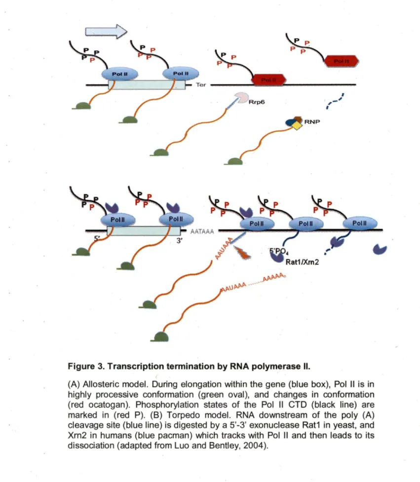
with processing (Carroll et al., 2004; Steinmetz et al., 2001 ). The maturation of non-coding RNA is then completed by the 3' end trimming of exonucleases and the assembly of the snoRNPs complex. Recently, it was proposed that the recruitment of the machinery required for ~· end formation to the transcription termination site is dictated by the phosphorylation state of the Pol Il C-terminal demain (CTD) depends on the transcript size (Figure 3) (Gudipati et al., 2008; Vasiljeva et al., 2008). ln particular this idea states that in the early state of transcription,Ser5 residues of the CTD become hypermethylated and thus recruit the Nrd1 termination complex, which has a preference for the Ser5 phosphorylated CTDs (Gudipati et al., 2008). This binding triggers the formation of the 3' end of short RNA transcripts like snRNA, snoRNA or cryptic non-polyadenylated transcripts destined for degradation (Gudipati et al., 2008; Vasiljeva et al., 2008). As the transcript length increases, the phosphorylation of Pol 11 Ser5 decreases and the phosphorylation of Ser2 increases favoring the binding of the polyadenylation dependent processing machinery leading to the formation of mRNA 3' ends (Gudipati et al., 2008; Morlando et al., 2004; Vasiljeva et al., 2008). lt should be noted, however, that transcript size is in sufficient to indicate the nature of the 3' end. lt is likely that the combination of the sequence near the transcription termination site and the length of the transcript determines the final outcome. Once the 3' end is formed, transcription is terminated and Pol li
falls off its DNA template.
"anti-terminator" or 'allosteric" model proposes that transcription through the termination signal changes the properties of the elongating Pol 11 complex
(Figure 3) (Epshtein et al., 2007), perhaps by dissociation of positive elongation factors or recruitment of termination factors (Erie, 2002). ln this model termination apparently occurs without cleavage as in the case of snoRNA or snRNA termination (Osheim et al., 1999; Steinmetz et al., 2001; Tran et al., 2001; Egloff et al., 2008; Richard and Manley, 2009). ln the seco~d so called "torpedo" model cleavage of the nascent RNA transcript at the poly (A) site transmits a signal to Pol Il, leading to the destabilization of the elongation complex (Tollervey, 2004). This torpedo model is conserved between human and yeast (Luo and Bentley, 2004; West et al., 2004). Recently, it was shown that the 5' to 3' exoribonuclease Rat1 p in yeast and its homologue in human Xrn2 induce termination by degrading the 3' end RNA fragment generated by the poly (A) processing machinery (West et al., 2004). lt is believed the degradation of RNA near the RNAPll transcription complex destabilizes the complex and induces its dissociation from the DNA template. lt remains unclear however, how these long non-polyadenylated transcripts like the spliceosomal component U2snRNA (Abou Elela and Ares, 1998) or the telomerase RNA (TLC1) (Chapon et al., 1997) are terminated. These RNAs are too long for NRD1 dependent transcription termination and do not require polyadenylation for 3' end formation.
--->
RNP
u~ ···~
Figure 3. Transcription termination by RNA polymerase Il.
'
1 , - - '
'
(A) Allosteric model. During elongation within the gene (blue box), Pol Il is in highly processive conformation (green aval), and changes in conformation (red ocatogan). Phosphorylation states of the Pol Il CTD (black line) are marked in (red P). (8) Torpedo model. RNA downstream of the poly (A) cleavage site (blue line) is digested by a 5'-3' exonuclease Rat1 in yeast, and Xrn2 in humans (blue pacman) which tracks with Pol Il and then leads to its dissociation (adapted from Luo and Bentley, 2004).
4. RNA turnover and degradation
RNA turnover and degradation are obligatory processes of every mRNA in cell. RNA turnover defines the natural cycle of RNA degradation in which RNA is degraded after a certain time following its transcription (Lombardo et al., 1992; Zhai et al., 2008). The time it takes any specific RNA to naturally degrade determines its relative stability, influences the number of times it is translated and the amount of proteins it can produce. Therefore, the half-life of each RNA must be programmed as a factor of its function. For example, house-keeping genes are generally transcribed into mRNAs with long half-lives like glycolytic enzyme GAPDH mRNA with half-life >24h (Lekas et al., 2000). On the other hand, proteins that are required only at particular times during the cell cycle, or during differentiation or growth have short hait-lives such as c-myc mRNA (Loflin et al., 1999). The stability of the different mRNAs are generally determined by the length and structure of the 5' and 3' UTR as well as the length of the poly(A) tail (Bloch, 1999; Wang et al., 2005). The overall principals of mRNA degradation are conserved in bath yeast and mammals. These features normally influence the degradation of mRNA in the cytoplasm, which is tightly linked to translation and often initiated by the removal of the 3' poly(A) tail followed by the removal of the 5' cap and exoribonucleolytic degradation (Santiago et al., 1987; Stripecke et al., 1994; Wang et al., 2005; Zhai et al., 2008). However, certain RNAs contain special destabilization sequence elements that signal the recruitment of trans-acting factors like ribonculeases (Tourriere et al., 2002). Destabilization elements
like the AU rich (ARE) decay signais (Maitra et al., 2008) or stem loop structures like the iron response elements (IRE;:S) are usually found in the untranslated regions of mRNAs ( Rothenberger et al., 1990; Constable et al., 1992; Thomson et al., 1999; Cairo et al., 2002;). The presence of these signais induces endoribonucleolytic cleavage and rapid RNA decay. Changing the activity of these elements has strong impact on cell metabolism and may lead to' disease development (Palmer et al., 2008). RNA degradation can also be used as a quality contrai or surveillance mechanism to identify mRNAs in the cytoplasm lacking translation-termination codons (non-stop decay) (Vasudevan ~t al., 2002) or containing premature termination codons (nonsense decay) (Neu-Yilik and Kulozik, 2008) or that undergo translation stalling (no-go decay) (Passos et al., 2009).
RNA degradation can also occur in the nucleus (Kuai et al., 2005). ln yeast, nuclear degradation routinely eliminates excised pre-rRNA spacer fragments, introns, and short cryptic RNAs (Allmang et al., 1999; van Hoof et al., 2000; Mitchell et al., 2003; Peng et al., 2003; Gonzales et al., 2005). ln fact, given the large number of pre-rRNA, snRNA and snoRNA that is processed in the nucleolus or the nucleoplasm, one could imagine that the majority of RNA processing activities occur in the nucleus and not in the cytoplasm. ln addition to this routine disposai of unused RNA spacers, nuclear degradation may also serve as an early quality contrai surveillance mechanism to eliminate transcriptional, processing and assembly errors (Skruzny et al., 2009). For example, premature or aberrant transcriptional termination may alter mRNA 3'
end formation and thus lead to the recruitment of the 3' - 5' nuclear exosome complex that degrades the unwanted product (Arigo et al., 2006). Nuclear surveillance is particularly important to eliminate the RNA components of unassembled or incorrectly assembled RNP complexes. Assembly of defective RNA would lead to generation of faulty ribosomes or spliceosomes that may compromise the survival of the entire cell. lndeed, any mutation that prevents protein binding to rRNA, snRNA or snoRNA often leads to rapid RNA degradation by the nuclear exosome complex (Lee et al., 1995; Lee and Nazar, 1997; Good et al., 1997; Hilleren et al., 2001; Morlando et al., 2004; Passos et al., 2009; Skruzny et al., 2009) ln all cases, nuclear and cytoplasmic RNA decay and turnover are entirely dependent on the accuracy and efficiency of ribonucleases that quickly react to changes in inter- and intra-cellular conditions.
5. Ribonucleases
Ribonucleases (RNases) are enzymes that specifically cleave RNA phosphodiester bonds (Nicholson, 1999). Ribonucleases are divided into two classes, exoribonucleases and endoribonucleases (Nicholson, 1999). Most RNases are protein enzymes and will be discussed in details below. However, RNA based ribozyme activity has also been identified such as the tRNA processing RNase P and the mitochondrial RNA processing enzyme (MRP), which is required for pre-rRNA processing (Lindahl and Zengel, 1995; Reddy
and Shimba, 1995; Reilly and Schmitt, 1995; Tollervey, 1995). Ali known, exoribonucleases are protein enzymes that degrade RNA by using free 3' or 5'ends as entry sites, whereas endorinucle.ases recognize internai RNA sequence or structure (Deutscher, 1993; Virtanen and Astrom, 1997; Deutscher and Li, 2001; Brouwer et al., 2001; Andrade et al., 2009). These enzymes are found in all cells and each possesses special activity and specificity that suits particular cellular functions. The expression, specificity and recruitment of these ribonucleases plays an important raie in shaping the RNA degradation and turnover program of all cells.
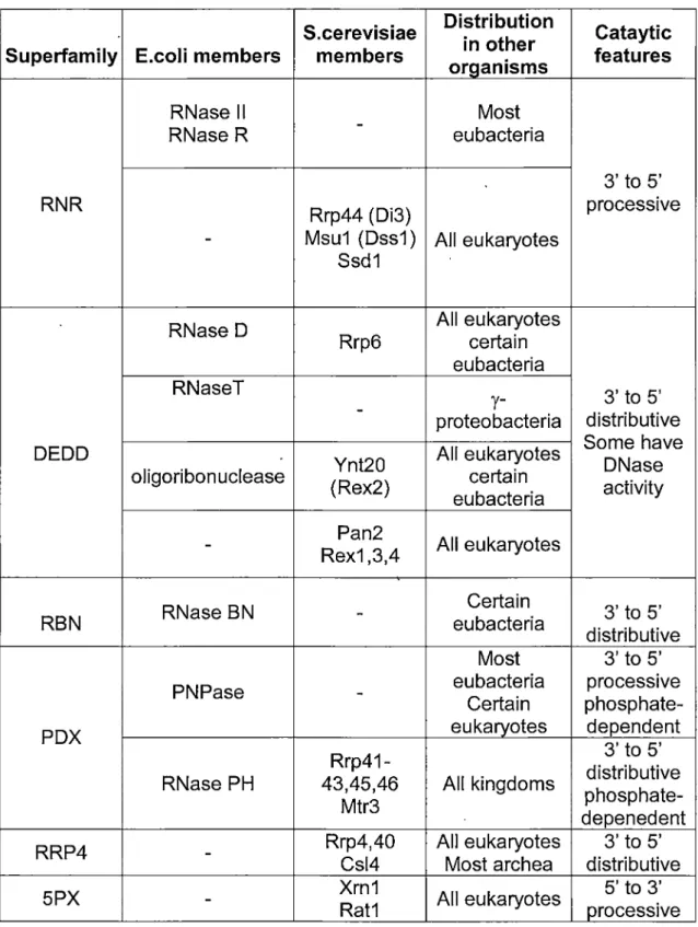
5.1 Exoribonucleases
Exoribonucleases are key components of the RNA-surveillance machinery and can be divided into 6 families based on their protein fold and activities (See table 1 ). These families (RNR, DEDD, RBN, PDX and RRP4) possess 3' - 5' exoribonuclease activity and one (5PX) includes RNases with 5' - 3' exoribonucleolytic activities (Zuo and Deutscher, 2001 ). ln yeast, 3' - 5' exoribonculeases in general function as integrated complexes called exosomes that can be found in the nucleus and / or the cytoplasm (Decker, 1998; Raijmakers et al., 2004; Lykke-Andersen et al., 2009). Nuclear and cytoplasmic exosomes share ten common components from 4 different families (RNR, DEDD, PDX and RRP4) (Schneider et al., 2009). However, the RNase Ski? are found exclusively in the cytoplasmic complex (Araki et al.,
2001) and the RNase Rrp6 and the putative nucleic-acid-binding protein Rrp47 is found only in the nuclear complex (Mitchell et al., 2003). Rrp6 is a key nuclear 3' - 5' nuclear exoriqonuclease (Burkard and Butler, 2000). lt directly contributes ta the hydrolytic activity of the nuclear exosome and confers distributive exonucleolytic activity on unstructured and poly (A)-extended RNA (Graham et al., 2009). The nuclear exosome functions in the 3' processing of the precursors of stable RNAs, including rRNA and many snoRNAs (Kent et al., 2009; Kufel et al., 2000; Torchet et al., 2002). The nuclear exosome is also responsible for the surveillance and degradation of aberrant nuclear precursors of many types of RNA including mRNAs, pre-tRNAs and pre-rRNAs.
The 5PX family currently contains two 5' - 3' exoribonucleases one is nuclear (Rat1 p) (Li et al., 2006), and the other is cytoplasmic (Xrn1 p). Xrn1 p is the main cytoplasmic mechanism for uncapped RNA and also plays a major raie in controlling the steady state level of mRNA in yeast (Brown et al., 2000; Long and McNally, 2003). lndeed, Xrn1 p acts at the final step of mRNA degradation, following the normal deadenylation and decapping of mRNA. On the other hand, deletion of the nuclear exoribonuclease Rat1 p impairs transcription termination and leads ta the accumulation of many processing by-products (Henry et al., 1994). Bath Rat1p and Xrn1p share similar substrate.
S.cerevisiae Distribution in other Superfamily E.coli members members organisms
RNase Il - Most
RNase R eubacteria
RNR Rrp44 (Di3)
- Msu1 (Dss1) Ali eukaryotes Ssd1
RNase D Rrp6 Ali eukaryotes certain eubacteria RNaseT
- proteobacteria
y-DEDD Ynt20 Ali eukaryotes
oligoribonuclease (Rex2) certain eubacteria Pan2 Ali eukaryotes
-
Rex1 ,3,4RNase BN Certain
RBN - eubacteria
Most PNPase - eubacteria Certain
PDX eukaryotes
Rrp41-RNase PH 43,45,46 Ali kingdoms Mtr3
RRP4 - Rrp4,40 Csl4 Ali eukaryotes Most archea
5PX
-
Xrn1 Rat1 Ali eukaryotesTable 1. Summary of exoribonuclease superfamilies. (Zuo and Deutscher, 2001)
Cataytic features 3' to 5' processive 3' to 5' distributive Sorne have DNase activity 3' to 5' distributive 3' to 5' processive phosphate-dependent 3' to 5' distributive phosphate-depenedent 3' to 5' distributive 5' to 3' processive
specificity and RNA that escapes Rat1 p degradation in the nucleus is seamlessly degraded by Xrn1 p in the cytoplasm (Poole and Stevens, 1995). ln general, it seems that nuclear and cytoplasmic degradation share overlapping substrate specificity.
5.2
EndoribonucleasesUntil recently, endoribonucleases were thought to play a minor raie in RNA degradation. The fact that endoribonucleases recognize specific internai sequences or structural motifs make them more specialized than exoribonucleases that can recognize any RNA with free termini (MacBeth and Patterson, 1998; Kennell, 2002; Saida and Odaert, 2007). Therefore, it was thought that endoribonucleases affect a small group of transcripts with limited impact on overall gene expression. However, it is becoming increasingly clear that despite the relatively high specificity of the ribonucleases, they can still maintain broad substrate specificity. lndeed, RNA mediated endoribonucleolytic cleavage of RNA that is often called RNA interfrerence (RNAi) is rapidly becoming the largest and most studied mechanism of RNA degradation (Yamaguchi and lnouye, 2009). ln yeast, there are 9 well characterized protein endoribonucleases with a broad range of activities including the debranching of excised intron lariats (e. g. Dbr1 p) (Khalid et al., 2005), RNA decay activities (e.g. Dis3p) (Schaeffer et al., 2009), 3' endo"ribonucleolytic, ribonuclease activities (e. g. Ngl2p) (Faber et al., 2002),
and RNase Ill cleavage activities (Rnt1 p) (Lamontagne et al., 2001 ). ln higher . eukaryotes, many of these enzymes are conserved; e.g RNase H or Dis3p,
while other have evolved new functions such as yeast RNase Ill (Rnt1 p) (Lamontagne et al., 2001; Carmel! and Hannon, 2004; Ji, 2008; Lebreton et al.-, 2008; Schultz and Champoux, 2008). ln higher eukaryotes orthologues of RNase Ill (Dicer and Drosha) induce and regulate the mechanism· of RNA interference (RNAi) that seems to govern most conditional mRNA degradation in mammalian cells (Carmel! and Hannon, 2004). This mechanism of RNA degradation is found in most eukaryotes with the notable exception of budding yeast. RNAi achieves RNA degradation through the formation of RNA duplex in trans. This process can be induced by the cleavage of long duplex RNA or through the processing of short structured pre-micro RNAs (miRNA) (Tijsterman and Plasterk, 2004; Filipowicz, 2005; Hutvagner, 2005; Kim et al., 2006a). ln all cases, the final outcome is the formation of short RNA duplex of 21-22 base pairs that is simultaneously integrated into the RNA-induced silencing complex (RISC) that dissociates the RNA duplex and pairs one of the two RNA strands with its complementary target (Ji, 2008; Kim et al., 2006a). Perfect pairing with the target induces enodribonucleolytic cleavage of the target RNA by a 5' end-dependent endoribonuclease called argonaute (Brodersen and Voinnet, 2009). lmperfect pairing of the RISC associated RNA strand with the 3' end of the target RNA does not solicit cleavage but instead inhibits mRNA translation (Pillai et al., 2005; Pillai et al., 2007). ln this
way, the cell can conditionally target endoribonucleolytic cleavage of any RNA using the same set of endoribonucleases.
6. RNase Ill family
Members of the RNase Ill family are found in all species examined with the exception of archaebacteria, where the functions of RNase Ill are carried-out by the bulge-helix-bulge nuclease (BHB) (Lamontagne et al., 2001 ). Ali proteins classified into this family show homology with the structural elements of the founding member, Escherichia coli RNase Ill (Nicholson, 1996, 1999). These structural elements include a nuclease domain (NUGD) that exhibits a conserved signature motif, and a dsRNA binding domain (dsRBD) that contains a motif specific to the dsRNA binding protein (dsRBP) family. The RNase Ill family can be divided into 4 classes, based on additional protein features and organizations (Figure 4) (Lamontagne et al., 2001 ). Glass 1 includes bacterial enzymes that possess a single N-terminal NUGD and a G-terminal dsRBD (Gan et al., 2006). Glass Il enzymes are identified by the presence of a highly variable N-terminal extension and include fungal RNase Ill (Lamontagne and Abou Elela, 2001 ). Glass Ill enzymes contain two NUGDs and indude plant and vertebrate enzymes (Lee et al., 2003b). Glass IV includes the RNAi enzyme Dicer, which possesses a N-terminal helicase domain (Lee et al., 2004). The sequence homology between orthologues varies between 84% to 20%, depending on the evolutionary distance. Most
RNase llls display low sequence specificity in vitro and usually cleave any duplex RNA with low sequence complexity (Lamontagne and Abou Elela, 2004). ln contrast, RNase llls are highly specific and mostly target short RNA hairpins in vivo. This surprisingly high in vivo specificity prevents complementation, even between closely related species.
6.1 Protein structure of budding yeast RNase Ill
Budding Yeast Rnt1 p is a 471 aa protein (54.5 kDa) that exhibits the features of class Il RNase llls. The dsRBD motif is located at the C-terminus (positions 372-440) and has about 25% identity with other RNase llls. A unique 32 aa extensi.on at the C-terminus is required for nucleolar localization (Lamontagne et al., 2000). The Rnt1 p 162 aa NUCD contains the RNase Ill signature sequence implicated in catalysis. ln addition, Rnt1 p possesses a 199 aa terminal domain (term), that is unique to eukaryotic RNase llls. This N-terminal extension has no apparent functional motifs and it is not highly conserved, even among members of the Saccharomyces species. Deletion of the N-term renders Rnt1 p sait sensitive and reduces cleavage efficiency both
in vivo and in vitro..: Rnt1 p functions as a homodimer formed by interactions between the dsRBD and the N-term (Lamontagne and Abou E;lela 2001 ).
C l ass 1
(RNase 1 1)Class
( Rn~1 p)Class Il
(Drosha)C l ass IV
(Dilcer) F PAZ NUC 2o m a œn
Figure 4: Schematic representation of the RNase Ill family.
UC · 1
dsRBD
Green boxes represent the dsRNA binding domain (dsRBD), yellow boxes represent the amino acid residues that extend beyond the dsRBD (CTE}, red boxes represent the nuclease domain (NUCD). Blue boxes represent the N-terminal domain.
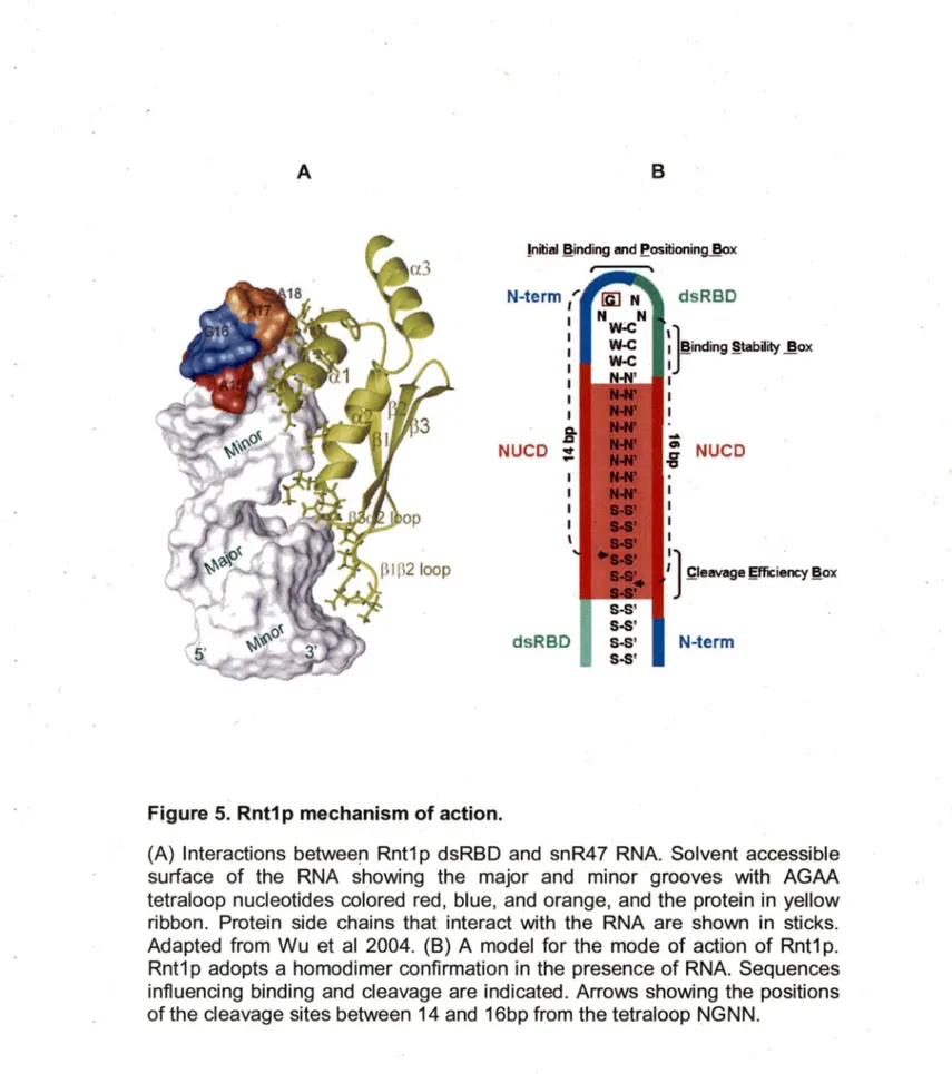
Based on the crystal structure of two bacterial RNase llls, specifically those of Aquifex aeolicus (Gan et al., 2006), the Rnt1 p homodimer is predicted to form an antiparallel dimer (Figure 58). If Rnt1 p folds like bacterial RNase Ill, the two NUCDs of the homodimer would form a valley supported by a ball-and-socket junction involving highly conserved amino acids. Secondary structure prediction suggests that Rnt1 p maintains the conserved
ocpppoc
structure of dsRBD and of several of the seven helices in the bacterial NUCD. Recently, crystal and solution structures of Rnt1 p dsRBD confirmed the presence of the classicalocpppoc
structure, and revealed an additional helix near the C-terminus(oc
3) unique to Rnt1 p (Figure 5 A) (Leulliot et al., 2004; Wu et al., 2004). The solution structure of the dsRBD RNA complex indicates that the additional helix is not located near the RNA, but that it could influence the binding of RNA tooc
1.Studies of E.coli RNase Ill suggest that the substrate selection is influenced by antideterminant nucleotides (Rudinger et al., 1996; Zhang and Nicholson, 1997). This means that the absence of a nucleotide or structure, and n_ot its presence, triggers RNA binding and cleavage. As more RNaselll are tested, it is becoming increasingly clear that eukaryotic RNase llls possess a different mechanism of substrate selectivity. Unlike other RNase Ill, Rnt1 p recognizes substrates with conserved stem-loop structures. Most Rnt1 p substrates exhibit conserved AGNN tetraloop structure (Lamontagne et al. 2001; Lebars et al., 2001; Lamontagne et al., 2003; Lamontagne et al., 2004).
A
B!nitial ~inding and f!ositioning~x
N-term (
NUCD
dsRBD
Figure 5. Rnt1 p mechanism of action.
S-S' S-S' S-S' S-S'
dsRBD
: f
inding §tability J!ox1
-i
NUCD1
1
')
_çleavage gfficiency ~ox .N-term
(A) Interactions betweeri Rnt1 p dsRBD and snR47 RNA. Solvent accessible surface of the RNA showing the major and minor grooves with AGAA tetraloop nucleotides colored red, blue, and orange, and the protein in yellow ribbon. Protein side chains that interact with the RNA are shown in sticks. Adapted from Wu et al 2004. (8) A model for the mode of action of Rnt1 p. Rnt1p adopts a homodimer confirmation in the presence of RNA. Sequences influencing binding and cleavage are indicated. Arrows showing the positions of the cleavage sites between 14 and 16bp from the tetraloop NGNN.
Rnt1 p cleaves at a fixed distance from the conserved loop, generating a product with staggered ends (Figure 58). The solution structures of two different substrates of Rnt1 p reveal a common fold for the terminal loop with
.
the universal G in syn conformation and extensive base stacking (Lebars et al., 2001 ). The structure suggests that Rnt1 p recognizes the shape of the tetraloop for the initial interaction with its substrate. This is in marked contrast with the recognition mode of most dsRNA binding proteins including RNase Ill, which interact primarily with the minor groove of the double helix and recognize the shape of the A-form dsRNA (Wu et al., 2004).
6.2 Cellular functions of yeast RNase Ill
RNT1 is not an essential gene but its deletion causes severe growth defects, temperature sensitivity, hypersensitivity to hèavy metals and sporulation defects (Abou Elela et al., 1996; Abou Elela and Ares, 1998). Normal wild type cell doubling is around 2-3 hours at 30°C, however cells lacking Rnt1 p will divide in around 7 hours at 26°C (Abou Elela and Ares, 1998). Deletion of Rnt1 p also affects cell cycle progression and nuclear division; yet this effect has been shown to be independent of Rnt1 p cleavage activity (Catala et al., 2004 ). Rnt1 p localizes to various places depending on the phases of the cell cycle; in G1 till the end of S phase, Rnt1 p is localized to the nucleolus, in G2 phase to the end of mitosis, Rnt1 p is present at the nucleoplasm. However, Rnt1 pis not detected in the cytoplasm, even when it is overexpressed. Rnt1 p
is also important for the co-localisation of nuclear proteins implicated in the maturation of rRNA. Rnt1 p is shown to bind to Gar1 p and this interaction is required for the nuclear localization of Gar1 p, Nhp2 and Cbp5p that are involved in the processing of H/ACA snoRNAs (Tremblay et al., 2002b).
The primary function of Rnt1 p is the processing of pre-rRNA. This processing is achieved by the cleavage of a stem-loop structure at the 3' external transcript spacer (3'ETS) of the pre-rRNA (Abou Elela et al., 1996). Recenlty, it has been shown that Rnt1 p is not only implicated in the maturation of rRNA but also its transcription. Rnt1 p interacts with two subunits of the polymerase 1
(Pol 1-) (Catala et al 2008). Moreover deletion of Rnt1 p inhibits the synthesis of rRNA and alters the conformation of the chromatin at the ribosomal DNA locus (Catala et al., 2008). Finally, Rnt1 p is also required for the termination of rDNA transcription by Pol I (Prescott et al., 2004; Kawauchi et al., 2008).
Other functions of Rnt1 p include the processing of all Pol 11 transcribed snRNAs ( Chanfreau et al., 1997; Abou Elela and Ares, 1998; Seipelt et al., 1999) and a number of snoRNAs (Chanfreau et al., 1998a). lnitially, Rnt1 p cleaves a stem-loop structure capped with the canonical AGNN tetraloop at the 3'end of the snoRNAs. This cleavage allows the trimming by the exosome and further assembly of the Ribonucleoproteins (RNP) to complete the maturation. ln addition to the role of Rnt1 p in the maturation of non-coding RNAs, Rnt1 p can also trigger mRNA degradation. ln this case, Rnt1 p cleavage occurs in the coding sequence of the mRNA to contrai its
Larose et al., 2007). The presence of a Rnt1 p cleavage signal in an mRNA coding sequence triggers the degradation of the nuclear fraction of the RNA presumably by the nuclear exosome. lt is not clear, however, how Rnt1 p differentiates between mRNA scheduled for degradation from mRNA exported ta the cytoplasm for translation. lt is also unclear how Rnt1 p cleavage is activated by environmental and cellular conditions.
7. Aim of the project
ln the beginning of this study only a handful of Rnt1 p cleavage signais in non-coding RNA were known and the mechanism by which the enzyme selects its substrate was restricted an apparent preference for AGNN hairpins. Therefore, we aimed at understanding the mechanism by which Rnt1 p identifies its substrate and developed tools ta find and characterize new Rnt1 p substrates. This essential biochemical approach led us ta discover new forms of Rnt1 p substrates that do not require an AGNN tetraloop for cleavage and identified new and unexpected functions of Rnt1 p in transcription termination. The initial work succeeded in identifying Rnt1 p cleavage sites in ail known non-coding RNAs and revealed a new mechanism by which long-range interaction induces excision of intron embedded snoRNA. Examination of the newly discovered Rnt1 p cleavage sites suggested that the enzyme uses a flexible substrate recognition mechanism that tolerates broad variation in primary and tertiary structures. Based on this finding, we modified our
model for substrate recognition and scanned the yeast genome for new forms of cleavage targets that are not associated with snoRNAs. Surprisingly, we uncovered a large number of Rnt1 p cleavage sites in intergenic sequences. Biochemical and genetic studies of the intergenic cleavage signais suggesteçJ a new raie of Rnt1 p in inducing transcription termination of long non-polyadenylated RNAs. ln addition, this study directly linked dsRNA specific endoribonucleolytic activity to the transcription complex. The central findings of this thesis challenge the common views of distinct RNA transcription, processing and decay and provide a model in which gene expression is not simply defined by the decision to transcribe a gene but rather by the stability of the nascent transcripts.
ARTICLE 1
Ghazal, G., Ge, D. Gervais-Bird, J., Gagnon, J., and Abou Elela, S. (2005) Genome-wide prediction and analysis of yeast RNase 111-dependent snoRNA processing signais. Mol Cell Biol. 2005 April; 25(8): 2981-2994.
Préambule
Dans cette étude, nous avons cherché de nouveaux substrats de Rnt1 p en examinant le profil d'expression de tous les snoRNAs en présence et en absence de Rnt1 p. En parallèle, nous avons développé un programme qui identifie les signaux de clivage de Rnt1 p connus
à
proximité des séquences des snoRNAs. Une combinaison d'approchesin si/ico et in vitro a identifié tous les substrats connus de Rnt1 p et révélé 7 nouveaux substrats snoRNAs. Une enquête minutieuse du rôle de Rnt1 p dans la maturation de ces snoRNAs a montré que les signaux de clivage de Rnt1 p sont plus grands que ce que l'on croyait auparavant. Ce travail montre l'implication de Rnt1 p dans la maturation des différentes organisations des snoRNAs. Fait intéressant, nous avons montré que la maturation des snoRNAs encodés dans les introns des gènes ribosomales représente un nouveau mécanisme qui coordonne la production des isoformes des protéines ribosomales et de leurs snoRNAs associés. J'ai effectué toutes les expériencesà
l'exception de l'analyse bioinformatique et un des Northern blots montré dans la Figure 3.ARTICLE 1
Ghazal, G., Ge, D. Gervais-Bird, J., Gagnon, J., and Abou Elela, S. (2005) Genome-wide prediction and analysis of yeast RNase 111-dependent snoRNA processing signais. Mol Cell Biol. 2005 April; 25(8): 2981-2994.
Summary
ln this study we have searched for new Rnt1 p substrates by examining the expression ·profile of all known snoRNAs before and after the deletion of Rnt1 p. ln parallel, we have developed a program that identifies Rnt1 p cleavage signais near known snoRNA sequences. A combined in silico and in vitro approach identified all known substrates of Rnt1 p and revealed 7 new snoRNA associated substrates. Careful investigation of the raie of Rnt1 p in the maturation of these snoRNAs showed that Rnt1 p cleavage signais are larger than what was previously believed. This work shows the implication of Rnt1 p in the processing of different snoRNAs organization. lnterestingly, we have shown that processing of snoRNAs imbedded in the introns of ribosomal genes represents a new mechanism that coordinates the production of ribosomal protein isoforms and their associated snoRNAs. 1 conducted all experimental data with the exception of the bioinformatic analysis and one of the Nothern blots shown in Figure 3.
Genome-wide prediction and analysis of yeast RNase 111-dependent snoRNA processing signais
Ghada Ghazal, Dongling Ge, Julien Gervais-Bird, Jules Gagnon, and Sherif Abou Elela*
RNA Group /Groupe ARN
Département de Microbiologie et d'lnfectiologie,
Faculté de Médecine, Université de Sherbrooke,
Sherbrooke, Québec, Canada J1 H 5N4
Running Title: Genomic Approach for the Identification of Rnt1 p Substrates
Keywords: RNase Ill, Rnt1 p, snoRNA, dsRNA, Processing.
Ward Count: 54,942
Phone: (819) 564-5275.
Fax: (819) 564-5392.
E-mail: [email protected].
Abstract
ln Saccharomyces cerevisiae, the maturation of bath pre-ribosomal RNA (pre-rRNA) and pre-small nucleolar RNAs (pre-snoRNAs) involves common factors, thereby providing a potential mechanism for the co-regulation of snoRNA and rRNA synthesis. ln this study we examined the global impact of the dsRNA specific ribonuclease Rnt1 p, which is required for pre-rRNA processing, on the maturation of all known snoRNAs. ln silico searches for Rnt1 p cleavage signais, and genome-wide analysis of the Rnt1 p-dependent expression profile, identified 7 new Rnt1 p substrates. lnterestingly, two of the newly identified Rnt1 p-dependent snoRNAs snR39 and snR59 are located in the introns of the ribosomal proteins (r-protein) genes RPL?A and RPL?B. ln vitro and in vivo experiments indicated that snR39 is normally processed from the lariat of RPL 7 A, suggesting that the expressions of RPL 7 A and snR39 are linked. ln contrast, snR59 is produced by a direct cleavage of the RPL?B pre-mRNA indicating that a single pre-pre-mRNA transcript cannot be spliced to produce a mature RPL?B mRNA and processed by Rnt1 p to produce a mature snR59 simultaneously. The results presented here reveal a new raie of yeast RNase Ill in the processing of intron-encoded snoRNAs that permits independent regulation of the hast mRNA and its associated snoRNA.
Introduction
Bacterial pre-rRNA processing is carried out by a defined set of nucleases (Apirion, 1983; Apirion and Gegenheimer, 1981; Apirion and Miczak, 1993; Perry, 1976; Srivastava and Schlessinger, 1990). Key among this set is RNase Ill, initially isolated by its ability to bind and cleave duplex RNA (Robertson, 1967; Robertson et al., 1968). RNase Ill generates the immediate precursors to the mature 16S and 23S rRNA from the primary transcripts by cleaving within two extended RNA duplexes formed by long range interactions that pair the
.
termini of each rRNA (Bram et al., 1980; Young and Steitz, 1978). These long-range interactions provide a simple method of coordinating the processing events at both ends of the transcript. ln eukaryotes, pre-rRNA processing is more complex and requires many more snoRNAs and protein components with overlapping functions (Eichler and Craig, 1994; Fromont-Racine et al., 2003; Granneman and Baserga, 2004; Pederson, 1998; Reeder, 1990). For example, the removal of the 5' external transcribed spacer (ETS) requires 4 snoRNAs (U3, snR30, U14, and snR10), and about 64 snoRNAs are required for rRNA modifications (Lafontaine and Tollervey, 1995; Venema and Tollervey, 1995). snoRNAs are divided in two major subclasses: the first includes box CID snoRNAs that mostly function as a guide for the methylation of rRNA (Bachellerie and Cavaille, 1997; Kiss-Laszlo et al., 1996; Kiss-Laszlo et al., 1998; Tycowski et al., 1996); while, the second includes H/ACA snoRNAs that guide RNA pseudouridine formation (Lafontaine et al., 1998; Ni et al., 1997; Watkins et al., 1998). Most mammalian snoRNAs are encoded within intron
sequences and are processed from either unspliced precursors or lariat species (Hirose et al., 2003; Hirose and Steitz, 2001; Zhou et al., 2004). ln yeast, most snoRNAs are transcribed either as independent units, or as a part of polycistronic transcript, while only 7 of the 66 known snoRNAs are located in the introns of mRNAs (Filipowicz et al., 1999; Petfalski et al., 1998; Tollervey and Kiss, 1997). Several polycistronic snoRNAs, and few monocistronic ones are processed by Rnt1 p, the orthologue of the bacterial RNase Ill (Lamontagne et al., 2001 ), which is also required for the processing of the pre-rRNA's 3' end (Abou Elela et al., 1996; Chanfreau et al., 1998a; Chanfreau et al., 1998b; Kufel et al., 1999; Lee et al., 2003a). Following processing by Rnt1 p, the RNAs are trimmed by exonucleases producing the mature ends (Kufel et al., 2000; van Hoof et al., 2000).
Unlike other RNase llls, Rnt1 p recognizes substrates with conserved stem-loop structures and has a low affinity for generic RNA duplexes (Lamontagne and Abou Elela, 2004). Most Rnt1 p substrates exhibit a conserved AGNN tetraloop structure (Chanfreau et al., 2000; Lamontagne et al., 2003; Lebars et al., 2001; Wu et al., 2001 ). Rnt1 p, cleaves at a fixed distance from the conserved loop, generating a product with staggered ends (Lamontagne et al., 2003). Mutations (Lamontagne et al., 2003), chemical protection assay (Lamontagne and Abou Elela, 2004), chemical interference (Chanfreau et al., 2000) and NMR analysis (Lamontagne et al., 2003) indicate that Rnt1 p binding and cleavage are regulated by reactivity epitopes grouped into three