For Peer Review. Do not distribute. Destroy after
use.
1
Dual-specificity phosphatase 3 deletion protects female, but not
1
male mice, from endotoxemia- and polymicrobial-induced septic
2shock
34
Running title: Sex-dependent DUSP3’s role in sepsis 5
6 7 8
Maud Vandereyken*§, Prathiba Singh*§, Caroline Wathieu*, Sophie Jacques*, Tinatin
9
Zurashvilli*, Lien Dejager†‡, Mathieu Amand*, Lucia Musumeci*, Maneesh Singh*, Michel
10
Moutschen*, Claude Libert†‡ and Souad Rahmouni*¶
11 12
* Immunology and Infectious Disease Unit, GIGA-Research, University of Liège, Belgium.
13
† Inflammation Research Center, VIB, B-9052 Ghent, Belgium;
14
‡ Department of Biomedical Molecular Biology, Ghent University, B-9000 Ghent, Belgium;
15 16 17
§ Contributed equally to this work
18 19 ¶ Corresponding Author: 20 Dr Souad Rahmouni 21 University of Liège 22
Immunology and Infectious Diseases Research Unit
23
GIGA B34, Avenue de l’Hôpital, 1,
For Peer Review. Do not distribute. Destroy after
use.
2 B-4000 Liège - Belgium 25 Tel: +32 4 366 28 30 / Fax: +32 4 366 45 34 26e-mail address: srahmouni@ulg.ac.be
27 28
Authors email address
29
Maud Vandereyken (maud.vandereyken@gmail.com), Pratibha Singh
30
(pratibhacdri@gmail.com), Caroline Wathieu (caroline.wathieu@student.ulg.ac.be), Sophie
31
Jacques (sjacques@student.ulg.ac.be), Tinatin Zurashvilli (tinazurashvili@hotmail.com), Lien
32
Dejager (lien.dejager@gmail.com), Mathieu Amand (Mathieu.Amand@lih.lu), Lucia
33
Musumeci (lmusumeci@ulg.ac.be), Maneesh Singh (maneesh@mit.edu), Michel Moutschen
34
(michel.moutschen@ulg.ac.be), Claude Libert (claude.libert@irc.vib-ugent.be), Souad
35 Rahmouni(srahmouni@ulg.ac.be) 36 37 38 39 40 41 42 43 44 45 46 47 48 49
For Peer Review. Do not distribute. Destroy after
use.
3 Abstract 50 51DUSP3, is a small dual specificity phosphatase of poorly known physiological functions and
52
for which only few substrates are known. Using DUSP3-deficient mice, we recently reported
53
that DUSP3 deficiency confers resistance to endotoxin- and polymicrobial-induced septic
54
shock. We showed that this protection was macrophage-dependent. In this work, we further
55
investigate the role of DUSP3 in sepsis tolerance and show that the resistance is
sex-56
dependent. Using adoptive transfer experiments and ovariectomized (OVX) mice, we
57
highlighted the role of female sex hormones in the phenotype. Indeed, in OVX female and
58
male mice, the dominance of M2-like macrophages observed in DUSP3-/- female mice was
59
reduced suggesting a role of this cell subset in sepsis tolerance. At the molecular level,
60
DUSP3 deletion was associated with oestrogen-dependent decreased phosphorylation of
61
ERK1/2 and Akt in peritoneal macrophages stimulated ex vivo by LPS. Our results
62
demonstrate that oestrogens may modulate M2-like responses during endotoxemia in a
63
DUSP3-dependent manner.
64 65 66
Key words: DUSP3, sepsis, endotoxemia, LPS, female sex hormones, oestrogen,
67 macrophages 68 69 70 71 72 73 74
For Peer Review. Do not distribute. Destroy after
use.
4 Introduction 75 76Sepsis and septic shock are complex clinical syndromes that arise when the local body
77
response to pathogens becomes systemic and injures its own tissues and organs (1). When
78
infection occurs, bacterial components such as LPS, are recognized by the host and
79
inflammation is initiated. TLR4 pathway is activated and triggers the release of cytokines,
80
chemokines and nitric oxide (NO) (2, 3). Systemic release of pro-inflammatory cytokines
81
causes large-scale of cellular and tissue injuries, leading to microvascular disruptions, severe
82
organ dysfunctions and eventually death (4). Sepsis occurrence and outcome depend on
83
pathogen characteristics but also on risk factors such as age or sex (1). Indeed, women are
84
better protected against infection and sepsis compared to men. Women younger than 50 years
85
show a lower incidence of severe sepsis and a better survival compared to age-matched men.
86
This may be explained by the influence of female sex hormones on the immune system
87
responses (5).
88 89
DUSP3, or Vaccinia-H1-related (VHR), is an atypical dual specificity phosphatase of 21kDa.
90
The phosphatase contains one catalytic domain but lacks a binding domain (6). DUSP3
91
broader catalytic site allows the protein to dephosphorylate both Tyr and
phospho-92
Thr residues (7). The MAPK ERK1/2 and JNK were the first reported DUSP3 substrates (8–
93
10). Other substrates such as the EGFR and ErbB2 tyrosine receptors (11) and STAT5
94
transcription factor (12) were also reported. DUSP3 physiological functions started to be
95
elucidated thanks to the knockout mouse we have generated. Studies from our laboratory
96
using DUSP3-/- mice showed that DUSP3 plays an important role in platelets biology, in
97
monocytes and macrophages and in endothelial cells (13–15). In platelets, DUSP3 plays an
98
important role in arterial thrombosis and platelet activation through GPVI and CLEC-2
For Peer Review. Do not distribute. Destroy after
use.
5
signalling pathways (14). DUSP3 plays also an important role in endothelial cells and
100
angiogenesis and seems to act as a pro-angiogenic factor (16). Surprisingly, this function was
101
not correlated with reduced tumour or metastatic growth. Indeed, in an experimental
102
metastasis model using Lewis lung carcinoma cells (LLC), we found that DUSP3 plays rather
103
an anti-tumour role since DUSP3-/- mice were more sensitive to LLC metastatic growth when
104
compared to WT littermates. This enhanced tumour growth in DUSP3-/- mice was associated
105
with higher recruitment of M2-like macrophages (Vandereyken et al, under revision).
106
Previous studies from our laboratory and others showed that DUSP3 was downregulated in
107
some human cancers and upregulated in others (reviewed in (16, 17)). Further studies are
108
required to better understand the role of this phosphatase in cancer biology.
109
DUSP3 plays also an important role in immune cell functions. In T cells, DUSP3 can be
110
activated by ZAP-70 tyrosine kinase after TCR triggering (18). This activation, through
111
tyrosine phosphorylation of DUSP3, allows the targeting of the MAPK ERK1/2 and the
112
activation of its downstream signalling pathway. Moreover, in Jurkat leukemia T cells,
113
DUSP3 targets ERK and JNK, but not p38. Together, these data suggest that DUSP3 controls
114
T cell physiological functions at least partially through the MAPKs ERK and JNK (8). In
115
innate immune cells, we recently showed that DUSP3 is the most highly expressed atypical
116
DUSP in human monocytes. This was also true in mice (15). These findings suggested to us
117
that DUSP3 could play an important role in innate immune responses. Indeed, using DUSP3
-/-118
mice, we found that DUSP3 deletion conferred resistance of female mice to LPS-induced
119
endotoxemia and to polymicrobial infection-induced septic shock. This protection was
120
macrophage dependent since a higher percentage of M2-like macrophage subset was found in
121
DUSP3-/- mice. Moreover, the resistance was also associated with a decreased
122
phosphorylation of the tyrosine kinases ERK1/2 and a subsequent decrease in TNF-α
123
production (15).
For Peer Review. Do not distribute. Destroy after
use.
6
In this study, we report that DUSP3 deletion does not protect male mice from LPS-induced
125
endotoxemia and CLP-induced septic shock and that this protection was female sex hormones
126
dependent. Furthermore, we report that sepsis resistance was associated with a higher
127
percentage of M2-like macrophages in peritoneal cavity of DUSP3-/- female mice but not with
128
decreased pro-inflammatory cytokines production. We also showed that sepsis resistance in
129
females, but not in males or in OVX females, was associated with decreased ERK1/2, PI3K
130
and Akt activation.
131 132 133 134 135 136 137 138 139 140 141 142 143 144 145 146 147 148 149
For Peer Review. Do not distribute. Destroy after
use.
7
Material and methods
150 151
Mice and ethic statement
152
C57BL/6 (CD45.2)-DUSP3-/- mice were generated by homologous recombination as
153
previously reported (13). These mice were backcrossed with C57BL/6-CD45.2 mice (Charles
154
River) to generate heterozygotes that were mated to generate DUSP3+/+ and DUSP3
-/-155
littermate colonies used for experimentation. Age matched male and female DUSP3+/+ and
156
DUSP3-/- mice were used in all the experiments. Mice were kept in ventilated cages under
12-157
hours dark/12-hours light cycle in an SPF animal facility and received food and water and
158
libitum. Health status was evaluated every 3 months and mice were always found free of
159
specific pathogens.
160
All mouse experiments and procedures were approved by the animal ethics committees of the
161
Universities of Ghent and Liege and were carried out according to their guidelines.
162 163
Cecal ligation and puncture and in vivo LPS challenge
164
Cecal ligation and puncture (CLP) was performed as previously described (19). For LPS
165
challenge, mice were i.p. injected with 6mg/kg of LPS. Body temperature was monitored
166
using a rectal thermometer at various times after LPS injection and after CLP. Death of mice
167
was recorded and the data were analysed for statistical significance of differences between the
168
experimental groups.
169 170
Mice irradiation and bone marrow transplantation
171
10-12 weeks old C57BL/6 (CD45.2) donor mice were killed by cervical dislocation. Tibiae
172
and femurs were collected and BM cells were flushed with PBS. BM cells (10x106) were
173
immediately i.v. injected to 6-8 weeks old lethally irradiated (866, 3cGy) C57BL/6 (CD45.1)
For Peer Review. Do not distribute. Destroy after
use.
8
recipient mice. 4 weeks later, transplantation efficiency was evaluated on the basis of the ratio
175
of CD45.2 to CD45.1 cells in the blood of transplanted mice.
176 177
Female ovariectomy and in vivo oestrogen complementation
178
4 weeks old females were anesthetized using ketamine/xylazine (150 mg/kg and 20 mg/kg). A
179
vertical incision of 2-3 cm was performed in the middle of the back. 1 cm lateral of the
180
midline, another incision of 2-3 mm was performed in the fascia. Adipose tissue surrounding
181
ovary was pulled out and ovary was removed after clamping. The same operation was realized
182
for contralateral ovary. The incision in fascia was closed with stitches and the skin incision
183
with clips. Shame operated mice were used as a control. All above procedures were applied to
184
these mice except the removal of ovaries. For in vivo oestrogen complementation, 2 weeks
185
after surgery, subcutaneous implants for controlled release of 17β-oestradiol (1.5µg/day)
186
(Belma technologies) were applied to OVX mice and were kept for 3 weeks before sacrifice.
187 188
Antibodies and reagents
189
The following materials were from Cell Signalling Technology Inc: anti-phospho-Akt
190
(Ser473), anti-Akt, anti-phospho-ERK1/2 (Thr202/Tyr204), anti-ERK, anti-phospho-PI3K
191
p85 (Tyr458)/p55 (Tyr199), anti-PI3K p85, anti-phospho-GSK3α/β (Ser21/9). Anti-GSK3α/β
192
was from Santa Cruz. Anti-GAPDH antibody was from Sigma. HRP-conjugated anti-goat
193
antibody was from Dako. HRP-conjugated anti-mouse antibody was from GE healthcare.
194
HRP-conjugated anti-rabbit antibody was from Merck Millipore. APC-anti-CD45.1 (A20) and
195
PerCp-Cy5.5-anti-CD45.2 (104), FITC-anti-CD11b, APC-Cy7-anti-Ly6G, PE-anti-CD3,
196
PerCp-anti-CD8, FITC-anti-CD4, Biotin-anti-B220 and streptavidin-PE-Cy7 were all from
197
BD Biosciences. APC–anti-F4/80, PerCp-Cy5-anti-NK1.1, and PerCP–Cy5.5–anti-CD11b
For Peer Review. Do not distribute. Destroy after
use.
9
were from eBiosciences. PE-Cy-anti-Ly6G antibody was from BioLegend. LPS from
199
Escherichia coli serotype O111:B4 was from Sigma and was diluted in pyrogen-free PBS.
200 201
Animal blood sampling and plasma preparation
202
Peripheral blood was drawn in EDTA-coated tubes (BD Microtainer K2E tubes; BD
203
Biosciences) by puncturing the heart with 26G needle. Centrifugation was performed twice at
204
800g for 15 min at RT. Plasma samples were separated in sterile Eppendorf tubes, aliquoted
205
in small volumes, and stored at -80°C until used.
206 207
Meso Scale Discovery electrochemiluminescence assay
208
MSD assay was performed according to manufacturer’s instructions (Mesoscale Discovery).
209
Briefly, plasma was diluted 15 and 15.000 times for TNF and IL-6 respectively. For IL-10
210
and IFN-γ, samples were diluted twice. Samples were loaded on 96 well plates, incubated 2h
211
at RT and washed. Detection antibodies were added for 2h at RT. Signal detection was
212
measured within 15 minutes after read buffer addition using MSD instrument.
213 214
Isolation and stimulation of thioglycollate elicited peritoneal macrophages
215
Peritoneal washes were performed 4 days after intraperiteonal injection of 1 mL of 4%
216
thioglycollate broth (Sigma). 5 mL of PBS-EDTA 0.6 mM were injected twice in the
217
peritoneal cavity using an 18G needle and then collected. Peritoneal macrophages were
218
selected by adherence to tissue culture plastic dishes in complete RPMI 1640 medium.
219
Peritoneal macrophages were stimulated with LPS 1µg/mL during 15, 30 or 60 minutes or
220
during 8 and 24h hours, depending on the experiment performed
221 222
Phenotyping and flow cytometry.
For Peer Review. Do not distribute. Destroy after
use.
10
Peritoneal washes were centrifuged 10 min at 350g and the pellet was re-suspended in PBS.
224
For surface cell staining, cells were incubated for 15 min with anti-CD16/CD32 (Fcγ III/IIR)
225
before labelling for 30 min with specific antibodies for 30 min at 4°C. Cells were then washed
226
and fixed with 1% paraformaldehyde solution. Cells were next analysed on FACSCanto II
227
(Becton Dickson) using FlowJo (Tree Star).
228 229
Protein extraction and Western blot
230
For Western blot experiments, cells were stimulated for the indicated time points and lysis
231
was performed with RIPA buffer (50 mM Tris-HCl (pH = 8.0), 150 mM NaCl, 1% NP-40,
232
0.5% sodium deoxycholate, 0.1% SDS, 1 mM orthovanadate, complete protease inhibitor
233
cocktail tablets EDTA free and 1 mM phenylmethylsulfonyl fluoride) on ice during 20 min.
234
Lysates were next clarified by centrifugation at 19.000g during 20 min at 4°C. The resulting
235
supernatants were collected and protein concentrations were determined using the
236
colorimetric Bradford reagent (Bio-Rad). Proteins were next denatured at 95°C in Laemmli
237
buffer (40% glycerol; 8% SDS 5%; 20% B-mercaptoethanol; 20% Tris-HCl 0.5 M pH6.8;
238
0.05% bromophenol blue and water) during 5 min.
239
Denatured samples were run on 10% SDS-PAGE gel and transferred onto nitrocellulose
240
membranes. To block the non-specific binding sites, membranes were incubated for one hour
241
at room temperature in Tris-buffered saline-Tween 20 containing 5% of non-fat milk or 3%
242
BSA (bovine serum albumin). Membranes were incubated overnight with primary antibody at
243
4°C. Membranes were next washed thrice in Tris-buffered saline-Tween and incubated with
244
HRP-conjugated secondary antibody during one hour at room temperature. The blots were
245
developed by enhanced chemiluminescence (ECL kit, Amersham) according to the
246
manufacturer’s instructions.
247 248
For Peer Review. Do not distribute. Destroy after
use.
11
RNA purification, reverse transcription, and real-time PCR
249
RNA was extracted from PMs using the miRNeasy Mini Kit (Qiagen) and cDNA was
250
synthesized using Expand reverse transcriptase (Roche) according to the recommendations of
251
the manufacturer. cDNA was amplified using Sybr Green PCR Master Mix (Roche) and 0.3
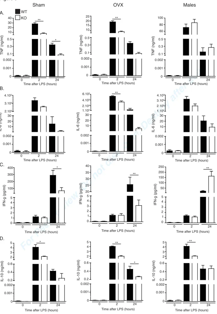
252
mM specific primers for Arginase 1 (Arg1), iNOS, and 2-microglobulin ( 2M). All
253
quantitative PCR were performed on a LightCycler System for RealTime PCR (Roche). The
254
ratio between the expression level of the gene of interest and b2M in the sample was defined
255
as the normalization factor. Relative mRNA quantities for Arg1 and iNOS were determined
256
using the ΔCq method. All primers were from Eurogentec. Sequences were as follow:
257
inducible NO synthase (iNOS): FW, GCTTCTGGTCGATGTCATGAG-39, RV,
59-258
TCCACCAGGAGATGTTGAAC-39; Arg1: FW, 59-CAGAAGAATGGAAGAGTCAG-39,
259 RV, 59-AGATATGCAGGGAGTCACC-39; and b2M: FW, 59-260 CACCCCACTGAGACTGATACA-39, RV, 59-TGATGCTTGATCACATGTCTCG-3. 261 262 Statistical analysis 263
The student t-test was used to assess statistical differences between different groups. Survival
264
differences after LPS challenge and CLP were analysed by Kaplan-Meier analysis with log
265
rank test. Results were considered as significant if p-value < 0.05. Results are presented as
266
mean ± SEM. Prism software (GraphPad) was used to perform statistical analysis.
267
* = p < 0.05, ** = p < 0.01, *** = p < 0.001.
268 269 270
For Peer Review. Do not distribute. Destroy after
use.
12 Results 271 272DUSP3-/- female, but not male, mice are resistant to LPS-induced endotoxemia and to
CLP-273
induced septic shock
274
In a previous study, we showed that DUSP3 deletion protected mice from LPS-induced
275
endotoxemia and polymicrobial infection-induced septic shock (15). Only female were used
276
in the first study. To investigate whether the protection observed is a general feather of
277
DUSP3 deletion or sex dependent, we challenged DUSP3-/- males with a lethal dose of LPS
278
(i.p. injection of 6 mg/kg) and compared their survival to females and to WT control
279
littermates of both sex. Body temperature was also monitored. As expected and previously
280
reported, 90% of DUSP3-/- female mice were resistant to LPS while only 5% of DUSP3+/+
281
female mice survived the challenge (15). Interestingly, DUSP3+/+ and DUSP3-/- male mice
282
were equally sensitive to LPS-induced death (Fig. 1A). Body temperature of all groups of
283
mice, but not DUSP3-/- females, decreased after LPS injection. 24h later, almost all DUSP3
-/-284
females recovered while the other groups remained hypothermic (Fig. 1B). These results were
285
further confirmed in the cecal ligation and puncture (CLP) model performed on DUSP3+/+ and
286
DUSP3-/- males and females. As expected, only 10% of DUSP3+/+ and DUSP3-/- male mice
287
and DUSP3+/+ female mice were still alive by the end of the experiment whereas 70% of
288
DUSP3-/- female mice survived (Fig. 1C.). The body temperature of each group dropped after
289
surgery and only DUSP3-/- female mice recovered (Fig. 1D). These results indicate a sex
290
specific response to septic shock in DUSP3-/- mice.
291 292
Ovariectomized DUSP3-/- mice are sensitive to LPS-induced death
293
Male and female sex hormones receptors have been identified on immune cells suggesting
294
direct effects of androgen and oestrogen on these cells (20). Sexual steroid hormones have
For Peer Review. Do not distribute. Destroy after
use.
13
been recognized to influence numerous immune pathophysiological processes (21). To
296
elucidate the effect of female sex hormones, we ovariectomized (OVX) 4 weeks old
297
DUSP3+/+ and DUSP3-/- mice (OVX mice). As controls, another group of 4 weeks old
298
DUSP3+/+ and DUSP3-/- were sham operated. To assess the ovariectomy’s efficiency, we
299
checked the presence and the size of the uterus. Successful OVX mice were deprived of
300
normal uterus development whereas sham operated mice presented a normally developed
301
uterus (Fig. 1E). 6 weeks after surgery, sham and OVX mice were challenge with 6 mg/kg of
302
LPS and survival and temperature were monitored (Fig. 1F and 1G). Ovariectomy impaired
303
the observed endotoxemia resistance of DUSP3-/- mice, whereas sham operated DUSP3
-/-304
mice were still fully protected from endotoxin-induced death. These data demonstrate that
305
female sex hormones are involved in the observed resistance of DUSP3-/- female mice to
LPS-306
induced lethality.
307 308
DUSP3-/- female bone marrow cells rescue DUSP3+/+ female, but not male mice from
LPS-309
induced lethality
310
We previously showed that adoptive transfer of DUSP3-/- female bone marrow cells or
311
monocytes to DUSP3+/+ female mice was sufficient to transfer resistance to LPS-induced
312
lethality (15). We therefore investigated whether this is also true when recipient mice are
313
males. To generate chimeric mice, 10x106 bone marrow cells (BM) from DUSP3-/-
C57BL/6-314
CD45.2 female mice were intravenously injected into lethally irradiated DUSP3+/+
C57BL/6-315
CD45.1 recipient male and female mice (DUSP3-/- > M-DUSP3+/+ and DUSP3-/- >
F-316
DUSP+/+, respectively). As a control, DUSP3+/+ females BMs were transplanted into lethally
317
irradiated DUSP3+/+ male or female mice (DUSP3+/+ > M-DUSP3+/+ and DUSP3+/+ >
F-318
DUSP3+/+, respectively). Successful hemato-lymphoid reconstitution was verified by flow
319
cytometry 3 to 4 weeks after the transplantation. 95% of peripheral blood cells were CD45.2
For Peer Review. Do not distribute. Destroy after
use.
14
positive (Fig. 1H and 1I). Moreover, in recipient mice, the expression of DUSP3 in peritoneal
321
macrophages was abolished in the recipient mice transplanted with DUSP3-/- BM cell
322
suspension, as showed by DUSP3 immunoblotting (Fig. 1J). 4 weeks after BM
323
transplantation, 6 mg/kg of LPS were i.p. injected into recipient mice and survival was
324
monitored during 8 days (Fig. 1K). Interestingly, more than 70% of the chimeric DUSP3-/- >
325
F-DUSP+/+ mice survived up to the end of the experiment compared to 9% of DUSP3+/+ >
F-326
DUSP3+/+ mice. On the other hand, all DUSP3-/- > M-DUSP3+/+ and DUSP3+/+ >
M-327
DUSP3+/+ mice died within 4 days after LPS injection (Fig. 1K). These data suggest that, in
328
the absence of DUSP3, both female sex hormones and myeloid cells are required for
329
resistance to LPS shock.
330 331
DUSP3-deletion-induced LPS shock resistance in female mice, but not in male, OVX and wild
332
type mice, is associated with increased M2-like macrophages in the peritoneal cavity.
333
We have previously reported that DUSP3 is expressed in several immune cells where it plays
334
an important role in macrophage and in T cell functions (15)(18). Since sepsis involves the
335
participation of both innate and adaptive immune cells (22), we investigated whether DUSP3
336
deletion-associated survival to shock, in females, was linked to unbalanced contribution of
337
one cell type or another in LPS-resistant compared to LPS-sensitive mice. We found that, at
338
basal levels as well as after LPS injection, percentage of CD19posB, CD4posT, CD8posT,
339
macrophages (Ly6GnegCD11bposF4/80pos), Neutrophils (F4/80neg/CD11bposLy6Gpos), NK
340
(CD3negNK1.1pos) and NKT (CD3posNK1.1pos) cells were equal between males and females of
341
both genotypes (Fig. 2A). LPS injection induced a significant reduction of T cells and
342
macrophages, increased neutrophils infiltration on the peritoneal cavity and had no significant
343
impact on the percentage of NK, NKT and B cells (Fig. 2A).
For Peer Review. Do not distribute. Destroy after
use.
15
We previously reported that increased survival of DUSP3-/- female mice after LPS and CLP
345
was associated with a higher percentage of M2-like macrophages in the peritoneal cavity of
346
these mice compared to DUSP3+/+ females (15). To investigate if this is associated to
DUSP3-347
deficient female survival, we phenotyped DUSP3+/+ and DUSP3-/- peritoneal macrophages
348
from male and female mice (both sham operated and OVX) challenged with LPS based on the
349
characterisation previously reported by Ghosn et al (23). M1 macrophages are
350
F4/80intCD11bintLy6Gneg, whereas M2-like macrophages are F4/80hiCD11bhiLy6Gneg (Fig. 2B
351
and 2C). We confirmed previous findings showing that the percentage of M2-like
352
macrophages was higher in the peritoneal cavity of DUSP3-/- female mice compared to
353
littermate controls 2h and 24h after LPS injection (Fig. 2B and 2C). Interestingly, we
354
observed that the percentage of M2-like macrophages in male mice was slightly lower
355
compared to DUSP3-/- female mice 2h after LPS challenge. This difference was exacerbated
356
at 24h after LPS injection. There was not significant difference for the percentage of M2-like
357
macrophages between DUSP3+/+ and DUSP3-/- male mice. Similarly, there was no difference
358
in the percentage of M1-like macrophages at 2h and 24h after LPS injection between
359
DUSP3+/+ and DUSP3-/- female mice. However we noticed a slight increase in the percentage
360
of M1-like macrophages in males compared to female mice 2h after LPS injection. This
361
difference was accentuated, though not significantly, at 24h (Fig. 2B and 2C). For the OVX
362
mice, 2h after LPS injection, the percentage of M1-like macrophages (F4/80intCD11bint) was
363
higher in DUSP3+/+ and DUSP3-/- OVX mice compared to DUSP3-/- sham mice. The
364
difference was maintained at 24h, although not significantly (Fig. 2B and 2C). M2-like
365
macrophages percentage was equal in DUSP3+/+ and DUSP3-/- OVX mice compared to
366
DUSP3+/+ and DUSP3-/- sham mice 2h after LPS injection. However 24h after LPS challenge,
367
the percentage of M2-like macrophages in the peritoneal cavity of OVX mice decreased, but
368
did not reach statistical significance when compared to DUSP3-/- sham mice (Fig. 2B and
For Peer Review. Do not distribute. Destroy after
use.
16
2C). These data suggest that M2-like macrophages could be involved in the resistance to
LPS-370
induced endotoxemia. To further characterise these cells, we measured the relative expression
371
of genes associated with M1-like and M2-like PMs, namely, Nos2 and Arg1. At basal levels,
372
none of the transcript was detected (data not shown). 2h after LPS challenge, Arg1 expression
373
increased significantly in DUSP3-/- sham compared to DUSP3+/+ sham (Fig. 2D). In males and
374
OVX groups, Arg1 was detected but at significantly lower levels compared to sham operated
375
female mice. 24h after LPS injection, level of Arg1 increased dramatically in DUSP3-/- sham
376
group compared to all the other groups (Fig. 2D). Nos2 levels were low 2h after LPS injection
377
but increased significantly 22h later in sham operated female mice of both genotypes, though,
378
the increase was more significant in DUSP3+/+ female mice (Fig. 2D). Altogether, these data
379
suggest that M2-like macrophages and female hormones could be involved in
DUSP3-380
induced resistance to LPS-induced endotoxemia.
381 382
DUSP3-KO female mice survival to LPS is not due to a modification in pro-inflammatory
383
cytokines production
384
We previously reported that DUSP3-/- female survival to LPS was associated with decreased
385
systemic TNF level compared to DUSP3+/+ mice (15). Therefore, we wanted to know whether
386
the susceptibility of DUSP3-/- male and OVX mice to LPS-induced death could be linked to
387
differential expression of TNF or to other pro-inflammatory cytokines such as IL6, IFN and
388
IL10. We measured and compared plasma levels of these four cytokines at basal levels, at 2h
389
and 24h after LPS challenge in all group of mice, using MSD assay. For TNF, there was no
390
difference between DUSP3+/+ and DUSP3-/- males. However and as previously reported (15)
391
there was a significant decrease of this cytokine in DUPS3-/- females compared to DUSP3+/+
392
female mice 2h and 24h after LPS challenge (Fig. 3A). Compared to DUSP3+/+ mice, DUSP3
-393
/- mice of both sex had a slight, but not significant decrease of IL6 2h after LPS injection (Fig.
For Peer Review. Do not distribute. Destroy after
use.
17
3B). These differences were maintained in OVX mice groups (Fig. 3A and 3B). For IFN ,
395
secretion was equal in all groups of mice 2h after LPS challenge. However, at 24h after LPS
396
injection, IFN levels were lower in DUSP3-/- females sham and OVX compared to DUSP3+/+
397
females sham and OVX. There was however, a 10-fold decrease of IFN in all OVX mice,
398
regardless of their genotype. In males, the level of IFN was significantly higher in DUSP3
-/-399
than in the littermates controls at 24h but not at 2h after LPS injection (Fig. 3C). Finally, the
400
level of IL10 was lower in DUSP3-/- mice compared to controls regardless of sex or type of
401
surgery (Fig. 3D). Altogether, these data strongly suggest that DUSP3 deletion-induced
402
female mice resistance to LPS-induced shock is not a consequence of the observed
403
modifications of the measured cytokines.
404 405
DUSP3-deletion alters ERK1/2 and PI3K/Akt phosphorylation magnitudes and kinetics in
406
oestrogen-depend manner.
407
We have previously reported that, although DUSP3 is ubiquitously expressed protein, the
408
level of expression vary significantly between cell types (15)(14) and during cell cycle
409
progression (24). We therefore investigated whether its expression vary between males and
410
females and if it changes in response to LPS or after ovariectomy. As shown in figure 5A,
411
DUSP3 expression level was similar in males and females and was not influenced by LPS or
412
OVX (Fig. 5A).
413
We have previously reported that DUSP3 deletion in female mice macrophages was
414
associated with decreased ERK1/2 phosphorylation levels after ex vivo LPS stimulation (15).
415
To investigate if this alteration was also associated with the sex-specific resistance to septic
416
choc, DUSP3+/+ and DUSP3-/- peritoneal macrophages from sham or OVX mice were
417
stimulated ex vivo with LPS (1 μg/mL) at different time points and cell lysates were probed
418
with phospho-specific ERK1/2 antibodies. As expected, ERK1/2 phosphorylation was
For Peer Review. Do not distribute. Destroy after
use.
18
significantly lower in DUSP3-/- sham peritoneal macrophages at all time points compared to
420
DUSP3+/+ macrophages. Interestingly, in OVX mice, LPS stimulation led to an equal ERK1/2
421
activation in both DUSP3-/- and DUSP3+/+ peritoneal macrophages as demonstrated by the
422
observed phosphorylation levels. There was no difference of ERK1/2 phosphorylation in male
423
mice from both genotypes (Fig. 4B and 4C).
424
The observed reduced phosphorylation of ERK1/2 in DUSP3-/- sham mice suggests that
425
DUSP3 could be targeting either ERK1/2 upstream kinase or one of ERK1/2 phosphatases.
426
Therefore we analysed MAPKK MEK1/2 activation following ex vivo LPS stimulation (1
427
μg/mL) of peritoneal macrophages. MEK1/2 kinetic phosphorylation was equal between
428
DUSP3+/+ and DUSP3-/- sham mice of both sex (Fig. 4D and 4E), suggesting that MEK1/2 is
429
not targeted by DUSP3.
430 431
The PI3K/Akt pathway is another important pathway activated after TLR4 triggering (25).
432
We therefore investigated whether DUSP3 deletion could impact this pathway after activation
433
with LPS and whether the kinetic and magnitude of this activation could be sex dependent.
434
PI3K and Akt activations were evaluated using phospho-specific antibodies and Western blot
435
after ex vivo LPS stimulation (1 μg/mL) of peritoneal macrophages at different time points.
436
Interestingly, PI3K and Akt activations decreased in DUSP3-/- sham peritoneal macrophages
437
compared to DUSP3+/+ peritoneal macrophages at all time points. This difference was
438
abolished in OVX mice since the phosphorylation level of PI3K and Akt remained equal
439
between DUSP3+/+ and DUSP3-/- peritoneal macrophages. The activation of GSK3
440
downstream target of Akt was, however, not affected by DUSP3 deficiency neither in sham
441
nor OVX mice (Fig. 5A and 5B). There was no difference in PI3K and Akt activations in
442
male peritoneal macrophages after LPS stimulation. PI3K and Akt were equally activated at
For Peer Review. Do not distribute. Destroy after
use.
19
all time points in DUSP3+/+ and DUSP3-/- LPS- stimulated peritoneal macrophages. GSK3
444
activation was not affected by DUSP3 deficiency (Fig. 5A and 5B).
445
These data suggest that DUSP3 affects ERK1/2, PI3K and Akt activation probably in concert
446
with estrogens. To investigate this hypothesis, DUSP3-/- and DUSP3+/+ female mice were
447
ovarictomized at the age of 4 weeks. 2 weeks later, half of the mice from each group were
448
complemented with estrogen using subcutaneous implant for controlled release of
17β-449
oestradiol (1.5µg/day). Mice were kept for 3 weeks before sacrifice. Peritoneal macrophages
450
were stimulated ex vivo with LPS (1 μg/mL) at different time points and cell lysates were
451
probed with ERK1/2, anti-ERK, PI3K, anti-PI3K,
anti-phospho-452
Akt and anti-Akt antibodies. As shown in figure 6, oestrogen complementation reduced
453
significantly the phosphorylation levels of ERK1/2 and Akt in DUSP3-/-, but not in DUSP3+/+,
454
peritoneal macrophages (Fig.6A and 6B). These data clearly suggest that DUSP3-dependent
455
reduced phosphorylation of ERK1/2 and Akt are oestrogen dependent.
456 457 458 459 460 461
For Peer Review. Do not distribute. Destroy after
use.
20
Discussion
462
It is well recognized that immune responses to infection are sex dependent. Indeed stronger
463
immune responses confer to women protection against infections and sepsis (26). Several
464
epidemiological studies have been performed and showed a greater incidence of sepsis in
465
males compared to females (27). Consequently, compared to males, there are less female
466
hospitalizations associated with infections. In addition, male sex, and presence of
467
comorbidities were commonly reported independent predictors of post-acute mortality in
468
sepsis survivors (28). Interestingly, many of the differences between males and females in
469
response to infections become apparent at puberty (29). In line with this, women younger than
470
50 years show lower incidence of severe sepsis and better survival compared to age-matched
471
men (30). Altogether, these observations suggest a role for sexual hormones in the protection
472
from severe infections and sepsis. This hypothesis has been supported by the finding that
473
receptors for reproductive hormones are present in a variety of immune cell types (31). On the
474
other hand, estrogen have been demonstrated to increase resistance to several bacterial
475
infections whereas the removal of endogenous estrogens have been shown, for example, to
476
markedly increase the severity of Mycobacterium avium infections, an effect that can be
477
reversed after 17 -estradiol replacement (32, 33). The role of female reproductive hormones
478
in susceptibility to acute infection and sepsis is still however poorly understood.
479
In the present study, we report that DUSP3 deletion confers resistance to LPS-induced
480
lethality and to polymicrobial-induced septic shock in female mice but not in males. We
481
demonstrated that this protection is female sexual hormone and monocyte/macrophage
482
dependent. Indeed, ovariectomy induced a loss of resistance. On the other hand, DUSP3
-/-483
monocytes transfer to WT females was sufficient to transfer the resistance to WT recipient
484
mice (15). This protection was, however, not due to decreased TNF production as suggested
For Peer Review. Do not distribute. Destroy after
use.
21
by our previous study (15). To our knowledge, this is the first report demonstrating a
486
signalling molecule-induced synergistic immunoprotective effect of monocytes/macrophages
487
and female sexual hormones against sepsis.
488
The observed resistance to LPS-induced septic shock of DUSP3-/- female mice was associated
489
with a modest increase of M2-like macrophages in the peritoneal cavity of mice. This
490
observation was strengthened by the increase of Arg1 gene expression in DUSP3-/- females
491
but not in males or ovarictomized mice. Arg1 is indeed a known marker for M2-like
492
macrophages (34). DUSP3-deficient mice ovariectomy induced a loss of resistance to
LPS-493
induced death with no difference in M2-like macrophage percentage between control groups
494
and OVX-DUSP3-/- mice. Together with the fact that the percentage of M2-like macrophages
495
was also equal in both DUSP3+/+ and DUSP3-/- male mice, it suggests that female sex
496
hormones may influence macrophage alternative activation. Our observations are in line with
497
studies showing that oestrogens influence numerous immunological processes, among which
498
monocytes and macrophages physiological functions (35). Indeed, ovarian sex hormones
499
modulate monocyte adhesion and chemotaxis, TLR expression, cytokines production as well
500
as phagocytosis activity (36). Moreover several evidences suggest that oestrogens also
501
influence macrophage polarization. ER-α knockout mice undergo a decrease of alternative
502
activated macrophages (36). ER-α-deficient macrophages are indeed refractory to
IL-4-503
induced alternative activation as demonstrated by a decrease of IL-4R and STAT6
504
phosphorylation in these cells (37). Oestrogens have also been reported to increase the
505
expression of the transcription factor IRF4 (interferon regulatory factor-4) involved in
506
alternative activation of macrophages (38). Using transcriptomic assay, we did not observe
507
differences in IL4, IL4R or IRF4 expression levels between DUSP3-KO males and females
508
neither at basal levels nor after LPS challenge (data not shown). On the other hand, TNF
509
production does not seem to play a role in the observed phenotype since ovariectomy of
For Peer Review. Do not distribute. Destroy after
use.
22
DUSP3-/- mice did not influence the level of this pro-inflammatory cytokine, although mice
511
succumb to endotoxemia. These data were rather surprising since sex steroids are known to
512
regulate pro- and anti-inflammatory cytokine levels released by macrophages. On the other
513
hand, female sex hormones are known to negatively regulate TNF production (39), one of the
514
most important cytokines in sepsis (40, 41). The change of TNF production, as well as the
515
observed change in IFN , IL6, IL-10 and perhaps other cytokines upon DUSP3 deletion
516
should be therefore considered as an independent phenomenon not related to DUSP3-/- female
517
mice survival to sepsis.
518
How does DUSP3 regulate macrophage alternative activation in a female sexual hormone
519
dependent manner is a complex question to answer. The molecular mechanisms involved are
520
probably linked to the observed decrease of ERK1/2 and Akt/PI3K activations. Upon ex vivo
521
LPS stimulation, DUSP3-/- female peritoneal macrophages showed reduced phosphorylation
522
of both ERK1/2 and Akt when compared DUSP3+/+ female macrophages. These differences
523
were not observed in macrophages from OVX DUSP3-/- mice but were maintained in DUSP3
-524
/- OVX mice under oestrogen complementation. Together, these data suggest that, under
525
inflammatory conditions, oestrogen controls macrophage polarization through
DUSP3-526
ERK1/2-Akt signalling pathway axis.
527
ERK1/2 has been previously reported to play a role in macrophage polarization through
528
mTOR signalling pathway (42). Indeed, ERK1/2 phosphorylates and dissociates the tuberous
529
sclerosis protein (TSC) complex leading to its inactivation and subsequent activation of
530
mTOR (42), constitutive activation of which leads to decreased IL-4-induced M2 polarization
531
in TSC-deficient mice (42)(43). The role of sex hormones has not been investigated in these
532
studies. In our model, it would be interesting to investigate whether the observed lower
533
phosphorylation of ERK1/2 found in DUSP3-/- female peritoneal macrophages could lead to
For Peer Review. Do not distribute. Destroy after
use.
23
TCS activation and consequently to M2 polarization. On the other hand, it has been reported
535
that, upon TLR4 stimulation, PI3K engagement is followed by Akt and mTORC1 activation
536
due to TSC inactivation by Akt (44). This may lead to M1 macrophages polarization (44)(45).
537
Similarly to ERK decreased phosphorylation, decreased PI3K/Akt activation may lead to TSC
538
activation and shifts macrophage polarization towards a M2 phenotype.
539
Another important question raised by our study is how does DUSP3 deletion lead to
540
decreased activation of the ERK1/2 and Akt signalling molecules under the control of
541
oestrogen. Decreased phosphorylation of these kinases clearly suggests that they are not
542
directly targeted by DUSP3. The observed decreased phosphorylation could be due to reduced
543
activation of specific ERK1/2 and PI3K/Akt yet unknown phosphatase. Indeed, preliminary
544
data from our laboratory show that pervanadate (non-specific protein tyrosine phosphatases
545
inhibitor) treatment of LPS-stimulated peritoneal macrophages restores ERK1/2
546
phosphorylation while okadaic acid (inhibitor of Ser/Thr PP1/PP2A), at low and high
547
concentrations, did not (data not shown). Further investigations using, among others,
548
phosphoproteomic approaches are required to confirm this hypothesis and identify the specific
549
substrate(s) for DUSP3 and assess the exact role of this phosphatase in TLR4 signalling under
550
the influence of female sex hormones.
551
In summary, we identified DUSP3 dual-specificity phosphatase as a new key signalling
552
molecule playing an important role in macrophage alternative activation and sexual
553
dimorphism in innate immune response to infection. Our data suggest that DUSP3 inhibition,
554
combined to oestrogen administration, may lead to protection from sepsis and septic shock.
For Peer Review. Do not distribute. Destroy after
use.
24
Consent for publication: not applicable
556
557
Availability of data and materials: not applicable 558
559
Funding and Acknowledgements 560
561
This work was supported by the Fonds Léon Fredericq and Centre anticancereux près de
562
l’ULg and by the Fond National de la Recherche Scientifique (FRS-FNRS) (to SR). MV and
563
MA are FNRS-Télévie PhD fellows.
564
We are thankful to the GIGA-animal, GIGA-imaging and GIGA-immunohistochemistry core
565
facilities for technical assistance and help.
566 567
Author Contributions:
568
S.R designed the research. M.V., C.W., P.S., M.A., L.M. M.S and L.D., performed the
569
experiments. S.R. and C.L. analyzed data. S.R. and M.V. wrote the manuscript.
570 571
Competing Financial Interests statement: The authors declare that they have no competing
572 financial interests. 573 574 575 576 577 578 579 580
For Peer Review. Do not distribute. Destroy after
use.
25 References 581 5821. Singer, M., C. S. Deutschman, C. W. Seymour, M. Shankar-Hari, D. Annane, M. Bauer, R.
583
Bellomo, G. R. Bernard, J.-D. Chiche, C. M. Coopersmith, R. S. Hotchkiss, M. M. Levy, J. C.
584
Marshall, G. S. Martin, S. M. Opal, G. D. Rubenfeld, T. van der Poll, J. Vincent, and D. C.
585
Angus. 2016. The Third International Consensus Definitions for Sepsis and Septic Shock
586
(Sepsis-3). Jama 315: 801–10.
587
2. Rittirsch, D., M. A. Flierl, and P. A. Ward. 2009. Harmful molecular mechanisms in sepsis.
588
Nat Rev Immunol 8: 776–787.
589
3. Cohen, J. 2002. The immunopathogenesis of sepsis. Nature 420: 885–891.
590
4. Seeley, E. J., M. a. Matthay, and P. J. Wolters. 2012. Inflection points in sepsis biology:
591
from local defense to systemic organ injury. AJP Lung Cell. Mol. Physiol. 303: L355–L363.
592
5. Angele, M. K., S. Pratschke, W. J. Hubbard, and I. H. Chaudry. 2014. Gender differences
593
in sepsis: cardiovascular and immunological aspects. Virulence 5: 12–9.
594
6. Ishibashi, T., D. P. Bottaro, a Chan, T. Miki, and S. a Aaronson. 1992. Expression cloning
595
of a human dual-specificity phosphatase. Proc. Natl. Acad. Sci. U. S. A. 89: 12170–4.
596
7. Yuvaniyama, J., Denu, J. M., Dixon, J. E. & Saper, M. A. 1996. Crystal structure of the
597
dual specificity protein phosphatase VHR. Science (80-. ). 272: 1328–1331.
598
8. Alonso, a, M. Saxena, S. Williams, and T. Mustelin. 2001. Inhibitory role for dual
599
specificity phosphatase VHR in T cell antigen receptor and CD28-induced Erk and Jnk
600
activation. J. Biol. Chem. 276: 4766–71.
601
9. Todd, J. L., J. D. Rigas, L. A. Rafty, and J. M. Denu. 2002. Dual-specificity protein
602
tyrosine phosphatase VHR down-regulates c-Jun N-terminal kinase ( JNK ). Oncogene .
603
10. Todd, J. L., K. G. Tanner, and J. M. Denu. 1999. Extracellular Regulated Kinases ( ERK )
604
1 and ERK2 Are Authentic Substrates for the Dual-specificity Protein-tyrosine Phosphatase.
605
274: 13271–13280.
606
11. Wang, J.-Y., C.-L. Yeh, H.-C. Chou, C.-H. Yang, Y.-N. Fu, Y.-T. Chen, H.-W. Cheng,
607
C.-Y. F. Huang, H.-P. Liu, S.-F. Huang, and Y.-R. Chen. 2011. Vaccinia H1-related
608
phosphatase is a phosphatase of ErbB receptors and is down-regulated in non-small cell lung
609
cancer. J. Biol. Chem. 286: 10177–84.
610
12. Hoyt, R., W. Zhu, F. Cerignoli, A. Alonso, T. Mustelin, and M. David. 2007. Cutting
611
edge: selective tyrosine dephosphorylation of interferon-activated nuclear STAT5 by the
612
VHR phosphatase. J. Immunol. 179: 3402–6.
613
13. Amand, M., C. Erpicum, K. Bajou, F. Cerignoli, S. Blacher, M. Martin, F. Dequiedt, P.
614
Drion, P. Singh, T. Zurashvili, M. Vandereyken, L. Musumeci, T. Mustelin, M. Moutschen,
For Peer Review. Do not distribute. Destroy after
use.
26
C. Gilles, A. Noel, and S. Rahmouni. 2014. DUSP3/VHR is a pro-angiogenic atypical
dual-616
specificity phosphatase. Mol. Cancer 13: 108.
617
14. Musumeci, L., M. J. Kuijpers, K. Gilio, A. Hego, E. Théâtre, L. Maurissen, M.
618
Vandereyken, C. V Diogo, C. Lecut, W. Guilmain, E. V Bobkova, J. A. Eble, R. Dahl, P.
619
Drion, J. Rascon, Y. Mostofi, H. Yuan, E. Sergienko, T. D. Y. Chung, M. Thiry, Y. Senis, M.
620
Moutschen, T. Mustelin, P. Lancellotti, J. W. M. Heemskerk, L. Tautz, C. Oury, and S.
621
Rahmouni. 2015. Dual-specificity phosphatase 3 deficiency or inhibition limits platelet
622
activation and arterial thrombosis. Circulation 131: 656–68.
623
15. Singh, P., L. Dejager, M. Amand, E. Theatre, M. Vandereyken, T. Zurashvili, M. Singh,
624
M. Mack, S. Timmermans, L. Musumeci, E. Dejardin, T. Mustelin, J. a. Van Ginderachter, M.
625
Moutschen, C. Oury, C. Libert, and S. Rahmouni. 2015. DUSP3 Genetic Deletion Confers
626
M2-like Macrophage-Dependent Tolerance to Septic Shock. J. Immunol. 194: 4951–4962.
627
16. Amand Mathieu, Erpicum Charlotte, Gilles Christine, Noel Agnes, R. S. 2016. functional
628
analysis of dual specificity phosphatases in angiogenesis. Methods Mol Biol 1447: 331–349.
629
17. Pavic, K., G. Duan, and M. Köhn. 2015. VHR/DUSP3 phosphatase: structure, function
630
and regulation. FEBS J. 282: 1871–1890.
631
18. Alonso, A., S. Rahmouni, S. Williams, M. van Stipdonk, L. Jaroszewski, A. Godzik, R. T.
632
Abraham, S. P. Schoenberger, and T. Mustelin. 2003. Tyrosine phosphorylation of VHR
633
phosphatase by ZAP-70. Nat. Immunol. 4: 44–48.
634
19. Rittirsch, D., M. S. Huber-lang, M. a Flierl, and P. a Ward. 2009. Immunodesign of
635
experimental sepsis by cecal lingation and puncture. Nat protoc 4: 31–36.
636
20. Klein, S. L., and C. W. Roberts. 2010. Sex hormones and immunity to infection,.
637
21. Verthelyi, D. 2001. Sex hormones as immunomodulators in health and disease. Int.
638
Immunopharmacol. 1: 983–993.
639
22. Hotchkiss, R. S., G. Monneret, and D. Payen. 2013. Sepsis-induced immunosuppression:
640
from cellular dysfunctions to immunotherapy. Nat. Rev. Immunol. 13: 862–874.
641
23. Ghosn, E. E. B., A. A. Cassado, G. R. Govoni, T. Fukuhara, Y. Yang, D. M. Monack, K.
642
R. Bortoluci, S. R. Almeida, L. A. Herzenberg, and L. A. Herzenberg. 2010. Two physically,
643
functionally, and developmentally distinct peritoneal macrophage subsets. Proc. Natl. Acad.
644
Sci. U. S. A. 107: 2568–73.
645
24. Rahmouni, S., F. Cerignoli, A. Alonso, T. Tsutji, R. Henkens, C. Zhu, C. Louis-dit-Sully,
646
M. Moutschen, W. Jiang, and T. Mustelin. 2006. Loss of the VHR dual-specific phosphatase
647
causes cell-cycle arrest and senescence. Nat. Cell Biol. 8: 524–531.
648
25. Laird, M. H. W., S. H. Rhee, D. J. Perkins, A. E. Medvedev, W. Piao, M. J. Fenton, and S.
649
N. Vogel. 2009. TLR4/MyD88/PI3K interactions regulate TLR4 signaling. J. Leukoc. Biol.
650
85: 966–77.
For Peer Review. Do not distribute. Destroy after
use.
27
26. Straub, R. H. 2007. The complex role of estrogens in inflammation. Endocr. Rev. 28: 521–
652
574.
653
27. De La Rica, A. S., F. Gilsanz, and E. Maseda. 2016. Epidemiologic trends of sepsis in
654
western countries. Ann. Transl. Med. 4: 325–325.
655
28. Shankar-Hari, M., M. Ambler, V. Mahalingasivam, A. Jones, K. Rowan, and G. D.
656
Rubenfeld. 2016. Evidence for a causal link between sepsis and long-term mortality: a
657
systematic review of epidemiologic studies. Crit. Care 20: 101.
658
29. Beery, T. . 2003. Sex differences in infection and sepsis. Crit Care Nurs Clin North Am.
659
15: 55–62.
660
30. Wichmann, M. W., D. Inthorn, H. J. Andress, and F. W. Schildberg. 2000. Incidence and
661
mortality of severe sepsis in surgical intensive care patients: the influence of patient gender on
662
disease process and outcome. Intensive Care Med 26: 167–172.
663
31. Angele, MK, Schwacha MG, Ayala A, C. I. 2000. Effect of gender and sex hormones on
664
immune responses following shock. Shock 14: 81–90.
665
32. Tsuyuguchi, K., K. Suzuki, H. Matsumoto, E. Tanaka, R. Amitani, and F. Kuze. 2001.
666
Effect of oestrogen on Mycobacterium avium complex pulmonary infection in mice. Clin Exp
667
Immunol 123: 428–434.
668
33. Leone, M., J. Textoris, C. Capo, and J. Mege. 2012. Sex Hormones and Bacterial
669
Infections. Culture 15: 100–0.
670
34. Murray, P. J., and T. A. Wynn. 2011. Protective and pathogenic functions of macrophage
671
subsets. Nat. Rev. Immunol. 11: 723–737.
672
35. Fairweather, D., and D. Cihakova. 2009. Alternatively activated macrophages in infection
673
and autoimmunity. J. Autoimmun. 33: 222–230.
674
36. Bolego, C., A. Cignarella, B. Staels, and G. Chinetti-Gbaguidi. 2013. Macrophage
675
function and polarization in cardiovascular disease a role of estrogen signaling? Arterioscler.
676
Thromb. Vasc. Biol. 33: 1127–1134.
677
37. Ribas, V., B. G. Drew, A. Le, T. Soleymani, P. Daraei, D. Sitz, D. C. Henstridge, M. a
678
Febbraio, C. Sylvia, K. S. Korach, S. J. Bensinger, L. Andrea, K. Chen, A. Richlitzki, D. E.
679
Featherstone, V. Ribas, B. G. Drew, J. a Le, T. Soleymani, P. Daraei, D. Sitz, and L.
680
Mohammad. 2012. Myeloid-specific estrogen receptor deficiency impairs metabolic
681
homeostasis and accelerates atherosclerotic lesion development. Proc. Natl. Acad. Sci. 109:
682
645–645.
683
38. Carreras E, Turner S, Frank MB, Knowlton N, Osban J, Centola M, Park CG, Simmons
684
A, Alberola-lla J, K. S. 2010. Estrogen receptor signaling promotes dendritic cell
685
differentiation by increasing expression of the transcription factor IRF4. Blood 115: 238–246.
For Peer Review. Do not distribute. Destroy after
use.
28
39. Angele, M. K., M. W. Knöferl, M. G. Schwacha, A. Ayala, W. G. Cioffi, K. I. Bland, and
687
I. H. Chaudry. 1999. Sex steroids regulate pro- and anti-inflammatory cytokine release by
688
macrophages after trauma-hemorrhage. Am. J. Physiol. 277: C35–C42.
689
40. Srivastava, S., M. N. Weitzmann, S. Cenci, F. P. Ross, S. Adler, and R. Pacifici. 1999.
690
Estrogen decreases TNF gene expression by blocking JNK activity and the resulting
691
production of c-Jun and JunD. J. Clin. Invest. 104: 503–513.
692
41. Ray, P., S. K. Ghosh, D.-H. Zhang, and A. Ray. 1997. Repression of interleukin-6 gene
693
expression by 17β-estradiol: FEBS Lett. 409: 79–85.
694
42. Ma, L., Z. Chen, H. Erdjument-Bromage, P. Tempst, and P. P. Pandolfi. 2005.
695
Phosphorylation and functional inactivation of TSC2 by ERK: Implications for tuberous
696
sclerosis and cancer pathogenesis. Cell 121: 179–193.
697
43. Byles, V., A. J. Covarrubias, I. Ben-sahra, and D. W. Lamming. 2013. The TSC-mTOR
698
pathway regulates macrophages polarization. Nat. Commun. 4.
699
44. Inoki, K., Y. Li, T. Zhu, J. Wu, and K.-L. Guan. 2002. TSC2 is phosphorylated and
700
inhibited by Akt and suppresses mTOR signalling. Nat. Cell Biol. 4: 648–57.
701
45. Hospital, G., T. Street, T. Gard, E. A. Hoge, and C. Kerr. 2015. Control of macrophage
702
metabolism and activation by mTOR and Akt signaling. Semin. Immunol. 27: 286–296.
703 704 705 706 707 708
For Peer Review. Do not distribute. Destroy after
use.
29 Figure legends 709 710Figure 1: Female sex hormones and myeloid cells are required for DUSP3 deletion-induced
711
resistance to endotoxemia and septic shock. (A) DUSP3+/+ male (n = 12) and female (n = 17),
712
DUSP3-/- male (n = 13) and female (n = 19) mice were i.p injected with 6 mg/kg of LPS.
713
Percent survival was assessed twice a day for 10 days. (B) Body temperature of DUSP3+/+ and
714
DUSP3-/- mice before, 6 h, and 24 h after LPS injection. (C) DUSP3+/+ male (n = 10) and
715
female (n = 11) and DUSP3-/- male (n = 9) and female (n = 11) mice were subjected to CLP
716
(one puncture with 21-gauge needle). Survival was documented twice a day for 7 days. (E-G)
717
DUSP3+/+ and DUSP3-/- were sham operated (n= 9 for DUSP3+/+ and n=8 for DUSP3-/-) or
718
OVX (n=9 for DUSP3+/+ and n=11 for DUSP3-/-) 4 weeks after birth. (E) Representative
719
macroscopic view of uterus after sham surgery or OVX is shown in. (F) 6 weeks after
720
surgery, mice were i.p injected with 6 mg/kg LPS. Percent survival was assessed twice a day
721
for 5 days. (G) Body temperature of DUSP3+/+ and DUSP3-/- mice before, 8 h, and 24 h after
722
LPS injection. (H-K) 10x106 bone marrow cells (BM) from DUSP3-/- C57BL/6-CD45.2
723
female mice were intravenously injected into lethally irradiated DUSP3+/+ C57BL/6-CD45.1
724
recipient male and female mice (DUSP3-/- > M-DUSP3+/+ and DUSP3-/- > F-DUSP+/+,
725
respectively). As control, DUSP3+/+ females BMs were transplanted into lethally irradiated
726
DUSP3+/+ male or female mice (DUSP3+/+ > M-DUSP3+/+ and DUSP3+/+ > F-DUSP3+/+,
727
respectively). (H) Representative dot plot of CD45.1 and CD45.2 immune cells in BM
728
transplanted mice. (I) Percentage of CD45.1 and CD45.2 immune cells in all transplanted
729
mice. (J) Western blot was performed on peritoneal cells from transplanted mice using
anti-730
DUSP3 antibody. Anti-GAPDH was used as a loading control. Each line corresponds to one
731
mouse. Line 1: lysate from peritoneal cavity cells of DUSP3+/+ mouse. Lines 2-8: ♀ DUSP3
-/-732
into ♂-DUSP3+/+. Lines 9-14: ♀ DUSP3-/- into ♀-DUSP3+/+ (K). Transplanted mice survival
For Peer Review. Do not distribute. Destroy after
use.
30
after LPS i.p. injection (6mg/mL). Data are presented as mean ± SEM. Survival data were
734
compared usingKaplan–Meir with log-rank test. *p< 0.05, ***p <0,001, ***p <0,001.
735 736
Figure 2. DUSP3-deletion-induced LPS shock resistance in female mice, but not in male,
737
OVX and wild type mice, is associated with increased M2-like macrophages in the peritoneal
738
cavity. (A) Peritoneal cells harvested from PBS and 24h LPS-challenged DUSP3+/+ and
739
DUSP3-/- mice were analyzed by flow cytometry to evaluate the percentage of T, B, NK,
740
NKT, neutrophil and macrophage cell populations. For lymphocyte and NK cell phenotyping,
741
cells were stained using PE-anti-CD3, FITC-anti-CD4, PE-Cy7-anti-B220 and
PerCp-Cy5-742
anti-NK1.1. FSC and SSC were used for gating on live cells and lymphocyte populations.
743
CD4 T cells were B220neg/NK1.1neg/CD3pos/CD4pos. CD8 T cells were
744
B220neg/NK1.1neg/CD3pos/CD8pos. B cells were B220pos/NK1.1neg/CD3neg. NK cells were
745
B220neg/CD3negNK1.1pos and NK-T cells were B220neg/CD3posNK1.1pos. For neutrophils and
746
macrophages, phenotyping was performed using PerCP-Cy5.5-anti-CD11b,
APC-Cy7-anti-747
Ly6G and APC–anti-F4/80. Neutrophils were F4/80neg/CD11bpos/Ly6Gpos while macrophages
748
were considered as Ly6Gneg/F4/80pos/CD11bpos. Percentage of the indicated cell population
749
out of live cells (total live cells for macrophages and neutrophils analysis and leucocytes gate
750
for the analysis of lymphocytes and neutrophils) are presented as histogram of means (n=3 in
751
each group) ± SEM. (B) Peritoneal cells from PBS or LPS (24h) injected DUSP3+/+ and
752
DUSP3-/- male mice, DUSP3+/+ and DUSP3-/- sham operated or OVX female mice were
753
analysed to discriminate between M1-like macrophages (F4/80intCD11bint) and M2-like
754
macrophages (F4/80hiCD11bhi). Analysis was performed on Ly6Gneg live cell gate.
755
Representative dot plot from each group of mice is shown. (C) Quantification of M1-like and
756
M2-like macrophages out of total live Ly6Gneg cells. Results are presented as means SEM.
757
N=6-10 mice per group. (D) Quantitative RT-PCR analysis for the expression of Arg1 and